Báo cáo y học: "Elimination of rheumatoid synovium in situ using a Fas ligand ''''gene scalpel" pptx
Bạn đang xem bản rút gọn của tài liệu. Xem và tải ngay bản đầy đủ của tài liệu tại đây (1.11 MB, 9 trang )
Open Access
Available online />R1235
Vol 7 No 6
Research article
Elimination of rheumatoid synovium in situ using a Fas ligand
'gene scalpel'
Haidi Zhang
1
, Guangping Gao
2
, Gilda Clayburne
3
and H Ralph Schumacher
2,3
1
Division of Pharmaceutics and Industry Pharmacy, School of Pharmacy, Long Island University, Brooklyn, NY, USA
2
Department of Medicine, School of Medicine, University of Pennsylvania, Philadelphia, PA, USA
3
Veterans Affairs Medical Center in Philadelphia, Philadelphia, PA, USA
Corresponding author: Haidi Zhang,
Received: 14 Sep 2004 Revisions requested: 1 Nov 2004 Revisions received: 29 Jul 2005 Accepted: 3 Aug 2005 Published: 8 Sep 2005
Arthritis Research & Therapy 2005, 7:R1235-R1243 (DOI 10.1186/ar1811)
This article is online at: />© 2005 Zhang et al.; licensee BioMed Central Ltd.
This is an Open Access article distributed under the terms of the Creative Commons Attribution License ( />2.0), which permits unrestricted use, distribution, and reproduction in any medium, provided the original work is properly cited.
Abstract
Surgical synovectomy to remove the inflammatory synovium can
temporarily ameliorate rheumatoid inflammation and delay the
progress of joint destruction. An efficient medically induced
programmed cell death (apoptosis) in the rheumatoid synovium
might play a role similar to synovectomy but without surgical
tissue damage. Gene transfer of Fas ligand (FasL) has
increased the frequency of apoptotic cells in mouse and rabbit
arthritic synovium. In this study, we investigated whether
repeated FasL gene transfer could remove human inflammatory
synovial tissue in situ and function as a molecular synovectomy.
Briefly, specimens of human synovium from joint replacement
surgeries and synovectomies of rheumatoid arthritis (RA)
patients were grafted subcutaneously into male C.B-17 severe
combined immunodeficiency (SCID) mice. Injections of a
recombinant FasL adenovirus (Ad-FasL) into the grafted
synovial tissue at the dosage of 10
11
particles per mouse were
performed every two weeks. Three days after the fifth virus
injection, the mice were euthanized by CO
2
inhalation and the
human synovial tissues were collected, weighed and further
examined. Compared to the control adenovirus-LacZ (Ad-LacZ)
and phosphate buffered saline (PBS) injected RA synovium, the
Ad-FasL injected RA synovium was dramatically reduced in size
and weight (P < 0.005). The number of both synoviocytes &
mononuclear cells was significantly reduced. Interestingly, an
approximate 15-fold increased frequency of apoptotic cells was
observed in RA synovium three days after Ad-FasL injection,
compared with control tissues. In summary, our in vivo
investigation of gene transfer to human synovium in SCID mice
suggests that repeated intra-articular gene transfer of an
apoptosis inducer, such as FasL, may function as a 'gene
scalpel' for molecular synovectomy to arrest inflammatory
synovium at an early stage of RA.
Introduction
Rheumatoid arthritis (RA) is a potentially very disabling dis-
ease that is characterized by chronic synovitis, a hyperplasic
synovial membrane, and finally cartilage and bone destruction.
Overgrowth of fibroblast-like synoviocytes as well as their
secretion of an impressive array of cytokines/chemokines,
adhesion molecules and proteases play important roles in the
pathogenesis of rheumatoid joint destruction [1-3]. Surgical
synovectomy to remove the inflammatory synovium can tempo-
rarily ameliorate rheumatoid inflammation and delay the
progress of joint destruction [4]. An efficient medically
induced programmed cell death (apoptosis) in the inflamma-
tory synovium [5-7] might play a role similar to surgical
synovectomy.
Hyperplasia of the rheumatoid synovium may result from the
imbalance between cell proliferation and apoptosis. Mutations
in tumor suppressor genes such as p53, and elevated expres-
sion of proto-oncogenes and apoptosis inhibitors, such as c-
myc, c-fos, c-ras, c-jun, and bcl-2 in RA synoviocytes, may lead
to inadequate apoptosis and tumor-like proliferation of rheu-
matoid synoviocytes [8-11]. Thus, the induction of apoptosis
by gene transfer of an apoptosis inducer or growth modulator
in a sense or anti-sense orientation may function as a 'gene
Ad-FasL = recombinant FasL adenovirus; Ad-LacZ = LacZ adenovirus; bp = base pair; FasL = Fas ligand; PBS = phosphate buffered saline; RA =
rheumatoid arthritis; RT-PCR = reverse transcription-polymerase chain reaction; SCID = severe combined immunodeficiency; TUNEL = terminal deox-
yribonucleotidyl transferase-mediated dUTP nick end labeling; X-gal = 5-bromo-4-chloro-3-indolyl β-d-galactopyranoside.
Arthritis Research & Therapy Vol 7 No 6 Zhang et al.
R1236
scalpel' for decreasing or eliminating the hyperplasia of the
synovial membrane.
Fas, a membrane-bound death molecule, is highly expressed
in RA synovial lining and sub-lining cells compared to osteoar-
thritis and normal synovial tissues [11]. Between 30% and
90% of cells in RA synovium are positive for Fas antigen, but
Fas ligand (FasL) mRNA has been detected only in mononu-
clear cells in RA synovium [10]. The upregulation of Fas-medi-
ated apoptosis in inflammatory synovium has been established
by intra-articular administration of adenovirus mediated FasL
gene [5,12], anti-Fas monoclonal antibodies [13-15], and
FasL transfectants [16]. Current reports indicate that a medi-
cally induced upregulation of the Fas-mediated apoptosis
pathway in inflammatory synoviocytes may provide a novel
therapeutic strategy for RA treatment [5,12-18].
Compared to other current available gene delivery vehicles,
adenoviral vector has high transduction efficiency into human
and rabbit fibroblast-like synoviocytes in vitro and into the syn-
ovium of mice, rabbits, guinea pigs and rhesus monkeys in vivo
(5,12,19,7). Thus, recombinant adenovirus could be an ideal
vector for identifying dose dependent effects of certain trans-
genes expressed in arthritic joints, although it would not be an
ideal vehicle for molecular synovectomy in clinical administra-
tion because of its strong immunogenicity.
Induction of apoptosis in RA synoviocytes by gene transfer
may be an efficient approach for the treatment of synovitis
because the inflammatory cytokines/chemokines and pro-
teases/adhesion molecules in RA synovium will be limited
once the producing cells have died. Determination of the
effects of FasL gene transfer on human inflammatory synovium
in vivo is an important step in progressing from mouse/rabbit
gene therapies to human gene therapy. A novel experimental
system incorporating the grafting of RA synovial tissue into
severe combined immunodeficiency (SCID) mice [20-22] has
provided an in vivo model for the evaluation of our hypothesis;
the repeated gene transfer of an apoptosis inducer such as
FasL, mediated by an efficient vehicle, might function as a
gene scalpel for the removal of inflammatory synovium in situ.
Materials and methods
Rheumatoid arthritis synovial tissues
Synovium and cartilage were obtained from RA patients who
were diagnosed according to the 1987 revised criteria of the
American College of Rheumatology [23] and who underwent
joint replacement surgeries and synovectomies. The fresh RA
synovium and cartilage used for grafting into SCID mice were
obtained from the same RA patient at the time of surgery, in
accordance with the Institutional Review Board approved
study protocol at University of Pennsylvania. The human tis-
sues were kept on ice during the procedures. RA synovium
from the knee, hip, or shoulder joints of six patients were exam-
ined for the effects of FasL gene transfer in vivo in SCID mice.
Animal model
Male C.B-17 SCID mice (Taconic, Germantown, NY, USA)
aged 6 to 7 weeks were housed in the Research Animal Facil-
ity at the Veterans Affairs Medical Center in Philadelphia.
SCID mice were kept at 72–75°F and handled under specific
pathogen-free conditions. During surgical procedures, the
mice were anesthetized intra-peritoneally with ketamine 2 mg
and xylazine 0.4 mg in 0.2 ml PBS per mouse. RA synovial tis-
sues were cut into 2 × 3 × 3 mm
3
pieces. Synovium alone or
synovium mixed with cartilage were grafted into SCID mice at
200 mg tissue per mouse subcutaneously on their backs
approximately 30 minutes after the synovial tissues were
removed from patients. The entire procedure was performed
under sterile conditions.
Adenovirus preparation
The FasL adenovirus (Ad-FasL) and the LacZ adenovirus (Ad-
LacZ) were generated as described previously [5,24] based
on human type 5 adenovirus vector with the deletion of E1a,
E1b, and a portion of the E3 region. The cDNA of FasL or LacZ
was inserted between the cytomegalovirus enhancer/pro-
moter and simian virus 40 late gene polyadenylation signal,
respectively, in the 5' inverted terminal repeat regions of ade-
novirus type 5 at the place of the E1 deletion. Stock viruses
were amplified in 293 cells with a variety of virus titer and
serum concentrations. After 36 to 60 h of virus infection, cells
were harvested and lysed by freeze and thaw cycles and puri-
fied through a two-round of cesium chloride gradient centrifu-
gation. The cesium chloride was removed by a BioRad
desalting column (Bio-Rad Laboratories, Hercules, CA, USA).
The viruses were diluted with 10% glycerol in PBS (pH 7.4) to
2–4 × 10
12
particles per ml, aliquoted and frozen at -80°C until
use. The transducibility of Ad-LacZ and Ad-FasL was evalu-
ated by detection of β-galactosidase transgene expression
and apoptosis induction in human synovial fibroblasts in vitro.
Tissue X-gal staining
To detect the adenovirus vector mediated dose dependent
transgene expression in vivo, RA synovium alone or with car-
tilage in vivo in the SCID mice were injected with Ad-LacZ
at 10
9
, 10
10
or 10
11
particles per mouse into the middle of
the grafted tissues. Three days after virus injection, the syn-
ovium alone or with cartilage was collected and fixed in 4%
paraformaldehyde on ice for 2 h, then washed three times
with PB S and stained in a solution of 5 mM K
4
Fe(CN)
6
, 5 mM
K3 Fe(CN)
6
, 2 mM MgCl
2
in PBS containing 0.5 mg/ml of
X-gal stain (5-bromo-4-chloro-3-indolyl-b-D-galactopyrano-
side; Sigma Chemical Co., St Louis, MO, USA) overnight at
37°C. Samples were then washed three times with PBS,
photographed and embedded in paraffin blocks.
Treatment of the rheumatoid arthritis-SCID model
To identify the clinical potential of FasL gene transfer, RA syn-
ovium from six patients was grafted into SCID mice at 200 mg
tissue per mouse subcutaneously. Two weeks after
Available online />R1237
engraftment, 10
11
particles per mouse of Ad-FasL or Ad-LacZ
in 0.1 ml PBS, or 0.1 ml PBS only, were injected into the
engrafted tissue location in the SCID mice. The injections
were repeated every two weeks for a total of five times. Mice
were euthanized by CO
2
inhalation three days after the fifth
virus injection. The human synovial tissues were collected,
weighed, and used for further examinations. Ten samples from
each treatment group were examined. The weight of each
sample was measured individually for each group as shown in
Table 1. Comparative statistical analysis of the effects of Ad-
LacZ and Ad-FasL treatments on the weight of RA synovium
was performed using the rank sum test. Some grafted tissues
were collected three days after the first virus treatment for the
detection of apoptosis and Fas and FasL protein expression in
synovial cells.
Tissue hematoxylin and eosin staining
RA synovium and cartilage removed from the SCID mice were
fixed in 10% phosphate-buffered formalin. After paraffin
embedding, tissue sections (6 µm) were stained with hema-
toxylin and eosin for morphological evaluation.
Reverse transcription-PCR
Total RNA was isolated from the grafted RA synovium using
the Ultraspectrum RNA System (Biotech Laboratories, Hou-
ston, TX, USA). The specific first-strand cDNA synthesis and
amplification were performed in the one tube for two-step
reactions with the Promega Access Systems (Promega Corp.,
Madison, WI, USA). The specific primers used for detection of
the transgenic FasL mRNA expression correspond to the cod-
ing region sequence of FasL cDNA (forward primer: 5'-
TCAGCTCTTCCACCTGCAG-3') and poly(A) region
sequence of the viral vector (reverse primer: 5'-CACTGCAT-
TCTAGTTGTGG-3') to generate a 688 base-pair (bp) DNA
fragment. The pair of primers corresponding to β-actin cDNA
(forward primer: 5'-GAAATCGTGCGTGACATTAAG-3' ;
reverse primer: 5'-CTAGAAGCATTTGCGGTGGACG-3')
was used to generate a 505 bp cDNA product as an internal
control to evaluate the quality of RNA samples. Briefly, the
first-strand cDNA was synthesized for 45 minutes at 48°C,
and then amplified through 35 cycles at 94°C for 1 minute,
55°C for 1 minute and 72°C for 2 minutes. The final concen-
tration of reagents was 1.5 mM MgSO
4
, 0.2 mM each dNTP,
1 µM each primer, 0.1u/ µl AMV reverse transcriptase and
0.1u/ µl Tfl DNA polymerase obtained from Promega Corp.
Subsequently, aliquots of the PCR products were fractionated
by electrophoresis on a 1% agarose gel and visualized by
ethidium bromide staining. The PCR products of transgenic
FasL cDNA were then purified from the gel using the QIAquick
Gel Extraction Kit (QIAGEN, Valencia, CA, USA) and were
confirmed by sequencing analysis using BigDye Terminator
Ready Reaction Kits (PE Biosystems, Foster City, CA, USA).
Immunohistochemistry
An immunohistochemistry staining system from Santa Cruz
(Santa Cruz Biotechnology, Inc., Santa Cruz, CA, USA) was
used for detection of Fas and FasL protein in RA synovium.
Grafted RA synovium samples from SCID mice were snap-fro-
Table 1
Effect of repeated Ad-FasL or Ad-LacZ treatment on the weight of RA synovium
Ad-LacZ treated Ad-FasL treated
Weight (mg) Rank no. Weight (mg) Rank no.
162.00 16 17.00 4
122.00 13 72.00 11
171.00 17 29.00 6
159.00 15 62.00 9
203.00 20 11.00 3
184.00 19 46.00 8
98.00 12 34.00 7
177.00 18 10.00 2
156.00 14 0.00 1
68.00 10 25.00 5
n1 = 10 T1 = 154 n2 = 10 T2 = 56
P < 0.005 (Rank sum test
a
)
The repeated gene transfer of recombinant FasL adenovirus (Ad-FasL) and LacZ adenovirus (Ad-LacZ) into rheumatoid arthritis (RA) synovium in
severe combined immunodeficiency (SCID) mice are the same as described in Fig. 5. Ten samples per treatment group were examined. After two
months of treatment with viruses, the RA synovium samples were collected. The weight of each sample was measured individually for each group.
a
The rank sum test was used to determine the P-value (P < 0.005).
Arthritis Research & Therapy Vol 7 No 6 Zhang et al.
R1238
zen. The cryosections (6 µm) were fixed in cold acetone and
treated with 3% hydrogen peroxide, blocked with 5% non-fat
milk and 10% goat or horse serum, and incubated with the
affinity-purified polyclonal rabbit anti-human Fas antibody or
goat anti-FasL of mouse, rat and human antibody at room tem-
perature for 1 h. Purified rabbit immunoglobulin G (IgG) or
goat IgG was used as a control. After three washes with PBS,
biotinylated goat anti-rabbit or horse anti-goat IgG was then
added to the tissue. Positive staining was viewed subse-
quently using an avidin-peroxidase and diaminobenzidine color
detection system combined with a hematoxylin counter-stain.
Terminal deoxyribonucleotidyl transferase-mediated
dUTP nick end labeling
An ApopTag fluorescein in situ apoptosis detection kit (Inter-
gen Company, Purchase, NY, USA) was used to detect apop-
totic cells in RA synovium. The formalin-fixed and paraffin-
embedded tissue sections (6 µm) were permeabilized with
proteinase K (20 µg/ml) and then the 3'-OH ends of frag-
mented DNA were labeled with terminal deoxyribonucleotidyl
transferase. The incorporated digitonigen-dUTP was detected
by incubation with fluorescein-conjugated anti-digitonigen
antibody at room temperature for 60 minutes, and positive
reactions were revealed under fluorescence microscopy using
appropriate excitation and emission filters after propidium
iodide counter-staining and mounting (Vector Laboratories
Inc., Burlingame, CA, USA). The apoptotic cells and total syn-
ovial cells were counted blindly to the specific treatments at
10 high power microscopic fields randomly selected for each
section. Three sections at multiple layers were examined for
each sample. The percentage of apoptotic cells was recorded
and represented as mean and standard deviation for each
group.
Results
Adenovirus vector mediated dose dependent transgene
expression in human synovial tissues in vivo in SCID
mice
We tested Ad-LacZ transgene expression at the dosage of
10
9
, 10
10
, or 10
11
particles per injection to determine the suit-
able experimental dosage of Ad-FasL in the in vivo RA-SCID
model. The implanted human synovium only or with cartilage
was harvested three days after virus injection and examined by
X-gal staining (Fig. 1). The results show that the adenovirus
vector mediates high dose dependent transgene expression in
human synovium (Fig. 1a–d) but not in cartilage (Fig. 1e,f) in
vivo three days after virus injection. The adenovirus vector
infected synoviocytes, lymphocytes, macrophages, granulo-
cytes and endothelial cells in human synovium, as indicated by
LacZ transgene expression in these cell types (Fig. 1g–k). The
most efficient dosage of adenovirus vector mediated gene
transfer into human synovium in vivo was 10
11
particles of
virus per injection (Fig. 1a). This virus dosage was selected for
further in vivo experiments with Ad-FasL.
Time dependent FasL transgene expression in engrafted
human synovium in vivo in SCID mice
To investigate the adenovirus vector mediated, time depend-
ent FasL transgene expression in engrafted human synovium
in vivo, 10
11
virus particles of Ad-FasL per dose were injected
into the middle of the implanted human synovium in SCID
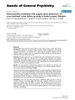
Figure 1
Adenovirus vector mediated LacZ transgene expression in synovial tissuesAdenovirus vector mediated LacZ transgene expression in synovial tis-
sues. Synovium alone or synovium with cartilage from joint replacement
surgery and synovectomy was implanted subcutaneously on the lower
backs of C.B-17 severe combined immunodeficiency (SCID) mice.
LacZ adenovirus (Ad-LacZ) in 0.1 ml PBS was injected at a dosage of
10
11
, 10
10
, or 10
9
particles into the middle of the grafted tissues. An
equal amount of PBS was injected as a control. Three days after virus
injection, synovial tissues were collected and LacZ transgene expres-
sion in the synovial tissues was detected by X-gal staining. (a) The
dose of 10
11
virus particles of Ad-LacZ per injection resulted in high
transgene expression in human synovium; (b) the dose of 10
10
virus
particles of Ad-LacZ per injection resulted in lower transgene expres-
sion; (c) the dose of 10
9
virus particles of Ad-LacZ per injection failed
to induce LacZ transgene expression, similar to (d) PBS injected con-
trol tissues. (e) The expression of LacZ transgene only was apparent in
synovium but not in cartilage three days after injection of 10
11
virus par-
ticles (the red arrow indicates synovium; the white arrow indicates carti-
lage). (f) There was no evidence of chondrocyte infection by Ad-LacZ in
cartilage, as demonstrated by X-gal/hematoxylin and eosin staining
(original magnification 200×). A variety of cell types including (g) syn-
oviocytes, (h) lymphocytes, (i) macrophages, (j) granulocytes and (k)
endothelial cells could be infected by adenovirus vector, indicated by
LacZ transgene expression detected with X-gal staining (g-k) (original
magnification 400×).
Available online />R1239
mice. The same dosage of Ad-LacZ was injected into the
synovium in vivo as a control. Tissue samples were harvested
on days 2, 5, 7, 10 and 20. Transgenic FasL mRNA expression
in tissue samples was detected by RT-PCR (Fig. 2). No trans-
genic FasL mRNA was detected in Ad-LacZ injected tissues
samples (Fig. 2, lanes 7–11). The transgenic FasL mRNA was
highly expressed on days 2 to 10 and obviously decreased on
day 20 after virus injection (Fig. 2, lanes 2–6), which suggests
that repeated administration of FasL gene transfer would be
necessary to maintain a high level of FasL transgene expres-
sion in vivo. This experiment was repeated and the same gene
expression profile was observed. Therefore, we decided to
perform repeated Ad-FasL treatment every two weeks in our in
vivo experiments.
FasL gene transfer increases the expression of FasL
protein and the frequency of apoptotic cells in RA
synovium
Fas protein is highly expressed in RA synovial lining cells and
almost all kinds of inflammatory cells infiltrating the synovium,
but FasL protein is poorly expressed in RA synovium [11]. We
determined Fas and FasL protein expression in engrafted RA
synovium by immunohistochemistry using polyclonal anti-Fas
and anti-FasL antibodies. Our experimental results (Fig. 3a,b)
are similar to those published by another group [11]. Three
days after Ad-FasL gene transfer into RA synovium in vivo,
augmented FasL protein expression was observed (Fig. 3c),
and an increased frequency of apoptotic cells in RA synovium
was detected by terminal deoxyribonucleotidyl transferase-
mediated dUTP nick end labeling (TUNEL) staining (Fig. 3f).
DNA fragmentation was determined in the location of the
nucleus using a Leica TCS SPII laser scanning confocal
microscope (Leica Microsystems, Heidelberg, Germany) (Fig.
3g–i). Few apoptotic cells were detected in RA synovium in
the SCID mouse in vivo two weeks after tissue grafting. Three
days after Ad-FasL gene transfer, the frequency of apoptotic
cells increased 15 to 20-fold (Fig. 4). The frequency of apop-
totic cells in PBS treated RA synovium underwent almost no
change, and the frequency of apoptotic cells in Ad-LacZ
injected RA synovium increased only slightly (Fig. 4).
Elimination of inflammatory RA synovium in vivo
Upregulation of Fas/FasL interaction in arthritic joints by virus
vector mediated FasL gene transfer or anti-Fas antibody treat-
ment intra-articularly can induce apoptosis in inflammatory
synoviocytes, as has been identified by many groups [5,12-
18]. Here the effect of continuous activation of Fas/FasL inter-
action through a repeated FasL gene transfer was explored to
determine if this treatment could result in the death of
inflammatory synoviocytes in RA synovium and elimination of
RA tissue in vivo. The in vivo experiments were performed as
explained in the Materials and methods section. Compared to
the control Ad-LacZ treated RA synovium, the Ad-FasL
injected RA-synovium was dramatically reduced in size (Fig.
5b) and weight (Table 1).
A significance test for the effects of Ad-LacZ and Ad-FasL
treatments on the weight of RA synovium was performed using
the rank sum test (n1 = n2 = 10, T1 = 154, T2 = 56, P <
0.005) (Table 1). Along with the approximate 15-fold increase
in the frequency of apoptotic cells observed in the RA syn-
ovium three days after after Ad-FasL injection (Fig. 4) the
number of both synoviocytes and mononuclear cells was
greatly decreased in RA synovium after two months of treat-
ment with repeated FasL gene transfers (Fig. 5d) compared
with Ad-LacZ treated RA synovium (Fig. 5c). The RA synovium
partially recovered, exhibiting features of normal human syn-
ovium after the two-month treatment with Ad-FasL (Fig. 5d).
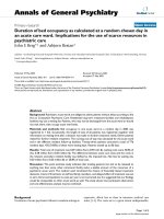
Figure 2
Time dependent transgenic Fas ligand (FasL) mRNA expression in human synovium in vivoTime dependent transgenic Fas ligand (FasL) mRNA expression in
human synovium in vivo. The 10
11
virus particles of recombinant FasL
adenovirus (Ad-FasL) and LacZ adenovirus (Ad-LacZ) were injected
into the grafted human synovium in severe combined immunodeficiency
(SCID) mice in vivo. The injected tissue samples were harvested at
days 2, 5, 7, 10, and 20 after virus injection. Total tissue RNA was iso-
lated and the first-strand cDNA of transgenic FasL was detected by RT-
PCR as described in Materials and methods. (a) Lane 1, PBS injected
tissue; lanes 2–6, Ad-FasL infected tissues harvested on day 2, 5, 7,
10, and 20, respectively, after virus injection; lanes 7–11, the Ad-LacZ
infected tissues harvested at the same time points. Ethidium bromide
staining of fractionated DNA at 688 base pairs (bp) represents trans-
genic FasL mRNA expression; the DNA fragments at 505 bp represent
β-actin expression as the internal control for the quality of mRNA sam-
ples. (b) Transgenic FasL mRNA in vivo was highly expressed on days
2–10 (lanes 2–5), and obviously decreased on day 20 (lane 6), indi-
cated by the ratio of luminosity between transgenic FasL cDNA and the
endogenous β-actin cDNA.
Arthritis Research & Therapy Vol 7 No 6 Zhang et al.
R1240
Discussion
Molecular surgery to remove the pathological cells and tissues
in situ by gene transfer could be an important part of molecular
medicine. Molecular synovectomy using gene scalpels simpli-
fies the current problems of gene delivery, gene target and
gene expression regulation involved in human gene therapy.
Because the induction of apoptosis in inflammatory synovio-
cytes is the main goal for intra-articular gene transfer, a tran-
sient, localized, immune tolerant and dose dependent gene
transfer may be achieved by direct intra-articular injection of
apoptosis inducers carried by a suitable vector, controlled to
infect only synovial cells but not chondrocytes in cartilage. In
this study, we have established that the inflammatory synovium
from RA patients can be effectively reduced in vivo in a RA-
SCID mouse model by repeated, locally administered adeno-
virus vector mediated FasL gene transfer, strongly supporting
the gene scalpel concept.
C.B-17 SCID mice have severe combined immunodeficiency,
resulting from a mutation on chromosome 16 responsible for
deficient activity of an enzyme involved in DNA repair [25]. We
chose to use C.B-17 SCID mice in this study because this
strain has successfully hosted human synovium and has pro-
vided an ideal in vivo experimental environment to test how
human inflammatory synovium responds to novel therapeutic
reagents [21-23]. Unlike C.B-17 SCID beige mice, C.B-17
SCID mice have no deficiency of macrophages and natural
killer cells, which is important for our experimental design
because macrophage phagocytosis of apoptotic cells [26-28]
is considered as one of the important mechanisms involved in
the molecular synovectomy procedure. In theory, inflammatory
synoviocytes in implanted RA synovium in SCID mice may
consist of antigen specific human lymphocytes, proliferated
human fibroblast-like synoviocytes, and infiltrated mouse mac-
rophages and so on. An artificial feature of this model,
however, is the absence of circulating human blood compo-
nents when studying the properties of rheumatoid synovium
[21]. In our experiments, the inflammatory histological features
of rheumatoid synovium are preserved in implanted RA syn-
ovium in SCID mice. This combination of RA synovium and
SCID mice allows for repeated FasL gene transfer mediated
by adenovirus vector into RA synovium in vivo because the
C.B-17 SCID mice lack an immune response to the adenovi-
rus vector.
Figure 3
Detection of apoptotic cells and Fas/Fas ligand (FasL) protein expression in grafted rheumatoid arthritis (RA) synoviumDetection of apoptotic cells and Fas/Fas ligand (FasL) protein expression in grafted rheumatoid arthritis (RA) synovium. The RA synovium in severe
combined immunodeficiency (SCID) mice was harvested approximately two weeks after engraftment without or with three day treatment with 10
11
particles of recombinant FasL adenovirus (Ad-FasL), LacZ adenovirus (Ad-LacZ), or PBS before it was collected. (a-c) Fas and FasL protein expres-
sion in RA synovium in vivo were examined using an immunohistochemistry staining system. (a) Fas protein is highly expressed on synoviocytes in
RA synovium (original magnification 400×). (b) FasL protein is lacking in RA synoviocytes treated with Ad-LacZ (original magnification 400×); (c)
but a significant increase in FasL protein expression in RA synoviocytes appears three days after Ad-FasL injection (original magnification 400×). (d-
i) Apoptotic cells in RA synovium were examined using the ApopTag fluorescein in situ apoptosis detection kit (Intergen). The red propidium iodide
counter-staining indicates the nucleus of all synovial cells in RA synovium. The green fluorescein stains the fragmented DNA in the nucleus. (d)
Apoptotic cells in PBS treated RA synovium are almost undetectable (original magnification 200×). (e) A slightly increased apoptotic cell population
was seen in Ad-LacZ treated RA synovium (original magnification 200×), (f) while around a 15-fold increased frequency of apoptotic cells in RA syn-
ovium was observed in Ad-FasL treated RA synovium (original magnification 200×; arrows point to TUNEL-positive cells). (g-i) The location of the
fragmented DNA was observed using a laser scanning confocal microscope. (g) The nucleus of synovial cells in RA synovium was stained with pro-
pidium iodide (red; bar = 15 µm). (h) Fragmented DNA was stained with fluorescein (green; bar = 15 µm). (i) The localization of the fragmented
DNA was determined by the overlap of the fluorescein staining and propidium iodide counter-staining (bar = 15 µm), which characterizes cell death.
Available online />R1241
The elimination of synoviocytes from RA synovium after multi-
ple applications of FasL gene transfer results in a significant
reduction in RA synovium size and weight in vivo in the RA-
SCID mice (Fig. 5; Table 1) along with dramatic synovial cell
death detected by TUNEL staining (Figs 3g–I and 4). Recom-
binant adenovirus is a highly efficient vector for gene transfer
into the synovium [5,7,12,19]. Adenovirus vector can infect a
wide variety of cell types in human synovium including synovi-
ocytes, lymphocytes, macrophages, granulocytes and
endothelial cells, as determined by X-gal staining (Fig. 1g–k).
In recent studies, endothelial cells were reported to constitu-
tively express FasL and release soluble FasL [29-31]. These
cells are not sensitive to Fas-mediated apoptosis and may play
a role in the negative regulation of inflammation [30]. The
mediation of cell death for lymphocytes and granulocytes
strongly involves the Fas/FasL apoptosis pathway [32-35].
Primary cultured macrophages can survive 1 to 2 weeks after
infection with 10
4
particles per cell of Ad-FasL (data not
shown). Fas-associated death domain-like interleukin 1beta-
converting enzyme (FLICE)-inhibitory protein (Flip), a negative
regulator of Fas-induced apoptosis, is upregulated when
monocytes differentiate into macrophages, which may confer
the resistance macrophages have to Fas-mediated apoptosis
[36,37]. The survival of macrophages after FasL gene transfer
may be necessary for the phagocytosis of apoptotic cells dur-
ing 'molecular synovectomy' using the 'FasL gene scalpel'. On
the other hand, the survival of macrophages carrying viral vec-
tor antigens may activate the naive lymphocytes and initiate an
anti-virus immune response if the adenovirus vector was used
for clinical administration. Thus, the development of an
immune tolerant gene delivery vehicle is an important task for
the clinical use of FasL gene scalpel.
Compared with Ad-LacZ gene transfer, Ad-FasL gene transfer
induces a significantly increased frequency of apoptotic cells
in RA synovium (Figs 3e,f and 4), even though adenovirus vec-
tor itself shows a slight effect on enhancing apoptosis in syn-
oviocytes (Fig. 4) and certain other kinds of cells [38,39]. From
the point of view that repeated FasL gene transfer can remove
RA synovium in vivo (Fig. 5b; Table 1), the FasL gene may
function as a sharp scalpel for molecular synovectomy. It has
been reported that Fas-mediated apoptosis is associated with
activation of the Fas-associated death domain protein/Cas-
Figure 4
Efficient increase of apoptosis incidence in rheumatoid arthritis (RA) synoviocytes by administration of recombinant FasL adenovirus (Ad-FasL)Efficient increase of apoptosis incidence in rheumatoid arthritis (RA)
synoviocytes by administration of recombinant FasL adenovirus (Ad-
FasL). The percentage of apoptotic cells in grafted RA synovium in
severe combined immunodeficiency (SCID) mice is compared to that in
the RA synovium three days after treatments with Ad-FasL, LacZ aden-
ovirus (Ad-LacZ) and PBS. Total synoviocytes and apoptotic cells were
counted in 10 high power fluorescent microscopic fields randomly
selected from each section, and three sections at multiple layers for
each sample were examined. The percentage of apoptotic cells was
recorded and is represented as the mean and standard deviation for
each group. Open bars represent the percentage of apoptotic cells in
RA synovium two weeks after engraftment without treatment; striped
bars represent the percentage of apoptotic cells in RA synovium three
days after Ad-FasL, Ad-LacZ and PBS injections.
Figure 5
Reduction of the size and cell density of rheumatoid arthritis (RA) syn-ovium in vivo by repeated Fas ligand (FasL) gene transferReduction of the size and cell density of rheumatoid arthritis (RA) syn-
ovium in vivo by repeated Fas ligand (FasL) gene transfer. (a) RA syn-
ovium, 200 mg tissue per mouse, was grafted into the back of each
severe combined immunodeficiency (SCID) mouse subcutaneously.
Two weeks after engraftment, 10
11
particles of recombinant FasL aden-
ovirus (Ad-FasL) or Ad-LacZ adenovirus (Ad-LacZ) in 0.1 ml PBS were
injected into the engrafted RA synovium in the SCID mice. Injections
were repeated every two weeks for a total of five times. Three days after
the fifth injection, the mice were euthanized and the RA synovium sam-
ples were collected, weighed, and examined with hematoxylin and eosin
(H&E) staining. (b) A comparative view of the size of the grafted RA
synovium after two months of repeated treatment with control Ad-LacZ
(tissue on the left side) and Ad-FasL (tissue on the right side). (c) H&E
staining shows that both synoviocytes and mononuclear cells are
greatly decreased in Ad-FasL treated RA synovium, in which a partial
recovery of the normal features of synovium was observed (original
magnification 200×); (d) but the Ad-LacZ treated RA synovium main-
tains the inflammatory infiltrates (original magnification 200×)
Arthritis Research & Therapy Vol 7 No 6 Zhang et al.
R1242
pase-8/Caspase-3/poly(ADP-ribose)polymerase pathway and
the c-Jun amino-terminal kinase/activator protein-1 pathway
[40,41]. The former is thought to be the major signaling path-
way required for Fas-mediated apoptosis in RA synoviocytes;
the latter appears to be involved in the pathogeneses of inflam-
mation by inducing pro-inflammatory cytokine/chemokine pro-
duction [40,42]. Thus, the clinical potential of a FasL gene
scalpel will depend on the clarification of the side effects of
FasL intra-articular gene transfer. The combined administra-
tion of the intra-articular gene transfer of FasL with other genes
and/or certain anti-inflammatory drugs might be an ideal com-
plementary therapeutic approach for the local treatment of RA
and other arthropathies.
A subset of chondrocytes located in the superficial zone of
cartilage expresses the Fas antigen, and activation of the Fas
receptor triggers apoptosis in these cells [43,44]. An adeno-
virus mediated short-term (24 h) FasL gene transfer in vivo in
the rabbit knee only transduced synovium, but it did not affect
the viability of chondrocytes in cartilage [12]. Our in vivo
experiments with Ad-LacZ gene transfer on human synovium
and cartilage in SCID mice presents similar results (Fig. 1e,f),
but after a long-term (4 weeks) in vitro culture the chondro-
cytes in the superficial zone of cartilage were infected by Ad-
LacZ 24 h after virus infection (unpublished data). This phe-
nomenon suggests that the loss of cartilage matrix may have
occurred during long-term in vitro culture. The cartilage matrix
may be important for protection of chondrocytes from infection
by adenovirus vector mediated gene transfer. Thus, the most
suitable period for the treatment of rheumatoid arthritis with
gene transfer would be in the early stage of the disease when
the cartilage matrix is undamaged. The effects of long-term
gene transfer of an apoptosis inducer such as FasL on the
chondrocytes in cartilage in vivo require further investigation.
Currently, potential strategies for the treatment of RA using
intra-articular gene transfer are focused on reducing the pro-
duction and activation of inflammatory cytokines/chemokines
and proteases/adhesion molecules, and inducing apoptosis of
inflammatory synoviocytes. The latter may be more efficient in
suppressing the inflammation by removing the producing and
reactive cells responding to pro-inflammatory molecules. This
potential therapeutic approach using 'gene scalpels' may
function as a molecular synovectomy, not only for the treat-
ment of RA, but also for the treatment of other arthropathies.
Conclusion
Our study elucidates firstly the in vivo therapeutic potential of
molecular synovectomy using 'gene scalpels' for the treatment
of RA locally through repeated intra-articular gene transfer of
an apoptosis inducer. This approach may more efficiently
arrest intra-articular inflammation and suppress the progress
of RA by removing the cells producing and/or responding to
pro-inflammatory molecules in arthritic joints.
Competing interests
The author(s) declare that they have no competing interests.
Authors' contributions
GG provided the adenoviruses for study. GC contributed to
the experiments and generated the histology data. RS coordi-
nated the collection of the synovial tissues from patients in the
clinic, carried out the pathological diagnosis of the human
samples and provided advisory support for the study. HZ con-
ceived of the study, participated in its design and coordination,
carry out the in vivo study and molecular examinations, and
drafted the manuscript. All authors read and approved the final
manuscript.
Acknowledgements
This work was supported by grants 2T32AR07442-12 and R03
AR47983 from the National Institutes of Health/National Institute of
Arthritis and Musculoskeletal and Skin Diseases (NIAMS), a research
award from the University of Pennsylvania Research Foundation, and an
investigator award from the Arthritis Foundation. We thank Dr Robert A
Eisenberg for the advisory support with the NIH training grant; Dr James
M Wilson for critical support with the collaboration; Drs William V Wil-
liams and Shigekazu Nagata for cells and reagents; Drs Philip Cohen
and William V Williams for helpful discussion; Ms Marie Sieck for pho-
tographs; the Animal Facility at Veterans Affairs Research Center in Phil-
adelphia and the Department of Orthopaedic Surgery at University of
Pennsylvania Health System for assistance and cooperation with animal
care and human sample collection.
References
1. Yocum DE, Lafyatis R, Remmers EF, Schumacher HR, Wilder RL:
Hyperplastic synoviocytes from rats with streptococcal cell
wall-induced arthritis exhibit a transformed phenotype that is
thymic-dependent and retinoid inhibitable. Am J Pathol 1988,
132:38-48.
2. Harris EDJr: Rheumatoid arthritis: Pathophysiology and impli-
cations for therapy. N Engl J Med 1990, 322:1277-1289.
3. Firestein GS: Invasive fibroblast-like synoviocytes in rheuma-
toid arthritis. Arthritis Rheum 1996, 39:1781-1790.
4. Cozzolino F, Gigliotti S, Giuzio E, Angrisani C: Surgical synovec-
tomy in the treatment of rheumatoid arthritis: the results
obtained in a controlled study. Chirurgia Degli Organi di
Movimento 1991, 76:341-346.
5. Zhang H, Yang Y, Horton J, Samoilova EB, Judge TA, Turka LA,
Wilson JM, Chen Y: Amelioration of collagen-induced arthritis
by CD 95 (Apo-1/Fas)-Ligand gene transfer. J Clin Invest
1997, 100:1-7.
6. Nishioka K, Hasunuma T, Kato T, Sumida T, Kobata T: Apoptosis
in rheumatoid arthritis: a novel pathway in the regulation of
synovial tissue. Arthritis Rheum 1998, 41:1-9.
7. Goossens PH, Schouten GJ, 't Hart BA, Bout A, Brok HP, Kluin
PM, Breedveld FC, Valerio D, Huizinga TW: Feasibility of adeno-
virus-mediated nonsurgical synovectomy in collagen-induced
arthritis-affected rhesus monkeys. Hum Gene Ther 1999,
10:1139-1149.
8. Migita K, Honda S, Yamasaki S, Hirai Y, Fukuda T, Aoyagi T, Kita
M, Ida H, Tsukada T, Kawakami A, et al.: Regulation of rheuma-
toid synovial cell growth by ceramide. Biochem Biophys Res
Commun 2000, 269:70-75.
9. Michael VV, Alisa KE: Cell cycle implications in the pathogene-
sis of rheumatoid arthritis. Frontiers Biosci 2000, 5:D594-601.
10. Asahara H, Hasunuma T, Kobata T, Inoue H, Muller-Ladner U, Gay
S, Sumida T, Nishioka K: In situ expression of protooncogenes
and Fas/Fas ligand in rheumatoid arthritis synovium. J Rheum
1997, 24:430-435.
Available online />R1243
11. Zhang X, Jiang M, Zheng D: Rheumatoid arthritis synoviocyte
hyperplasia and expression of fas and bcl-2 genes. Chung-
Hua i Hsueh Tsa Chih 1998, 78:175-178.
12. Yao Q, Glorioso JC, Evans CH, Robbins PD, Kovesdi I, Oligino TJ,
Ghivizzani SC: Adenoviral mediated delivery of FAS ligand to
arthritic joints causes extensive apoptosis in the synovial
lining. J Gene Med 2000, 2:210-219.
13. Iwakura Y, Tosu M, Youshida E, Takiguchi M, Sato K, Kitajiam I,
Nishioka K, Yamamoto K, Takeda T, Hatanaka M: Induction of
inflammatory arthropathy resembling rheumatoid arthritis in
mice transgenic for HTLV-I. Science 1991, 253:1026-1028.
14. Fujisawa K, Asahara H, Okamoto K, Aono H, Hasunuma T, Kobata
T, Iwakura Y, Yonehara S, Sumida T, Nishioka K: Therapeutic
effect of the anti-Fas antibody on arthritis in HTLV-I tax trans-
genic mice. J Clin Invest 1996, 98:271-278.
15. Ogasawara J, Watanabe-Fukunaga R, Adachi M, Matsuzawa A,
Kasugai T, Kitamura Y, Itoh N, Suda T, Nagata S: Lethal effect of
the anti-Fas antibody in mice. Nature 1993, 364:806-809.
16. Okamoto K, Asahara H, Kobayashi T, Matsuno H, Hasunma T,
Kobata T, Sumida T, Nishioka K: Induction of apoptosis in the
rheumatoid synovium by Fas Ligand gene transfer. Gene
Therapy 1998, 5:331-338.
17. Zhang H, Gao G, Williams W, Wilson J, Schumacher HR: Induc-
tion of apoptosis in synovial fibroblasts from rheumatoid
arthritis patients by Fas ligand gene transfer [abstract]. Arthri-
tis Rheum 1998, 41:S142.
18. Wakisaka S, Suzuki N, Takeba Y, Shimoyama H, Nagafuchi H, Tak-
eno M, Saito N, Yokoe T, Kaneko T, Asai T, Sakane T: Modulation
by proinflammatory cytokines of Fas/Fas ligand-mediated
apoptotic cell death of synovial cells in patients with rheuma-
toid arthritis (RA). Clin Exp Immunol 1998, 114:119-128.
19. Ikeda T, Kubo T, Arai Y, Nakanishi T, Kobayashi K, Takahashi K,
Imanishi J, Takigawa M, Hirasawa Y: Adenovirus mediated deliv-
ery to the joints of guinea pigs. J Rheumatol 1998,
25:1666-1673.
20. SaKai K, Matsuno H, Morita I, Nezuka T, Tsuji H, Shirai T, Yonehara
S, Hasunuma T, Nishioka K: Potential withdrawal of rheumatoid
synovium by the induction of apoptosis using a novel in vivo
model of rheumatoid arthritis. Arthritis Rheum 1998,
41:1251-1257.
21. Geiler T, Kriegsmann J, Keyszer GM, Gay RE, Gay S: A new
model for rheumatoid arthritis generated by engraftment of
rheumatoid synovial tissue and normal human cartilage into
SCID mice. Arthritis Rheum 1994, 37:664-71.
22. Jorgensen C, Demoly P, Noel D, Mathieu M, Piechaczyc M, Gougat
C, Bousquet J, Sany J: Gene transfer to human rheumatoid syn-
ovial tissue engrafted in SCID mice. J Rheumatol 1997,
24:2076-2079.
23. Arnett FC, Edworthy SM, Bloch DA, McShane DJ, Fries JF, Cooper
NS, Healey LA, Kaplan SR, Liang MH, Luthra HS, et al.: The Amer-
ican Rheumatism Association 1987 revised criteria for the
classification of rheumatoid arthritis. Arthritis Rheum 1988,
31:315-24.
24. Gao GP, Yang YP, Wilson JM: Biology of adenoviral vectors
deleted of E1 and E4 for liver-directed gene therapy. J Virol
1996, 70:8934-8943.
25. Jorgensen C, Bologna C, Sany J: Autoimmunity. Insights pro-
vided by the SCID mouse model. Revue Du Rhum 1995,
62:519-524.
26. Dini L, Ruzittu MT, Falasca L: Recognition and phagocytosis of
apoptotic cells. Scanning Microsc 1996, 10:239-251.
27. Fadok VA, Savill JS, Haslett C, Bratton DL, Doherty DE, Campbell
PA, Henson PM: Different populations of macrophages use
either the votronectin receptor or the phosphatidylserine
receptor to recognize and remove apoptotic cells. J Immunol
1992, 149:4029-4035.
28. Rossi AG, McCutcheon JC, Roy N, Chilvers ER, Haslett C, Drans-
field I: Regulation of macrophage phagocytosis of apoptotic
cells by camp. J Immunol 1998, 160:3562-3568.
29. Mogi M, Fukuo K, Yang J, Suhara T, Ogihara T: Hypoxia stimu-
lates release of the soluble form of Fas ligand that inhibits
endothelial cell apoptosis. Lab Invest 2001, 81:177-184.
30. Walsh K, Sata M: Negative regulation of inflammation by Fas
ligand expression on the vascular endothelium. Trends Cardi-
ovasc Med 1999, 9:34-41.
31. Sata M, Suhara T, Walsh K: Vascular endothelial cells and
smooth muscle cells differ in expression of Fas and Fas ligand
and in sensitivity to Fas ligand-induced cell death: implications
for vascular disease and therapy. Thromb Vasc Biol 2000,
20:309-316.
32. Lundy SK, Boros DL: Fas ligand-expressing B-1a lymphocytes
mediate CD4(+)-T-cell apoptosis during schistosomal infec-
tion: induction by interleukin 4 (IL-4) and IL-10. Infect Immun
2002, 70:812-819.
33. Liles WC, Kiener PA, Ledbetter JA, Aruffo A, Klebanoff SJ: Differ-
ential expression of Fas (CD95) and Fas ligand on normal
human phagocytes: implications for the regulation of apopto-
sis in neutrophils. J Exp Med 1996, 184:429-440.
34. Hanna N, Vasquez P, Pham P, Heck DE, Laskin JD, Laskin DL,
Weinberger B: Mechanisms underlying reduced apoptosis in
neonatal neutrophils. Pediatr Res 2005, 57:56-62.
35. O'Flaherty E, Wong WK, Pettit SJ, Seymour K, Ali S, Kirby JA: Reg-
ulation of T-cell apoptosis: a mixed lymphocyte reaction
model. Immunology 2000, 100:289-299.
36. Perlman H, Pagliari LJ, Georganas C, Mano T, Walsh K, Pope RM:
FLICE-inhibitory protein expression during macrophage differ-
entiation confers resistance to fas-mediated apoptosis. J Exp
Med 1999, 190:1679-1688.
37. Perlman H, Pagliari LJ, Liu H, Koch AE, Haines GK, Pope RM 3rd:
Rheumatoid arthritis synovial macrophages express the Fas-
associated death domain-like interleukin-1beta-converting
enzyme-inhibitory protein and are refractory to Fas-mediated
apoptosis. Arthritis Rheum 1999, 44:21-30.
38. Teramoto S, Matsuse T, Matsui H, Ohga E, Ishii T, Nagase T, Ouchi
Y: Recombinant E1-deleted adenoviral vectors induce apopto-
sis in a rat airway epithelial mucous goblet cell line. Japan J
Pharmacol 1999, 81:73-78.
39. Teramoto S, Matsuse T, Matsui H, Ohga E, Ishii T, Ouchi Y:
Recombinant E1-deleted adenovirus vector induces apoptosis
in two lung cancer cell lines. Eur Respir J 1999, 13:1125-1132.
40. Kobayashi T, Okamoto K, Kobata T, Hasumuna T, Nishioka K: Apo-
modulation as a novel therapeutic concept for the regulation
of apoptosis in rheumatoid synoviocytes. Curr Opinin
Rheumatol 1999, 11:188-193.
41. Okamoto K, Kobayashi T, Kobata T, Hasumuna T, Kato T, Sumida
T, Nishioka K: Fas-associated death domain protein is a Fas-
mediated apoptosis modulator in synoviocytes. Rheumatol
2000, 39:471-480.
42. Naumann M, Rudel T, Wieland B, Bartsch C, Meyer TF: Coordi-
nate activation of activator protein 1 and inflammatory
cytokines in response to Neisseria gonorrhoeae epithelial cell
contact involves stress response kinases. J Exp Med 1998,
188:1277-1286.
43. Lotz M, Hashimoto S, Kuhn K: Mechanisims of chondrocyte
apoptosis. Osteoarthritis Cartilage 1999, 7:389-391.
44. Hashimoto S, Setareh M, Ochs RL, Lotz M: Fas/Fas ligand
expression and induction of apoptosis in chondrocytes. Arthri-
tis Rheum 1997, 40:1749-1755.