Báo cáo y học: " TGF β-induced cartilage repair is maintained but fibrosis is blocked in the presence of Smad7" pps
Bạn đang xem bản rút gọn của tài liệu. Xem và tải ngay bản đầy đủ của tài liệu tại đây (1.07 MB, 8 trang )
Open Access
Available online />Page 1 of 8
(page number not for citation purposes)
Vol 8 No 3
Research article
TGF β-induced cartilage repair is maintained but fibrosis is
blocked in the presence of Smad7
Esmeralda N Blaney Davidson, Elly L Vitters, Wim B van den Berg and Peter M van der Kraan
Experimental Rheumatology and Advanced Therapeutics, St. Radboud University Medical Centre Nijmegen, Geert Grooteplein 26-28, 6525 GA
Nijmegen, The Netherlands
Corresponding author: Esmeralda N Blaney Davidson,
Received: 9 Jan 2006 Revisions requested: 3 Feb 2006 Revisions received: 23 Feb 2006 Accepted: 7 Mar 2006 Published: 29 Mar 2006
Arthritis Research & Therapy 2006, 8:R65 (doi:10.1186/ar1931)
This article is online at: />© 2006 Blaney Davidson et al.; licensee BioMed Central Ltd.
This is an open access article distributed under the terms of the Creative Commons Attribution License ( />),
which permits unrestricted use, distribution, and reproduction in any medium, provided the original work is properly cited.
Abstract
Cartilage damage in osteoarthritis (OA) is considered an
imbalance between catabolic and anabolic factors, favoring the
catabolic side. We assessed whether adenoviral overexpression
of transforming growth factor-β (TGFβ) enhanced cartilage
repair and whether TGFβ-induced fibrosis was blocked by local
expression of the intracellular TGFβ inhibitor Smad7. We
inflicted cartilage damage by injection of interleukin-1 (IL-1) into
murine knee joints. After 2 days, we injected an adenovirus
encoding TGFβ. On day 4, we measured proteoglycan (PG)
synthesis and content. To examine whether we could block
TGFβ-induced fibrosis and stimulate cartilage repair
simultaneously, we injected Ad-TGFβ and Ad-Smad7. This was
performed both after IL-1-induced damage and in a model of
primary OA. In addition to PG in cartilage, synovial fibrosis was
measured by determining the synovial width and the number of
procollagen I-expressing cells. Adenoviral overexpression of
TGFβ restored the IL-1-induced reduction in PG content and
increased PG synthesis. TGFβ-induced an elevation in PG
content in cartilage of the OA model. TGFβ-induced synovial
fibrosis was strongly diminished by simultaneous synovial
overexpression of Smad7 in the synovial lining. Of great interest,
overexpression of Smad7 did not reduce the repair-stimulating
effect of TGFβ on cartilage. Adenoviral overexpression of TGFβ
stimulated repair of IL-1- and OA-damaged cartilage. TGFβ-
induced synovial fibrosis was blocked by locally inhibiting TGFβ
signaling in the synovial lining by simultaneously transfecting it
with an adenovirus overexpressing Smad7.
Introduction
Osteoarthritis (OA) is a degenerative joint disease character-
ized by cartilage breakdown, synovial fibrosis, and bone spurs.
An imbalance between catabolic and anabolic factors favoring
the catabolic side is very likely involved in the pathological fea-
tures of OA.
Currently, many attempts are being made to repair the carti-
lage that has been damaged in OA. One approach focuses on
shifting the metabolic imbalance back by stimulating the ana-
bolic side. Transforming growth factor-β (TGF-β) is one of the
anabolic factors involved in cartilage maintenance and
appears to be a good candidate for cartilage repair. TGF-β is
a stimulator of extracellular matrix production, like collagen
type II and proteoglycan (PG), in chondrocytes and it down-
regulates matrix-degrading enzymes [1]. High amounts of
TGF-β are stored in healthy cartilage [2-6], whereas in OA car-
tilage the expression of TGF-β is reduced [7]. Injection of TGF-
β into naive murine knee joints results in an increase in PG
content of the articular cartilage [8]. Moreover, in murine
experimental rheumatoid arthritis, injection of TGF-β protected
cartilage from PG loss [9]. In addition, TGF-β counteracts the
anabolic factor interleukin-1 (IL-1), which is a very potent
inducer of cartilage degradation [10,11] both in vivo and in
vitro [1,12-16]. These data indicate that TGF-β has great
potential as a tool for stimulating cartilage repair.
To obtain sufficient amounts of TGF-β in the joint for a pro-
longed period of time, an adenovirus can be used as a vehicle.
In vitro, chondrocytes that are transfected with an adenovirus
encoding TGF-β responded by elevation of PG and collagen
production [17]. We wanted to assess whether adenoviral
overexpression of TGF-β in the synovial lining could stimulate
repair of damaged cartilage in vivo.
IL-1 = interleukin-1; Luc = luciferase; OA = osteoarthritits; PBS = phosphate-buffered saline; pfu = plaque-forming unit; PG = proteoglycan; TGF-β
= transforming growth factor-β
Arthritis Research & Therapy Vol 8 No 3 Blaney Davidson et al.
Page 2 of 8
(page number not for citation purposes)
Unfortunately, introducing high amounts of TGF-β into a knee
joint has adverse effects. Administration of 20 ng TGF-β is
already sufficient to result in an increased cellularity of the syn-
ovial lining, expansion of fibroblast population in the synovial
connective tissue, and continued collagen deposition [18].
Injection of high amounts of TGF-β, either as a bolus injection
or via adenoviral transfection, results in marked hyperplasia of
the synovium and chondro-osteophyte formation [8,18-21].
This illustrates that the use of TGF-β for cartilage repair can
result in side effects that are deleterious for future therapeutic
applications.
The aim of this study was to use TGF-β as a cartilage repair
factor but at the same time to prevent the TGF-β-induced
fibrotic side effect. Therefore, we examined the effect of aden-
oviral overexpression of active TGF-β on cartilage repair and
additionally studied whether simultaneous Smad7 overexpres-
sion could block TGF-β-induced fibrosis. Smad7 is an intrac-
ellular molecule that inhibits the TGF-β signaling pathway.
TGF-β binds to its type II receptor, which then forms a complex
with the type I TGF-β receptor.
Subsequently, the intercellular signaling molecule Smad2 or
Smad3 gets phosphorylated, forms a complex with common
Smad, Smad4, and shuttles to the nucleus for transcription
[22]. Smad7 inhibits Smad2 and Smad3 phosphorylation,
thereby preventing further signaling [23,24].
To both stimulate cartilage and block side effects, we took
advantage of the fact that adenoviruses, once injected into the
murine knee joint, transfect the synovial lining but do not pen-
etrate the cartilage [25]. We co-transfected the synovial lining
with an adenovirus overexpressing TGF-β and an adenovirus
overexpressing Smad7. The transfected synovial lining cells
will produce TGF-β but due to an intercellular signaling block
caused by Smad7, will no longer respond to this factor.
We show that adenoviral overexpression of TGF-β results in
increased PG content of the cartilage both after IL-1-induced
damage and in a spontaneous model of experimental OA. In
both cases, the TGF-β-induced fibrosis can be prevented by
simultaneous Smad7 overexpression.
Materials and methods
Animals
C57Bl/6 mice (10 weeks old) and STR/ort mice (4 weeks old)
were used. Mice were kept in filter-top cages with woodchip
bedding under standard pathogen-free conditions. They were
fed a standard diet and tap water ad libitum. The local animal
committee had approved this study.
Stimulation of cartilage repair by TGF-β after IL-1 insult
To assess whether adenoviral overexpression of TGF-β could
stimulate cartilage repair, we inflicted cartilage damage in 73
C57Bl/6 mice by intra-articular injection of 10 ng IL-1 (R&D
Systems, Inc., Minneapolis, MN, USA). Two days after IL-1
injection, PG synthesis will have reached a low point [11]. At
this time point, an adenovirus overexpressing active TGF-β
(Ad-TGF-β
223/225
) was injected intra-articularly (plaque-form-
ing units [pfu] 10
7
/6 µl) and compared with a control virus (Ad-
del 70-3). Four days after the primary insult, 53 mice were
used for patellae isolation for PG synthesis measurement by
35
SO
4
2-
incorporation. The other 20 mice were used for isola-
tion of whole knee joints for histology.
Blocking TGF-β-induced fibrosis
To block TGF-β-induced fibrosis, 24 C57Bl/6 mice were
injected intra-articularly with adenoviruses in the combinations
of Ad-TGF-β
223/225
+ Ad-luciferase (Ad-luc), Ad-Smad7 + Ad-
luc, and Ad-TGF-β
223/225
+ Ad-Smad7 (at a pfu of 0.5 × 10
7
per adenovirus in 6 µl) or Ad-luc alone (at a total pfu of 10
7
) as
a control. After 14 days, when synovial fibrosis can be
observed histologically, knee joints were isolated for histology.
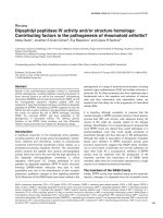
Figure 1
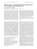
Effect of adenoviral transforming growth factor-β (TGF-β) expression on proteoglycan (PG) synthesis and content in cartilageEffect of adenoviral transforming growth factor-β (TGF-β) expression on
proteoglycan (PG) synthesis and content in cartilage. (a) PG synthesis
was measured by
35
SO
4
2-
incorporation into patellar cartilage 4 days
after interleukin-1 (IL-1) injection. (CPM = counts per minute) PG syn-
thesis increased after IL-1 injection (p < 0.005), and this increase was
boosted by TGF-β (p < 0.0005 compared with IL-1). (b) PG content of
cartilage was measured by Safranin O staining intensity of the cartilage
in Safranin O/Fast Green stained sections. The mean PG content of
non-treated knee joints was set at 100%. After IL-1 exposure, a clear
reduction in PG content was observed (p < 0.05). By adenoviral
expression of TGF-β after IL-1-induced damage, the PG content of the
cartilage was almost normal. Error bars display standard error.
Available online />Page 3 of 8
(page number not for citation purposes)
Simultaneously stimulating cartilage repair and blocking
of fibrosis
To make sure that Smad7 did not interfere with TGF-β-stimu-
lated PG synthesis, we assessed whether stimulation of carti-
lage repair was not blocked by co-transfection with Ad-
Smad7, and cartilage damage was again introduced in 48
C57Bl/6 mice by intra-articular injection with 10 ng IL-1. After
2 days, mice were injected with adenoviruses in the combina-
tions of Ad-TGF-β
223/225
+ Ad-luc, Ad-TGF-β
223/225
+ Ad-
smad7, or Ad-luc alone. Four days after IL-1 injection, 24 mice
were used for isolation of patellae for
35
SO
4
2-
incorporation
measurements. After 2 weeks, the other mice were used for
isolation of knee joints for histological assessment of fibrosis.
Cartilage repair while blocking fibrosis in spontaneous
OA
To test whether we could stimulate cartilage repair in a spon-
taneous experimental OA model while preventing fibrosis, we
extended our experiment to STR/ort mice. STR/ort mice
develop OA spontaneously and show pathological changes by
8 weeks of age. We injected adenoviruses intra-articularly into
the knee joint of 24 4-week-old STR/ort mice and repeated
this injection after 2 weeks. The adenoviruses were injected in
the combinations of Ad-TGF-β
223/225
+ Ad-luc, Ad-smad7 +
Ad-luc, and Ad-TGF-β
223/225
+ Ad-smad7 at a pfu of 0.5 × 10
7
per adenovirus or Ad-luc at a pfu of 10
7
alone as a control.
Four weeks after the first injection, knee joints were isolated
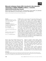
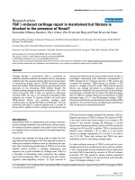
Figure 2
Synovial fibrosis was assessed in knee joints 2 weeks after intra-articular injection of Ad-transforming growth factor-β (TGF-β) combined with Ad-luciferase or Ad-Smad7Synovial fibrosis was assessed in knee joints 2 weeks after intra-articular injection of Ad-transforming growth factor-β (TGF-β) combined with Ad-
luciferase or Ad-Smad7. As a measure of fibrosis, the synovial width opposite the growth plates in the femur was measured (a) In addition, the
amount of cells staining positive in immunohistochemically stained sections for procollagen type I was calculated with a computerized imaging sys-
tem (b). The data are represented as an increase of the viral control. Histological representations of the measurements give an indication of actual
thickness (c) and procollagen positive cells (d) There were no differences between viral and non-injected controls. TGF-β overexpression resulted in
an increase in synovial thickness and number of procollagen type I-positive cells (p < 0.05). By co-expression with Smad7, most of the TGF-β-
induced fibrosis was prevented (p < 0.05). Error bars display standard error.
Arthritis Research & Therapy Vol 8 No 3 Blaney Davidson et al.
Page 4 of 8
(page number not for citation purposes)
for histological analysis of synovial fibrosis and PG content of
the cartilage.
Histology
Knee joints of mice were dissected and fixed in phosphate-
buffered formalin for 7 days. Thereafter, they were decalcified
in 10% formic acid for 1 week. Knee joints were dehydrated
with an automated tissue-processing apparatus (Tissue Tek
VIP, Sakura, Ramsey, MN, USA) and embedded in paraffin.
Coronal whole knee joint sections of 7 µm were made. Sec-
tions were stained with Safranin O and Fast Green.
Immunohistochemistry
Sections were deparaffinized and washed with phosphate-
buffered saline (PBS). For antigen unmasking, sections were
incubated in citrate buffer (0.1 M sodium citrate + 0.1 M citric
acid) for 2 hours. Endogenous peroxidase was blocked with
1% hydrogen peroxide in methanol for 30 minutes. Thereafter,
sections were blocked with 5% normal serum of the species
in which the secondary antibody was produced. Specific pri-
mary antibodies against procollagen type I (2 µg/ml) (Santa
Cruz Biotechnology, Inc., Santa Cruz, CA, USA) were incu-
bated overnight at 4°C. After extensive washing with PBS, the
appropriate biotin-labeled secondary antibody was used
(DakoCytomation, Glostrup, Denmark) for 30 minutes at room
temperature followed by a biotin-streptavidine detection sys-
tem according to manufacturer's protocol (Vector Laborato-
ries, Burlingame, CA, USA). Bound complexes were visualized
using DAB (3,3'-diaminobenzidine) reagent, counterstained
with haematoxylin, dehydrated, and mounted with Permount.
PG synthesis
For measurement of PG synthesis,
35
SO
4
2-
was incorporated
into isolated patellae. Immediately after isolation, the patellae
were placed in Dulbecco's modified Eagle's medium with gen-
tamicin (50 mg/ml) and pyruvate. After half an hour, medium
was replaced by medium containing
35
SO
4
2-
(20 µCi/ml) and
incubated for 3 hours at 37°C in 5% CO
2
. Patellae were then
further prepared for measurement of
35
SO
4
2-
incorporation in
the articular cartilage as previously described [26].
PG content
PG content was measured in sections stained with Safranin O
and Fast Green, using a computerized imaging system as pre-
viously described [27]. Briefly, Safranin O stains PGs in the
cartilage red. The amount of PGs is determined by a compu-
terized calculation of the amount of blue light passing through
the red-stained cartilage. An increase in PGs leads to more
intense red staining and reduced blue light passing through.
The PG content of the tibia was calculated by the average of
three sections per joint.
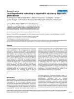
Figure 4
Cartilage damage was introduced by interleukin-1 (IL-1) injection, and 2 days later transforming growth factor-β (TGF-β) was injected intra-articularly combined with Ad-luciferase or Ad-Smad7Cartilage damage was introduced by interleukin-1 (IL-1) injection, and
2 days later transforming growth factor-β (TGF-β) was injected intra-
articularly combined with Ad-luciferase or Ad-Smad7. Two weeks after
adenoviral transfection, knee joints were isolated for assessment of
fibrosis. (a) Synovial thickness had increased significantly (p < 0.005)
after exposure to TGF-β. The TGF-β-induced increase was reduced by
simultaneous overexpression of Smad7 (p < 0.05). (b) The number of
procollagen type I-expressing cells had increased after TGF-β expo-
sures accordingly (p < 0.0005). This was also partially blocked by
Smad7 (p < 0.005). Error bars display standard error.
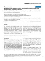
Figure 3
Proteoglycan (PG) synthesis in patellar cartilage after exposure to both transforming growth factor-β (TGF-β) and Smad7Proteoglycan (PG) synthesis in patellar cartilage after exposure to both
transforming growth factor-β (TGF-β) and Smad7. PG synthesis was
measured by
35
SO
4
2-
incorporation into patellar cartilage 4 days after
interleukin-1 (IL-1) injection. (CPM = counts per minute) Simultaneous
injection of Ad-TGF-β and Ad-Smad7 significantly increased PG syn-
thesis compared with IL-1 + control virus (p < 0.005) and therefore did
not block
35
SO
4
2-
incorporation. Error bars display standard error.
Available online />Page 5 of 8
(page number not for citation purposes)
Measurement of fibrosis
Sections were stained immunohistochemically for procollagen
type I as a measure of fibrosis. Subsequently, the amount of
cells that stain positive in the synovial tissue was determined.
A blinded observer selected the synovial tissue in three sec-
tions per knee joint. A computerized imaging system subse-
quently determined the amount of positive cells in the selected
area. The obtained values were averaged per knee joint.
In addition, synovial hyperplasia was assessed by measure-
ment of synovial thickness. This was determined in sections
stained with Safranin O and Fast Green. The thickness of the
synovial tissue was measured with a computerized imaging
system again in three sections per knee joint and averaged per
joint as previously described [27] (Qwin; Leica Imaging Sys-
tems Ltd., Cambridge, UK). In short, the width of the joint from
bone edge to joint capsule, minus the width of the joint space
itself, was determined.
Statistical analysis
Results were analyzed with a Student's t test and stated sig-
nificant if the p value was lower than 0.05.
Results
Overexpression of TGF-β stimulates cartilage repair
Knee joints that are injected with IL-1 initially show a reduced
incorporation of
35
SO
4
2-
into their patellar cartilage. After 2-3
days, this has reached a low point, and thereafter the incorpo-
ration rapidly increases above normal incorporation levels. By
day 4, the incorporation of
35
SO
4
2-
had significantly increased
to 165% of the normal value (p < 0.005). On day 2, an aden-
ovirus encoding active TGF-β was injected into the knee joint.
The TGF-β overexpression boosted the incorporation of
35
SO
4
2-
beyond the already elevated incorporation to 200% of
the normal value (Figure 1a). As a result, the overexpression of
TGF-β after IL-1 injection almost completely restored the sig-
nificantly reduced PG content of cartilage (96% of normal
control, p < 0.0005) (Figure 1b). These data show that aden-
oviral overexpression of TGF-β can stimulate cartilage repair.
TGF-β induces synovial fibrosis that can be blocked with
Smad7
Intra-articular injection of Ad-TGF-β resulted in a significant
increase of synovial thickness (2.5-fold the width of controls)
2 weeks after injection. The percentage of cells expressing
procollagen I increased accordingly (2.45-fold the amount of
positive cells in controls) (Figure 2). By simultaneous overex-
pression of Ad-TGF-β and Ad-Smad7, the TGF-β-induced syn-
ovial thickness had been significantly reduced (p < 0.05).
Sixty-five percent of the increased thickness and 65% of the
elevated number of procollagen I-positive cells had been
blocked by co-expression of Smad7 (Figure 2). Smad 7 itself
had no effect on synovial thickness or procollagen I expres-
sion. This illustrates that the TGF-β-induced fibrosis can be
significantly blocked by co-transfection with Ad-Smad7.
TGF-β overexpression repairs cartilage while fibrosis is
blocked with Smad7
To use the combination of TGF-β and Smad7 as a potential
therapeutic intervention, we had to make sure that Smad7
overexpression did not interfere with the beneficial effect of
TGF-β on cartilage repair. Therefore, TGF-β and Smad7 ade-
noviruses were injected 2 days after IL-1 injection. This com-
bination turned out to be beneficial for PG synthesis and could
still induce a significant increase in PG synthesis (80%
increase of PG synthesis compared with IL-1 alone, p <
0.005). Simultaneously overexpressing Smad7 and TGF-β did
not result in blocking of the repair-stimulating effects of TGF-
β on cartilage (Figure 3).
Adenoviral overexpression of TGF-β resulted in a significant
increase of 3.5 times as many cells expressing procollagen
type I as the IL-1 control alone (p < 0.005). By co-expression
of Smad7, 38% of the increase was blocked (p < 0.005). The
synovial tissue had expanded significantly to almost four times
in width after TGF-β overexpression compared with IL-1 +
control virus (p < 0.0005). Almost half of the increase was sig-
nificantly blocked by simultaneous exposure of the synovial
cells to TGF-β and Smad7 (p < 0.005) (Figure 4).
These data show that after IL-1-induced cartilage damage, the
TGF-β-induced fibrosis could still be blocked by Smad7 over-
expression without interfering with the effect TGF-β elicits on
cartilage.
Figure 5
Proteoglycan (PG) content of osteoarthritits (OA) cartilage in STR/ort miceProteoglycan (PG) content of osteoarthritits (OA) cartilage in STR/ort
mice. STR/ort mice (4 weeks old) were injected intra-articularly with
Ad-transforming growth factor-β (TGF-β) combined with Ad-Smad7 or
Ad-luciferase and injected again 2 weeks later. Four weeks after the
first injection, knee joints were isolated and the effect of TGF-β overex-
pression on PG content was assessed. TGF-β exposure resulted in a
significantly higher PG content of cartilage than luciferase-injected con-
trols (p < 0.05). Co-expression with Smad7 had no effect on the PG
content. Error bars display standard error.
Arthritis Research & Therapy Vol 8 No 3 Blaney Davidson et al.
Page 6 of 8
(page number not for citation purposes)
In experimental OA, simultaneous overexpression of
TGF-β and Smad7 increased PG content of cartilage and
prevented synovial fibrosis
The experiments conducted so far had been done in a rela-
tively simple model introducing cartilage damage by injection
of IL-1. However, in OA we are dealing with a more complex
situation. We introduced combined overexpression of TGF-β
and Smad7 in STR/ort mice. These mice develop OA sponta-
neously; therefore, they can be used as a model of primary OA.
In STR/ort mice, OA progresses relatively slowly. Therefore,
we examined the final result of TGF-β and Smad7 overexpres-
sion 2 weeks after the second of (in total) two viral injections.
STR/ort mice that had been injected with the adenovirus for
TGF-β (combined with a control virus) alone displayed a sig-
nificantly higher PG content in cartilage than did controls with-
out TGF-β overexpression (p < 0.05) (Figure 5). However,
these mice had massive synovial hyperplasia. The synovial
thickness had increased significantly to 4.4 times the width of
non-injected controls, and almost every cell expressed procol-
lagen type I (p < 0.005) (Figure 6).
Combining overexpression of TGF-β and Smad7 managed to
maintain a significantly higher PG content in cartilage than
controls (p < 0.005) (Figure 5). More than half of the increased
synovial thickening that had been caused by TGF-β was inhib-
ited significantly by overexpression of Smad7 (p < 0.005). The
amount of procollagen type I-positive cells had been reduced
accordingly (Figure 6).
These data clearly indicate that PG synthesis can be stimu-
lated while inhibiting an increase of synovial fibrosis in an
experimental model of OA.
Discussion
The cartilage damage in OA is thought to be a consequence
of a misbalance between anabolic and catabolic factors, favor-
Figure 6
Blocking of transforming growth factor-β (TGF-β)-induced synovial fibrosis in osteoarthritits (OA)Blocking of transforming growth factor-β (TGF-β)-induced synovial fibrosis in osteoarthritits (OA). STR/ort mice (4 weeks old) were injected intra-
articularly with Ad-TGF-β combined with Ad-Smad7 or Ad-luciferase (Ad-luc) and injected again 2 weeks later. Two weeks after the last injection,
knee joints were isolated for histology and synovial fibrosis was assessed. Synovial thickness was determined by measuring the width of the syn-
ovium opposite the growth plates in the femur. (a) Synovial thickness had increased significantly after adenoviral TGF-β overexpression (p < 0.005).
More than 50% of this increase in width was prevented by simultaneous expression of Smad7 (p < 0.005). The number of procollagen type I-
expressing cells (b) had increased as well after exposure to TGF-β (p < 0.0005). This was also inhibited by more than 50% by co-expression of
Smad7. Histological representations of the measurements give an indication of actual thickness (c) and procollagen positive cells (d) Error bars dis-
play standard error.
Available online />Page 7 of 8
(page number not for citation purposes)
ing the catabolic side. In this study, we used TGF-β as the ana-
bolic factor for cartilage repair. TGF-β has been reported to
enhance periosteal chondrogenesis in explants in a dose-
dependent manner [28]. Morales and colleagues [4,5] demon-
strated that TGF-β increased PG synthesis and suppressed
its degradation in articular cartilage organ cultures. In addition,
van Beuningen and colleagues [8] showed that in vivo TGF-β
injections result in prolonged elevation of PG synthesis and
PG content of cartilage in mice. These studies indicate that
TGF-β has good potential for repairing cartilage. We showed
that adenoviral overexpression of TGF-β was indeed able to
boost cartilage repair in vivo. In vitro, it had already been
shown that chondrocytes exposed simultaneously to IL-1 and
TGF-β could reverse the IL-1-induced suppression of PG
incorporation in their extracellular matrix [15]. Supportive of
our findings, van Beuningen and colleagues [13] demon-
strated that, in vivo, TGF-β also counteracted deleterious
effects of IL-1 on cartilage PG synthesis and PG content. In
the current study, we first damaged cartilage by IL-1 injection
and subsequently overexpressed TGF-β. In this way, we could
assess whether TGF-β was able to restore, instead of prevent,
cartilage damage. We introduced TGF-β via adenoviral over-
expression, thereby gaining prolonged high expression of
TGF-β, instead of via a bolus injection that results in short
TGF-β exposure. This way, we were able to demonstrate
increased PG synthesis and higher PG content in cartilage not
only in a clean setting introducing cartilage damage with IL-1
but also in a spontaneous OA model.
The drawback of using TGF-β is that it can have adverse
effects in joints. TGF-β is a known inducer of fibrosis in various
tissues, and synovial tissue is no exception [8,21]. We took
advantage of the fact that adenoviruses transfect only the syn-
ovial lining. In addition, we profited from the fact that Smad7 is
an intercellular inhibitor of TGF-β. Smad7 stays inside the cell
that is transfected with the adenovirus encoding Smad7.
Because the synovial lining is where TGF-β induces synovial
fibrosis, by co-transfection with Smad7, the lining appeared to
be less sensitive to TGF-β-induced fibrosis. The reduction of
TGF-β-induced fibrosis was not optimal and resulted in only a
partial block of the fibrosis. This is likely due to the fact that not
all cells in the synovial lining will be targeted. By optimizing
this, we might be able to target every single one of the synovial
lining cells and thereby fully block the TGF-β-induced fibrosis.
We have previously demonstrated that blocking TGF-β with
Ad-Smad7 in OA resulted in reduction of the synovial fibrosis
that was induced by the OA process itself [27]. Now we com-
bined the Smad7 adenovirus with Ad-TGF-β to block the TGF-
β-induced fibrosis. We showed that the Ad-TGF-β transfec-
tion was still functional in combination with Smad7. Moreover,
we demonstrated for the first time that adenoviral overexpres-
sion of TGF-β could stimulate repair of damaged cartilage and
that co-expression with Smad7 could prevent a great deal of
the TGF-β-induced synovial fibrosis. Combining Smad7 and
TGF-β resulted in a higher PG synthesis after IL-1 insult than
did TGF-β alone. This is likely due to the reduced synovial
fibrosis when combined with Smad7.
Unfortunately, synovial fibrosis is not the only side effect of
TGF-β overexpression in knee joints. TGF-β can induce oste-
ophyte formation [8,19,20,29-31]. In the case of OA, TGF-β
can aggravate the osteophyte formation that already occurs.
We show that it is possible to target synovial cells to prevent
fibrosis. In a similar fashion, we could potentially target the
mesenchymal stem cells that eventually form the osteophytes
after TGF-β exposure. This could be an option when key play-
ers of osteophyte formation are identified and can be blocked
selectively.
Conclusion
We demonstrated that adenoviral overexpression of TGF-β
increases PG synthesis and PG content in cartilage, even in
experimental OA. In addition, co-transfecting the synovial lin-
ing with Ad-Smad7 can block the fibrosis that is induced by
TGF-β overexpression.
Competing interests
The authors declare that they have no competing interests.
Authors' contributions
ENBD participated in the animal experiments and immunohis-
tochemistry, carried out the histological measurements, ana-
lyzed the data, and drafted the manuscript. ELV participated in
the animal experiments, carried out histological processing of
the knee joints, participated in immunohistochemistry, and per-
formed 35S-sulphate measurements. PMK conceived of the
study, participated in the design and coordination, and helped
draft the manuscript. WBB participated in study design and
revision of the final manuscript. All authors read and approved
the final manuscript.
Acknowledgements
We thank Dr. C.D. Richards for the gift of the TGF-β adenovirus. ENBD
and PMK were financially supported by the Dutch Arthritis Association
"Reumafonds."
References
1. Edwards DR, Murphy G, Reynolds JJ, Whitham SE, Docherty AJ,
Angel P, Heath JK: Transforming growth factor beta modulates
the expression of collagenase and metalloproteinase inhibi-
tor. EMBO J 1987, 6:1899-1904.
2. Pedrozo HA, Schwartz Z, Gomez R, Ornoy A, Xin SW, Dallas SL,
Bonewald LF, Dean DD, Boyan BD: Growth plate chondrocytes
store latent transforming growth factor (TGF)-beta 1 in their
matrix through latent TGF-beta 1 binding protein-1. J Cell
Physiol 1998, 177:343-354.
3. Redini F, Min W, Demoor FM, Boittin M, Pujol JP: Differential
expression of membrane-anchored proteoglycans in rabbit
articular chondrocytes cultured in monolayers and in alginate
beads. Effect of transforming growth factor-beta 1. Biochim
Biophys Acta 1997, 1355:20-32.
4. Morales TI, Joyce ME, Sobel ME, Danielpour D, Roberts AB:
Transforming growth factor-beta in calf articular cartilage
organ cultures: synthesis and distribution. Arch Biochem Bio-
phys 1991, 288:397-405.
Arthritis Research & Therapy Vol 8 No 3 Blaney Davidson et al.
Page 8 of 8
(page number not for citation purposes)
5. Morales TI: Transforming growth factor-beta 1 stimulates syn-
thesis of proteoglycan aggregates in calf articular cartilage
organ cultures. Arch Biochem Biophys 1991, 286:99-106.
6. Burton-Wurster N, Lust G: Fibronectin and proteoglycan syn-
thesis in long term cultures of cartilage explants in Ham's F12
supplemented with insulin and calcium: effects of the addition
of TGF-beta. Arch Biochem Biophys 1990, 283:27-33.
7. Verdier MP, Seite S, Guntzer K, Pujol JP, Boumediene K: Immu-
nohistochemical analysis of transforming growth factor beta
isoforms and their receptors in human cartilage from normal
and osteoarthritic femoral heads. Rheumatol Int 2003,
25:118-124.
8. van Beuningen HM, van der Kraan PM, Arntz OJ, van den Berg
WB: Transforming growth factor-beta 1 stimulates articular
chondrocyte proteoglycan synthesis and induces osteophyte
formation in the murine knee joint. Lab Invest 1994,
71:279-290.
9. Glansbeek HL, van Beuningen HM, Vitters EL, van der Kraan PM,
van den Berg WB: Stimulation of Articular Cartilage Repair in
Established Arthritis by Local Administration of Transforming
Growth Factor-beta into Murine Knee Joints. Lab Invest 1998,
78:133-142.
10. Tetlow LC, Adlam DJ, Woolley DE: Matrix metalloproteinase and
proinflammatory cytokine production by chondrocytes of
human osteoarthritic cartilage: associations with degenera-
tive changes. Arthritis Rheum 2001, 44:585-594.
11. van Beuningen HM, Arntz OJ, van den Berg WB: In vivo effects of
interleukin-1 on articular cartilage. Prolongation of proteogly-
can metabolic disturbances in old mice. Arthritis Rheum 1991,
34:606-615.
12. Hui W, Rowan AD, Cawston T: Transforming growth factor
beta1 blocks the release of collagen fragments from boving
nasal cartilage stimulated by oncostatin M in combination with
IL-1alpha. Cytokine 2000, 12:765-769.
13. van Beuningen HM, van der Kraan PM, Arntz OJ, van den Berg
WB: In vivo protection against interleukin-1-induced articular
cartilage damage by transforming growth factor-beta 1: age-
related differences. Ann Rheum Dis 1994, 53:593-600.
14. van Beuningen HM, van der Kraan PM, Arntz OJ, van den Berg
WB: Protection from interleukin 1 induced destruction of artic-
ular cartilage by transforming growth factor beta: studies in
anatomically intact cartilage in vitro and in vivo. Ann Rheum Dis
1993, 52:185-191.
15. Chandrasekhar S, Harvey AK: Transforming growth factor-beta
is a potent inhibitor of IL-1 induced protease activity and carti-
lage proteoglycan degradation. Biochem Biophys Res Com-
mun 1988, 157:1352-1359.
16. Iqbal J, Dudhia J, Bird JL, Bayliss MT: Age-related effects of TGF-
beta on proteoglycan synthesis in equine articular cartilage.
Biochem Biophys Res Commun 2000, 274:467-471.
17. Shuler FD, Georgescu HI, Niyibizi C, Studer RK, Mi Z, Johnstone
B, Robbins RD, Evans CH: Increased matrix synthesis following
adenoviral transfer of a transforming growth factor beta1 gene
into articular chondrocytes. J Orthop Res 2000, 18:585-592.
18. Allen JB, Manthey CL, Hand AR, Ohura K, Ellingsworth L, Wahl
SM: Rapid onset synovial inflammation and hyperplasia
induced by transforming growth factor beta. J Exp Med 1990,
171:231-247.
19. Bakker AC, van de Loo FAJ, van Beuningen HM, Sime P, van Lent
PLEM, van der Kraan PM, Richards CD, van den Berg WB: Over-
expression of active TGF-beta-1 in the murine knee joint: evi-
dence for synovial-layer-dependent chondro-osteophyte
formation. Osteoarthritis Cartilage 2001, 9:128-136.
20. van Beuningen HM, Glansbeek HL, van der Kraan PM, van den
Berg WB: Differential effects of local application of BMP-2 or
TGF-beta 1 on both articular cartilage composition and osteo-
phyte formation. Osteoarthritis Cartilage 1998, 6:306-317.
21. van Beuningen HM, Glansbeek HL, van der Kraan PM, van den
Berg WB: Osteoarthritis-like changes in the murine knee joint
resulting from intra-articular transforming growth factor-beta
injections. Osteoarthritis Cartilage 2000, 8:25-33.
22. Zimmerman CM, Padgett RW: Transforming growth factor beta
signaling mediators and modulators. Gene 2000, 249:17-30.
23. Hayashi H, Abdollah S, Qiu YB, Cai JX, Xu YY, Grinnell BW, Rich-
ardson MA, Topper JN, Grimbrone MA, Wrana JL, et al.: The MAD
related protein SMAD7 associates with the TGF-b receptor
and functions as an antagonist of TGF-b signaling. Cell 1997,
89:1165-1173.
24. Nakao A, Afrakhte M, Moren A, Nakayama T, Christian JL, Heuchel
R, Itoh S, Kawabata M, Heldin NE, Heldin CH, ten Dijke P: Identi-
fication of Smad7, a TGF-beta-inducible antagonist of TGF-
beta signalling. Nature 1997, 389:631-635.
25. Maroudas A: Transport of solutes through cartilage: permeabil-
ity to large molecules. J Anat 1976, 122:335-347.
26. Scharstuhl A, van Beuningen HM, Vitters EL, van der Kraan PM,
van den Berg WB: Loss of TGF-b counteraction on IL-1-medi-
ated effects in cartilage of old mice. Ann Rheum Dis 2002,
61:1095-1098.
27. Scharstuhl A, Vitters EL, van der Kraan PM, van den Berg WB:
Reduction of osteophyte formation and synovial thickening by
adenoviral overexpression of transforming growth factor
beta/bone morphogenetic protein inhibitors during experi-
mental osteoarthritis. Arthritis Rheum 2003, 48:3442-3451.
28. Miura Y, Fitzsimmons JS, Commisso CN, Gallay SH, O'Driscoll
SW: Enhancement of periosteal chondrogenesis in vitro.
Dose-response for transforming growth factor-beta 1 (TGF-
beta 1). Clin Orthop 1994, 301:271-280.
29. van den Berg WB, van Osch GJ, van der Kraan PM, van Beuningen
HM: Cartilage destruction and osteophytes in instability-
induced murine osteoarthritis: role of TGF beta in osteophyte
formation? Agents Actions 1993, 40:215-219.
30. van den Berg WB: The role of cytokines and growth factors in
cartilage destruction in osteoarthritis and rheumatoid arthritis.
Z Rheumatol 1999, 58:136-141.
31. van den Berg WB: Growth factors in experimental osteoarthri-
tis: transforming growth factor beta pathogenic? J Rheumatol
Suppl 1995, 43:143-145.