Báo cáo khoa học: "Does the surgeon still have a role to play in the diagnosis and management of lymphomas?" docx
Bạn đang xem bản rút gọn của tài liệu. Xem và tải ngay bản đầy đủ của tài liệu tại đây (221.18 KB, 4 trang )
BioMed Central
Page 1 of 4
(page number not for citation purposes)
World Journal of Surgical Oncology
Open Access
Research
Does the surgeon still have a role to play in the diagnosis and
management of lymphomas?
Gareth Morris-Stiff*
1
, Peipei Cheang
1
, Steve Key
1
, Anju Verghese
2
and
Timothy J Havard
1
Address:
1
Department of Surgery, Royal Glamorgan Hospital, Ynysmaerdy, Llantrisant, UK and
2
Department of Pathology, Royal Glamorgan
Hospital, Ynysmaerdy, Llantrisant, UK
Email: Gareth Morris-Stiff* - ; Peipei Cheang - ; Steve Key - ;
Anju Verghese - ; Timothy J Havard -
* Corresponding author
Abstract
Background: Over the course of the past 40 years, there have been a significant number of changes in
the way in which lymphomatous disease is diagnosed and managed. With the advent of computed
tomography, there is little role for staging laparotomy and the surgeon's role may now more diagnostic
than therapeutic.
Aims: To review all cases of lymphoma diagnosed at a single institution in order determine the current
role of the surgeon in the diagnosis and management of lymphoma.
Patients and methods: Computerized pathology records were reviewed for a five-year period 1996 to
2000 to determine all cases of lymph node biopsy (incisional or excisional) in which tissue was obtained
as part of a planned procedure. Cases of incidental lymphadenopathy were thus excluded.
Results: A total of 297 biopsies were performed of which 62 (21%) yielded lymphomas. There were 22
females and 40 males with a median age of 58 years (range: 19–84 years). The lymphomas were classified
as 80% non-Hodgkin's lymphoma, 18% Hodgkin's lymphoma and 2% post-transplant lymphoproliferative
disorder. Diagnosis was established by general surgeons (n = 48), ENT surgeons (n = 9), radiologists (n =
4) and ophthalmic surgeons (n = 1). The distribution of excised lymph nodes was: cervical (n = 23), inguinal
(n = 15), axillary (n = 11), intra-abdominal (n = 6), submandibular (n = 2), supraclavicular (n = 2), periorbital
(n = 1), parotid (n = 1) and mediastinal (n = 1). Fine needle aspiration cytology had been performed prior
to biopsy in only 32 (52%) cases and had suggested: lymphoma (n = 10), reactive changes (n = 13), normal
(n = 5), inadequate (n = 4). The majority (78%) of cervical lymph nodes were subjected to FNAC prior to
biopsy whilst this was performed in only 36% of non-cervical lymphadenopathy.
Conclusion: The study has shown that lymphoma is a relatively common cause of surgical
lymphadenopathy. Given the limitations of FNAC, all suspicious lymph nodes should be biopsied following
FNAC even if the FNAC is reported normal or demonstrating reactive changes only. With the more
widespread application of molecular techniques, and the development of improved minimally-invasive
procedures, percutaneous and endoscopic techniques may come to dominate, however, at present; the
surgeon still has an important role to play in the diagnosis if not treatment of lymphomas.
Published: 4 February 2008
World Journal of Surgical Oncology 2008, 6:13 doi:10.1186/1477-7819-6-13
Received: 14 May 2007
Accepted: 4 February 2008
This article is available from: />© 2008 Morris-Stiff et al; licensee BioMed Central Ltd.
This is an Open Access article distributed under the terms of the Creative Commons Attribution License ( />),
which permits unrestricted use, distribution, and reproduction in any medium, provided the original work is properly cited.
World Journal of Surgical Oncology 2008, 6:13 />Page 2 of 4
(page number not for citation purposes)
Background
Lymphomas are a heterogeneous family of malignant
neoplasia of the reticuloendothelial system, which may be
divided into two main subtypes; Hodgkin's lymphoma
(HL), eponymous to the nineteenth century Guy's pathol-
ogist Thomas Hodgkin, and non-Hodgkin's lymphoma
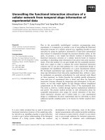
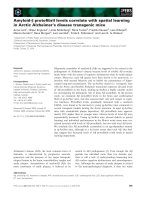
(NHL). The incidence of NHL increased over the 1980s
decade from 120 to 320 registrations per year whereas the
incidence of HL has remained static at around 80 cases per
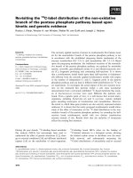
year in Wales as illustrated in Figure 1[1].
The surgeon's role in the diagnosis and management of
lymphomas, in particular HL, was stimulated by a report
from Stanford University in the late 1960s which showed
that the performance of a staging laparotomy altered the
stage of disease in 42% of cases, up regulating in 28% and
down regulating in 14% of cases [2]. The procedure con-
sisted of liver and lymph node biopsies together with
splenectomy. In addition to allowing accurate staging, the
splenectomy was believed to debulk the disease mass and
offer a more precise target for radiotherapy.
The advent of computed tomography brought about the
demise of staging laparotomies and splenectomy is now
limited to symptomatic splenomegaly and occasionally
hyposplenism. Computed tomography is rapid, non-
invasive and allows assessment of both thoracic and
abdominal compartments. However, a tissue diagnosis is
still required to allow accurate cellular classification of the
lymphomas.
Fine needle aspiration cytology (FNAC) was developed at
the turn of the century and has become a popular diagnos-
tic tool as it is rapid, painless, safe, inexpensive, does not
require any anaesthetic or hospital admission and leaves
no scar [3]. In addition to confirming the diagnosis of
lymphomas, one of the important roles of FNAC is the
exclusion of metastatic squamous carcinoma as this
requires an alternative therapeutic approach. There is a
question as to the accuracy of FNAC in the diagnosis of
lymphomas as the tumours often contain malignant and
reactive elements and the FNAC may only have sampled
the reactive regions leading to false negative results.
Another disadvantage of FNAC of lymphomas is that it
does not provide the cellular architecture required for the
accurate subtyping of the lymphoma.
As a result of the deficiencies of FNAC, lymph node exci-
sion is required and is the recommended second line diag-
nostic procedure. In addition to providing a greater
volume of tissue for histological evaluation subtype clas-
sification, it also provides a baseline against which the
effects of chemotherapy may be judged.
The aim of this study was to examine whether the 21
st
cen-
tury surgeon still has a role to play in the diagnosis and
management of lymphoma.
Patients and methods
The study was a retrospective study of all patients under-
going lymph node biopsy at the Royal Glamorgan Hospi-
tal (formerly known as East Glamorgan Hospital) for the
five-year period 1996 to 2000. Patients were identified
from the computerised records of the pathology depart-
ment. All cases of lymph node biopsy were collected (exci-
sional and incisional) however patients in whom
lymphadenopathy was an incidental finding were
excluded and thus the cohort consisted of patients in
whom the aim of surgery was lymph node biopsy.
For each patient the following information was collected:
patient demographics, location of lymphadenopathy,
findings of lymph node biopsy, performance or not of
FNAC and findings of FNAC.
Results
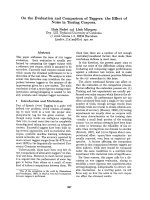
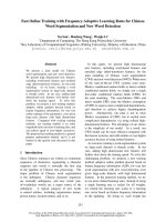
The study population comprised 297 patients undergoing
lymph node biopsy (Figure 2). Lymphoma was confirmed
in 62 patients, representing 21% of all biopsies. There
were 40 males and 22 females of median age 58 years
(range 19–84 years). The lymphomas were classified into
80% NHL, 18% HL and 2% post-transplant lymphopro-
liferative disorder.
Diagnosis was established mainly by general surgeons (n
= 48), ENT surgeons (n = 9), radiologists (n = 4) and oph-
thalmic surgeons (n = 1). The anatomical distribution of
the excised lymph nodes is detailed in Table 1. The com-
monest locations for lymphadenopathy were cervical (n =
23), inguinal (n = 15), and axillary (n = 11).
Diagnosis of lymphoma in Wales over the period 1980–1990Figure 1
Diagnosis of lymphoma in Wales over the period 1980–1990.
HD = Hodgkin's disease, NHL = Non-Hodgkin's lymphoma.
0
50
100
150
200
250
300
350
Number of New Registrations in Wales
1980 1981 1982 1983 1984 1985 1986 1987 1988 1989 1990
HD NHL
World Journal of Surgical Oncology 2008, 6:13 />Page 3 of 4
(page number not for citation purposes)
Fine needle aspiration cytology had been performed prior
to biopsy in only 32 (52%) cases out of the total of 62
with a final diagnosis of lymphoma. The findings of
FNAC were: lymphoma (n = 10); reactive changes (n =
13); normal (n = 5); inadequate (n = 4). The remaining 30
patients proceeded to biopsy without FNAC. FNAC was
performed in 18 of 23 patients with cervical lymphaden-
opathy but in only 14 of 39 of individuals with non-cervi-
cal lymphadenopathy. The time interval between
performance of FNAC and histological confirmation of
the biopsy specimens was less than one month in 81% of
cases and less than six weeks in all cases. In cases of delay
more than one month, delays were due to patient non-
compliance.
Discussion
The study has confirmed that lymphoma is a common
cause of surgical lymphadenopathy, representing the his-
tological diagnosis in 21% of all lymph node biopsy spec-
imens. The ratio of HL to NHL in this study was identical
to the current trend in lymphoma incidence in Wales with
a ratio of 1:4 [1].
The locations of lymphomatous nodes corresponded to
the distribution of lymphadenopathy as a whole, with the
majority of palpable nodes being in the cervical, inguinal
and axillary chains and as such were amenable to simple
excision. The majority of lymph node biopsies were per-
formed mainly by general surgeons whilst ENT and oph-
thalmic surgeons performed a total of ten biopsies. The
remaining four lymphomas were biopsied using ultra-
sound-guidance by radiologists.
Fine needle aspiration cytology was performed in little
over half of the cases although this was performed in 81%
of head and neck lymphadenopathy in accordance with
practice guidelines [4]. The importance of performing an
FNAC in patients with cervical lymphadenopathy prior to
embarking on an excisional biopsy relates to the fact that,
for those patients found to have squamous carcinoma
metastases from a head and neck primary, open biopsy
leads to a significantly higher local treatment failure rate
which may in turn be associated with an adverse effect on
survival [5,6].
The accuracy of FNAC in the diagnosis of lymphoma has
previously been questioned [7]. The lymphomatous proc-
ess may involve the node focally and may not involve all
the nodes that appear to be enlarged. Other factors that
influence the diagnostic specificity and sensitivity of
FNAC in the diagnosis of lymphoma include; necrosis in
involved nodes; the presence of dual pathology and scle-
rosis/fibrosis in involved nodes leading to insufficient
diagnostic material.
Other disadvantages of FNAC are lack of material for an
accurate typing of lymphoma due to lack of tissue for
immunohistochemistry [5]. Low grade lymphomas are
difficult to diagnose even on excisional biopsies and spe-
cial staining techniques are required to differentiate
between a florid follicular hyperplasia and a follicular
lymphoma.
In this study, lymphomas were correctly identified by
FNAC in only 31% of cases. The commonest diagnosis, in
40% of FNACs was reactive changes whilst the remaining
cases were equally divided between normal and inade-
quate. All patients with FNACs not diagnostic of lym-
phoma went on to lymph node biopsy because of
suspicious clinical histories or persisting lymphadenopa-
thy. The performance of FNAC was not regarded as being
compulsory at the start of this observational study but
became standard practice, and more recently the perform-
ance of FNAC under ultrasound-guidance was introduced
in order to maximize the likelihood of correctly targeting
the suspicious lymph node.
Table 1: Anatomical location of lymphomatous lymph nodes (n =
62)
Anatomical location Number of cases
Cervical 23
Inguinal 15
Axillary 11
Intra-abdominal 6
Supraclavicular 2
Submandibular 2
Parotid 1
Peri-orbital 1
Mediastinal 1
Findings of lymph node biopsies (n = 279)Figure 2
Findings of lymph node biopsies (n = 279).
22%
21%
19%
10%
5%
5%
4%
2%
2%
10%
Nor mal Lymphoma Hyper plasia
SCC Melanoma Ad enocar cinoma
Gr anulomatous Fatty Unsatisfactor y
Other
World Journal of Surgical Oncology 2008, 6:13 />Page 4 of 4
(page number not for citation purposes)
The uses of flow cytometry, immunohistochemistry, and
molecular studies such as polymerase chain reaction and
fluorescent in-situ hybridization have significantly
increased the yield of FNAC [8-10]. Furthermore, the
more recent introduced technique of core biopsy has been
shown to be of benefit over FNAC in the diagnosis of lym-
phoma especially when performed under ultrasound-
guidance combined with advanced molecular techniques
[11-13].
One area not explored by this study but which may be of
increasing importance in the future is the role of endos-
copy and laparoscopy in obtaining biopsy material. The
advent of endoscopic ultrasound-guided FNAC allows tar-
geting of mediastinal and intra-abdominal lymphadenop-
athy, which can be performed without the morbidity
associated with trans-cavity radiological sampling or open
surgical biopsy [14-16]. For lesions outside the reach of
the endoscope, laparoscopy may play an increasing role
[17,18] as it allows access to perihepatic and perisplenic in
addition to retroperioneal lymphadenopathy. Thus upper
gastrointestinal surgeons with training in these tech-
niques may have an increasing role in the diagnosis of
lymphomas. In cases of intrathoracic lympahadopathy,
newer minimally-invasive techniques such as mediasinos-
copy; thoracoscopy are also now well established and pro-
vide adequate tissue for sub-typing [19]. Although not
performed by 'general surgeons', they do represent a sur-
gical biopsy.
Conclusion
All patients presenting with lymphadenopathy should
undergo FNAC, this being of critical importance for cervi-
cal lesions as lymphadenopathy presenting in this region
may represent metastases from primary squamous cell
carcinomas of the head and neck. Given the limitations of
FNAC, all suspicious lymph nodes should be biopsied if
the FNAC is reported normal or demonstrates reactive
changes only, this being performed mainly by general sur-
geons. Thus at present the 'surgeon' still has a role to play
in the diagnosis of lymphoma.
Advancements in diagnostic methods has meant that
many superficial lesions traditionally requiring open exci-
sion biopsy may now be able to be diagnosed accurately
by image-guided core biopsy, thus reducing the role of the
surgeon. However, on the contrary, deep-seated lesions
previously targeted by radiologists may now be more
accurately approached by minimally-invasive surgical
techniques and so a new role is likely to evolve for the sur-
geon in the diagnosis of lymphoma.
Competing interests
The author(s) declare that they have no competing inter-
ests.
Authors' contributions
GMS developed the concept, and prepared the draft man-
uscript. PC and SK provided the pathological data and
helped in preparing the manuscript, AV and TGH
reviewed and edited the manuscript and helped in prepar-
ing the final version. All authors read and approved final
manuscript.
References
1. Welsh Cancer Intelligence & Surveillance Unit. In Cancer reg-
istration in Wales 1974–1990 Cardiff, WCISU; 1999.
2. Glatstein E, Guernsey JM, Rosenberg SA, Kaplan HS: The value of
laparotomy and splenectomy in the staging of Hodgkin's dis-
ease. Cancer 1969, 24:709-718.
3. Buley ID: Fine needle aspiration of lymph nodes. J Clin Pathol
1998, 51:881-885.
4. Gleeson M, Herbert A, Richards A: Management of lateral neck
masses in adults. Br Med J 2000, 320(7248):1521-1524.
5. Lefebvre JL, Coche-Dequeant B, Van JT, Buisset E, Adenis A: Cervi-
cal lymph nodes from an unknown primary tumor in 190
patients. Am J Surg 1990, 160:443-446.
6. Janot F, Klijanienko J, Russo A, Mamet JP, de Braud F, El-Naggar AK,
Pignon JP, Luboinski B, Cvitkovic E: Prognostic value of clinico-
pathologic parameters in head and neck squamous cell car-
cinoma: a prospective analysis. Br J Cancer 1996, 73:531-538.
7. Lioe TF, Elliott H, Allen DC, Spence RA: The role of fine needle
aspiration cytology (FNAC) in the investigation of superficial
lymphadenopathy; uses and limitations of the technique.
Cytopathol 1999, 10(5):291-297.
8. Gong JZ, Williams DC Jr, Liu K, Jones C: Fine-needle aspiration in
non-Hodgkin lymphoma: evaluation of cell size by cyomor-
phology and flow cytometry. Am J Clin Pathol 2002, 117:880-888.
9. Austin RM, Birdsong GG, Sidawy MK, Kaminsky DB: Fine needle
aspiration is a feasible and sccurate technique in the diagno-
sis of lymphoma. J Clin Oncol 2005, 23:9029-9030.
10. Fraga M, Forteza J: Diagnosis of Hodgkin's disease: an update
on histopathological and immunophenotypical features. His-
tol Histopathol 2007, 22:923-935.
11. Ravinsky E, Morales C: Diagnosis of lymphoma by image-guided
needle biopsies: fine needle aspiration biopsy, core biopsy or
both? Acta Cytol 2005, 49:51-57.
12. Kim BM, Kim EK, Kim MJ, Yang WI, Park CS, Park S: Sonographi-
cally guided core biopsy of cervical lymphadenopathy in
patients without known malignancy. J Ultrasound Med 2007,
26:585-591.
13. Vandervelde C, Kamani T, Varghese A, Ramesar K, Grace R, Howlett
DC: A study to evaluate the efficacy of image-guided core
biopsy in the diagnosis and management of lymphoma –
results in 103 biopsies. Eur J Radiol 2007 in press. doi:10.1016/
j.ejrad.2007.05.016.
14. Emery SC, Savides TJ, Behling CA: Utility of immediate evalua-
tion of endoscopic ultrasound-guided transesophageal fine
needle aspiration of mediastinal lymph nodes. Acta Cytol 2004,
48:630-634.
15. Eloubeidi MA, Vilmann P, Wiersema MJ: Endoscopic ultrasound-
guided fine-needle aspiration of celiac lymph nodes. Endos-
copy 2004, 36:901-908.
16. Pugh JL, Jhala NC, Eloubeidi MA, Chhieng DC, Eltoum IA, Crowe DR,
Varadarajulu S, Jhala DN: Diagnosis of deep-seated lymphoma
and leukemia by endoscopic ultrasound-guided fine-needle
aspiration biopsy. Am J Clin Pathol 2006, 125:703-709.
17. Silecchia G, Raparelli L, Perrotta N, Fantini A, Fabiano P, Monarca B,
Basso N: Accuracy of laparoscopy in the diagnosis and staging
of lymphoproliferative diseases. World J Surg 2003, 27:653-658.
18. Casaccia M, Torelli P, Cavaliere D, Panaro F, Nardi I, Rossi E, Spriano
M, Bacigalupo A, Gentile R, Valente U: Laparoscopic lymph node
biopsy in intra-abdominal lymphoma: high diagnostic accu-
racy achieved with a minimally invasive procedure. Surg
Laparosc Endosc Percutan Tech 2007, 17:175-178.
19. Massone PP, Lequaglie C, Magnani B, Ferro F, Cataldo I: The real
impact and usefulness of video-assisted thoracoscopic sur-
gery in the diagnosis and therapy of clinical lymphadenopa-
thies of the mediastinum. Ann Surg Oncol 2003, 10:1197-1202.