Báo cáo khoa học: "Primary multifocal osseous Hodgkin''''s lymphoma" ppt
Bạn đang xem bản rút gọn của tài liệu. Xem và tải ngay bản đầy đủ của tài liệu tại đây (1.96 MB, 6 trang )
BioMed Central
Page 1 of 6
(page number not for citation purposes)
World Journal of Surgical Oncology
Open Access
Review
Primary multifocal osseous Hodgkin's lymphoma
Clare R Langley*
1
, Simon JW Garrett
2
, Jill Urand
3
, Janice Kohler
3
and
Nick MP Clarke
3
Address:
1
Orthopaedic Department, Basingstoke and North Hampshire Foundation Trust, Aldermaston Road, Basingstoke, Hampshire, RG24
9NA, UK,
2
Royal Bournemouth Hospital, Castle Lane East, Bournemouth, Dorset, BH7 7DW, UK and
3
Southampton University Hospitals NHS
Trust, Tremona Road, Southampton, SO16 6YD, UK
Email: Clare R Langley* - ; Simon JW Garrett - ; Jill Urand - ;
Janice Kohler - ; Nick MP Clarke -
* Corresponding author
Abstract
Background: Hodgkin's disease (HD) most commonly presents with progressive painless
enlargement of peripheral lymph nodes, especially around the cervical region. A few children have
systemic symptoms and weight loss. At the time of diagnosis, osseous involvement is uncommon
Case presentation: A case is described of Primary Multifocal Osseous Hodgkin's Lymphoma in
a seven-year-old boy. He presented with a painful swelling in the sternum, and further investigations
revealed deposits in his L1 vertebra, the left sacro-iliac joint and the right acetabulum.
Conclusion: The clinical, radiological and histological features of this disease can mimic other
medical conditions, including Tuberculosis, making the diagnosis difficult and often leading to delays
in treatment. This is a very rare condition and we believe this to be the youngest reported case in
the literature.
Background
Hodgkin's disease (HD) most commonly presents with
progressive painless enlargement of peripheral lymph
nodes, especially around the cervical region. A few chil-
dren have systemic symptoms and weight loss. At the time
of diagnosis, osseous involvement is uncommon and
even in the late stages only 9–35% of cases have any bony
involvement [1]. It is therefore extremely rare for patients
to present with primary Hodgkin's disease of the bone. If
there is no associated extra-osseous involvement, the con-
dition is referred to as primary osseous Hodgkin's lym-
phoma (POHL). It is termed primary multifocal osseous
Hodgkin's lymphoma, if more than one osseous site is
involved. The clinical, radiological and histological fea-
tures of POHL can mimic other medical conditions,
thereby making the diagnosis difficult, often leading to
delays in treatment
We present a case of a seven-year-old boy diagnosed with
primary multifocal osseous Hodgkin's lymphoma. We
believe this to be the youngest such reported case and only
the third ever reported paediatric case of primary multifo-
cal osseous Hodgkin's lymphoma in the English litera-
ture.
Case presentation
A seven year old male presented with a painless, firm 3 cm
mass overlying his sternum. He was clinically well, apy-
rexial with no history of weight loss. Initial investigations
revealed an elevated CRP (27.5), ESR (90), white cell
Published: 17 March 2008
World Journal of Surgical Oncology 2008, 6:34 doi:10.1186/1477-7819-6-34
Received: 17 July 2007
Accepted: 17 March 2008
This article is available from: />© 2008 Langley et al; licensee BioMed Central Ltd.
This is an Open Access article distributed under the terms of the Creative Commons Attribution License ( />),
which permits unrestricted use, distribution, and reproduction in any medium, provided the original work is properly cited.
World Journal of Surgical Oncology 2008, 6:34 />Page 2 of 6
(page number not for citation purposes)
count (22.3) with a neutrophilia (17.0) and a hypochro-
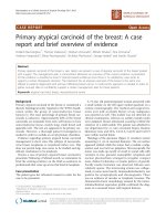
mic microcytic anaemia (Hb: 9.3). Technecium
99
bone
scan (Figure 1) revealed increased uptake in the sternum,
L1 vertebra, the left sacro-iliac joint and the right acetabu-
lum.
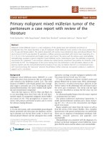
A CT scan of the chest (Figure 2) and sternum confirmed
the presence of a sternal mass with no underlying soft tis-
sue involvement. A fine needle biopsy of the sternal mass
showed an inflammatory infiltrate. Bone marrow aspi-
rates and trephine from the sternum showed a reactive
marrow with no evidence of malignancy. Both ultrasound
of the abdomen and echocardiogram were normal. On
the basis of these results a provisional diagnosis of multi-
focal osteomyelitis was made and the patient was started
on antibiotic treatment (Benzylpenicillin, Flucloxacillin
and Fusidic Acid). Despite this treatment his white cell
count and inflammatory markers continued to rise.
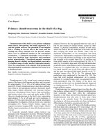
Five weeks after discharge he represented because of an
enlarging sternal mass and the development of back pain with no associated neurology (Figure 3). He remained
well with no weight loss or signs and symptoms of sys-
temic disease. An open biopsy of the chest wall mass and
a CT guided biopsy of the L1 spinal lesion were per-
formed. These revealed macroscopically caseous material.
Mantoux and Heaf tests were negative. An MRI scan of the
lumbar spine (Figure 4) showed loss of height of L1 with
disease extending bilaterally to the pedicles of T12 and L2.
There was a soft tissue mass anterior and posterior to L1
causing spinal stenosis and impingement on the conus.
Radiologically this was thought to resemble Potts disease
and the macroscopic appearance of the lumbar specimen
Plain radiographs (AP and Lateral) of lumbar spineFigure 3
Plain radiographs (AP and Lateral) of lumbar spine.
Demonstrate destruction of the L1 vertebra (arrow).
Axial CT of chestFigure 2
Axial CT of chest. Demonstrating the sternal mass (arrow)
but no underlying soft tissue involvement.
Bone scan at time of presentationFigure 1
Bone scan at time of presentation. Demonstrating
increased uptake in the sternum, L1 vertebra, left sacro-iliac
joint and right acetabulum.
World Journal of Surgical Oncology 2008, 6:34 />Page 3 of 6
(page number not for citation purposes)
suggested a diagnosis of tuberculosis. Triple therapy was
commenced (Rifampicin, Isoniazid and Pyrazinamide).
The specimens were negative for acid fast bacilli, Ziehl-
Neelson stain for TB was negative and no organisms were
cultured. Subsequent histology from the sternal mass
showed Hodgkin's lymphoma (Figure 5). Cells within the
specimen were positive for CD30 and CD20 (Figure 6).
Treatment with chemotherapy was started following the
current UKCCSG (United Kingdom Childrens Cancer Sur-
vey Guidelines) for Hodgkin's disease, and antitubercu-
lous treatment was stopped. Staging showed no
lymphadenopathy in the chest or abdomen. Two weeks
after commencement of chemotherapy a dramatic
decrease in soft tissue involvement around the spinal cord
was seen on a repeat MRI. (Figure 7). The time from pres-
entation to diagnosis was two months.
MRI Thoraco-lumbar spineFigure 7
MRI Thoraco-lumbar spine. Performed after commence-
ment of chemotherapy regime. This illustrates the decrease
in size of L1 lesion and reduction in impingement on the
conus.
Histology from sternal aspirateFigure 5
Histology from sternal aspirate. Illustrates mixed inflam-
matory cells, lacunar cells (green arrow) and Hodgkin cells
(black arrow).
MRI (T1 and T2 weighted images) of lumbar spineFigure 4
MRI (T1 and T2 weighted images) of lumbar spine.
Investigation undertaken 6 weeks after presentation showing
loss of height of L1 with surrounding soft tissue mass and
impingement on the conus.
Histology from sternal aspirate with stain for CD30Figure 6
Histology from sternal aspirate with stain for CD30.
The large cells (arrow) are positive for CD30 found on the
surface of Reed-Sternburg cells in Hodgkins Lymphoma.
World Journal of Surgical Oncology 2008, 6:34 />Page 4 of 6
(page number not for citation purposes)
Discussion
The incidence of skeletal Hodgkin's disease varies from
9–14% during the course of the disease with up to
30–50% at post mortem [1]. Skeletal involvement may
present in four different ways: POHL (either solitary or
multifocal); simultaneously in osseous and non-osseous
sites; or recurrence of disease at osseous sites. We consider
that in order to make a diagnosis of POHL there should
not be any signs or symptoms of systemic disease at the
time of presentation or at the time of staging. Historically
it was felt that primary Hodgkin's of the bone did not
occur and that bony involvement was a feature of haema-
tological dissemination of the disease, thereby implying a
less favourable prognosis [2]. Granger et al., [3] reported a
5 year survival of just 4.2% with 80% of deaths occurring
within first 3 years. POHL must therefore be distinguished
from systemic HD with diffuse bone marrow involvement
(Ann Arbor IV, see table 1), as it appears that POHL may
have a better prognosis than systemic Hodgkin's with
bony involvement [4]. The most recent case reported in
the literature regards a 51 year old female who presented
with left hip pain and was subsequently lymphadenopa-
thy in the cervical and inguninal nodes. She was staged as
VIB. The question remains as to whether POHL has a bet-
ter prognosis than HD with bony involvement [5].
There are thirty three clearly reported cases of primary
osseous lymphoma at either single or multiple sites, in all
ages, in the scientific literature since 1927. Table 2 details
these 32 cases as well as this current case. It does not
include patients who presented with disease at non
osseous and osseous sites, or those patients in whom
hodgkins disease disseminated to the bone. At least 7 of
these cases were reported prior to 1954 when CT, MRI and
PET scanning was not available, so it cannot be stated for
certain, whether these cases had any evidence of lymphad-
enopathy within the chest or abdomen. We are uncertain
therefore, whether these are true cases of POHL.
The two cases presented by Ostrowski et al., [6] were
among 25 patients diagnosed with osseous Hodgkin's dis-
ease from a group of over 500 patients known to have had
Hodgkin's lymphoma, at the Mayo clinic between 1927
and 1996. Five of the twenty five had POHL; three had dis-
ease at a single bony site and two had multifocal bony dis-
ease.
Gross et al., [7] presented two cases in adolescents (12
years and 17 years) who presented in a very similar pat-
tern to ours. Both presented with back pain and raised
inflammatory markers. Investigation revealed widespread
osseous involvement. In the case of the 17 year old, treat-
ment was delayed by a misdiagnosis of eosinophilic gran-
uloma. The 12 year old is one of two other paediatric cases
of primary multifocal osseous Hodgkin's lymphoma that
we have identified. The other case was an 11 year old girl
with disease in the thoracic spine, pelvis and left femur
[1].
There have been two paediatric cases identified by our lit-
erature review of patients with POHL at a single site. A
case report by Citow JS et al., [8] of a 54 year old female,
with back pain and spinal cord compression, thought to
be secondary to tuberculosis. Only when antituberculous
treatment failed, did re-examination and investigations
reveal POHL as the cause.
Radiologically, bony lesions of Hodgkin's disease may be
lytic, sclerotic or mixed. One study showed that 75% were
lytic, 13.6% mixed and 11.4% mixed [3]. When they
involve the vertebral column, the disease can spread from
one vertebral body to another across the intra-vertebral
disc space and cause destruction of the disc [9].
In all reported cases, the correct diagnosis was only
reached after extensive and repeated investigations and
review of the histology. The average time to diagnosis
from initial presentation was 6–8 months. The most fre-
quent misdiagnosis was osteomyelitis. Our case high-
lights the difficulties in diagnosing this rare form of
Hodgkin's disease.
TB in England has increased by 25 per cent over the last 10
years. Most TB in England occurs among people in inner
cities – two in every five cases are in London (see table 3).
Conclusion
This case indicates that Primary Multifocal OHL may
present in childhood. It demonstrates the difficulties in
reaching a definitive diagnosis, and the need to continu-
ally evaluate patients and diagnoses, especially when
patients fail to respond to initial therapies.
Competing interests
The author(s) declare that they have no competing inter-
ests.
Table 1: Staging of Lymphoma: Ann Arbor classification
Stage I Disease in a single lymph node region
Stage II Disease in two or more regions on the same side of the
diaphragm
Stage III Disease in lymph node regions on both sides of the
diaphragm
Stage IV Diffuse or disseminated involvement of one or more
extralymphatic organs or tissues with or without associated lymph
node enlargement.
World Journal of Surgical Oncology 2008, 6:34 />Page 5 of 6
(page number not for citation purposes)
Authors' contributions
CL, SG, JU and JK all contributed to the literature review.
CL,SG and NC have written and revised the manuscripts.
All authors read and approved final manuscript for publi-
cation.
Acknowledgements
Written consent was obtained from the patient for publication of this case
report.
Table 2: A table of cases in the literature who presented with Hodgkin's lymphoma disease at single or multiple bony sites.
Year Author and
[reference]
Age
(years)
Gender
(M/F)
Site(s) Therapy Outcome
1927 Gerbert et al. [10] 42 M T4-T8 XRT Alive at 10mo LTFU
1936 Gerbert et al. [10] 39 F L humerus Surgery DOD 12mo
1943 Gerbert et al. [10] 5 F L scapula XRT NED
1958 Gerbert et al. [10] 53 M L Humerus, L Ilium XRT DOD at 4mo
1960 Ostrowski et al. [6] 73 F R Femur XRT DOD at 4 yrs
1968 Ostrowski et al. [6] 34 M L Humerus XRT AWD at 10 yrs
1979 Gerbert et al. [10] 25 F L humerus XRT NED 4.5 yrs
1982 Chan et al. [1] 12 M R tibia XRT & CT Alive at 66mo
1982 Chan et al. [1] 18 M R ulna, L tibia and fibula XRT & CT Alive at 18mo
1982 Chan et al. [1] 20 M T11 XRT & CT Alive at 15mo
1982 Chan et al. [1] 11 F T8-10, L femur, Pelvis XRT & CT Alive at 7mo
1982 Chan et al. [1] 68 F R SI joint None Died 2mo
1982 Chan et al. [1] 29 M T10-T12 Surgery ?
1982 Chan et al. [1] 45 M Sternum Surgery, XRT & CT Alive at 10mo
1982 Chan et al. [1] 21 F Sternum Surgery & XRT Alive at 24mo
1982 Chan et al. [1] 41 M Skull, ischium, L spine XRT Died 3mo
1982 Chan et al. [1] 17 F T7 – T8 XRT Died 24mo
1982 Chan et al. [1] 9 M R tibia Surgery & XRT Died 26mo
1982 Chan et al. [1] 27 F R tibia XRT Died 3mo
1982 Chan et al. [1] 62 M L humerus Surgery Died 1 mo
1982 Chan et al. [1] 40 M T2 Surgery Died 8mo
1982 Chan et al. [1] 24 F L femur Surgery & XRT Died 12mo
1989 Mac Cormick et al. [11] 61 M T spine, R 12
th
rib, R clavicle CT remission
1991 Gross et al [7] 17 F T10, L4, 11
th
rib, L ilium CT, XRT, BMT DOD
1991 Gross et al [7] 12 F T11-12, L3-4, L scapula, L10
th
rib, L
ilium, L acetabulum, L femur
CT AWD at 2.5 yrs
1993 Borg et al [12] 31 M Sacrum CT & XRT NED 5 yrs
1995 Ostrowski et al. [6] 61 F T11 resection & XRT AWD at 22 mo
1995 Fried et al. [13] 21 F L clavicle CT NED 36mo
1995 Gerbert et al. [10] 63 M L femur, R ilium CT & XRT NED 6mo
1996 Citow et al [8] 54 F T4, T5 Surgery, CT & XRT AWD at 36 mo
1999 Gerbert et al. [10] 21 M R femur, R tibia CT & XRT NED 48mo
2006 Chandra et al [5] 51 F L ileum CT & XRT Alive
2006 Present case 7 M L1, sternum, Lt SI joint, Rt
acetabulum
CT AWD
CT – chemotherapy
AWD – alive with disease
XRT – Radiotherapy
DOD – died of disease
LTFU – lost to follow up
NED – no evidence of disease
Table 3: TB in England has increased by 25 per cent over the last
10 years. Most TB in England occurs among people in inner cities
– two in every five cases are in London. Tuberculosis in the UK
(2004 statistics) from global health facts.
New cases 7101
New case rate (per 100,000) 12
People with TB 5497
TB prevalence (per 100, 000) 9
TB deaths 710
Death rate (per 100, 000) 1
Publish with BioMed Central and every
scientist can read your work free of charge
"BioMed Central will be the most significant development for
disseminating the results of biomedical research in our lifetime."
Sir Paul Nurse, Cancer Research UK
Your research papers will be:
available free of charge to the entire biomedical community
peer reviewed and published immediately upon acceptance
cited in PubMed and archived on PubMed Central
yours — you keep the copyright
Submit your manuscript here:
/>BioMedcentral
World Journal of Surgical Oncology 2008, 6:34 />Page 6 of 6
(page number not for citation purposes)
References
1. Chan KW, Rosen G, Miller DR, Tan CT: Hodgkin's disease in ado-
lescents presenting as a primary bone lesion. Am J Ped Hematol
Oncol 1982, 4:7-11.
2. Horan FT: Bone involvement in Hodgkin's disease. Br J Surg
1969, 56:277-281.
3. Granger W, Whitaker P: Hodgkin's disease in both with special
reference to periosteal reaction. Br J Radiol 1967, 40:939-948.
4. Stuhlbarg J, Ellis FW: Hodgkin's disease of the bone: favourable
prognostic significance? Am J Roentgenol Radium Ther Nucl Med
1965, 93:568-572.
5. Chandra D, Ewton A, Baker K: Hodgkin's disease presenting
with osseous involvement. Am J Hematol 2006, 81:550-551.
6. Ostrowski ML, Inwards CY, Strickler JG, Witzig TE, Wenger DE,
Unni KK: Osseous Hodgkin's disease. Cancer 1999,
85:1166-1178.
7. Gross SB, Robertson WW Jr, Lange BJ, Bunin NJ, Drummond DS:
Primary Hodgkin's disease of bone. A report of two cases in
adolescents and review of the literature. Clin Orthopaed Rel Res
1992, 283:276-280.
8. Citow JS, Rini B, Wollmann R, Macdonald R: Isolated, primary
extranodal Hodgkin's disease of the spine: Case report. Neu-
rosurgery 2001, 49:453-456.
9. McElwain TJ, Selby P: Hodgkin's disease 1st edition. Oxford: Blackwell
S.C. Publications; 1987:105-106. 141–146
10. Gebert C, Hardes J, Ahrens H, Buerger H, Winkelmann W, Gosheger
G: Primary multifocal osseous Hodgkin disease: a case
report and review of the literature. J cancer Res Clin Oncol 2005,
131:163-168.
11. MacCormick R, Covert A, Gross M: Primary bone involvement
in Hodgkin's disease. CMAJ 1989, 140:1059-1060.
12. Borg MF, Chowdhury AD, Bhoopal S, Benjamin CS: Bone involve-
ment in Hodgkin's disease. Australasian Radiology 1993, 37:63-66.
13. Fried G, Ben Arieh Y, Haim N, Dale J, Stein M: Primary Hodgkin's
disease of the bone. Med Paediatr Oncol 1995, 24:204-207.