Báo cáo khoa học: "An unusual case of low-grade tubulopapillary adenocarcinoma of the sinonasal tract" doc
Bạn đang xem bản rút gọn của tài liệu. Xem và tải ngay bản đầy đủ của tài liệu tại đây (567.1 KB, 3 trang )
BioMed Central
Page 1 of 3
(page number not for citation purposes)
World Journal of Surgical Oncology
Open Access
Case report
An unusual case of low-grade tubulopapillary adenocarcinoma of
the sinonasal tract
Ashish Bansal*
1
, Keloth E Pradeep
2
and Krishna P Gumparthy
1
Address:
1
Department of Histopathology, Wirral Hospitals NHS Trust, Upton, Wirral, CH49 5PE, UK and
2
Department of Histopathology,
Wrexham Maelor Hospital, Wrexham, UK
Email: Ashish Bansal* - ; Keloth E Pradeep - ;
Krishna P Gumparthy -
* Corresponding author
Abstract
Background: Low-grade papillary adenocarcinomas of the sinonasal tract are rare neoplasms.
Over recent years, little doubt remains that this tumour represents a separate entity based on
morphology, ultrastructural features and behaviour. We outline a case of this rare entity displaying
a not hitherto described immunophenotype.
Case presentation: A 32 year old man presented recurrent epistaxis was evaluated with
endoscopy which revealed a well circumscribed pedunculated mass lesion in left nares. The mass
was arising from the nasal septum which was excised along with the mass. The biopsy revealed low-
grade, non-intestinal type sinonasal tubulopapillary adenocarcinoma.
Conclusion: TTF-1 immunoreactivity in absence of thyroid or pulmonary primary in the present
case remains an enigma. However, this raises the possibility of the utility of this antibody to predict
a better clinical outcome in the subset of low grade non-intestinal sinonasal adenocarcinoma. More
cases of similar morphological appearance may need to be examined for TTF-1 immunoreactivity
and clinically followed up to establish this theory.
Background
Sinonasal adenocarcinomas are rare tumours accounting
for 0.4% [1] of all human neoplasms, of which adenocar-
cinoma accounts for 13% [2]. We outline a case of this
rare entity displaying an unusual immunophenotype.
Case presentation
A 32 year old man who had recurrent episodes of epistaxis
was seen in the ENT outpatient clinic. Flexible endoscopy
revealed deviation of the nasal septum to the left. Arising
from the posterior end of the left nasal septum was a
pedunculated well-circumscribed lesion. Magnetic reso-
nance imaging revealed no other abnormalities. At opera-
tion, a lobulated solid mass was seen. The mucosa
anterior to the mass had become detached. The underly-
ing bone was removed but did not look involved. Postop-
erative recovery was uneventful and he was discharged the
next day. The lesion was suspected to be a haemangioma.
Previous episodes of epistaxis were treated with silver
nitrate cautery. The patient has no significant past medical
history. He is a non-smoker, was not on any regular med-
ication and had no relevant occupational history. Subse-
quently, the patient had two further operations. Firstly,
removal of the posterior aspect of the nasal septum was
performed four months after removal of this mass. Sec-
ondly, a biopsy of the nostril was undertaken. The former
Published: 20 May 2008
World Journal of Surgical Oncology 2008, 6:54 doi:10.1186/1477-7819-6-54
Received: 3 November 2007
Accepted: 20 May 2008
This article is available from: />© 2008 Bansal et al; licensee BioMed Central Ltd.
This is an Open Access article distributed under the terms of the Creative Commons Attribution License ( />),
which permits unrestricted use, distribution, and reproduction in any medium, provided the original work is properly cited.
World Journal of Surgical Oncology 2008, 6:54 />Page 2 of 3
(page number not for citation purposes)
revealed mucosal fragments incorporating seromucinous
glands with intervening chronic inflammation of the
stroma but no evidence of residual adenocarcinoma. The
latter showed inflammatory granulation tissue around
suture granulomata from previous surgery. Since initial
presentation over two years ago, the patient remains free
of recurrence or metastatic disease and does not have any
lesions in his lungs or thyroid gland.
Macroscopically, two yellow-white polypoid fragments of
tissue, measuring 10 and 4 mm in maximum dimension
were received. Histologically, these fragments were partly
covered by focally ulcerated squamous epithelium. The
underlying stroma was infiltrated by a neoplasm with a
complex papillary and tubular configuration, lined by
moderately dysplastic pale columnar epithelium with
intervening spindle shaped cells(Figure 1 and 2).
Immunohistochemical labelling revealed diffuse positiv-
ity with antibodies to EMA, CAM 5.2, CK 7, CK 19 and
TTF-1 (Figure 3). The cells were negative with CK 20, CEA,
S-100 protein, thyroglobulin, SMA and p63. The appear-
ances were consistent with a low-grade, non-intestinal
type sinonasal tubulopapillary adenocarcinoma.
Discussion
As described recently [3], low-grade tubulopapillary aden-
ocarcinoma represents a distinctive sinonasal adenocarci-
noma. Historically, one of the earliest classifications was
based on whether the tumour arose from the surface
mucosal epithelium or from submucosal seromucinous
glands [4]. However, this separation was flawed in that
the latter are direct invaginations of the former. Subse-
quently, some pathologists began to classify these
tumours solely as high-grade or low-grade adenocarcino-
mas based on their histological appearance [5]. In view of
the histological resemblance of sinonasal adenocarcino-
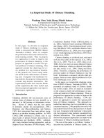
High power photomicrograph (×250): complex tubules and papillae lined by mild/moderately dysplastic pale columnar cellsFigure 2
High power photomicrograph (×250): complex tubules and
papillae lined by mild/moderately dysplastic pale columnar
cells.
Low Power photomicrograph (×40) of this entity: low-grade non-intestinal tubulopapillary adenocarcinoma of the sinona-sal tract with overlying surface squamous epitheliumFigure 1
Low Power photomicrograph (×40) of this entity: low-grade
non-intestinal tubulopapillary adenocarcinoma of the sinona-
sal tract with overlying surface squamous epithelium.
Immunohistochemical nuclear positivity for thyroid transcrip-tion factor 1 (TTF-1)Figure 3
Immunohistochemical nuclear positivity for thyroid transcrip-
tion factor 1 (TTF-1).
Publish with BioMed Central and every
scientist can read your work free of charge
"BioMed Central will be the most significant development for
disseminating the results of biomedical research in our lifetime."
Sir Paul Nurse, Cancer Research UK
Your research papers will be:
available free of charge to the entire biomedical community
peer reviewed and published immediately upon acceptance
cited in PubMed and archived on PubMed Central
yours — you keep the copyright
Submit your manuscript here:
/>BioMedcentral
World Journal of Surgical Oncology 2008, 6:54 />Page 3 of 3
(page number not for citation purposes)
mas to intestinal and submucosal seromucinous glands,
classifications [6] have tended to categorise such tumours
into intestinal and non-intestinal types. The current WHO
classification [7] of these tumours considers two catego-
ries: intestinal and non-intestinal types of high and low
grade sub-types. In addition, sinonasal tumours of the sal-
ivary gland type are identified too. The high grade types in
both groups of adenocarcinomas and the overall category
of intestinal type are described to have a worse prognosis.
The importance of recognition and separation of this neo-
plasm from other types of sinonasal adenocarcinoma is
critical as it virtually never metastasizes and has an excel-
lent prognosis. Unlike this case, Franchi et al. [8], have
recently described two cases positive for basal cell mark-
ers, demonstrating that at least a subset of these tumours
are most likely salivary-type in origin. With the possible
exception of a low proliferation index, immunohisto-
chemical markers have so far proved unhelpful. Immuno-
histochemistry for intestinal type adenocarcinoma is
known to reveal positivity for pancytokeratin, EMA,
B72.3, BerEP4, Leu M1, CK20, CDX2 and variable CK7
immunoreactivity. In this case, the tumour showed dif-
fuse positivity with antibodies to EMA, CAM 5.2, CK7,
CK19 and TTF-1 and no expression (negative) with CK 20,
CEA, S-100 protein, thyroglobulin, SMA and p63.
Conclusion
There is no published data on the role of TTF-1 in adult
primary nasal adenocarcinomas. To date, we are unaware
of any occult thyroid or pulmonary tumours in our
patient to explain the TTF-1 immunoreactivity. The signif-
icance of this unexpected immunohistochemical labelling
remains an enigma. However, this unusual TTF-1 positiv-
ity raises the possibility of the utility of this antibody to
predict a better clinical outcome in the subset of low grade
non-intestinal sinonasal adenocarcinoma. More cases of
similar morphological appearance may need to be exam-
ined for TTF-1 immunoreactivity and clinically followed
up to establish this theory.
Competing interests
The authors declare that they have no competing interests.
Authors' contributions
AB conducted a literature search, took the photomicro-
graphs and drafted the manuscript; KEP edited the manu-
script; KPG is the consultant who reported the biopsies
and proofread the final manuscript. All authors read and
approved the final manuscript.
Acknowledgements
Written informed consent was obtained from the patient to publish this
case report.
We wish to thank Dr T R Helliwell (Head & Neck specialist) for reviewing
this case and corroborating the diagnosis.
References
1. Abecasis J, Viana G, Pissarra C, Pereira T, Fonseca I, Soares J: Aden-
ocarcinomas of the nasal cavity and paranasal sinuses: a clin-
icopathological and immunohistochemical study of 14 cases.
Histopathology 2004, 45:254-259.
2. Harbo G, Grau C, Bundgaard T, Overgaard M, Elbrønd O, Søgaard H,
Overgaard J: Cancer of the nasal cavity and paranasal sinuses.
Acta Oncol 1997, 36:45-50.
3. Skalova A, Cardesa A, Leivo I, Pfaltz M, Ryska A, Simpson R, Michal
M: Sinonasal tubulopapillary low-grade adenocarcinoma.
Histopathological, immunohistochemical and ultrastruc-
tural features of poorly recognised entity. Virchows Arch 2003,
443:152-158.
4. Kleinsasser O: Terminal tubulus adenocarcinoma of the nasal
seromucous glands. A specific entity. Arch Otorhinolaryngol 1985,
241:183-193.
5. Heffner DK, Hyams VJ, Hauck KW, Lingeman C: Low-grade aden-
ocarcinoma of the nasal cavity and paranasal sinuses. Cancer
1982, 50:312-322.
6. Franquemont DW, Fechner RE, Mills SE: Histologic classification
of sinonasal intestinal-type adenocarcinoma. Am J Surg Pathol
1991, 15:368-375.
7. Barnes L, Eveson JW, Reichart P, Sidransky D, (Eds): World Health
Organization Classification of Tumours. Pathology and Genetics of Head
and Neck Tumours Lyon: IARC Press; 2005:22-23.
8. Franchi A, Palomba A, Massi D, Biancalani M, Sardi I, Gallo O, Santucci
M: Low-grade salivary type tubulopapillary adenocarcinoma
of the sinonasal tract. Histopathology 2006, 48:881-884.