Báo cáo khoa học: "bcl-2 expression is not associated with survival in metastatic cutaneous melanoma: A historical cohort study" pptx
Bạn đang xem bản rút gọn của tài liệu. Xem và tải ngay bản đầy đủ của tài liệu tại đây (762.61 KB, 7 trang )
BioMed Central
Page 1 of 7
(page number not for citation purposes)
World Journal of Surgical Oncology
Open Access
Research
bcl-2 expression is not associated with survival in metastatic
cutaneous melanoma: A historical cohort study
Marília B Espíndola*
1,2
and Oly C Corleta
1,2
Address:
1
Graduate Program in Medicine: Surgery, Universidade Federal do Rio Grande do Sul, Porto Alegre, RS, Brazil and
2
Rua Ramiro Barcelos
2300, Porto Alegre, RS, Brazil
Email: Marília B Espíndola* - ; Oly C Corleta -
* Corresponding author
Abstract
Background: Programmed cell death (apoptosis) has been implicated in tumor development and
may affect the metastatic potential of tumor cells. The role of bcl-2, a proto-oncogene that inhibits
apoptosis, has been studied in several malignancies, including cutaneous melanoma (CM). The
purpose of this study was to evaluate the immunohistochemical expression of bcl-2 in 35 regional
lymph node, 28 subcutaneous and 17 visceral CM metastases, correlating the findings with patient
survival.
Methods: In a historical cohort study patient survival was correlated with the expression of bcl-2
in regional lymph node, subcutaneous and visceral metastases of CM. Eighty slides containing
surgical specimens from 50 patients diagnosed with stage III and IV CM, 28 male (56%) and 22
female (44%), were analyzed. Mean age at diagnosis was 43 years (16–74 years; median = 42 years).
Mean Breslow depth was 5.01 mm (0.4–27.5 mm). The slides were submitted to
immunohistochemical reaction using anti-bcl-2 monoclonal antibody and classified according to the
degree of staining (< 5%; 5 to 50%; or > 50% of tumor cells stained). The relationship between bcl-
2 protein expression and survival for each type of metastasis, gender and age at initial diagnosis was
analyzed.
Results: Mean overall survival was 33.9 months after the diagnosis of the initial metastatic lesion
(range: 0 to 131 months). Twenty-four out of 50 patients (48%) had died from CM by the end of
the study period. bcl-2 expression was detected in 74.3, 85.7 and 82.4% of lymph node,
subcutaneous and visceral metastases, respectively. After univariate and multivariate analyses, no
correlation was found between positive bcl-2 expression and overall survival for the types of
metastases evaluated.
Conclusion: The immunohistochemical expression of bcl-2 in metastasis alone is not a prognostic
marker for CM.
Background
The incidence of cutaneous melanoma (CM) has
increased steadily in the last few years [1]. Sun exposure
and sunburn, with subsequent genetic damage caused by
ultraviolet radiation [2], play a major role in this increase.
The prognosis of CM is positive in its initial stages; how-
Published: 20 June 2008
World Journal of Surgical Oncology 2008, 6:65 doi:10.1186/1477-7819-6-65
Received: 10 December 2007
Accepted: 20 June 2008
This article is available from: />© 2008 Espíndola and Corleta; licensee BioMed Central Ltd.
This is an Open Access article distributed under the terms of the Creative Commons Attribution License ( />),
which permits unrestricted use, distribution, and reproduction in any medium, provided the original work is properly cited.
World Journal of Surgical Oncology 2008, 6:65 />Page 2 of 7
(page number not for citation purposes)
ever, the five-year survival rate is only 41% in patients
with regional lymph node metastases. A maximum sur-
vival of 2 years has been reported for patients with distant
metastases [3].
Despite major advances in cancer treatment, surgery is still
the treatment of choice for CM, since chemotherapy and
radiation therapy generally produce low response rates
[4,5]. Since the ultimate goal of non-surgical treatments is
to induce apoptosis in tumor cells, this physiological
event has recently received much attention [6]. Defects in
the regulation of apoptosis have been implicated in tumor
progression, metastatic spread and resistance to chemo-
therapy [7,8]. In recent years, several biomolecules,
including B cell lymphoma/leukemia-2 (bcl-2), have been
studied in CM, in an attempt to determine which lesions
are more likely to respond to non-surgical treatments [9].
The bcl-2 gene is located on chromosome segment
18q21.3, in a telomere-centromere orientation [9]. The
bcl-2 protein is an integral part of the cell membrane, with
a molecular weight of 26 kDa, and it is found in the cell
nucleus, mitochondria and endoplasmic reticulum [10].
bcl-2 acts as an apoptosis inhibitor, without any influence
on cell proliferation [9].
The results obtained so far for the role of bcl-2 in CM are
controversial. Although some authors have described an
increase in bcl-2 during the progression of normal
melanocytes to melanomas, others have observed the
opposite [11-15]. Grover et al. [11] have reported a lower
survival rate in CM patients with regional lymph node
metastasis and positive bcl-2 expression.
The purpose of the present study was to evaluate the rela-
tionship between the immunohistochemical expression
of bcl-2 and survival in patients with regional lymph
node, subcutaneous and visceral metastases of CM.
Patients and methods
In this historical cohort study, the survival of patients with
CM represents the outcome, and the expression of bcl-2 in
regional lymph node, subcutaneous and visceral metas-
tases of CM is the variable of interest.
Eighty slides containing surgical specimens from 50
patients treated at three institutions between 1990 and
2007, 28 male (56%) and 22 female (44%), were ana-
lyzed. All patients had been diagnosed with stage III
(regional metastases) and stage IV (distant metastases)
CM [16]. In all cases, initial resection was performed at
one of the participating hospitals. Exclusion criteria were
previous diagnosis of other types of cancer, simultaneous
diagnosis of secondary neoplasm, previous radiation ther-
apy, chemotherapy or resection of metastases prior to
diagnosis, initial surgery for metastasis at a different insti-
tution, incomplete resection of metastases, or death due
to causes other than CM. The study was approved by the
Hospital de Clínica de Porto Alegre Research Ethics Com-
mittee (IRB equivalent).
Table 1 shows the distribution of patients in terms of pri-
mary diagnosis and location of tumor and first metastasis.
Mean age at initial diagnosis was 43 years, ranging from
16 to 74 years, with a median of 42 years. Mean Breslow
depth was 5.01 mm, ranging from 0.4 to 27.5 mm. Clark's
level ranged from II to V, of which level IV was the most
prevalent (22 cases – 44%).
If more than one metastasis was present in the same site,
only the first metastasis at each site was evaluated for bcl-
2 protein expression. Thirty patients did not receive any
treatments other than surgery. Of the remaining 20
patients, nine received chemotherapy, with regimens that
included the following drugs: dacarbazine (DTIC), car-
mustine (BCNU), verapamil, cisplatin and tamoxifen, in
varying combinations and for one to five treatment cycles.
One patient received hyperthermic isolated lower limb
perfusion with melphalan; complete response was
observed immediately after treatment, but a relapse
occurred later. Five patients were treated with interferon
for 1 to 5 months. Five patients received radiation ther-
apy, of whom four were treated for central nervous system
(CNS) metastases and one for disease in the axilla, all with
palliative intent.
Immunohistochemistry
Sections of paraffin-embedded metastasis specimens were
initially stained with hematoxylin-eosin for evaluation of
tumor representation. The chosen blocks were sliced into
4-μm sections and stored in an incubator at 56°C for 24
h. After deparaffinization and hydration by immersion in
xylol and decreasing concentrations of ethanol (100 to
20%) in room temperature, antigenic recovery was carried
out using the microwave irradiation method. After that,
the specimens were rinsed in tap water and distilled water
and immersed in PBS buffer for 5 min. To block tissue
enzymes that could interfere with the reaction, the endog-
enous peroxidase method was employed. To block unspe-
cific reactions that could yield false-positive results,
powder milk was used, with rehydration in 5% PBS buffer
for 40 min, washing in tap water and distilled water and
immersion in PBS for 5 min. The specific antibody reac-
tion was carried out with bcl-2 antibody (IgG 1, kappa,
280 mg/L – Monoclonal Mouse Anti-Human bcl-2 Onco-
protein Clone 124 Code no. M0887 Lot 018; Dako Cor-
poration, Carpinteria, CA, USA) diluted in PBS buffer
(1:500). The sections were then stored in a dark chamber
for 1 hour at 37°C or left at 4°C in a refrigerator over-
night. The slides were individually washed three times for
World Journal of Surgical Oncology 2008, 6:65 />Page 3 of 7
(page number not for citation purposes)
5 min in PBS buffer. Secondary antibody (DAKO/LSAB –
Dako Liquid DAB Large Volume Substrate-Chromogen
System Code N°: K3466; Dako Corporation, Carpinteria,
CA, USA) was applied for 30 minutes, followed by three
5-min baths with PBS buffer. The DAB kit was used for
development. Counterstaining was carried out with Harris
hematoxylin for 20 s after serial rinsing with tap water, 2%
ammonia solution, ethanol and xylol.
The slides were evaluated by two independent patholo-
gists and classified according to staining intensity (%
stained cells), as follows:
- 0 (negative): less than 5% of tumor cells stained with bcl-
2;
- I (weakly positive): 5 to 50% of tumor cells stained with
bcl-2;
- II (strongly positive): over 50% of tumor cells stained
with bcl-2.
Since statistical analysis did not reveal differences
between groups I and II, they were considered as one
group (bcl-2-positive) for the present analysis. Results are
presented as arithmetic means, standard deviation, medi-
ans, and percentage rates. The chi-square test, Fisher's
exact test, log-rank test, and Kaplan Meier method were
used to evaluate the relationship between bcl-2 protein
expression and survival for the three types of metastases
(regional lymph node, subcutaneous and visceral metas-
tases), gender and age at initial diagnosis, with 95% con-
fidence intervals. Univariate and multivariate Cox
regression tests were performed to evaluate the interaction
between gender, age at first metastasis, survival, type of
metastasis, and bcl-2 expression. A P < 0.05 was consid-
ered to be statistically significant.
Results
Mean overall survival was 33.9 months after the diagnosis
of the initial metastatic lesion (range: 0 to 131 months).
Twenty-four out of 50 patients (48%) had died from CM
by the end of the study period. Of the 26 surviving
patients, nine (36%) had imaging exams suggestive of
multiple metastatic lesions or a single unresectable meta-
static lesion. The mean disease-free interval (from initial
diagnosis to the diagnosis of the first metastasis) was 17.6
months (0 to 83 months) (median of 11.5 months and
standard deviation of 21.5 months). Five patients (10%)
had visceral and subcutaneous metastases simultane-
ously, and one (2%) had the three types of metastasis
since the start of metastatic disease.
Results concerning the immunohistochemical expression
of bcl-2 and deaths according to the three types of metas-
tasis are shown in table 2. Figure 1 shows the different
degrees of immunostaining observed in metastasis speci-
mens, including intense (A-C), weak (D-F) and absence
(G-I) of immunostaining. Fisher's exact test did not reveal
an association between metastasis site and death (P = 1 for
lymph node metastases, P = 0.613 for subcutaneous
metastases, and P = 0.576 for visceral metastases). Table 3
shows chi-square test results for the expression of bcl-2 in
the three types of metastases, revealing no difference
between the sites. Fisher's exact test confirmed that the
metastasis sites were similar in terms of bcl-2 protein
expression, survival, and presence of unresected meta-
static disease by the end of the study (Fisher's P = 1.0, P =
0.333, and P = 0.429 for lymph node, subcutaneous, and
visceral metastases, respectively).
Survival was correlated with age > 60 years at initial diag-
nosis (log rank = 6.17; P = 0.130) and male gender (log
rank = 3.17; P = 0.0752) in patients with subcutaneous
metastases. None of the other comparisons between sur-
vival, age, gender, and bcl-2 expression were statistically
significant. Similar results were obtained using univariate
analysis (P > 0.05) and Cox's multivariate analysis.
Table 1: Distribution of 50 patients with cutaneous melanoma in
terms of primary diagnosis and location of tumor and first
metastasis
Primary tumor diagnosis Number
Superficial spreading CM 21
Nodular CM 15
Amelanotic 5
Acral 2
Unknown 7
Tumor location
Trunk 30
Lower limbs 13
Upper limbs 4
Head and neck 3
Site of first metastasis
Lymph node 35
Subcutaneous 28
Visceral 17
Uterine adnexa 1
Small intestine 4
Adrenal gland 2
Omentum 1
Colon 1
Lung 3
Liver 2
Testis 1
Breast 2
World Journal of Surgical Oncology 2008, 6:65 />Page 4 of 7
(page number not for citation purposes)
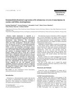
Photomicrograh 200×Figure 1
Photomicrograh 200×. A) Lymph node B) visceral and C) subcutaenous metastases in patients with cutaneous melanoma
showing intense immunostaining (brown spots). D) Lymph node, E) subcutaneous and F) visceral mestastases in patients with
cutaneous melanoma showing weak expression of bcl-2 protein. Absence of bcl-2 expression in G) lymph node H) subcutane-
ous melanoma and I) visceral melanoma metastases.
World Journal of Surgical Oncology 2008, 6:65 />Page 5 of 7
(page number not for citation purposes)
Discussion
Apoptosis, or programmed cell death, has recently
become the focus of great interest [7,9,17]. Discovery of
the bcl-2 gene, an apoptosis inhibitor, in translocation
14;18 (q32;q21) in follicular B-cell lymphomas, followed
by the identification of the BCL-2 family in the study of
Caenorhabditis elegans, opened new perspectives for the
study of tissue morphogenesis and oncogenesis [9,11,18].
Inhibition of apoptosis can occur in any phase of the cell
cycle, although the exact mechanism through which bcl-2
inhibits apoptosis is not fully understood [19]. In the
presence of bcl-2 overexpression, the ability of the cell to
remove genetic and cell damage through apoptosis is lim-
ited. Despite the fact that bcl-2 does not act directly on cell
proliferation, its overexpression enables tumors to
progress to highly malignant phenotypes and to become
more resistant to chemotherapy and apoptosis-inducing
radiation therapy, with subsequent metastatic spread and
tumor progression [8,19,20].
Several different tissues express the bcl-2 protein. In some
types of neoplasms originating from these tissues (such as
lymph nodes with breast cancer metastasis), a relation-
ship between bcl-2 expression and longer disease-free sur-
vival has been observed. On the other hand, in other
tumor types, there is an inverse relationship between pos-
itive bcl-2 expression and prognosis, such as in prostate
cancer, ovarian cancer, non-small cell lung cancer, follicu-
lar thyroid cancer, neuroblastoma, and breast cancer
[14,15]. In the case of CM, this relationship is still contro-
versial and undefined.
Confounding variables such as sample size, gender and
age should be considered when studying prognostic fac-
tors in CM. The relatively small number of cases analyzed
in our study is due mainly to the absence of appropriate
protocols for the management of CM in our setting, mak-
ing it more difficult to gather patient information. More-
over, it is often not possible to retrieve data from
inaccurate patient charts, and pathology divisions, at least
in our institutions, lack an appropriate database, which
would facilitate the access to patient charts using pathol-
ogy diagnosis as a search variable.
The ages of 50 and 60 years have been used as cutoff
points for the evaluation of CM patients [3,21-23]. In this
study, we observed that patients older than 60 years were
at greater risk for subcutaneous metastases of CM, and
also that age had no effect on the survival of patients with
other types of metastasis. The larger tumor thickness
found in patients older than 60 years at diagnosis, as well
as the accumulation of genetic damage acquired through-
out the years, may account for these findings [24]. Fernan-
dez-Pol and Douglas related the presence of bcl-2,
mitochondrial integrity and carcinogenesis with human
aging [25]. Garbe and Blum noticed that most melanomas
were diagnosed between the sixth and seventh decades of
life, with only 22% of the cases diagnosed before the age
of 40 [26].
We were unable to evaluate the histological characteristics
of primary tumors because this information was not avail-
able for all cases. Also, we did not analyze the relationship
between location of the primary lesion, survival and bcl-
2. Nevertheless, the characteristics of the primary lesion
lose some importance once the first metastasis is diag-
nosed [3,22,27-29]. The importance of data about the pri-
mary tumor is in fact controversial, with different authors
reporting conflicting results about the role of pathology
Table 2: Immunohistochemical expression of bcl-2 and deaths in patients with cutaneous melanoma
Lymph node metastases Subcutaneous metastases
a
Visceral metastases
(n = 35) (n = 28) (n = 17)
No. % No. % No. %
bcl-2-negative 9 25.7 4 14.3 3 17.6
Deaths 4 11.4 3 10.7 1 5.9
bcl-2-positive
Intense staining 17 48.6 20 71.4 11 64.7
Weak staining 9 25.7 4 14.3 3 17.6
Deaths 12 83.3 13 84.6 8 89
a
Stage IV [16]
Table 3: Chi-square test results for expression of bcl-2 in three
types of metastases
bcl-2 positive (%) bcl-2 negative (%) Total (%)
Lymph node 26 (74.3) 9 (25.7) 35 (100)
Subcutaneous 24 (85.7) 4 (14.3) 28 (100)
Visceral 14 (82.3) 3 (17.7) 17 (100)
Total 64 (80.0) 16 (20.0) 80 (100)
χ
2
= 1.34; P = 0.510.
World Journal of Surgical Oncology 2008, 6:65 />Page 6 of 7
(page number not for citation purposes)
data and primary site location in CM prognosis
[1,3,21,23,27,30,31].
In addition, the expression of bcl-2 in CM metastases was
evaluated without comparison to bcl-2 expression in nor-
mal melanocytes. That comparison was not performed
because this is a retrospective study, based on paraffin
block specimens and on medical records. It was therefore
not possible to obtain normal skin or nevus samples from
the patients enrolled in the study. Nevertheless, bcl-2
expression in our study was similar to that described in
previous studies employing immunohistochemical meth-
ods [14,15,32,33].
We did not observe a relationship between bcl-2 expres-
sion and survival in the three types of CM metastasis. This
could be explained in three distinct ways: first, there is no
correlation between survival and the immunohistochem-
ical expression of bcl-2 in CM; second, this correlation
exists, but was not demonstrated in the present study due
to the small sample size and short follow-up time; and
third, the interaction between the other members of the
BCL-2 family may have neutralized the expression of bcl-
2. The regulation of proapoptotic and antiapoptotic com-
ponents in the BCL-2 family is complex. Several members
of this family are still being discovered, such as Bim and
GRS [34,35], and the complex interactions between BCL-
2 family members are only partially understood. In the
future, a deeper understanding of these interactions, and
mainly of their functions (rather than only of their pres-
ence), may allow for an adequate use of BCL-2 family
members as effective predictors of survival in CM.
It has been shown that the treatment of melanoma cells
with oligonucleotides targeting the reduction of bcl-2
expression rendered those cells more sensitive to chemo-
therapy [5,36-41]. These oligonucleotides are chemically
modified, single-stranded DNA that complement specific
codons of the messenger-RNA of a target gene, and which
are capable of inhibiting the expression of this particular
gene [5]. These experiments demonstrate that bcl-2 pro-
tein plays an important, although not fully understood,
role in chemoresistance in CM and several other tumors
[37,38].
Conclusion
The present results suggest that the immunohistochemical
expression of bcl-2 in metastases alone is not a prognostic
marker for CM. Further knowledge of the actions and rela-
tions between BCL-2 family members is necessary to
define the exact role of the bcl-2 protein and of other BCL-
2 family members in the pathogenesis, prognosis, and
response of metastatic CM to new treatments.
Competing interests
The authors declare that they have no competing interests.
Authors' contributions
MBE participated in study design, was in charge of data
collection and analysis and helped to draft the manu-
script, OCC conceived of the study, and participated in its
design and data analysis and helped to draft the manu-
script. Both authors read and approved the final version of
the manuscript.
Acknowledgements
The authors wish to thank Dr. Luis Fernando Rivero, Professor of Pathol-
ogy of at Universidade Federal do Rio Grande do Sul (UFRGS), for his sup-
port and guidance in the interpretation of immunohistochemical data.
The study was approved by the ethics committee.
References
1. Markowitz JS, Cosimi LA, Carey RW, Kang S, Padyk C, Sober AJ,
Cosimi AB: Prognosis after initial recurrence of cutaneous
melanoma. Arch Surg 1991, 126:703-708.
2. Gilchrest BA, Eller MS, Geller AC, Yaar M: The pathogenesis of
melanoma induced by ultraviolet radiation. NEJM 1999,
340:1341-1348.
3. Stadelmann WK, Rapaport DP, Soong S-J, Reintgen DS, Buzaid AC,
Balch CM: Prognostic Clinical pathologic features. In Cutaneous
melanoma 3rd edition. Edited by: Balch CM, Houghton AN, Sober AJ,
Soong S-J. St. Louis, Missouri: Quality Medical Publishing, Inc;
1998:11-35.
4. Rünger TM, Emmert S, Schadendorf D, Diem C, Epe B, Hllfritsch D:
Alterations of repair in melanoma cell lines resistant to Cis-
platin, Fotemustine, or Etoposide. J Invest Dermatol 2000,
114:34-39.
5. Jansen B, Schlagbauer-Wadl H, Brown BD, Bryan RN, van Elsas A,
Müller M, Wollf K, Eichler HG, Pehamberger H: bcl-2 antisense
therapy chemosensitizes human melanoma in SCDI mice.
Nat Med 1998, 4:232-234.
6. Miller LJ, Marx J: Apoptosis [editorial]. Science 1998, 281:1301.
7. Meterissian SH: Apoptosis: its role in the progression of and
chemotherapy for carcinoma. J Am Col Surg 1997, 184:658-666.
8. Wong CW, Lee A, Shientag L, Yu J, Dong Y, Kao G, Al-Mehdi AB,
Bernhard EJ, Mushel RJ: Apoptosis: an early event in metastatic
inefficiency. Cancer Res 2001, 61:333-338.
9. Korsmeyer SJ: Bcl-2 initiates a new category of oncogenes:
regulators of cell death. Blood 1992, 80:879-886.
10. Reed JA, Albino AP: Update of diagnostic and prognostic mark-
ers in cutaneous malignant melanoma. Dermatol Clin 1999,
17:631-643.
11. Grover R, Wilson GD: Bcl-2 expression in malignant
melanoma and its prognostic significance. Eur J Surg Oncol
1996, 22:347-349.
12. Mikhail M, Velasquez E, Shapiro R, Berman R, Pavlik A, Sorhaindo L,
Spira J, Mir C, Panageas KS, Polsky D, Osman I: PTEN expression
in melanoma: relationship with patient survival, Bcl-2
expression, and proliferation. Clin Cancer Res 2005,
11:5153-5157.
13. Tron VA, Krajewski S, Klein-Parker H, Li G, Ho VC, Reed JC: Immu-
nohistochemical analysis of bcl-2 protein regulation in cuta-
neous melanoma. Am J Pathol 1995, 146:643-650.
14. Tang L, Tron VA, Reed JC, Mah KJ, Krajewska M, Li G, Zhou X, Ho
VC, Trotter MJ: Expression of apoptosis regulators in cutane-
ous malignant melanoma. Clin Cancer Res 1998, 4:1865-1871.
15. Leiter U, Schmid RL, Kaskel P, Peter RA, Krähn G: Antiapoptotic
bcl-2 and bclxL in advanced melanoma. Arch Dermatol Res 2000,
292:225-232.
16. Balch CB, Buzaid AC, Atking MB, Cascinelli N, Coit DG, Fleming ID,
Houghton A Jr, Kirkwood JM, Mihm MF, Morton DL, Reintgen D,
Ross MI, Sober A, Soong S-J, Thompson JA, Thompson JF, Gershen-
wald JE, McMasters KM, for the AJCC Melanoma Staging Committee:
Publish with Bio Med Central and every
scientist can read your work free of charge
"BioMed Central will be the most significant development for
disseminating the results of biomedical research in our lifetime."
Sir Paul Nurse, Cancer Research UK
Your research papers will be:
available free of charge to the entire biomedical community
peer reviewed and published immediately upon acceptance
cited in PubMed and archived on PubMed Central
yours — you keep the copyright
Submit your manuscript here:
/>BioMedcentral
World Journal of Surgical Oncology 2008, 6:65 />Page 7 of 7
(page number not for citation purposes)
A New American Joint Committee on Cancer staging sys-
tem for cutaneous melanoma. Cancer 2000, 88:1484-1491.
17. Carson DA, Ribeiro JM: Apoptosis and disease. Lancet 1993,
341:1251-1254.
18. Plettenberg A, Ballaun C, Pammer J, Mildner M, Strunk D, Weninger
W, Tschachler E: Human melanocytes and melanoma cells
constitutively express the bcl-2 proto-oncogene in situ and in
cell culture. Am J Pathol 1995, 146:651-659.
19. Lu Q-L, Abel P, Foster C, Lalani E-N: bcl-2: role in epithelial dif-
ferentiation and oncogenesis. Hum Pathol 1996, 27:102-110.
20. Takaoka A, Adachi M, Okuda H, Sato S, Yawata A, Hinoda Y,
Takayama S, Reed JC, Imai K: Anti-cell death activity promotes
pulmonary metastasis of melanoma cells. Oncogene 1997,
14:2971-2977.
21. Drzewiecki KT, Andersen K: Survival with malignant melanoma
– A regression analysis of prognostic factors. Cancer 1982,
49:2414-2419.
22. Gershenwald JE, Sussman JJ, Lee JE: Melanoma. In The MD Anderson
surgical oncology handbook 2nd edition. Edited by: Feig BW, Berger DH,
Fuhrman GM. Philadelphia: Lippincott Williams and Wilkins;
1999:38-68.
23. Barth A, Wanek LA, Morton CL: Prognostic factors in 1,521
melanoma patients with distant metastases. J Am Coll Surg
1995, 181:193-201.
24. Osborne JE, Hutchinson PE: Clinical correlates of Breslow thick-
ness of malignant melanoma. Br J Dermatol 2001, 144:476-483.
25. Fernandez-Pol JA, Douglas MG: Molecular interactions of cancer
and age. Hem/Oncol Clin North Am 2000, 14:25-44.
26. Garbe C, Blum A: Epidemiology of cutaneous melanoma in
Germany and worldwide [abstract]. Skin Pharmacol Appl Skin
Physiol 2001, 14:280-290.
27. Ryan L, Kramar A, Borden E: Prognostic factors in metastatic
melanoma.
Cancer 1993, 71:2995-3005.
28. Eberlein TJ: Prognosis of patients with advanced melanoma. J
Am Coll Surg 1995, 181:263-265.
29. Presant CA, Bartolucci AA, Southeastern Cancer Study Group:
Prognostic factors in metastatic malignant melanoma – the
southeastern cancer study group experience. Cancer 1982,
49:2192-2196.
30. Shaw HM, McGovern VJ, Milton GW, Faraco GA, McCarthy WH:
Histologic features of tumors and the female superiority in
survival from malignant melanoma. Cancer 1980,
45:1604-1608.
31. Balch CM, Soong SJ, Gershenwald JE, Thompson JF, Reintgen DS,
Cascinelli N, Urist M, MacMasters KM, Ross MI, Kirkwood JM, Atkins
MB, Thompson JA, Coit DG, Byrd D, Desmond R, Zhang Y, Liu PY,
Lyman GH, Morabito A: Prognostic factors analysis of 17,600
melanoma patients: validation of the American Joint Com-
mittee on Cancer melanoma staging system. J Clin Oncol 2001,
19:3622-3634.
32. Collins KA, White WL: Intercellular adhesion molecule 1
(ICAM-1) and bcl-2 are differentially expressed in early
evolving malignant melanoma. Am J Dermatopathol 1995,
17:429-438.
33. Morales-Ducret CRJ, Rijn M van de, LeBrun DP, Smoller BR: Bcl-2
expression in primary malignancies of the skin. Arch Dermatol
1995, 131:909-912.
34. O'Connor L, Strasser A, O'Reilly LA, Hausmann G, Adams JM, Cory
S, Huang DC: Bim: a novel member of the bcl-2 family that
promotes apoptosis. EMBO J 1998, 17:384-395.
35. Kenny JJ, Knobloch TJ, Augustus M, Carter KC, Rosen CA, Lang JC:
GRS, a novel member of the bcl-2 gene family, is highly
expressed in multiple cancer cell lines and in normal leuko-
cytes. Oncogene 1997, 14:997-1001.
36. Jansen B, Wacheck V, Heere-ress E, Schlagbauer-Wadl H, Hoeller C,
Lucas T, Hoermann M, Hollenstein U, Wolff K, Pehamberger H:
Chemosensitisation of malignant melanoma by BCL2 anti-
sense therapy. Lancet 2000, 356:1728-1733.
37. Letai A: Pharmacological manipulation of Bcl-2 family mem-
bers to control cell death.
J Clin Invest 2005, 115:2648-2655.
38. Alvarez MG, Besa PC: Molecular basis of cancer and clinical
applications. Surg Clin North America 2000, 80:443-457.
39. Wolter KG, Verhaegen M, Fernandez Y, Nikolovska-Coleska Z, Rib-
lett M, de la Vega CM, Wang S, Soengas MS: Therapeutic window
for melanoma treatment provided by selective effects of the
proteasome on Bcl-2 proteins. Cell Death Differ 2007,
14(9):1605-1616.
40. Tarhini AA, Kirkwood JM: Oblimersen in the treatment of met-
astatic melanoma. Future Oncol 2007, 3:263-271.
41. Mena S, Benlloch M, Ortega A, Carretero J, Obrador E, Asensi M, Pet-
schen I, Brown BD, Estrela JM: Bcl-2 and glutathione depletion
sensitizes B16 melanoma to combination therapy and elimi-
nates metastatic disease. Clin Cancer Res 2007, 9:2658-2666.