Báo cáo y học: "Interleukin-15 and interferon-γ participate in the cross-talk between natural killer and monocytic cells required for tumour necrosis factor production" docx
Bạn đang xem bản rút gọn của tài liệu. Xem và tải ngay bản đầy đủ của tài liệu tại đây (534.52 KB, 11 trang )
Open Access
Available online />Page 1 of 11
(page number not for citation purposes)
Vol 8 No 4
Research article
Interleukin-15 and interferon-γ participate in the cross-talk
between natural killer and monocytic cells required for tumour
necrosis factor production
Isidoro González-Álvaro
1
, Carmen Domínguez-Jiménez
1
, Ana M Ortiz
1
, Vanessa Núñez-González
1
,
Pedro Roda-Navarro
2,3
, Elena Fernández-Ruiz
2
, David Sancho
4
and Francisco Sánchez-Madrid
4
1
Servicio de Reumatologia, Hospital Universitario de la Princesa, c/ Diego de León 62, 28006 Madrid, Spain
2
Unidad de Biología Molecular, Hospital Universitario de la Princesa, c/ Diego de León 62, 28006 Madrid, Spain
3
Current address: Department of Pathology, University of Cambridge, Tennis Court Road, Cambridge, CB2 1QP, UK
4
Servicio de Inmunologia, Hospital Universitario de la Princesa, c/ Diego de León 62, 28006 Madrid, Spain
Corresponding author: Francisco Sánchez-Madrid,
Received: 7 Mar 2006 Revisions requested: 23 Mar 2006 Revisions received: 31 Mar 2006 Accepted: 11 Apr 2006 Published: 9 May 2006
Arthritis Research & Therapy 2006, 8:R88 (doi:10.1186/ar1955)
This article is online at: />© 2006 González-Álvaro et al.; licensee BioMed Central Ltd.
This is an open access article distributed under the terms of the Creative Commons Attribution License ( />),
which permits unrestricted use, distribution, and reproduction in any medium, provided the original work is properly cited.
Abstract
We have characterized the lymphocyte subset and the receptor
molecules involved in inducing the secretion of TNF by
monocytic cells in vitro. The TNF secreted by monocytic cells
was measured when they were co-cultured with either resting or
IL-15-stimulated lymphocytes, T cells, B cells or natural killer
(NK) cells isolated from the peripheral blood of healthy subjects
and from the synovial fluid from patients with inflammatory
arthropathies. Co-culture with IL-15-activated peripheral blood
or synovial fluid lymphocytes induced TNF production by
monocytic cells within 24 hours, an effect that was mainly
mediated by NK cells. In turn, monocytic cells induced CD69
expression and IFN-γ production in NK cells, an effect that was
mediated mainly by β
2
integrins and membrane-bound IL-15.
Furthermore, IFN-γ increased the production of membrane-
bound IL-15 in monocytic cells. Blockade of β
2
integrins and
membrane-bound IL-15 inhibited TNF production, whereas TNF
synthesis increased in the presence of anti-CD48 and anti-
CD244 (2B4) monoclonal antibodies. All these findings suggest
that the cross-talk between NK cells and monocytes results in
the sustained stimulation of TNF production. This phenomenon
might be important in the pathogenesis of conditions such as
rheumatoid arthritis in which the synthesis of TNF is enhanced.
Introduction
Rheumatoid arthritis (RA) is the most common chronic polyar-
thritis and the autoimmune foundation of its pathogenesis was
established in the mid-twentieth century [1]. The importance of
self-reactivity in RA was first suggested by the identification of
rheumatoid factor, and attention subsequently became
focused on T cells as the cornerstone in the aetiology and
pathogenesis of this condition [1]. Memory T lymphocytes
bearing different activation markers (CD69, CD71) form the
most prominent subset of infiltrating cells in rheumatoid syn-
ovium [2,3]. In addition, the strong genetic-link between RA
and class II MHC molecules suggests that CD4
+
T cells might
be important in the development of the disease [1]. However,
the low concentrations of T cell-derived cytokines such as IL-
2, coupled with the absence of T cell proliferation and clonal
expansion in the rheumatoid synovium, has attenuated the
interest in CD4
+
T cells in RA [4]. Furthermore, the efficacy of
anti-CD4 therapy in RA is far lower than that directed against
TNF, IL-1 or CD20 [5-7].
Although it is clear that TNF is currently the most important
cytokine in the pathogenesis of RA, the mechanisms involved
in the perpetuation of TNF production in the rheumatoid syn-
ovium are not yet fully understood [1,8]. In this regard, it has
been proposed that antigen-independent T lymphocyte activa-
tion might be involved in chronic TNF production in the rheu-
matoid synovium through cell–cell interactions [9-11]. In
addition, it has been suggested that natural killer (NK) cells
BSA = bovine serum albumin; EIA = enzyme immunoassay; FCS = fetal calf serum; IFN = interferon; IL = interleukin; mAb, monoclonal antibody; NK
= natural killer; PBL = peripheral blood lymphocytes; PBS = phosphate-buffered saline; RA = rheumatoid arthritis; SFL = synovial fluid lymphocytes;
TNF = tumour necrosis factor.
Arthritis Research & Therapy Vol 8 No 4 González-Álvaro et al.
Page 2 of 11
(page number not for citation purposes)
might also be involved in the intercellular contacts that induce
TNF production in monocytes and dendritic cells [12-14]. To
further understand the cellular and molecular interactions that
regulate TNF production by monocytes/macrophages, we
have studied the effect of different lymphocyte subsets in this
process, as well as the involvement of functional relevant
molecules.
Materials and methods
Antibodies and reagents
The mAbs TP1/55 (anti-CD69), HP2/6 (anti-CD4), Lia3/2
(anti-CD18), B942 (anti-CD8) and DR (anti-HLA-DR) have
been described previously [15,16]. The mAbs T3b (anti-CD3)
and BU12 (anti-CD19) were generously donated by Dr J De
Vries (DNAX, Palo Alto, CA, USA). The BAB281 (anti-
NKp46), MA152 (anti-NKp80), z199 (anti CD94/NKG2A) and
KD1 (anti-CD16) mAbs were kindly provided by Dr A Moretta
(Universita degli Studi di Genova, Genova, Italy). Phycoeryth-
rin-conjugated Leu-19 (anti-CD56), Leu-19 (anti-CD56 pure)
and isotype-matched controls were purchased from Becton
Dickinson (Mountain View, CA, USA). Anti-human NKG2D
(MAB139), blocking anti-human IL-15 (MAB647), anti-human
CD244 (2B4; MAB1039) and the negative control MAB002
mAb were obtained from R&D Systems (Abingdon, Oxon.,
UK). The anti-human CD244 (2-69) was from BD-Pharmigen
(San Diego, CA, USA) and the anti-human CD48 (156-4H9)
was from NeoMarkers (Freemont, CA, USA).
Recombinant human IL-15, IFN-γ, TNF and IL-1 were supplied
by PeproTech EC, Ltd (London, UK). FCS was purchased
from Boehringer Mannheim (Mannheim, Germany), RPMI
1640 medium, Dulbecco's modified Eagle's medium, penicillin
and streptomycin were provided by BioWhittaker (Verviers,
Belgium) and L-glutamine by Gibco BRL (Paisley, Renfrews-
hire, Scotland). Lipopolysaccharide was supplied by Sigma
Diagnostics (St Louis, MO, USA).
Isolation of lymphocyte subsets
Peripheral blood lymphocytes (PBL) were isolated from
healthy donors by Histopaque-1077 density-gradient centrifu-
Figure 1
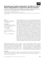
TNF release in co-cultures of IL-15 activated lymphocytes and monocytesTNF release in co-cultures of IL-15 activated lymphocytes and monocytes. (a) Peripheral blood lymphocytes (PBL), monocytes or culture of both cell
types were incubated with IL-15 (50 ng/ml; white column) or medium alone (black column) for 24 hours. As a positive control, monocytes were stim-
ulated with lipopolysaccharide (50 ng/ml; grey column in monocytes condition). To determine the effect of intercellular contacts between lym-
phocytes and monocytes in the presence of IL-15 (50 ng/ml), cells were separated by a 0.4 µm pore transwell (grey column in PBL + Mo condition).
TNF was measured in the cell-free supernatants with the use of an enzyme immunoassay. The data are shown as means ± SEM from five independ-
ent experiments. (b) PBL were stimulated with different doses of IL-15 (0.5 to 100 ng/ml) for 24 hours, and the cells were then washed and co-cul-
tured with autologous monocytes at a 10:1 ratio of PBL to monocytes for a further 24 hours. As a control, the cells in culture were separated by a
0.4 µm pore transwell. TNF was measured in the cell-free supernatants with the use of an enzyme immunoassay. The data are shown as means ±
SEM for eight independent experiments. (c) PBL were activated as described for (b) and then CD69 expression was analysed by flow cytometry. A
representative experiment is shown. The grey histogram depicts CD69 expression and the black solid-line histogram the negative control.
Available online />Page 3 of 11
(page number not for citation purposes)
gation (Sigma Diagnostics). This was followed by the removal
of monocytes by adhesion for 1 hour to Petri dishes (Costar,
Cambridge, MA, USA) in RPMI 1640 medium supplemented
with 10% FCS at 37°C. The lymphocyte-enriched fraction
contained less than 1% CD14
+
cells. CD4
+
and CD8
+
cells
were then obtained by negative selection with Subset Enrich-
ment Column kits (R&D Systems). Cell purity was determined
by flow cytometry and was always greater than 90% for CD4
+
and 95% for CD8
+
T lymphocytes. NK cells were purified by
negative selection with goat anti-mouse IgG Dynabeads
(Dynal Biotech, Oslo, Norway) previously coupled to anti-
CD3, anti-CD4 and anti-HLA-DR mAbs. After a second round
of selection with beads coupled to CD3 and CD19 mAbs
(Dynal Biotech) the cell population obtained was more than
95% CD56
+
with less than 1% CD3
+
cells.
In other experiments, PBL were depleted of T cells, B cells or
NK cells, and the NK-depleted PBL were obtained by incubat-
ing the PBL with immunomagnetic beads coupled to BAB281
(anti-NKp46), KD1 (anti-CD16) and Leu-19 (anti-CD56)
mAbs. This process was repeated and the cell population
obtained was less than 1% CD56
+
. B cell-depleted PBL and
T cell-depleted PBL populations were isolated by using the
same procedure with the anti-CD19 and anti-HLA-DR mAbs,
yielding a B cell-depleted PBL population that was less than
0.5% CD19
+
. When anti-CD3, anti-CD4 and anti-CD8 was
used, the T cell-depleted PBL population was less than 1%
CD3
+
.
Monocytic cells
Most experiments were performed with the human monocytic
leukaemic cell line THP-1 obtained from ATCC/LGC Promo-
chem (Barcelona, Spain). These cells were maintained in cul-
ture with RPMI 1640 medium supplemented with 10% heat-
inactivated FCS, penicillin (100 U/ml) and streptomycin (100
µg/ml) at 37°C in a humidified atmosphere consisting of 5%
CO
2
.
In experiments performed with human peripheral blood mono-
cytes, these cells were obtained with the following purification
procedure: peripheral blood mononuclear cells were obtained
by Histopaque-1077 density-gradient centrifugation and
resuspended in RPMI 1640 medium supplemented with 10%
FCS. A sample of this cellular suspension was analysed
through a Hitachi Coulter counter to determine the concentra-
tion of monocytes. A volume containing 10
5
monocytes was
then added to each well in 24-well plates (Costar) to allow the
attachment of monocytes and, after 1 hour at 37°C, wells were
washed three timed with RPMI 1640 medium. The population
attached to wells was more than 90% CD14
+
after cell
detachment and flow cytometry analysis. To perform co-cul-
ture assays, the autologous lymphocytes were treated as
described above and co-cultured with the monocytes at a
10:1 ratio (10
6
lymphocytes or subpopulations per 10
5
mono-
cytes attached at the wells of 24-well plates).
Patients and synovial fluid samples
Synovial fluid samples were obtained, with previous oral
informed consent, from patients attending our out-patient
clinic. Diagnoses included RA (n = 5), seronegative spondy-
loarthropathies (n = 6) and crystal-induced arthritis (n = 4).
Unfractionated or NK-depleted synovial fluid lymphocytes
(SFL) were purified as described above and this population
was, on average, less than 5% CD56
+
.
This study was approved by the ethics committee for clinical
research at Hospital Universitario de La Princesa.
Figure 2
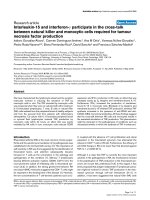
A subpopulation of IL-15-activated PBL induces TNF synthesis in monocytic cellsA subpopulation of IL-15-activated PBL induces TNF synthesis in monocytic cells. (a) Peripheral blood lymphocytes (PBL) were stimulated with IL-
15 at 50 ng/ml for 24 hours and then co-cultured with THP-1 cells for 24 hours; the ratio of lymphocytes to monocytes was 10:1. Under some con-
ditions, lymphocytes or THP-1 cells were fixed with 0.05% glutaraldehyde at 4°C for 30 to 45 s and washed intensively with sterile PBS before co-
culture. The data shown are the TNF concentration in the supernatant and are expressed as means ± SEM (n = 5). (b, c) PBL were stimulated as in
(a) and then incubated together with THP-1 cells for different durations (2 to 24 hours) (b) or at different cell ratios (1:1 to 50:1) (c). The data shown
are the TNF concentration in the supernatants and are expressed as means ± SEM (n = 5).
Arthritis Research & Therapy Vol 8 No 4 González-Álvaro et al.
Page 4 of 11
(page number not for citation purposes)
Cell-cell contact assays
PBL or different lymphocyte subsets were incubated for 24
hours in the presence of medium alone or with IL-15 (1 to 100
ng/ml). After being washed, the cells were resuspended in
medium and added to 24-well plates (Costar). Unless other-
wise stated, then THP-1 cells were added in the proportion 10
lymphocytes to 1 THP-1. As a negative control, lymphocyte–
THP-1 cell contact was prevented by using a 0.4 µm pore-size
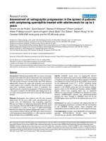
Figure 3
NK cells are the major lymphocyte subpopulation that induce TNF release by monocytesNK cells are the major lymphocyte subpopulation that induce TNF release by monocytes. (a) CD4 and CD8 T cells and natural killer (NK) cells were
isolated by negative selection from peripheral blood lymphocytes (PBL). Subsequently, purified cells or total PBL were incubated with IL-15 (50 ng/
ml; white bars), washed intensively and incubated together with THP-1 cells. To avoid intercellular contact where necessary, cells were separated by
a 0.4 µm pore semipermeable membrane (black bars). TNF was measured in cell-free supernatants harvested after 24 hours in co-culture. The data
are shown as means ± SEM (n = 5). (b) PBL were depleted of T cells, B cells or NK cells (PBL – T cells, PBL – B cells and PBL – NK cells, respec-
tively) as described in the Materials and methods section, and the total PBL or the different depleted PBL were then stimulated with 50 ng/ml IL-15
for 24 hours. After being washed, the different PBL groups were brought into contact with THP-1 cells (10:1 ratio of PBL to THP-1 cells; white bars)
or the cells were separated in culture by a 0.4 µm pore transwell (black bars) for 24 hours. The data show the TNF concentration in the supernatants
and are expressed as means ± SEM from five independent experiments. (c) PBL were depleted of NK cells and stimulated as described for (b). After
being washed, the different PBL groups were co-cultured with autologous monocytes (white column) at a 10:1 ratio of PBL to monocytes. As in (b),
black columns show TNF concentration in supernatants from conditions in which intercellular contacts was prevented by a 0.4 µm pore transwell.
The data show the TNF concentration in the supernatants and are expressed as means ± SEM from four independent experiments. (d) Synovial fluid
lymphocytes (SFL) obtained from knee effusions in different disorders were depleted of NK cells (SFL – NK) as described in the Materials and meth-
ods section. Then, both SFL and SFL-NK were allowed to contact with THP-1 cells (10:1 ratio of PBL to THP-1; white bars) or the cells were sepa-
rated in culture by a 0.4 µm pore transwell (black bars) for 24 hours. The data show the TNF concentration in the supernatants and are expressed as
means ± SEM from five samples from rheumatoid arthritis (RA), six samples from seronegative spondyloarthropathies (SSA) and four samples from
crystal-associated arthritis (CAA).
Available online />Page 5 of 11
(page number not for citation purposes)
transwell insert (Costar). In some experiments, lymphocytes or
THP-1 cells were fixed before cell co-culture (0.05% glutaral-
dehyde at 4°C for 30 to 45 seconds). After 24 hours the
supernatants were harvested and stored at -80°C until the
cytokines were quantified.
To investigate the involvement of different cell surface mole-
cules in these experiments, the following purified mAbs were
added: MAB002 (negative control), MAB647 (anti-IL15),
BAB281 (anti-NKp46), MA152 (anti-NKp80), MAB139 (anti-
NKG2D), z199 (anti-CD94/NKG2A), Lia3/2 (anti-CD18),
156-4H9 (anti-CD48) and 2-69 (anti-CD244). All mAbs were
used at a final concentration of 10 to 20 µg/ml.
Induction of IL-15 expression on THP-1 cells
THP-1 cells were stimulated with different concentrations of
IFN-γ (1 to 100 ng/ml), TNF (1 to 100 ng/ml), IL-1 (1 to 100
ng/ml) or medium alone for 24 hours; the expression of mem-
brane-bound IL-15 was then analysed by flow cytometry.
Flow cytometry analysis
Cells were incubated with the specific mAbs at 4°C for 30
minutes. After being washed in PBS, the cells were labelled
with fluorescein isothiocyanate-tagged goat anti-mouse Ig
(Dako, Salstrup, Denmark) for 30 minutes at 4°C. For double
staining, cells were additionally incubated for 15 minutes with
mouse serum diluted 1:100 (ICN Biomedicals Inc, Aurora,
OH, USA); they were washed and then incubated with a phy-
coerythrin-conjugated anti-CD56 mAb (Becton Dickinson) for
20 minutes. At least 5 × 10
3
cells were analysed with a FAC-
Scan flow cytometer (Becton Dickinson).
Quantification of cytokines in cell-free supernatant
Human TNF concentrations in supernatants were determined
by an enzyme immunoassay (EIA). In brief, 96-well high-bind-
ing EIA plates (Costar) were coated overnight at 4°C with 50
µl of MAB610 (R&D Systems) per well at 8 µg/ml in PBS, pH
7.4. Subsequently, each well was washed twice with 200 µl of
wash buffer (0.05% Tween 20 in PBS, pH 7.4) and blocked
for 1 hour by adding 200 µl of PBS containing 2% BSA at
37°C. After each step, the wells were washed three times with
200 µl of wash buffer; 50 µl of dilution buffer (0.1% BSA,
0.05% Tween20, 20 mM Trizma base, 150 mM NaCl, pH 7.3)
per well plus 50 µl of each sample or standard dilutions for
recombinant human TNF (10,000 to 39 pg/ml; R&D Systems)
were then added to the respective wells (in duplicate) and
incubated at room temperature for 2 hours. Bound TNF was
detected by incubation for 1 hour with, in each well, 50 µl of
BAF210 (R&D Systems) diluted to 200 ng/ml in dilution buffer
at room temperature. After washing, 100 µl streptavidin HRP
(Calbiochem, San Diego, CA) diluted 1:5,000 in dilution buffer
was added to each well for 20 minutes at room temperature;
the reaction was then developed with 100 µl 3,3',5,5' -tetram-
ethylbenzidine (Chemicon International Inc., Temecula, CA,
USA) per well. The optical density of each well was deter-
mined with a SpectraII microtitre plate reader (Innogenetics
Diagnóstica y Terapéutica, Barcelona, Spain) set to 450 nm,
with wavelength correction set to 550 nm. Cytokine values
were calculated from the standard curve. Samples that gener-
ated values higher than the highest standard were diluted
(1:1) in dilution buffer and assayed again.
Because TNF production can vary depending on the lym-
phocyte donor, in the experiments in which cell–cell interac-
tions were blocked with mAbs the results were normalized
with the following equation: TNF production = 100 × TNF
mAb
/TNF
medium
.
Human IFN-γ concentrations were measured with an EIA kit
from R&D Systems.
Statistical analysis
Statistical analysis was performed with Stata 9.1 for Windows
(StataCorp LP, College Station, TX, USA), by using one-way
analysis-of-variance model with Bonferroni multiple-compari-
son correction for multiple sample experiments and the Mann–
Whitney test for experiments with comparison between two
groups.
Table 1
TNF production in co-cultures of SFL and THP-1 cells: effect of NK cell depletion
Sample source TNF production (pg/ml) Inhibition (%)
SFL SFL – NK
Rheumatoid arthritis (n = 5) 9,237 ± 4,062 2,472 ± 2,472 73.2
Spondyloarthropathies (n = 6) 2,680 ± 503 1,407 ± 442 47.5
Crystal-associated arthritis (n = 4) 3,557 ± 1,402 2,478 ± 1,196 30.3
Total (n = 15) 5,187 ± 1,735 2,059 ± 491 60.3
a
Where errors are shown, data are means ± SEM. SFL, synovial fluid lymphocytes;
SFL-NK, synovial fluid lymphocytes depleted of natural killer (NK) cells.
a
Statistical significance: p = 0.039, Mann-Whitney test.
Arthritis Research & Therapy Vol 8 No 4 González-Álvaro et al.
Page 6 of 11
(page number not for citation purposes)
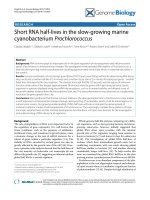
Figure 4
Reciprocal activation between NK and THP-1 cells, the role of IL-15 and IFN-γReciprocal activation between NK and THP-1 cells, the role of IL-15 and IFN-γ. (a) IL-15 and β
2
integrins are involved in the intercellular contact with
THP-1 cells that induces the expression of CD69 in natural killer (NK) cells. Peripheral blood lymphocytes (PBL) were co-cultured with THP-1 cells
(10:1 ratio of PBL to THP-1) for 24 hours in medium alone or in the presence of an anti-β
2
integrin mAb (Lia3/2) or an anti-IL-15 mAb (MAB647). As
a control, both cell lines were separated by a 0.4 µm pore transwell. A representative experiment of the five performed is shown. The histograms rep-
resent the CD69 expression in CD56
+
cells in the medium (grey histogram in all panels) or under the different conditions (solid black line in each
panel); a negative control is also shown (dotted histogram in all panels). (b) Intercellular contact between THP-1 and NK cells induces IFN-γ produc-
tion. NK cells were cultured in the presence of 50 ng/ml IL-15 for 24 hours, or in medium alone, and the NK cells were then washed and incubated
together with THP-1 cells. In some conditions the cells were separated by a 0.4 µm pore transwell and after 24 hours the supernatants were har-
vested to measure the IFN-γ content with the use of an enzyme immunoassay. The data show the IFN-γ concentrations and are expressed as means
± SEM from six independent experiments. One-way analysis-of-variance model with Bonferroni multiple-comparison correction was used to deter-
mine statistical significance. (c) IFN-γ increases IL-15 membrane expression in THP-1 cells. Cells were incubated with IFN-γ (100 ng/ml), TNF (100
ng/ml) or IL-1 (100 ng/ml) for 24 hours and then the membrane-bound IL-15 (mIL-15) was measured by indirect immunofluorescence and flow
cytometry. A representative experiment of the five performed is shown. Grey histograms represent mIL-15 on stimulated THP-1 cells, solid-line histo-
grams represent basal mIL-15 expression, and the dotted-line histogram is the negative control.
Available online />Page 7 of 11
(page number not for citation purposes)
Results
Characterization of a model of TNF production in co-
cultures of monocytic cells and IL-15-activated
peripheral blood lymphocytes
Different in vitro models have been described that have raised
the importance of intercellular contacts between activated
lymphocytes and monocytic cells in the perpetuation of rheu-
matoid synovitis [9-11]. We have used a model in which PBL
are activated with IL-15, a cytokine with a specific presence in
the RA microenvironment compared with other arthropathies
[17-19]. Neither PBL nor monocytes incubated separately
with IL-15 at 50 ng/ml were able to produce TNF (Figure 1a),
whereas lipopolysaccharide-activated monocytes secreted
large amounts of TNF (Figure 1a; monocytes, grey column). By
contrast, when PBL and monocytes were incubated together
in the presence of IL-15, a relevant TNF production was
observed (Figure 1a). When cell contact between the two cell
types was prevented by a 0.4 µm pore transwell, TNF synthe-
sis dropped markedly (Figure 1a; PBL+Mo, grey column). The
production of TNF in this model was dependent on the IL-15
dose (Figure 1b) and was correlated with the intensity of PBL
activation measured through CD69 expression on the subpop-
ulation that responded to IL-15 (Figure 1c).
Similar results were obtained with the monocytic cell line THP-
1 (Figure 2a–c). To determine whether TNF secretion was
produced by monocytic or lymphocytic cells, experiments
were performed with fixed cells. TNF concentration decreased
markedly when THP-1 cells were fixed with respect to their
basal condition, suggesting that monocytic cells were the
main source of this cytokine (Figure 2a). TNF release into the
supernatant was dependent on both time (Figure 2b) and the
ratio of IL-15-activated PBL to THP-1 cells (Figure 2c). Indeed,
TNF release was very inefficient at a ratio of 1:1 and reached
a 'plateau' at ratios above 20 activated PBL per THP-1 cell
(Figure 2c). These data suggest that the lymphocyte subset
that becomes activated by IL-15 and is able to induce TNF
production in macrophages seems to be a limiting factor.
NK cells induce TNF synthesis by monocytic cells
Additional experiments showed that the effect of purified NK
cells on TNF synthesis by THP-1 cells was similar to that of
unfractionated PBL (Figure 3a). In contrast, neither CD4
+
nor
CD8
+
T cells were able to induce any TNF synthesis (Figure
3a). Furthermore, when B cells or T cells were removed from
the PBL, their capacity to induce TNF synthesis remained
unaffected (Figure 3b). In contrast, the removal of NK cells
abrogated this effect almost completely (Figure 3b), a finding
that was reproduced when experiments were performed with
autologous peripheral blood monocytes (Figure 3c).
Similar results were obtained when we studied the effect of
SFL. Hence, when THP-1 cells were incubated with SFL
depleted of NK cells, TNF production was significantly lower
than that detected in co-cultures of THP-1 with complete SFL
(Table 1). To determine whether our findings were specific to
RA or were a common phenomenon in most inflammatory
arthropathies, we analysed data grouped by different disor-
ders. Interestingly, the highest TNF synthesis was observed in
co-cultures of RA SFL with THP-1 cells (Figure 3d). The inhi-
bition of TNF production, when SFL were depleted of NK cells,
was therefore stronger in samples from patients with RA
(about 75% inhibition) than in synovial fluid from seronegative
spondyloarthropathies (50%) or in samples from crystal-
induced arthritis (30%; Table 1).
Monocytic cells induce NK cell activation through
membrane-bound IL-15
Resting PBL and purified NK cells induce TNF synthesis in
monocytes and THP-1 cells to a smaller extent than those pre-
viously stimulated with IL-15 (Figure 1a, and data not shown).
We therefore assessed the possible effects of THP-1 cells on
NK cell activation by measuring the expression of CD69 in
these cells. More than 90% of NK cells expressed CD69 on
co-culture with this monocytic cell line (Figure 4a and Table 2).
This effect was almost totally abrogated when contact
between the two cell types was prevented with 0.4 µm tran-
swell inserts or partly prevented by the addition of antibodies
against β
2
integrins or of anti-IL-15 mAbs (Figure 4a and Table
2). Interestingly, this inhibition of CD69 expression was asso-
ciated with a poorer capacity to induce the synthesis of TNF
(Table 2).
In addition, whereas the incubation of resting PB NK cells
together with THP-1 cells induced IFN-γ production, this was
completely abrogated when both cell lines were separated by
a 0.4 µm transwell (Figure 4b). The IFN-γ produced by NK
cells prestimulated with IL-15 was significantly higher, but in
this case the prevention of intercellular contact with the use of
Table 2
Blockade of CD18 and mIL-15 decreases CD69 expression and TNF production
Substance Medium Transwell anti-CD18 anti-IL15
CD69 (RFI) 69.3 ± 14.9 21.9 ± 13
a
35.3 ± 10.4
a
39 ± 12.2
b
TNF (pg/ml) 9,687 ± 842 559 ± 139
a
3,594 ± 9,342
b
5,364 ± 841
b
The data shown are means ± SEM from eight independent experiments. m-IL-15, membrane-bound IL-15; RFI, relative fluorescence intensity.
a
Statistical significance: p < 0.01 by analysis of variance with Bonferroni multiple-comparison tests.
b
Statistical significance: p < 0.001 by analysis
of variance with Bonferroni multiple-comparison tests.
Arthritis Research & Therapy Vol 8 No 4 González-Álvaro et al.
Page 8 of 11
(page number not for citation purposes)
the transwell did not significantly decrease IFN-γ release (Fig-
ure 4b).
These findings support the notion that membrane-anchored IL-
15 participates in the activation of NK cells after co-culture
with THP-1 cells, as has been suggested in previous studies
with monocytes and synoviocytes [20,21]. We therefore stud-
ied whether the proinflammatory cytokines IFN-γ, IL-1 and TNF
modulate IL-15 expression on THP-1 cells, which is very low in
resting THP-1 cells. Unlike IFN-γ, neither TNF nor IL-1 was
able to induce significant expression of membrane-bound IL-
Figure 5
NK cell surface molecules involved in the cell–cell interaction that promotes TNF production by monocytesNK cell surface molecules involved in the cell-cell interaction that promotes TNF production by monocytes. (a) The effect of IL-15 on the expression
of cell surface molecules by natural killer (NK) cells. NK cells were cultured with IL-15 at 50 ng/ml or medium alone for 24 hours, and the expression
of different surface molecules was then assessed by flow cytometry. Grey-filled histograms represent the expression of each molecule on resting NK
cells, the grey-line histograms represent the expression on IL-15 activated NK cells, and the dotted-line histograms represent the negative control.
One representative experiment is shown. (b) Effect of different antibodies against NK cell surface molecules on TNF production in co-cultures of NK
and THP-1 cells. IL-15-stimulated NK cells were co-cultured with THP-1 cells at a 10:1 cell ratio for 24 hours in the presence of different monoclonal
antibodies (see the Materials and methods section for further information). The TNF concentration in cell-free supernatants was quantified with an
enzyme immunoassay. The results show the percentage TNF production and are expressed as means ± SEM for eight independent experiments
(see the Materials and methods section for definition). Staistical significance:*p < 0.01;
§
p < 0.05; analysis-of-variance test. (c) Expression of
CD244 and CD48 on monocytes and THP-1 cells. The solid-line histogram represents the expression of each molecule and the grey histogram the
negative control.
Available online />Page 9 of 11
(page number not for citation purposes)
15 in these cells (Figure 4c). Moreover, the effect of IFN-γ was
clearly dose-dependent (data not shown).
The role of NK-cell surface molecules in the induction of
TNF synthesis
Exposure to IL-15 increased the expression of CD69, CD56,
CD48 and NKG2D by NK cells, although this cytokine did not
have any significant effect on other surface molecules such as
NKG2A, CD244 (2B4), NKp46 or NKp80 (Figure 5a). To
assess the possible influence of these molecules, we per-
formed functional experiments with different mAbs. The mAbs
against NKp46, NKp80, NKG2A and NKG2D did not exert
any relevant effect on TNF production, whereas the blockage
of β
2
integrins significantly inhibited TNF release (Figure 5b).
In contrast, the mAbs against CD244 (2-69 mAb) and CD48
(the CD244 ligand) increased TNF synthesis (Figure 4b). It is
noteworthy that NK cells and monocytes express both CD48
and CD244, whereas THP-1 cells express only CD244 (Fig-
ure 5a and 5c). Incubation of each cell with anti-CD48 or anti-
CD244 mAb did not induce TNF release when these cells
were cultured alone (data not shown).
These data suggest that IL-15 enhances the expression of
several surface molecules in NK cells. Furthermore, some of
these could participate in the intercellular contacts that regu-
late TNF production by monocytic cells, such as β
2
integrins,
CD48 and CD244.
Discussion
A significant amount of evidence has accumulated supporting
the importance of intercellular contacts in the pathogenic
mechanisms underlying RA. Indeed, the importance of these
cell-cell interactions in the synthesis and release of pro-inflam-
matory cytokines and metalloproteinases has been highlighted
in several studies [9-11,20,22-24]. Although T cells were
thought to be responsible for these activating contacts, our
data indicate that NK cells are the main subset of lymphocytes
that induce TNF production by monocytic cells in this experi-
mental model of intercellular contact. In fact, considering that
NK cells compose about 10% of the PBL, our data suggest
that cellular ratios as low as 1 NK cell to 5 or 10 THP-1 cells
are able to induce TNF production. Therefore the effects pre-
viously assigned to T lymphocytes could indeed be mediated
mostly by NK cells. In this regard, although previous works
were described to be performed with purified T lymphocytes
(more than 90% CD3
+
cells), none of them actively employed
a strategy to deplete NK cells from their samples [9-11,20,22-
24]. Furthermore, here we provide solid evidence that mono-
cytic intercellular contacts with other subsets of PBLs (CD4
+
,
CD8
+
and B cells) do not induce TNF production.
Our results concur with a recent report describing that acti-
vated NK cells induce intracellular TNF expression in mono-
cytes [12]. However, in that work NK cells were purified by
positive selection, a procedure that may induce cellular signal-
ling. In contrast, our findings were obtained through negative
selection of different subpopulations, avoiding this problem. In
contrast, two previous studies described a bidirectional cross-
talk between NK cells and dendritic cells leading to mutual
activation, but they did not describe the molecules underlying
this phenomenon [13,14]. We show here that negatively
selected resting NK cells are able to induce TNF synthesis
because they are activated by coming into contact with mIL-
15 on monocytes. This interaction induces the expression of
CD69 on NK cells and also promotes them to synthesize IFN-
γ, which in turn upregulates the expression of mIL-15 in resting
monocytic cells. Our data therefore support the involvement of
monocytes and NK cells in a reciprocal activation loop in
which IL-15 and IFN-γ are critical for the sustained production
of TNF.
With regard to the specific role of NK cells in different rheu-
matic conditions, our data show that the capacity to induce
TNF release diminished when the SFL were depleted of NK
cells. Both effects, namely the induction of TNF synthesis and
its inhibition when NK cells were depleted from SFL, were par-
ticularly evident in samples from patients with RA. It is conceiv-
able that the activation of macrophages by NK cells, a normal
pathway during the initial immune response, might be exacer-
bated in RA. This might be the consequence of the increased
expression of NK-activating cytokines (IL-12, IL-15 and IL-18)
in these patients [25]. Indeed, we have already seen that in
patients with RA, the serum and synovial fluid levels of IL-15
are higher than in other inflammatory arthropathies [18,19].
Furthermore, a significant correlation between IL-15 serum
levels and the expression of mIL-15 on PB monocytes was
observed in patients with early arthritis (I Gonzalez-Alvaro, AM
Ortiz and Dominguez-Jimenez C, unpublished observation).
Accordingly, it would be of interest to determine whether an
increased expression of mIL-15 or IL-15 serum levels could
identify patients with a more severe disease progression.
We have also generated information about the molecules on
activated NK cells that promote TNF production in monocytes.
The interaction of CD244 expressed by THP-1 cells with its
CD48 ligand on NK cells seems to be involved in regulating
the TNF production mediated by intercellular contacts. How-
ever, the precise role of these molecules remains to be
defined, particularly given that CD244 is known to be an acti-
vating receptor in NK cells [26,27]. However, CD244 is also
thought to mediate inhibitory responses in the absence of the
signalling adaptor protein SAP [28]. Our data may support this
inhibitory role because the model renders higher TNF concen-
trations with THP-1 cells, which lack CD48, than in monocytes
that express both CD48 and CD244.
Thus, our findings show that NK cells can engage and stimu-
late monocytic cells, resulting in the synthesis of TNF. This
finding opens the possibility of exploring new therapeutic tar-
gets for RA and probably other chronic inflammatory disor-
Arthritis Research & Therapy Vol 8 No 4 González-Álvaro et al.
Page 10 of 11
(page number not for citation purposes)
ders. In this regard, these data further support the application
of IL-15 blockage as a treatment for RA, a strategy that has so
far provided satisfactory preliminary results [29].
Conclusion
The main new findings described in this study are as follows.
First, NK cells, rather than T lymphocytes, are the main lym-
phocyte subpopulation involved in the cell-contact-mediated
production of TNF that is induced in monocytic cells. This find-
ing may be relevant when considering the pathogenesis of
chronic synovitis and it seems to be particularly important with
regard to RA. Second, our findings suggest that mIL-15 and
IFN-γ contribute to the maintenance of a mutual activator loop
between NK cells and monocytes that may result in persistent
TNF synthesis. Third, our data also suggest that CD244 and
CD48 might regulate TNF production by monocytic cells.
Competing interests
The authors declare that they have no competing interests.
Authors' contributions
IG-A participated in the design of the study, performed statis-
tical analysis and drafted the manuscript. CD-J and VN-G puri-
fied cells and performed co-culture assays of both PBL and
SFL and performed enzyme immunoassays. AMO performed
the flow cytometry analysis. PR-N obtained purified NK cells.
EF-R and DS participated in the design of the study and
helped to draft the manuscript. FS-M participated in the
design of the study and its coordination and helped to draft the
manuscript. All authors read and approved the final
manuscript.
Acknowledgements
We thank Dr R Gonzalez-Amaro for critical review of the manuscript.
This work was supported by grants from the 'Instituto de Salud Carlos
III' (G03/0152 and 04/2009) to IG-A and from the 'Ministerio de Edu-
cación y Ciencia' (BFU2005-08435/BMC) and the 'Fundación Juan
March' (Ayuda a la Investigación Básica 2002) to FS-M. The work of
CD-J was supported by a grant from the 'Fundación Española de
Reumatología'.
References
1. Firestein GS: Evolving concepts of rheumatoid arthritis. Nature
2003, 423:356-361.
2. Cush JJ, Lipsky PE: Phenotypic analysis of synovial tissue and
peripheral blood lymphocytes isolated from patients with
rheumatoid arthritis. Arthritis Rheum 1988, 31:1230-1238.
3. Laffon A, Sanchez-Madrid F, Ortiz de Landazuri M, Jimenez Cuesta
A, Ariza A, Ossorio C, Sabando P: Very late activation antigen on
synovial fluid T cells from patients with rheumatoid arthritis
and other rheumatic diseases. Arthritis Rheum 1989,
32:386-392.
4. Fox DA: The role of T cells in the immunopathogenesis of
rheumatoid arthritis: new perspectives. Arthritis Rheum 1997,
40:598-609.
5. Bresnihan B, Alvaro-Gracia JM, Cobby M, Doherty M, Domljan Z,
Emery P, Nuki G, Pavelka K, Rau R, Rozman B, et al.: Treatment
of rheumatoid arthritis with recombinant human interleukin-1
receptor antagonist. Arthritis Rheum 1998, 41:2196-2204.
6. Edwards JC, Szczepanski L, Szechinski J, Filipowicz-Sosnowska
A, Emery P, Close DR, Stevens RM, Shaw T: Efficacy of B-cell-
targeted therapy with rituximab in patients with rheumatoid
arthritis. N Engl J Med 2004, 350:2572-2581.
7. Feldmann M: Development of anti-TNF therapy for rheumatoid
arthritis. Nat Rev Immunol 2002, 2:364-371.
8. Choy EH, Panayi GS: Cytokine pathways and joint inflamma-
tion in rheumatoid arthritis. N Engl J Med 2001, 344:907-916.
9. Isler P, Vey E, Zhang JH, Dayer JM: Cell surface glycoproteins
expressed on activated human T cells induce production of
interleukin-1β by monocytic cells: a possible role of CD69. Eur
Cytokine Netw 1993, 4:15-23.
10. McInnes IB, Leung BP, Sturrock RD, Field M, Liew FY: Inter-
leukin-15 mediates T cell-dependent regulation of tumor
necrosis factor-α production in rheumatoid arthritis. Nat Med
1997, 3:189-195.
11. Sebbag M, Parry SL, Brennan FM, Feldmann M: Cytokine stimu-
lation of T lymphocytes regulates their capacity to induce
monocyte production of tumor necrosis factor-α, but not inter-
leukin-10: possible relevance to pathophysiology of rheuma-
toid arthritis. Eur J Immunol 1997, 27:624-632.
12. Dalbeth N, Gundle R, Davies RJ, Lee YC, McMichael AJ, Callan
MF: CD56bright NK cells are enriched at inflammatory sites
and can engage with monocytes in a reciprocal program of
activation. J Immunol 2004, 173:6418-6426.
13. Gerosa F, Baldani-Guerra B, Nisii C, Marchesini V, Carra G,
Trinchieri G: Reciprocal activating interaction between natural
killer cells and dendritic cells. J Exp Med 2002, 195:327-333.
14. Piccioli D, Sbrana S, Melandri E, Valiante NM: Contact-depend-
ent stimulation and inhibition of dendritic cells by natural killer
cells. J Exp Med 2002, 195:335-341.
15. Sanchez-Madrid F, De Landazuri MO, Morago G, Cebrian M,
Acevedo A, Bernabeu C: VLA-3: a novel polypeptide associa-
tion within the VLA molecular complex: cell distribution and
biochemical characterization. Eur J Immunol 1986,
16:1343-1349.
16. Sanchez-Mateos P, Sanchez-Madrid F: Structure-function rela-
tionship and immunochemical mapping of external and intra-
cellular antigenic sites on the lymphocyte activation inducer
molecule, AIM/CD69. Eur J Immunol 1991, 21:2317-2325.
17. Cordero OJ, Salgado FJ, Mera-Varela A, Nogueira M: Serum
interleukin-12, interleukin-15, soluble CD26, and adenosine
deaminase in patients with rheumatoid arthritis. Rheumatol Int
2001, 21:69-74.
18. Gonzalez-Alvaro I, Ortiz AM, Garcia-Vicuna R, Balsa A, Pascual-
Salcedo D, Laffon A: Increased serum levels of interleukin-15
in rheumatoid arthritis with long-term disease. Clin Exp
Rheumatol 2003, 21:639-642.
19. Ortiz AM, Laffon A, Gonzalez-Alvaro I: CD69 expression on lym-
phocytes and interleukin-15 levels in synovial fluids from dif-
ferent inflammatory arthropathies. Rheumatol Int 2002,
21:182-188.
20. Miranda-Carus ME, Balsa A, Benito-Miguel M, Perez de Ayala C,
Martin-Mola E: IL-15 and the initiation of cell contact-dependent
synovial fibroblast-T lymphocyte cross-talk in rheumatoid
arthritis: effect of methotrexate. J Immunol 2004,
173:1463-1476.
21. Musso T, Calosso L, Zucca M, Millesimo M, Ravarino D, Giovarelli
M, Malavasi F, Ponzi AN, Paus R, Bulfone-Paus S: Human mono-
cytes constitutively express membrane-bound, biologically
active, and interferon-γ -upregulated interleukin-15. Blood
1999, 93:3531-3539.
22. Ribbens C, Dayer JM, Chizzolini C: CD40-CD40 ligand (CD154)
engagement is required but may not be sufficient for human T
helper 1 cell induction of interleukin-2- or interleukin-15-
driven, contact-dependent, interleukin-1β production by
monocytes. Immunology 2000, 99:279-286.
23. Wagner DH Jr, Stout RD, Suttles J: Role of the CD40-CD40 lig-
and interaction in CD4
+
T cell contact-dependent activation of
monocyte interleukin-1 synthesis. Eur J Immunol 1994,
24:3148-3154.
24. Lacraz S, Isler P, Vey E, Welgus HG, Dayer JM: Direct contact
between T lymphocytes and monocytes is a major pathway for
induction of metalloproteinase expression. J Biol Chem 1994,
269:22027-22033.
25. Liew FY, McInnes IB: The role of innate mediators in inflamma-
tory response. Mol Immunol 2002, 38:887-890.
Available online />Page 11 of 11
(page number not for citation purposes)
26. Assarsson E, Kambayashi T, Persson CM, Ljunggren HG, Cham-
bers BJ: 2B4 co-stimulation: NK cells and their control of adap-
tive immune responses. Mol Immunol 2005, 42:419-423.
27. Nakajima H, Cella M, Langen H, Friedlein A, Colonna M: Activating
interactions in human NK cell recognition: the role of 2B4-
CD48. Eur J Immunol 1999, 29:1676-1683.
28. Veillette A: SLAM family receptors regulate immunity with and
without SAP-related adaptors. J Exp Med 2004,
199:1175-1178.
29. Baslund B, Tvede N, Danneskiold-Samsoe B, Larsson P, Panayi G,
Petersen J, Petersen LJ, Beurskens FJ, Schuurman J, van de Win-
kel JG, et al.: Targeting interleukin-15 in patients with rheuma-
toid arthritis: a proof-of-concept study. Arthritis Rheum 2005,
52:2686-2692.