Báo cáo y học: "Inhibition of protein geranylgeranylation induces apoptosis in synovial fibroblasts" pdf
Bạn đang xem bản rút gọn của tài liệu. Xem và tải ngay bản đầy đủ của tài liệu tại đây (1.16 MB, 11 trang )
Open Access
Available online />Page 1 of 11
(page number not for citation purposes)
Vol 8 No 4
Research article
Inhibition of protein geranylgeranylation induces apoptosis in
synovial fibroblasts
Alison M Connor
1
, Stuart Berger
1
, Aru Narendran
2
and Edward C Keystone
3
1
The Wellesley Toronto Arthritis and Immune Disorder Research Centre, 101 College St. Toronto, Ontario, Canada M5G 1L7
2
Southern Alberta Children's Cancer Program, Alberta Children's Hospital, 1820 Richmond Road SW Calgary, Alberta, Canada T2T 5C7
3
The Rebecca MacDonald Centre for Arthritis and Autoimmune Disease, Mount Sinai Hospital, 60 Murray Street, Toronto, Ontario, Canada, M5T 3L9
Corresponding author: Edward C Keystone,
Received: 23 Feb 2006 Revisions requested: 21 Apr 2006 Revisions received: 1 May 2006 Accepted: 4 May 2006 Published: 14 Jun 2006
Arthritis Research & Therapy 2006, 8:R94 (doi:10.1186/ar1968)
This article is online at: />© 2006 Connor et al.; licensee BioMed Central Ltd.
This is an open access article distributed under the terms of the Creative Commons Attribution License ( />),
which permits unrestricted use, distribution, and reproduction in any medium, provided the original work is properly cited.
Abstract
Statins, competitive inhibitors of hydroxymethylglutaryl-CoA
reductase, have recently been shown to have a therapeutic
effect in rheumatoid arthritis (RA). In RA, synovial fibroblasts in
the synovial lining, are believed to be particularly important in the
pathogenesis of disease because they recruit leukocytes into
the synovium and secrete angiogenesis-promoting molecules
and proteases that degrade extracellular matrix. In this study, we
show a marked reduction in RA synovial fibroblast survival
through the induction of apoptosis when the cells were cultured
with statins. Simvastatin was more effective in RA synovial
fibroblasts than atorvastatin, and both statins were more potent
on tumor necrosis factor-α-induced cells. In contrast, in
osteoarthritis synovial fibroblasts, neither the statin nor the
activation state of the cell contributed to the efficacy of
apoptosis induction. Viability of statin-treated cells could be
rescued by geranylgeraniol but not by farnesol, suggesting a
requirement for a geranylgeranylated protein for synovial
fibroblast survival. Phase partitioning experiments confirmed
that in the presence of statin, geranylgeranylated proteins are
redistributed to the cytoplasm. siRNA experiments
demonstrated a role for Rac1 in synovial fibroblast survival.
Western blotting showed that the activated phosphorylated
form of Akt, a protein previously implicated in RA synovial
fibroblast survival, was decreased by about 75%. The results
presented in this study lend further support to the importance of
elevated pAkt levels to RA synovial fibroblast survival and
suggest that statins might have a beneficial role in reducing the
aberrant pAkt levels in patients with RA. The results may also
partly explain the therapeutic effect of atorvastatin in patients
with RA.
Introduction
Rheumatoid arthritis (RA) is a chronic inflammatory disease
causing progressive joint destruction, deformity and disability.
The pathogenesis of the rheumatoid joint involves hyperplasia
of the synovial lining cells, mononuclear cell infiltration and
new blood vessel formation within the synovium as well as the
destruction of cartilage and underlying bone as a conse-
quence of pro-inflammatory cytokines and proteases [1].
Much of the pathology is thought to be driven by cytokines,
particularly tumor necrosis factor α (TNF-α) [2].
Synovial tissue consists primarily of two distinct cell types: the
macrophage-like synoviocytes and synovial fibroblasts. The
synovial fibroblasts are important in all aspects of the patho-
genesis of arthritis. Hyperplasia of the synovial lining in RA is
due primarily to increases in the number of synovial fibroblasts.
Although the reason for this increase is currently unknown,
impaired apoptosis or senescence has been proposed to
explain their increased numbers [3].
The RA synovial fibroblast response to the macrophage-
derived cytokines TNF-α and IL-1 includes elevated expres-
sion of adhesion molecules, cytokines and chemokines. RA
synovial fibroblasts also secrete angiogenesis-promoting mol-
ecules such as vascular endothelial growth factor A and sev-
eral proteases, including matrix metalloproteinases,
CIA = collagen-induced arthritis; DMSO = dimethylsulfoxide; FLS = fibroblast-like synoviocytes; FPP = farnesylpyrophosphate; GGPP = geranylger-
anylpyrophosphate; HMG = hydroxymethylglutaryl; IL = interleukin; OA = osteoarthritis; PBS = phosphate-buffered saline; RA = rheumatoid arthritis;
siRNA = short interfering RNA; TNF-α = tumor necrosis factor-α; XTT = sodium 3-(1-(phenylamino-carbonyl)-3,4-tetrazolium)-bis (4-methoxy-6-nitro)
benzenesulfonic acid hydrate; TUNEL = TdT-mediated dUTP nick end labelling.
Arthritis Research & Therapy Vol 8 No 4 Connor et al.
Page 2 of 11
(page number not for citation purposes)
aggrecanases and cathepsins, that mediate extracellular
matrix degradation [4].
TNF-α is capable of signaling both cell-survival and cell-death
signals. The response of a cell to TNF-α depends on specific
adaptors and downstream signaling molecules [5]. The addi-
tion of TNF-α to RA synovial fibroblasts results in resistance to
apoptosis and hence to increased survival as well as prolifera-
tion [6]. Recent reports have indicated that it is possible to
reverse the survival response of RA synovial fibroblasts to
TNF-α by inhibiting the translocation of nuclear factor κB to
the nucleus [7], or ectopically expressing TIMP (tissue inhibi-
tor of metalloproteinases) 3 [8]. The ability to reverse resist-
ance of fibroblast-like synoviocytes (FLS) to apoptosis could
represent an important therapeutic target in arthritis [9].
Statins, competitive inhibitors of hydroxymethylglutaryl
(HMG)-CoA reductase, were initially designed as inhibitors of
cholesterol synthesis [10]. HMG-CoA reductase catalyzes the
conversion of HMG-CoA to mevalonate, a rate-limiting step in
cholesterol biosynthesis. However, statins seem to have anti-
inflammatory effects that cannot be accounted for by their
lipid-lowering abilities. These include the suppression of proin-
flammatory cytokine and chemokine production, immunomod-
ulation and the downregulation of endothelial cell activation
[11,12]. As a consequence of these properties, statin therapy
has been examined in several chronic immune-mediated
inflammatory diseases including experimental autoimmune
encephalomyelitis and arthritis. The statin simvastatin has
been shown to exhibit a therapeutic effect in the collagen-
induced arthritis (CIA) model of RA [13]. It was thought to
exert its effect through decreasing the viability of T helper type
1 cells and attenuating the interaction of T cells with macro-
phages. In contrast with these results, another study showed
that neither atorvastatin nor rosurvastatin had a beneficial
effect on the mouse CIA model of arthritis. The results of sim-
vastatin could be accounted for by severe side effects [14].
Nevertheless, atorvastatin was found to have a therapeutic
effect in patients with RA as well as beneficially influencing
inflammatory markers [15].
Some of the beneficial effects of statins can be explained by
the fact that the inhibition of mevalonate synthesis by statins
also prevents the synthesis of other intermediates along the
cholesterol biosynthetic pathway. These include the isopre-
noid intermediates geranylgeranylpyrophosphate (GGPP) and
farnesylpyrophosphate (FPP), which are required for the pre-
nylation of many cellular proteins such as the small GTP-bind-
ing proteins Ras, Rho and Rac [16]. Isoprenylation enables the
insertion of these proteins into the cellular membrane, where
they function in intracellular signal transduction pathways [17].
It is significant that statins have also been shown to induce
apoptosis, mediated by inhibition of the synthesis of FPP and
GGPP [18-20].
Because reduced apoptosis of synovial fibroblasts in RA is
thought to be a cause of synovial hyperplasia, statins could
theoretically reduce joint damage by interfering with synovial
fibroblast survival. The aim of the present study was therefore
to determine whether statins could substantially reduce RA
synovial fibroblast survival and whether one statin would be
particularly effective. Our data suggest that simvastatin is sig-
nificantly more effective than atorvastatin at decreasing the
viability of RA synovial fibroblasts, particularly in the presence
of TNF-α. This contrasts with the viability of synovial fibroblasts
derived from patients with osteoarthritis (OA), in whom no dif-
ferential effect of the statins was observed. The decreased via-
bility of RA synovial fibroblasts with statins was shown to be
mediated by the inhibition of membrane-associated geran-
ylgeranylated proteins and was associated with decreased
pAkt levels.
Materials and methods
Synovial tissue
The protocol for patient consent and the use of human tissues
was approved by ethics review committees at both the Univer-
sity Health Network and St Michael's Hospital. All tissue was
obtained with patient consent.
Cell culture
Synovial fibroblasts were isolated from synovial tissues of
patients with RA or OA removed at the time of arthroplasty, as
described previously [21]. Cells were maintained in Opti-MEM
supplemented with 4% fetal bovine serum and 1% antibiotic-
antimycotic (Life Technologies, Rockville, MD, USA) and were
cultured at 37°C in a humidified chamber containing 95% air,
5% CO
2
. Synovial fibroblasts were used at passages 2 to 4.
Unless indicated otherwise, cells used for Western blots were
plated at 10
5
cells per well in a six-well plate for 48 hours
before initiation of experiments.
Effect of statins on cellular viability
Stock solutions of lovastatin (Sigma-Aldrich, Oakville, Ontario,
Canada) and atorvastatin (Toronto Research Chemicals,
North York, Ontario, Canada) were prepared in dimethylsulfox-
ide (DMSO). Cerivastatin (Toronto Research Chemicals) was
prepared in PBS and simvastatin (Calbiochem, La Jolla, CA,
USA) was activated and neutralized in accordance with the
manufacturer's directions. Recombinant human TNF-α was
purchased from R&D Systems Inc. (Minneapolis, MN, USA).
GGPP and FPP, sodium 3-(1-(phenylamino-carbonyl)-3, 4-
tetrazolium)-bis (4-methoxy-6-nitro) benzenesulfonic acid
hydrate (XTT), and phenazine methosulfate (PMS) were
obtained from Sigma-Aldrich. Synovial fibroblasts (n = 6 RA, n
= 6 OA) were plated in 100 µl at a concentration of 3 × 10
4
cells/ml in a 96-well plate. Before treatment with statin or car-
rier control, they were cultured for 24 hours in the presence or
absence of 10 ng/ml TNF-α. Statins were then added to the
wells and cells were continued to be cultured for the duration
indicated in the figure legends. Where indicated, synovial
Available online />Page 3 of 11
(page number not for citation purposes)
fibroblast cultures were supplemented with 5 µM FPP or 5 µM
GGPP at the time of statin addition [22]. Cellular viability was
determined by the XTT assay on quadruplicate wells [23]. XTT
(1 mg/ml; 50 µl) and 1 µl of 1.25 mmol (PMS) were added to
each well and plates were cultured for a further 4 hours. Col-
our development was measured at 490 nm with a reference
wavelength of 650 nm.
Phase partitioning
RA synovial fibroblasts were incubated for 72 hours with 3 µM
simvastatin in the presence or absence of 5 µM FPP or 5 µM
GGPP. Integral membrane proteins were extracted by Triton
X-114 as described [24]. In brief, 200 µl of lysis solution (10
mM Tris-HCl pH 7.4, 150 mM NaCl, 1.0% Triton X-114) was
added to each well of a six-well culture plate and incubated on
ice for 30 minutes. Samples were scraped from the plate and
overlaid on a sucrose cushion. After a 3-minute incubation at
30°C they were centrifuged at 300 g for 3 minutes to separate
the phases. The upper aqueous phase was rinsed twice; then
Triton X-114 and buffer were added to the aqueous and deter-
gent phases, respectively, to yield equal volumes and approx-
imately the same salt and detergent concentration in both
fractions. Samples were separated on a 12% SDS-polyacryla-
mide gel. Fractionated proteins were transferred to a
poly(vinylidene difluoride) membrane (Immobilon-P; Millipore
Corp, Bedford, MA, USA). Membranes were blocked for 1
hour in blocking buffer (10 mM Tris pH 7.5, 2.5 mM EDTA,
100 mM NaCl, 0.1% Tween 20) containing 3% gelatin, and
then probed with anti-RhoA (Santa Cruz Biotechnology Inc.,
Santa Cruz, CA, USA) or anti-Rac1 (BD Biosciences, Missis-
sauga, Ontario, Canada) for 1 hour. After being washed, mem-
branes were incubated with a goat anti-mouse IgG conjugated
to horseradish peroxidase secondary antibody (Sigma-
Aldrich) for a further 1 hour, and bands were revealed by
enhanced chemiluminescence (ECL; Amersham Pharmacia
Biotech, Baie d'Urfe, Québec, Canada).
Cell Death Detection ELISA
Cytoplasmic histone-associated DNA fragments associated
with apoptotic cell death were determined quantitatively with
the Cell Death Detection ELISA
PLUS
(Roche Molecular Bio-
chemicals, Mannheim, Germany). Cells were plated at 1.5 ×
10
4
cells per well in a 24-well plate and treated for 72 hours
with DMSO or with 3 or 10 µM lovastatin in the presence or
absence of 5 µM FPP or 5 µM GGPP as indicated. Cells were
lysed with 200 µl of lysis buffer; 20 µl was then added to the
microtiter plate followed by 80 µl of immunoreagent. After
incubation for 2 hours at room temperature, the plate was
washed and developed with ABTS (2,2'-azino-bis(3-ethylben-
zthiazoline-6-sulfonic acid)) solution. Each experimental condi-
tion was performed in duplicate.
TUNEL (TdT-mediated dUTP nick end labelling) staining
of synovial fibroblasts
RA synovial fibroblast apoptotic nuclei were detected by a
TdT-FragEL DNA fragmentation kit in accordance with the
manufacturer's directions (Oncogene Research Products,
Cambridge, MA, USA). Cells were plated at 2 × 10
4
per cham-
ber of a two-well chamber slide and treated for 72 hours with
DMSO or 10 µM lovastatin in the presence or absence of 5
µM FPP or 5 µM GGPP as indicated in the figure legends.
After TUNEL staining, cells were counterstained with hematox-
ylin. This was performed on two different lines.
Analysis of phosphorylated proteins
RA synovial fibroblasts were treated with DMSO or 3 µM lov-
astatin for 48 hours and subsequently cultured for 15 minutes
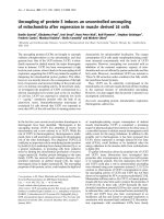
Figure 1
Statins decrease the viability of synovial fibroblasts in a concentration-dependent mannerStatins decrease the viability of synovial fibroblasts in a concentration-
dependent manner. Synovial fibroblasts from a patient with rheumatoid
arthritis were cultured for 96 hours in the presence of 1, 3 or 10 µM
lovastatin, atorvastatin, simvastatin or cerivastatin. XTT assays were
then performed on quadruplicate wells to determine the percentage
cellular viability of statin-treated cultures in comparison with those
treated with the vector. Results are representative of two experiments.
Figure 2
Statins decrease the viability of synovial fibroblasts in a time-dependent mannerStatins decrease the viability of synovial fibroblasts in a time-dependent
manner. Synovial fibroblasts were cultured for 24, 48, 72 and 96 hours
in the presence of 10 µM atorvastatin, 10 µM simvastatin or 3 µM ceriv-
astatin. XTT assays were then performed on quadruplicate wells to
determine the percentage cellular viability of statin-treated cultures in
comparison with those treated with the vector under identical condi-
tions. Results are representative of two experiments.
Arthritis Research & Therapy Vol 8 No 4 Connor et al.
Page 4 of 11
(page number not for citation purposes)
with 10 ng/ml TNF-α or 2 ng/ml IL-1 (R&D Systems Inc, Min-
neapolis, MN, USA). Cells were then rinsed twice with PBS
and lysed in 20 mM Tris pH 7.5, 150 mM NaCl, 1 mM EDTA,
1 mM EGTA, 1% Triton X-100, 2.5 mM sodium pyrophos-
phate, 1 mM β-glycerolphosphate, 0.5 µg/ml leupeptin, 0.2
mM phenylmethylsulphonyl fluoride. Protein concentrations
were determined by the micro bicinchoninic acid (BCA) pro-
tein assay kit (Pierce, Rockford, IL, USA) to enable compara-
ble sample loading on a 12% SDS-polyacrylamide gel. SDS-
PAGE was performed as described for the phase partitioning.
pJNK (Thr183/Tyr185), pERK and pAkt (Ser473) antibodies
were obtained from Cell Signaling Technology (Beverly, MA,
USA). Prehybridization and hybridization were performed in
accordance with the manufacturer's protocol. Blots were
scanned and band intensities were measured with the UN-
SCAN-IT version 5.1 software (Silk Scientific, Orem, UT,
USA). Relative amounts of protein were expressed as multi-
ples of the solvent control.
Blots were stripped between probings with Re-Blot-Plus
(Chemicon International, Inc., Temecula, CA, USA).
siRNA
To confirm the involvement of geranylgeranylated proteins in
FLS survival and pAkt expression, siRNA for Rac1 (target
sequence 5'-AACCGGTGAATCTGGGCTTAT-3') was trans-
fected into RA synovial fibroblasts (n = 3) plated at 6.0 × 10
4
cells per well of a six-well plate or 3 × 10
3
cells per well of a
96-well plate, at an siRNA to RNAiFect Reagent (Qiagen Inc.,
Mississauga, Ontario, Canada) ratio of 1:4.5. In preliminary
experiments, a non-silencing control siRNA labeled with Alexa
Fluor 488 was monitored by fluorescence microscopy. The
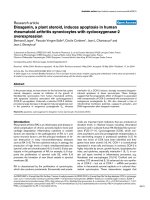
Figure 3
Statins are most effective at inhibiting the viability of TNF-α-stimulated RA synovial fibroblastsStatins are most effective at inhibiting the viability of TNF-α-stimulated RA synovial fibroblasts. Synovial fibroblasts derived from patients with rheu-
matoid arthritis (RA) (n = 6) and osteoarthritis (OA; n = 6) were stimulated or not with 10 ng/ml tumor necrosis factor-α (TNF-α) for 24 hours before
the addition of 3 or 10 µM atorvastatin or simvastatin. They were then maintained for a further 96 hours in the presence or absence of TNF-α and
statin. XTT assays were performed on quadruplicate wells. The percentage cellular viability was determined by comparing statin-treated cultures with
non-treated cultures in either the presence or absence of TNF-α as appropriate. Means and standard deviations are indicated by solid bars. *p <
0.05 for atorvastatin-treated versus simvastatin-treated RA fibroblasts.
+
p < 0.05 for RA versus OA TNF-α-stimulated fibroblasts.
Available online />Page 5 of 11
(page number not for citation purposes)
conditions used in this study resulted in a cell transfection effi-
ciency of greater than 95%. The medium was changed 18
hours after transfection. XTT assays were performed and cell
lysates were prepared 96 hours after transfection as
described above.
Statistics
Results are presented as means ± SD. Significant differences
between groups were analysed by using a two-tailed unpaired
Student t test; p < 0.05 was considered statistically
significant.
Results
Statins decrease synovial fibroblast viability
To determine whether statins influenced the viability of RA syn-
ovial fibroblasts, RA synovial fibroblasts were cultured in the
presence of increasing concentrations of statins ranging from
1 to 10 µM. After 96 hours, cell viability was determined by the
XTT assay. The results showed that all of the statins led to
reduced viability of RA synovial fibroblasts in a concentration-
dependent manner (Figure 1). However, it is noteworthy that
simvastatin and cerivastatin reduced FLS viability to a greater
extent than lovastatin or atorvastatin. Time-course experiments
with atorvastatin (10 µM), simvastatin (10 µM) and cerivastatin
(3 µM) revealed that reduced viability occurred in a time-
dependent manner (Figure 2). We chose to compare the
effects of atorvastatin and simvastatin because they had exhib-
ited different degrees of potency, both on RA synovial fibrob-
last viability in our preliminary studies and on the CIA mouse
model [14]. As shown in Figure 3, in comparison with non-
treated cells, simvastatin exhibited a greater effect on the via-
bility of RA synovial fibroblasts (n = 6) than atorvastatin both
at 3 µM (51 ± 18% versus 77 ± 13% viability, respectively; p
= 0.014) and at 10 µM (24 ± 13% versus 46 ± 19%, respec-
tively; p = 0.04). The effects of the statins were also examined
on OA synovial fibroblasts (n = 6). Although both simvastatin
and atorvastatin reduced the viability of OA synovial fibrob-
lasts, although to a smaller extent than that of RA synovial
fibroblasts, no significant differences were observed between
the statins at either concentration.
Statins decrease the viability of TNF-α-stimulated
synovial fibroblasts
Because TNF-α is an important driver of RA pathology, we
investigated the effects of statins on TNF-α-stimulated prolif-
eration of synovial fibroblasts. Preliminary time-course experi-
ments ranging from 24 to 96 hours (not shown) revealed that
similarly to non-treated synovial fibroblasts, those pretreated
with TNF-α exhibited decreased viability in a time-dependent
manner. As depicted in Figure 3, both statins were able to
decrease the viability of TNF-α-stimulated RA synovial fibrob-
lasts (n = 6). In TNF-α-stimulated RA synovial fibroblasts, sim-
vastatin caused a marked reduction in synovial fibroblast
viability compared with atorvastatin both at 3 µM (35 ± 11%
versus 67 ± 13%, respectively; p = 0.0008) and at 10 µM (2
± 5% versus 23 ± 6%, respectively; p = 8 × 10
-5
). Reduced
Figure 4
Geranylgeranylpyrophosphate (GGPP) restores the viability of synovial fibroblasts in the presence of statinsGeranylgeranylpyrophosphate (GGPP) restores the viability of synovial
fibroblasts in the presence of statins. Synovial fibroblasts were cultured
for 96 hours in the presence of 1, 3 or 10 µM lovastatin, atorvastatin or
simvastatin supplemented with 5 µM farnesylpyrophosphate (FPP) or 5
µM GGPP, or neither FPP nor GGPP (control). XTT assays were per-
formed on quadruplicate wells to determine the percentage cellular via-
bility of treated in comparison with non-treated cultures. Each treatment
was performed on two different synovial fibroblast lines. A representa-
tive experiment is shown. In a separate experiment, synovial fibroblasts
were pretreated with 10 ng/ml tumor necrosis factor-α for 24 hours
before the addition of 10 µM atorvastatin or simvastatin and 5 µM FPP
or 5 µM GGPP. They were cultured for a further 96 hours before an
XTT assay on quadruplicate wells.
Figure 5
Geranylgeranylated proteins are redistributed to the cytoplasm in the presence of statinGeranylgeranylated proteins are redistributed to the cytoplasm in the
presence of statin. Synovial fibroblasts were cultured for 72 hours in
the presence of 3 µM simvastatin supplemented or not with 5 µM far-
nesylpyrophosphate (FPP) or 5 µM geranylgeranylpyrophosphate
(GGPP). Phase separation of proteins was achieved by partitioning
with Triton X-114. The entire membrane and cytoplasmic fractions were
subjected to PAGE on a 12% gel. Blots were probed with mouse anti-
human RhoA or Rac1 followed by goat anti-mouse IgG conjugated to
horseradish peroxidase secondary antibody, and were revealed by
enhanced chemiluminescence.
Arthritis Research & Therapy Vol 8 No 4 Connor et al.
Page 6 of 11
(page number not for citation purposes)
RA synovial fibroblast viability was observed in TNF-α-stimu-
lated cells compared with unstimulated cells at 10 µM for both
simvastatin (p = 0.003) and atorvastatin (p = 0.02). As with
unstimulated OA synovial fibroblasts, simvastatin reduced the
viability of TNF-α-stimulated OA synovial fibroblasts (n = 6) to
a greater extent than atorvastatin, but no statistically significant
differences were observed at 3 µM. In contrast with RA syno-
vial fibroblasts, there were no differences between TNF-α-
stimulated and unstimulated OA synovial fibroblasts with
either statin at 3 µM or 10 µM. There was a significant differ-
ence in reduction in viability between TNF-α-stimulated RA
synovial fibroblasts compared with OA synovial fibroblasts at
10 µM, both with simvastatin (p = 0.02) and with atorvastatin
(p = 0.007). These differences were not explained by differ-
ences in the degree of stimulation of OA and RA synovial
fibroblasts to TNF-α (p = 0.72 for the atorvastatin control; p =
0.33 for the simvastatin control). Taken together, these results
show that although TNF-α stimulates OA and RA synovial
fibroblasts equally, RA synovial fibroblasts show a further
decrease in viability in the presence of statins.
Geranylgeranylpyrophosphate restores the viability of
RA synovial fibroblasts in the presence of statins
The decrease in cellular viability caused by statins has been
attributed to a lowering of intracellular FPP and GGPP con-
centrations [25]. This concept was examined in FLS by evalu-
ating the ability of exogenously added FPP and GGPP in the
presence of statins to rescue their viability. Our results demon-
strated that the statin-induced reduction in RA synovial fibrob-
last viability could be partly abrogated with 5 µM FPP and
completely abrogated with 5 µM GGPP (Figure 4). The com-
plete restoration of viability with GGPP suggests that the inhi-
bition of a geranylgeranylated protein in RA synovial
fibroblasts accounts for the reduced viability in the presence
of statins.
Geranylgeranylated proteins are redistributed to the
cytoplasm in the presence of statins
To explore the mechanism by which the inhibition of geran-
ylgeranylation leads to decreased RA synovial fibroblast viabil-
ity, proteins that normally associate with the cell membrane as
a consequence of geranylgeranylation were examined for
redistribution into the cytoplasm in the presence of statins.
Potential candidates for such redistribution are geranylgeran-
ylated members of the Rho subfamily of the Ras superfamily.
Members of the Rho family control several cellular processes
including transcriptional regulation, cell-cycle progression, cell
adhesion and apoptosis. Two geranylgeranylated members of
the Rho family that have been implicated in cell survival are
RhoA and Rac1. With the use of Triton X-114 partitioning, we
demonstrated a decrease in membrane-associated RhoA and
Rac1 and an increase in cytoplasm-associated RhoA and
Rac1 in the presence of simvastatin (Figure 5). The inclusion
of geranylgeranylpyrophosphate in the culture medium
restored RhoA and Rac1 to the membrane. The results sug-
gest that statins reduce RA synovial fibroblast viability by
affecting the normal cellular distribution of geranylgeranylated
proteins such as RhoA and Rac1.
Reduced RA synovial fibroblast viability with statins
results from apoptosis
To determine whether apoptosis accounts for the decrease in
RA synovial fibroblast viability we measured histone-bound
DNA fragments in cell lysates and used a TUNEL assay to
measure DNA fragments. Lysates prepared from RA synovial
fibroblasts grown in increasing concentrations of lovastatin
contained increasing amounts of histone bound DNA frag-
ments, suggesting the induction of apoptosis (Figure 6a). His-
tone-bound DNA fragments decreased about sixfold with the
addition of 5 µM FPP and returned to baseline with the addi-
tion of 5 µM GGPP (Figure 6b). Similar results were obtained
Figure 6
Cellular lysates of synovial fibroblasts cultured in lovastatin contain histone-bound DNA fragmentsCellular lysates of synovial fibroblasts cultured in lovastatin contain histone-bound DNA fragments. Cell lysates and corresponding culture superna-
tants were assayed by Cell Death Detection ELISA
PLUS
for the presence of histone-bound DNA fragments. (a) Synovial fibroblasts were cultured for
72 hours in the presence of 0, 3 or 10 µM lovastatin. The dark bars represent fragments present in the cell lysate indicative of apoptosis; the light
bars represent fragments present in the supernatant. Shown is a representative of three different experiments. (b) Synovial fibroblasts were cultured
for 72 hours in 10 µM lovastatin supplemented or not with 5 µM farnesylpyrophosphate (FPP) or 5 µM geranylgeranylpyrophosphate (GGPP).
Available online />Page 7 of 11
(page number not for citation purposes)
with the TUNEL assay (Figure 7). Taken together, these results
suggest that the reduced viability of synovial fibroblasts
induced by statins results from apoptosis brought about pri-
marily by the decrease in membrane-associated geranylgeran-
ylated proteins.
The Akt survival pathway is inhibited by statins in RA
synovial fibroblasts
We next examined intracellular signaling pathways implicated
in RA synovial fibroblast survival. Lovastatin decreased the
steady-state pAkt levels at least threefold, and TNF-α or IL-1
signalling through pAkt was also prevented (Figure 8). In con-
trast, neither the steady-state levels nor the TNF-α or IL-1β
induction of pJNK or pERK were affected by culturing RA syn-
ovial fibroblasts with 3 µM lovastatin. This suggests that the
Akt survival pathway is inhibited by statins in RA synovial
fibroblasts.
siRNA-directed inhibition of Rac1 reduces RA synovial
fibroblast viability and pAkt levels
To determine the relationship between the decrease in geran-
ylgeranylated proteins and pAkt levels, we used siRNA against
Rac1. Rac1 was selected because the expression of a domi-
nant-negative form of Rac1 has been shown to sensitize cells
to TNF-α-mediated apoptosis [26,27]. RA synovial fibroblasts
transfected with siRNA against Rac1 exhibited a decrease in
both Rac1 and pAkt protein levels in comparison with a control
siRNA against no known protein coding sequence (Figure
9a,b). Furthermore, synovial fibroblasts transfected with
siRNA against Rac1 exhibited decreased viability as deter-
mined in an XTT assay (n = 3; p < 0.01; Figure 9c). These
results suggest that the inhibition of Rac1 affects pAkt levels
and that Rac1 inhibition influences RA synovial fibroblast
viability.
Discussion
We have shown that statins decrease RA synovial fibroblast
viability in a concentration-dependent and time-dependent
manner. Simvastatin is significantly more effective than atorv-
astatin at inhibiting RA synovial fibroblast survival. Similar dif-
ferences in the efficacy of different statins have been reported
previously in tumor-specific apoptosis [28]. The difference
between simvastatin and atorvastatin was even greater in cells
preactivated with TNF-α. Although both statins also
decreased OA synovial fibroblast survival, there was no signif-
icant difference between them in their ability to reduce survival.
Moreover, they were not significantly more effective in reduc-
ing OA synovial fibroblast survival in the presence of TNF-α.
Taken together, these data suggest that in RA synovial fibrob-
lasts, statins are inhibiting TNF-α regulated synovial fibroblast
survival, enabling TNF-α to function as an apoptotic signal
rather than as a survival signal. Our results demonstrated that
all of the statins examined seem to exert their effects in a sim-
ilar manner through the inhibition of a geranylgeranyl pyro-
phosphate intermediate, resulting in a loss of membrane-
associated geranylgeranylated proteins and a subsequent
reduction in viability of synovial fibroblasts. All statins consist
of a dihydroxyheptanoic group that mimics mevalonate, but
they have different chemical substituents elsewhere on the
molecule that can lead to differences in their efficacy [29]. It is
therefore possible that differences in the efficacies of the stat-
ins on RA synovial fibroblast viability result from differences in
the molecules other than the dihydroxyheptanoic group. These
differences account for differences in the bioavailability of the
Figure 7
TUNEL assay confirms the presence of DNA fragments in synovial fibroblasts treated with lovastatinTUNEL assay confirms the presence of DNA fragments in synovial fibroblasts treated with lovastatin. DNA fragments in synovial fibroblasts treated
or not with 10 µM lovastatin for 72 hours were detected by a TdT-FragEL DNA fragmentation kit. After TdT-mediated dUTP nick end labelling
(TUNEL) staining, cells were counterstained with hematoxylin. TUNEL-positive cells have dark nuclear staining, whereas the nuclei of TUNEL nega-
tive cells stain blue. Asterisk, 5 µM geranylgeranylpyrophosphate (GGPP) or 5 µM farnesylpyrophosphate (FPP) were included with the lovastatin.
Arthritis Research & Therapy Vol 8 No 4 Connor et al.
Page 8 of 11
(page number not for citation purposes)
statins and the active metabolites generated by the cyto-
chrome P450 family of enzymes [30].
As far as we are aware, this is the first report to implicate ger-
anylgeranylated proteins in synovial fibroblast viability. The
presence of this pathway in both OA and RA synovial fibrob-
lasts suggests that it is a constitutive pathway in synovial
fibroblast biology. However, the inhibition by GGPP of the sta-
tin effect on TNF-α-stimulated RA synovial fibroblasts sug-
gests that in RA synovial fibroblasts, TNF-α is signaling
through the same pathway that is required for their overall via-
bility and that this pathway requires a functional geranylgeran-
ylated protein. This pathway differs in RA synovial fibroblasts
compared with OA synovial fibroblasts, as demonstrated by
the significant difference in the effect of statins between TNF-
α-stimulated and non-stimulated RA synovial fibroblasts not
seen with OA synovial fibroblasts.
The data in this study also show that the statins affect the Akt
pathway in RA synovial fibroblasts. Previous studies have
demonstrated that synovial tissue obtained from patients with
RA expresses higher levels of pAkt than that derived from
patients with OA [31]. It was also demonstrated that elevated
pAkt levels account for the anti-apoptotic response of synovial
fibroblasts to both TNF-α and transforming growth factor-β,
suggesting that elevated pAkt might contribute to the estab-
lishment of synovial hyperplasia observed in patients with RA
[31,32]. The phosphoinositide 3-kinase/Akt pathway is being
increasingly recognized as playing a major role in synovial
fibroblasts. It is used in response to TRAIL (TNF-related apop-
Figure 8
The steady-state and activated Akt pathway is affected by lovastatinThe steady-state and activated Akt pathway is affected by lovastatin. Rheumatoid arthritis synovial fibroblasts (2 lines shown) were cultured in the
presence of 3 µM lovastatin or DMSO (solvent control) for 48 hours. They were then treated with 2 ng/ml IL-1 or 10 ng/ml tumor necrosis factor-α
(TNF-α) for 15 minutes. (a) Cellular lysates were prepared, subjected to 12% PAGE and analysed by immunoblotting for pAkt, pJNK, pERK and
actin. D, dimethylsulfoxide (DMSO); 3L, 3 µM lovastatin; D, IL-1, cells cultured with DMSO then induced with IL-1; D, TNF, cells cultured with
DMSO then induced with TNF-α; 3L, IL-1, cells cultured with 3 µM lovastatin then induced with IL-1; 3L, TNF, cells cultured with 3 µM lovastatin
then induced with TNF-α (b-d) Bands were quantified by UN-SCAN-IT and corrected for loading by actin (b) pAkt, (c) pJNK, (d) pERK.
Available online />Page 9 of 11
(page number not for citation purposes)
tosis-inducing ligand) [33], the transmission of IL-18 signals
leading to VCAM (vascular cell adhesion molecule) expression
[34] and the production of IL-6 and IL-8 stimulated by IL-17
[35]. The results presented in the present study lend further
support to the importance of elevated pAkt levels to synovial
fibroblast survival and suggest that statins might have a bene-
ficial role in reducing the aberrant pAkt levels in patients with
RA.
Experiments with siRNA technology implicated Rac1 in syno-
vial fibroblast survival. Additional experiments are currently
under way to determine whether other geranylgeranylated pro-
teins contribute to synovial fibroblast survival. We also demon-
strated decreased levels of pAkt in these cells. These data
suggest that a survival signal is propagated in synovial fibrob-
lasts via a membrane-associated geranylgeranylated protein,
Rac1 with subsequent pAkt activation. These data are consist-
ent with other studies of Rac1-regulated Akt resulting in an
anti-apoptotic signal [36-38].
The results of this study have several implications for the ther-
apeutic use of statins in patients with RA. In the TARA study
of patients with RA, atorvastatin significantly improved the
swollen joint count, C-reactive protein, erythrocyte sedimenta-
tion rate, fibrinogen, soluble intercellular adhesion molecule 1
(sICAM1) and IL-6 [15]. Because synovial fibroblasts synthe-
size sICAM1 and IL-6 it is possible that the decreased level
observed results in part from atorvastatin-driven decreased
synovial fibroblast viability. If this were so, our studies suggest
that simvastatin might be more effective than atorvastatin in
patients with RA. Previous investigation demonstrated that the
administration of simvastatin in a CIA model resulted in
decreased proliferation of T cells, but it was not reported
whether this resulted from apoptosis [13]. There is the possi-
bility that the effect of statins might be cell type specific, a con-
cept supported by differential effects of statins on human lung
fibroblasts, human atrial myofibroblasts, and lymphoma tumour
cells compared with human pancreatic islets and microglia
[19,39-42].
Our results show that the pathway affected by statins in RA
synovial fibroblasts is also a TNF-α anti-apoptotic pathway.
We are currently dissecting these pathways with a view to dis-
cerning new therapeutic targets for RA.
Conclusion
Statins attenuate the insertion of geranylgeranylated proteins
into the plasma membrane with a resultant decrease in viability
and increased apoptosis of synovial fibroblasts. A significant
difference in the efficacy of simvastatin from that of atorvasta-
tin was observed in both activated and non-activated RA syn-
ovial fibroblasts but not OA synovial fibroblasts, suggesting a
fundamental difference in their intracellular signaling path-
ways. The intracellular redistribution of geranylgeranyl proteins
resulting from treatment with statin is correlated with
decreased activation of Akt, a pathway previously identified as
being anti-apoptotic in synovial fibroblasts. These results sug-
gest that statins, particularly simvastatin, might have a benefi-
Figure 9
siRNA-directed inhibition of Rac1 reduces synovial fibroblast viability and pAkt levelssiRNA-directed inhibition of Rac1 reduces synovial fibroblast viability and pAkt levels. Synovial fibroblasts were transfected with siRNA against Rac1
or a non-silencing RNA as control, and cultured for 96 hours at which point XTT assays were performed to measure cell viability and lysates were
prepared from wells that had been transfected in parallel. (a, b) Lysates were subjected to immunoblotting and probed with anti-Rac (a) or anti-pAkt
or actin (b) for normalization. (c) Effect of siRNA on cell viability. ** p < 0.01 for viability of synovial fibroblasts transfected with non-siRNA control
versus Rac1 siRNA. Shown is a representative of three independent experiments. siRNA transfection efficiency was greater than 95%.
Arthritis Research & Therapy Vol 8 No 4 Connor et al.
Page 10 of 11
(page number not for citation purposes)
cial role in reducing aberrant pAkt levels in the synovial
fibroblasts of patients with RA, with the resultant decrease in
synovial hyperplasia.
Competing interests
The authors declare that they have no competing interests.
Authors' contributions
AC participated in the design of the study, performed the
experiments and participated in the writing of the manuscript.
SB and AN participated in the design of the study and the writ-
ing of the manuscript. EK participated in the design of the
study, the analysis of data and the writing of the manuscript.
All authors read and approved the final manuscript.
Acknowledgements
We thank Dr E Bogochfor providing surgical samples and Ms K Griffith
Cunningham for coordinating the tissue collections. This study was
funded by The Younger Foundation.
References
1. Firestein GS: Evolving concepts of rheumatoid arthritis. Nature
2003, 423:356-361.
2. Feldmann M, Brennan FM, Foxwell BM, Taylor PC, Williams RO,
Maini RN: Anti-TNF therapy: Where have we got to in 2005? J
Autoimmun 2005:26-28.
3. Mor A, Abramson SB, Pillinger MH: The fibroblast-like synovial
cell in rheumatoid arthritis: akey player in inflammation and
joint destruction. Clin Immunol 2005, 115:118-128.
4. Davis LS: A question of transformation: the synovial fibroblast
in rheumatoid arthritis. Am J Pathol 2003, 162:1399-1402.
5. Gupta S, Gollapudi S: Molecular mechanisms of TNF-alpha-
induced apoptosis in aging human T cell subsets. Int J Bio-
chem Cell Biol 2005, 37:1034-1042.
6. Youn J, Kim HY, Park JH, Hwang SH, Lee SY, Cho CS, Lee SK:
Regulation of TNF-alpha-mediated hyperplasia through TNF
receptors, TRAFs, and NF-κB in synoviocytes obtained from
patients with rheumatoid arthritis. Immunol Lett 2002,
83:85-93.
7. Zhang HG, Huang N, Liu D, Bilbao L, Zhang X, Yang P, Zhou T,
Curiel DT, Mountz JD: Gene therapy that inhibits nuclear trans-
location of nuclear factor κB results in tumor necrosis factor
alpha-induced apoptosis of human synovial fibroblasts. Arthri-
tis Rheum 2000, 43:1094-1105.
8. Drynda A, Quax PH, Neumann M, van der Laan WH, Pap G,
Drynda S, Meinecke I, Kekow J, Neumann W, Huizinga TW, et al.:
Gene transfer of tissue inhibitor of metalloproteinases-3
reverses the inhibitory effects of TNF-alpha on Fas-induced
apoptosis in rheumatoid arthritis synovial fibroblasts. J
Immunol 2005, 174:6524-6531.
9. Pope RM: Apoptosis as a therapeutic tool in rheumatoid
arthritis. Nat Rev Immunol 2002, 2:527-535.
10. Endo A: The origin of the statins. Atheroscler Suppl 2004,
5:125-130.
11. Palinski W, Tsimikas S: Immunomodulatory effects of statins:
mechanisms and potential impact on arteriosclerosis. J Am
Soc Nephrol 2002, 13:1673-1681.
12. Blanco-Colio LM, Tunon J, Martin-Ventura JL, Egido J: Anti-inflam-
matory and immunomodulatory effects of statins. Kidney Int
2003, 63:12-23.
13. Leung BP, Sattar N, Crilly A, Prach M, McCarey DW, Payne H,
Madhok R, Campbell C, Gracie JA, Liew FY, McInnes IB: A novel
anti-inflammatory role for simvastatin in inflammatory arthritis.
J Immunol 2003, 170:1524-1530.
14. Palmer G, Chobaz V, Talabot-Ayer D, Taylor S, So A, Gabay C,
Busso N: Assessment of the efficacy of different statins in
murine collagen-induced arthritis. Arthritis Rheum 2004,
50:4051-4059.
15. McCarey DW, McInnes IB, Madhok R, Hampson R, Scherbakov O,
Ford I, Capell HA, Sattar N: Trial of Atorvastatin in Rheumatoid
Arthritis (TARA): double-blind, randomised placebo-controlled
trial. Lancet 2004, 363:2015-2021.
16. Goldstein JL, Brown MS: Regulation of the mevalonate
pathway. Nature 1990, 343:425-430.
17. Resh MD: Regulation of cellular signalling by fatty acid acyla-
tion and prenylation of signal transduction proteins. Cell
Signal 1996, 8:403-412.
18. Guijarro C, Blanco-Colio LM, Ortego M, Alonso C, Ortiz A, Plaza
JJ, Diaz C, Hernandez G, Egido J: 3-Hydroxy-3-methylglutaryl
coenzyme A reductase and isoprenylation inhibitors induce
apoptosis of vascular smooth muscle cells in culture. Circ Res
1998, 83:490-500.
19. van de Donk NW, Schotte D, Kamphuis MM, van Marion AM, van
Kessel B, Bloem AC, Lokhorst HM: Protein geranylgeranylation
is critical for the regulation of survival and proliferation of lym-
phoma tumor cells. Clin Cancer Res 2003, 9:5735-5748.
20. Bifulco M: Role of the isoprenoid pathway in ras transforming
activity, cytoskeleton organization, cell proliferation and
apoptosis. Life Sci 2005, 77:1740-1749.
21. Scott BB, Weisbrot LM, Greenwood JD, Bogoch ER, Paige CJ,
Keystone EC: Rheumatoid arthritis synovial fibroblast and
U937 macrophage/monocyte cell line interaction in cartilage
degradation. Arthritis Rheum 1997, 40:490-498.
22. Crick DC, Andres DA, Waechter CJ: Novel salvage pathway uti-
lizing farnesol and geranylgeraniol for protein isoprenylation.
Biochem Biophys Res Commun 1997, 237:483-487.
23. Roehm NW, Rodgers GH, Hatfield SM, Glasebrook AL: An
improved colorimetric assay for cell proliferation and viability
utilizing the tetrazolium salt XTT. J Immunol Methods 1991,
142:257-265.
24. Bordier C: Phase separation of integral membrane proteins in
Triton X-114 solution. J Biol Chem 1981, 256:1604-1607.
25. Perez-Sala D, Mollinedo F: Inhibition of isoprenoid biosynthesis
induces apoptosis in human promyelocytic HL-60 cells. Bio-
chem Biophys Res Commun 1994, 199:1209-1215.
26. Deshpande SS, Angkeow P, Huang J, Ozaki M, Irani K: Rac1
inhibits TNF-alpha-induced endothelial cell apoptosis: dual
regulation by reactive oxygen species. FASEB J 2000,
14:1705-1714.
27. Sanlioglu S, Luleci G, Thomas KW: Simultaneous inhibition of
Rac1 and IKK pathways sensitizes lung cancer cells to TNFal-
pha-mediated apoptosis. Cancer Gene Ther 2001, 8:897-905.
28. Wong WW, Tan MM, Xia Z, Dimitroulakos J, Minden MD, Penn LZ:
Cerivastatin triggers tumor-specific apoptosis with higher effi-
cacy than lovastatin. Clin Cancer Res 2001, 7:2067-2075.
29. Mason RP, Walter MF, Day CA, Jacob RF: Intermolecular differ-
ences of 3-hydroxy-3-methylglutaryl coenzyme areductase
inhibitors contribute to distinct pharmacologic and pleiotropic
actions. Am J Cardiol 2005, 96:11F-23F.
30. Schachter M: Chemical, pharmacokinetic and pharmacody-
namic properties of statins: an update. Fundam Clin Pharmacol
2005, 19:117-125.
31. Zhang HG, Wang Y, Xie JF, Liang X, Liu D, Yang P, Hsu HC, Ray
RB, Mountz JD: Regulation of tumor necrosis factor alpha-
mediated apoptosis of rheumatoid arthritis synovial fibrob-
lasts by the protein kinase Akt. Arthritis Rheum 2001,
44:1555-1567.
32. Kim G, Jun JB, Elkon KB: Necessary role of phosphatidylinositol
3-kinase in transforming growth factor beta-mediated activa-
tion of Akt in normal and rheumatoid arthritis synovial
fibroblasts. Arthritis Rheum 2002, 46:1504-1511.
33. Miyashita T, Kawakami A, Tamai M, Izumi Y, Mingguo H, Tanaka F,
Abiru S, Nakashima K, Iwanaga N, Aratake K, et al.: Akt is an
endogenous inhibitor toward tumor necrosis factor-related
apoptosis inducing ligand-mediated apoptosis in rheumatoid
synovial cells. Biochem Biophys Res Commun 2003,
312:397-404.
34. Morel JC, Park CC, Woods JM, Koch AE: A novel role for inter-
leukin-18 in adhesion molecule induction through NF κB and
phosphatidylinositol (PI) 3-kinase-dependent signal transduc-
tion pathways. J Biol Chem 2001, 276:37069-37075.
35. Hwang SY, Kim JY, Kim KW, Park MK, Moon Y, Kim WU, Kim HY:
IL-17 induces production of IL-6 and IL-8 in rheumatoid arthri-
tis synovial fibroblasts via NF-κB- and PI3-kinase/Akt-
dependent pathways. Arthritis Res Ther 2004, 6:R120-128.
Available online />Page 11 of 11
(page number not for citation purposes)
36. Nishida K, Kaziro Y, Satoh T: Anti-apoptotic function of Rac in
hematopoietic cells. Oncogene 1999, 18:407-415.
37. Murga C, Zohar M, Teramoto H, Gutkind JS: Rac1 and RhoG pro-
mote cell survival by the activation of PI3K and Akt, independ-
ently of their ability to stimulate JNK and NF-κB. Oncogene
2002, 21:207-216.
38. Gonzalez E, Kou R, Michel T: Rac1 modulates sphingosine 1-
phosphate-mediated activation of phosphoinositide 3-kinase/
Akt signaling pathways in vascular endothelial cells. J Biol
Chem 2006, 281:3210-3216.
39. Tan A, Levrey H, Dahm C, Polunovsky VA, Rubins J, Bitterman PB:
Lovastatin induces fibroblast apoptosis in vitro and in vivo. A
possible therapy for fibroproliferative disorders. Am J Respir
Crit Care Med 1999, 159:220-227.
40. Contreras JL, Smyth CA, Bilbao G, Young CJ, Thompson JA, Eck-
hoff DE: Simvastatin induces activation of the serine-threonine
protein kinase AKT and increases survival of isolated human
pancreatic islets. Transplantation 2002, 74:1063-1069.
41. Porter KE, Turner NA, O'Regan DJ, Balmforth AJ, Ball SG: Simv-
astatin reduces human atrial myofibroblast proliferation
independently of cholesterol lowering via inhibition of RhoA.
Cardiovasc Res 2004, 61:745-755.
42. Bi X, Baudry M, Liu J, Yao Y, Fu L, Brucher F, Lynch G: Inhibition
of geranylgeranylation mediates the effects of 3-hydroxy-3-
methylglutaryl (HMG)-CoA reductase inhibitors on microglia.
J Biol Chem 2004, 279:48238-48245.