Báo cáo khoa học: "An investigation of intensity-modulated radiation therapy versus conventional two-dimensional and 3D-conformal radiation therapy for early stage larynx cancer" ppt
Bạn đang xem bản rút gọn của tài liệu. Xem và tải ngay bản đầy đủ của tài liệu tại đây (854.55 KB, 9 trang )
RESEARC H Open Access
An investigation of intensity-modulated radiation
therapy versus conventional two-dimensional and
3D-conformal radiation therapy for early stage
larynx cancer
Daniel Gomez
1*
, Oren Cahlon
1
, James Mechalakos
2
, Nancy Lee
1
Abstract
Introduction: Intensity modulated radiation therapy (IMRT) has been incorporated at several institutions for early
stage laryngeal cancer (T1/T2N0M0), but its utility is controversial.
Methods: In three representative patients, multiple plans were generated: 1) Conventional 2D planning, with the
posterior border placed at either the anterior aspect ("tight” plan) or the mid-vertebral body ("loose” plan), 2) 3D
planning, utilizing both 1.0 and 0.5 cm margins for the planning target volume (PTV), and 3) IMRT planning,
utilizing the same margins as the 3D plans. A dosimetric comparison was performed for the target volume, spinal
cord, arytenoids, and carotid arteries. The prescription dose was 6300 cGy (225 cGy fractions), and the 3D and IMRT
plans were normalized to this dose.
Results: For PTV margins of 1.0 cm and 0.5 cm, the D95 of the 2D tight/loose plans were 3781/5437 cGy and
5372/5869 cGy, respectively (IM RT/3D plans both 6300 cGy). With a PTV margin of 1.0 cm, the mean carotid artery
dose was 2483/5671/5777/4049 cGy in the 2D tight, 2D loose, 3D, and IMRT plans, respect ively. When the PTV was
reduced to 0.5 cm, the the mean carotid artery dose was 2483/5671/6466/2577 cGy to the above four plans,
respectively. The arytenoid doses were similar between the four plans, and spinal cord doses were well below
tolerance.
Conclusions: IMRT provides a more ideal dose distribution compared to 2D treatment and 3D planning in regards
to mean carotid dose. We therefore recommend IMRT in select cases when the treating physician is confident with
the GTV.
Introduction
Larynx cancer is the most common head and neck
malignancy in the United States and approx imately half
of these malignancies present at an early stage (T1-
T2N0). Treatment of this early disease is controversial
because there are several effective treatment modalities
including radiation therapy, endoscopic resection and
open partial laryngectomy. No single modality has been
proven to be superior to the others [1]. The goal of any
therapy is cure with larynx preservation, high voice
quality, and minimal morbidity. Although endoscopic
resection has gained popularity over the past decade,
many still consider definitive radiation to be the main-
stay of therapy.
Many institutions, including our own, h ave recently
incorporated intensity modulated radiation therapy
(IMRT) into the treatment of early stage glottic cancer
for selected patients. IMRT has the capability of produ-
cing highly conformal dose distributions with steep dose
gradients to target areas of concern while sparing nearby
critical organs in the neck. In addition, IMRT may pro-
duce better target coverage, leading to improved local
control. However, a recent editorial questioned the role
* Correspondence:
1
Department of Radiation Oncology, Memorial Sloan-Kettering Cancer
Center, New York, NY, USA
Full list of author information is available at the end of the article
Gomez et al. Radiation Oncology 2010, 5:74
/>© 2010 Gomez et al; licensee BioMed Central Ltd. This is an Open Access article distributed under the terms of the Creative Commons
Attribution License ( which permits unrestricted use, distribution, and reproduction in
any medium, provided the original work is properly cited.
of IMRT in tr eating early larynx cancer and highlighted
potential pitfalls of using IMRT in this scenario [2].
The followin g study provides a dosimetric comparison
between IMRT, conventional techniques, and 3D plan-
ning for the treatment of early glottic cancer. We aim to
show that, at least in select cases such as bulkier lesions
or in patients with short, thick necks, IMRT can
improve t arget coverage while simultaneously minimiz-
ing the dose to sensitive structures in the neck. By
doing so, IMRT ma y be able to further improve local
control while minimizing toxicity for these patients.
Materials and methods
Three representative patients treated with definitive
radiation using IMRT at Memorial Sloan-Kettering Can-
cer Center in the last year were selected for a treatment
planning study. Criteria for inclusion were T1N0 or
T2N0 squamous cell carcinoma of the larynx. Two of
the patients selected had T1N0 tumors and one patient
had a bulky T2N0 tumor. Patients were staged with
direct laryngoscopy, computed tomography (CT) scan,
and positron emission tomography (PET) scans. Patients
were treated to the larynx without elective nodal
irradiation.
Patients were immobilized in the supine position with
a 5-point thermoplastic m ask. Treatment planning CT
scans were obtained from the top of the skull to the
lower part of the neck with a 3-mm slice thickness.
Intravenous contrast was used in two patients; one
patient did not receive it due to an iodine allergy. Three
different treatment plans were generated for each
patient: 1) 2D opposed laterals (single slice) assuming a
larynx contour and no CT, 2) 3D planning, using the
entire larynx as the clinical target volume (CTV), and 3)
IMRT, utilizing the same definition as 3D planning for
the CTV. The 3D and 2D plans utilize d the same beam
configuration, but the conformal plan used 3D informa-
tion to design apertures and normalize the plan. For
anteriorly located lesions, the plans include a centrally
placed 0.5 cm bolus on the skin over the treatment
field. The dose to the planning target volume (PTV) in
all patients was 6300 cGy in 225 cGy fractions, over a
course of 38 days. This dose schedule corresponds to a
nominal standard dose, which is used to compare the
effect of different dose regimens, of 1905 ret, the unit
for nominal standard dose.
2D Opposed laterals
Tosimulatethecaseinwhichasinglesliceplanis
developed from a larynx contour alone, a generic larynx
contour was drawn according to department guidelines
for these cases: 1 cm from the anterior skin surface, and
consist ing of two lobes at 150 and 210 degrees from the
vertical, each lobe being 1.8 cm in length for males or
2.2 cm in length for females. Right and left lateral treat-
ment fields were created using a number of different
wedge angles. The collimator angle was chosen such
that the posterior jaw of the lateral fields was parallel to
the cervical spine. In an attempt to cover the clinical
range of 2D larynx treatments among different institu-
tions, two different plans per patient were created: one
in which the posterior edge of the fields coincided with
the anterior surface of the vertebral bodies (referred to
as the tight clinical plan), and one in whic h the poster-
ior edge was in the middle of the vertebral body (labeled
the loose clinical plan). Dose was calculated without
inhomogeneity corrections and a dose of 100% was
assigned to the isocenter. The isodose line which cov-
ered the larynx and had a reasonably straight posterior
edge, at the discretion of the authors, was chosen as the
prescription isodose. The wedge angle chosen for the
plan was the one which concentrated a dose of 102-
105% anteriorly. Only the isocenter slice was used for
plan evaluation. Once an acceptable plan was obtained,
inhomogeneity calculations were turned on and the plan
was recalculated for comparison to the other plans.
3D planning/IMRT
Relevant structures were manually contoured on e ach
axial CT scan slice by a head and neck radiation oncolo-
gist. The gross tumor volume was defined as the bilat-
eral true vocal cords, to include any gross disease that
can be delineated by the treating radiation oncologist
(though it is often difficult to determine the region of
gross disease on imaging except in the case of bulkier
lesion s). The CTV for each plan consisted of the larynx
(false and true vocal cords, anterior and posterior com-
missure, arytenoids and aryepiglottic folds) as well as
the subglottic region, extending from the level of the
hyoid bone superiorly to the bottom of the cricoid carti-
lage inferiorly. Two PTV volumes were generated with
var ying margins from the clinical target volume, 1.0 cm
and 0.5 cm. A 1.0 cm margin is generally used to ensure
adequate coverage when there is greater uncertainty as
to patient setup. The 3D plans c onsisted of two fields,
with a beam configuration identical to the 2D plans, and
the IMRT plans consisted of 3-4 anterior fields. To
ensure that these plans were consistent with the 2D
plans, we visually verified that the PTV coverage super-
iorly was set below the level of the hyoid bone and
extended inferiorly to the level of the cricoids. The
entire spinal cord, the bilateral carotid arteries, and the
bilateral arytenoids were contoured as organs at risk
(OAR). The 3D plans were normalized such that the
PTV D95 was equal to the prescription dose. The IMRT
plans were created for each PTV and also normalized
such that the PTV D95 was equal to the prescription
dose and the maximum PTV dose was 105% or less.
Gomez et al. Radiation Oncology 2010, 5:74
/>Page 2 of 9
Optimization was performed by lowering the desired
mean dose to the carotid artery so that it did not exceed
105% of the prescription dose (PTV D95). Thus, con-
touring of the carotid arteries was critical in generating
the plan.
The beam arrangement for the 3D and IMRT p lans
was the same as those used for treatment. However, for
consistency, the plans were re-optimized to all conform
to the same constraints. All plans had four anterior obli-
que beams, two on each side. Since the primary pu rpose
of this exercise was to compare conventional techniques
for treating the larynx with 3D and IMRT, we did not
test different beam arrangements for the conformal
plans.
As an adjunct to the above analysis, we generated a
second plan of one of the three patients who had T1N0,
anteriorly-located disease, based on the premise that
these tumors often involve the anterior third of the true
vocal cord and thus sparing the arytenoids would in
turn reduce carotid dose. We reported this plan as
IMRT no arytenoids.
Plan evaluation
Plans were compared based on the following criteria:
CTV and PTV coverage as indicated by D95 and D90,
maximum PTV dose (Dmax), mean car otid artery dose,
mean arytenoid dose, and maximum spinal cord dose.
Results
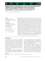
Table 1a depicts tumor coverage averaged over all three
patients with a 1.0 cm margin in the tight clinical 2D,
loose c linical 2D , and 3D/IMRT plans. The D95 ranged
from 3781 cGy in the tight clinical 2D plan (60% of the
prescription dose) to 6300 cGy in the IMRT and 3D
plans ( 100% of the prescription dose). The hot spot, as
measured by Dmax, was comparable in the four plans
butwashighestinthe3Dplan,at6913cGy.Table2a
demonstrates normal structure parameters of the four
different plans. The mean carotid dose was lowest in the
2D tight clinical plan, at 2483 cGy, and second lowest in
the IMRT plan, at 4049 cGy. The spinal cord doses were
all well below tolerance, with the maximum spinal cord
dose of 1437 cGy in the IMRT plan. The arytenoid
doses were comparable in all four plans, and ranged
from 6289 - 6500 cGy. Figure 1 is a graphical represen-
tation of key comparisons in Tables 1a and 2a.
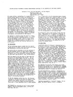
In the next step of analysis, we reduced the PTV mar-
gin from 1.0 cm to 0.5 cm, as there is no consensus
regarding the appropriate expansion that sho uld be uti-
lized in this disease. Target structure comparisons are
given in Table 1b. The margin contraction improved the
D95 of the tight and loos e clinical plans, now 5372 cGy
and 5869 cGy, respectively. However, the D95 was still
greatest in the IMRT and 3D plans, where it was opti-
mized to 6300 cGy. Table 2b depicts normal structure
doses between the four plans. While the carotid mean
dose was significant ly decreased in the IMRT plan by
reducing the margin size, from 4049 cGy to 2577 cGy,
as expected, the normal structure mean doses were
unchanged in the 2D plans, where the anterior and pos-
terior borders, and thus the dosimetry to normal struc-
tures, were independent of the size of the PTV
expansion. In a comparison between the IMRT and 3D
plans, the mean carotid artery dose remained substan-
tially better with IMRT (2577 cGy compared to 4371
cGy with 3D planning). The relative doses of the aryte-
noids and the spinal cord did not change by altering the
margin. Figure 2 demonstrates target and normal struc-
ture dosing with a 0.5 cm margin in graphical form.
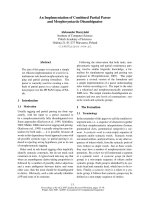
Figure 3 demonstrates axial slices from four different
plans: a) the tight clinical 2D plan, b) the loose clinical
2D plan, c) 3D plan, 0.5 cm margin and d) IMRT plan,
0.5 cm margin. This figure shows that the loose clinical
plan provides better target coverage than the tight clini-
cal plan, and similar coverage to the two conformal
plans, with the tradeoff of increased dose to the carotid
artery. Furthermore, because no optimization was per-
formed for the PTV in the 2D clinical plans, any rela-
tionship between the coverage of the PTV and the level
of expansion was dependent on the spatial relationship
between the expanded borders of the PTV and the pre-
determined boundaries of the clinical plan. Thus, it is
clear when examining the axial slices on Figures 3a and
3b that as the PTV expansion decreases in size (from
1.0 cm to 0.5 cm), t he percentage of target volume con-
tained within the pre-defined borders of the 2D clinical
plans increases. This increas ed coverage is the reason
why the PTV D95 and Dmax increase with both 2D
clinical plans as the CTV to PTV margin decreases.
When comparing the two conformal plans, we show
that both IMRT and 3D planning provide excellent cov-
erage to the target volume, and are optimized as such.
Table 1 Target Structure Dosing Comparison (PTV margin
in parentheses)
Dmax CTV D90 PTV D90 CTV D95 PTV D95
A
2D Tight (1 cm) 6713 6104 5782 5970 3781
2D Loose (1 cm) 6736 6233 6045 6111 5437
3D CRT (1 cm) 6913 6479 6437 6424 6300
IMRT (1 cm) 6610 6467 6431 6437 6300
B
2D Tight (5 mm) 6685 6104 5782 5970 5372
2D Loose (5 mm) 6711 6233 6045 6111 5869
3D CRT (5 mm) 6806 6439 6367 6404 6300
IMRT (5 mm) 6615 6428 6367 6400 6300
Gomez et al. Radiation Oncology 2010, 5:74
/>Page 3 of 9
However, utilizing various beam arrangements and
inverse planning, the IMRT plan provides more con-
formality in regards to the carotid arteries. As noted
above, the arytenoids receive similar doses, and the
spinal cord receives very low doses in both plans.
Table 3 compares the dose distributions to the carotid
arteries in a patient with T1N0 glottic cancer and an ante-
rior lesion. The 3D and IMRT plans were compared with a
0.5 cm margin, as these would give the lowest dose to the
carotid arteries and thus provide the most conservative
estimate of the advantages of sparing the arytenoid cartilage
in selected patient. Th e table demonstrates that sparin g the
arytenoids provides a substantial benefit (greater than 50%
in mean c arotid dose) compared to any other pla n.
Discussion
Early stage glottic c ancer is a highly curable malignancy
which can be treated with either larynx sparing surgery
(laser excision, cordectomy, or hemilarynge ctomy) or
radiation [1]. Because there is not a randomized trial to
guide treatment decisions, the management of this disease
remains controversial. However, at most institutions,
radiotherapy is still considered the mainstay of treatment.
Because both treatment modalities offer similar rates of
cure, decisions regarding which therapy to pursue often lie
on the anticipated toxicity profile of a particular regimen.
Other factors such as tumor location and extent of disease,
co-morbid illnesse s and physician and pa tient preference
also impact the final treatment decision. The ultimate goal
Table 2 Normal Structure Dosimetric comparison of Radiation Plans
A) 1 cm margin B) 0.5 cm margin
Arytenoid Mean
(cGy)
Carotid Mean
(cGy)
Spinal Cord Dmax
(cGy)
Arytenoid Mean
(cGy)
Carotid Mean
(cGy)
Spinal Cord Dmax
(cGy)
2D-Tight 6289 2483 228 6289 2483 228
2D-Loose 6351 5671 407 6351 5671 407
3D
wedges
6500 5777 374 6466 4371 251
IMRT 6500 4049 1437 6470 2577 1482
0
1000
2000
3000
4000
5000
6000
7000
8000
clinical-tight clinical-loose 3d wedges
(1cm margin)
IMRT(1 cm
margin)
PTV(1cm) D95
cord max dose
carotid mean
dose
PTV(1cm) dmax
Figure 1 Dosimetric Characteristics of Treatment Plans, One Centimeter PTV Margin (Dose in cGy on the Y-axis).
Gomez et al. Radiation Oncology 2010, 5:74
/>Page 4 of 9
of any therapy is cure, larynx preservation, high voice
quality and overall high quality of life.
Radiation therapy has typically been delivered using a
pair of lateral opposed, low energy photon fields that
encompass the entire larynx (cobalt to 6 MV) as seen i n
Figur e 3a and 3b. Typical field sizes range from 5 × 5 cm
to 6 × 6 cm. Fifteen or 30-degree wedges are often used
and improve the dose homogeneity throughout the vocal
cords, especially for mid and posterior tumors. The
superior and inferior borders are traditionally placed at
the top of the thyroid cartilage and bottom of the cricoid
cartilage, respectivel y. Anteriorly, a 1 cm flash with bolus
is used. Posteriorly, the field edge is usually placed
between the anterior edge of the vertebral body and the
middle of the vertebral body. This t reatment has consis-
tently produced excellent outcomes with local control
rates of 90-95% for T1 lesions and 75-80% for T2 lesions.
Given the excellent results with conventional treat-
ment, some have been reluctant to change technique. In
a recent editori al, Feigenberg et a l thoughtfully outlined
why IMRT offers little benefit and may in fact be of det-
riment [2]. The authors outline several key arguments in
their paper which we w ill address in our Discussion.
First, how can IMRT or any other form of conformal
radiation improve upon the excellent rates of local con-
trol already achieved with conventional techniques?
Second, will the routine use of IMRT lead to a higher
risk of marginal failures and lower rates of l ocal control
due to smaller planning target volumes? In addition, will
IMRT underdose the skin and anterior commissure due
to limitations in dose-calculating algorithms, resulting in
more local failures? Finally, can IMRT further reduce
the risk of major morbidity from the already low rate?
We have shown in this paper that, if a physician is
confident in the appropriate PTV to be used for treat-
ment planning, IMRT results in better target coverage
than conventional planning. In a tight clinic al plan with
the posterior border placed at the anterior edge of the
vertebral body, PTV coverage i s compromised. In this
study, the tight clinical plans resulted in a D95 of 60%
of prescription and loose clinical plans resulted in a D95
of 86% of prescription to the PTV. In contrast, the
IMRT plans were optimized to a D95 of 100% . Also, in
the superior-inferior direction, standard field sizes can
lead to tumor under-dosing, particularly for bulky T2
lesions with significant supra- or sub-glottic extension.
It is well documented that the local control rate after
definitive radiation is considerably lower for T2 tumors
than T1 tumors. This, in part at least, r esults from
inadequate target coverage for bulkier lesions, particu-
larly since a standard expansion from 5 × 5 cm (T1
tumors) to 6 × 6 cm (T2 tumors) is used with no
0
1000
2000
3000
4000
5000
6000
7000
8000
clinical-tight clinical-loose 3d
wedges(5mm
margin)
IMRT(5mm
margin)
PTV(5mm) D95
cord max dose
carotid mean
dose
PTV(5mm) Dmax
Figure 2 Dosimetric Characteristics of Treatment Plans, 0.5 cm PTV Margin (Dose in cGy on Y-axis).
Gomez et al. Radiation Oncology 2010, 5:74
/>Page 5 of 9
alteration of the posterior border. This “fi xe d” increase
in field size almost certainly does not adequately
account for the differences in the extent of tumor in al l
cases. Finally, it is evident that regardless of the PTV
expansion, the Dmax, and thus the magnitude of the
hotspotwaslesswiththeIMRTplans.Indeed,when
using a clinical wedge plan, the hot spots can approach
12%, which could also compromise long-term vocal
function.
Another situation in which IMRT may hold advan-
tages is in patients with thick, short necks. Because of
the difficulty with hyperextension, lateral beams cannot
cover the inferior aspect of the field due to shoulder
obstruction. In these cases, anterior oblique beams are
Figure 3 Representative Axial Slices of Four Different Plans, a) tight clinical 2D plan, b) loose clinical 2D plan, c) 3D plan with 0.5 c m
expansion from CTV to PTV, and d) IMRT plan with 0.5 cm expansion from CTV to PTV. The PTV is delineated in Figures 3c and 3d by
the dark blue thick line encompassing the larynx.
Table 3 Comparison of carotid artery doses in T1N0 patient with anterior lesion and arytenoid sparing (Prescription
dose 6300 cGy).
Bilateral Carotid Dmean (cGy) Bilateral Carotid Dmax (cGy)
Clinical Tight Plan 1493 5295
Clinical Loose Plan 5363 6302
3D Plan (0.5 cm margin) 3417 6365
IMRT plan (0.5 cm margin) 1946 5403
IMRT plan with arytenoid sparing 804 3032
Gomez et al. Radiation Oncology 2010, 5:74
/>Page 6 of 9
usually used to cover the inferior extent of the target
volume. This leads to increased dose to the lung apice s.
This strategy was indeed utilized on one of the patients
in this analysis. In addition to this study, other investiga-
tors have examined techniques to maximize the thera-
peutic ratio in the case of shoulder obstruction. For
example, Yom et al. reported outcome s using a “caudal
tilt” technique in the postlaryngectomy or pharyngect-
omy se tting. The te chnique involves the angling of non-
coplanar beams in t he caudal directio n while us ing 3D
planning to deliver dose inferior to the standard three-
field match line. The authors reported high 2-year locor-
egional c ontrol rates while shieldi ng a larger amount of
posterior lung as compared to the standard 3-field tech-
nique [3]. These same principles are used when altering
beam angles in the IMRT setting.
With proper target delineation and adequat e margins,
IMRT should not lead to higher rates of m arginal fail-
ures and may improve upon the already high rates of
local control. The concern about marginal failures was
present when IMRT was introduced into routine prac-
tice for each tumor site. However, there is no evidence
that IMRT leads to higher rates of marginal failures in
any disease site [4-6]. On the contrary, IMRT seems to
have increased tumor control in both prostate and head
and neck tumors by allowing for dose escalation and
better target coverage. A number of papers in the past
have shown that there is a dose response relationship
for larynx cancer, particularly in terms of utilizing a
higher dose per f raction [7-9]; thus, proper coverage of
the target is critical for definitive radiotherapy in order
to maximize local control and minimize patients who
will need a laryngectomy.
As a second analysis of this study, we compared IMRT
with a 3D c onformal technique. Indeed, many of the
issues pertaining to conventional techniques, such as
dose tradeoff between target structures and normal tis-
sue, and the need for larger margin volumes, would be
addressed by the latter technique, in which normal
structures could be specified and constraints set to
achieve dose escalation. Indeed, while caution should be
used and individualized based on the physician’scom-
fort level, CTV to PTV margins as low as 0.3 cm have
been utilized with conformal techniques.
We found in this study that, while IMRT and 3D con-
formal techniques were similar in terms of target cover-
age and the “ clinically meaningful” dose to normal
structures (the Dmax to the spinal cord was well below
tolerance in all techniques), IMRT demonstrated a sig-
nificant improvement in terms of the dose to the carotid
arteries. For example, a common belief is that, when CT
based planning is utilized, an appropriate CTV to PTV
margin is 0.5 cm. Even at these relatively tight margins,
the mean dose to the carotid arteries was almost 2000
cGy l ower when utilizing IMRT than the 3D plan. This
dose was lowered even further when an arytenoid spar-
ing plan was utilized, in the case of a patient with a
T1N0 lesion located anteriorly.
There is sufficient data that high dose radiation to the
carotid arteries can lead to vascular disease. Several
reports have shown that head and neck radiation using
conventional techniques can cause carotid artery steno-
sis and increase the risk of ischemic stroke [10-13]. Dor-
resteijn et al assessed 367 patients treated with
radiotherapy for head and neck tumors, including 162
patients with larynx carcinomas, and examined the risk
of ischemic stroke. The authors found that the relative
risk of developing an ischemic stroke in the patients
treated for larynx cancer was 5. 1, which reached statisti-
cal significance [11]. In a more recent study, Smith et al.
examined the risk of a cerebrovascular event in patients
older than 65 who previously received head and neck
radiotherapy. The authors found that the ten-year inci-
dence of cerebrovascular events was 34% in patients
treated with radiotherapy alone, c ompared to 25% and
26% in patients treated with surgery and radiation and
surgery alone, respectively [14].
Improving clinical toxicity outcomes by decreasing the
dose to normal structures has a precedent in head and
neck cancer. Most notably, IMRT is routinely used in
locally advanced disease to spare the parotid glands and
improve salivary function. More recently, investigators
from the Univer sity of Michi gan have al so shown that
IMRT can decrease the dose to the pharyngeal constric-
tor muscles, p otentially decreasing rates of long-term
dysphagia [15]. In the current dosimetric comparison,
we show that IMRT markedly reduces the dose to the
carotid arteries compared with conventional radiation
without compromising coverage of the PTV. Based on
this, it is reasonable to postulate that this reduction in
dosewilldecreasethefuturerateofradiationrelated
carotid artery disease.
Finally, the concern that IMRT will under-dose the
skin and anterior commissure is reasonable but the data
in this study suggests that with careful treatment plan-
ning, this can be avoided. We have shown that IMRT
provides at least equal overall coverage of the entire lar-
ynx when compared to 2D techniques, as delineated by
our PTV, which includes the a nterior portion of the
structure. Furthermore , as is the case with non-confor-
mal treatment planning, the routine use of bolus pro-
vides an additional safeguard to underdosing anteriorly,
though due to the unreliability of dose quantification in
the buildup region, the extent of dosimetric improve-
ment in this region is not clear.
It is important to note t hat our study complements
the data of a recent study by Rosenthal et al., which
demonstrated th at intensity-modulated radiation therapy
Gomez et al. Radiation Oncology 2010, 5:74
/>Page 7 of 9
consistently reduces radiation dose to the carotid
arteries, with no compromise in tumor coverage.
Furthermore, that study demonstrated that radiation
planning and trea tment times were similar using con-
ventional techniques versus IMRT [16]. Our study
expands on the previous one from a planning standpoint
by also including a 3D plan comparison and an analysis
of the arytenoid dose, and taken together the conclusion
of these two studies is that IMRT can spare normal tis-
sues in early stage laryngeal disease without a decrease
in tumor dose, both compared to conventional techni-
ques and 3D conformal therapy.
There are several limitations to the current study.
First, we comp ared techniques in only three patients. In
order to max imize the genera lity of our recommenda-
tions, we attempted to select patients with normal anat-
omy, and patients with both T1 and T2 disease. Second,
the impact of organ motion was not assessed in this
study. Clearly there is some degree of organ motion
when treating the larynx, and the typical boundaries
with conventional techniques account for this motion.
Whether there is a role for on-board imaging with
IMRT will be the subject of a future study. Third, we
assessed only one 3D conformal beam arrangement,
with the purpose being to compare conventional fields
with and without normal tissue optimization/CT plan-
ning. The addition of more beams to the 3D conformal
plans may offer a better dose distribution, though would
likely not have the nece ssary conformality needed to
spare the carotid arteries to the extent of IMRT, as
demonstrated in Figure 3d.
Finally, we have shown that IMRT provides dosimetric
advantages compared with both 2D and 3D conformal
techniques, but the clinical significance of such dose
reduction is not known. One criticism of our findings
may be that it is no surprise that when comparing target
and normal tissue dosimetry between two dimensional
and conformal techniques (3D and IMRT), the latter
methods provide a more optimal dose distribution from
a physics standpoint. However, we believe that this
study is important because it has shown that when
using “ appropriate” tumor margins for this disease,
IMRT can provide potential long-term clinical advan-
tages even i n the co ntext of the relativel y small f ields
and the unique anatomic relationships that are present
in the treatment volumes for early-stage glottic laryngeal
cancer. We also understand that the utili ty of IMRT in
this disease, and any recommendations that can be
drawn from this study, will depend on defining the ade-
quate target volume. Extrapolating from other head and
neck sites in which IMRT is utilized and in which excel-
lent rates of local control have been achieved, we believe
that CTV to PTV margins of 0.5 - 1.0 cm are reasonable
for glottic laryngeal cancer. With this underlying
assumption, our data supports the recommendation that
IMRT should be strongly considered for this cohort of
patients, Based on the dosimetric findings in this study,
a reasonable cost-effective treatment paradigm would
be: 1) A 2D “tight” clinical plan, a 3D conformal plan
with a 0.5 cm margin, or, ideally, an IMRT plan with
arytenoid sparing in the case of anteriorly located T1N0
disease, and 2) IMRT in the case of posteriorly located
or bulky T2 lesions, where this techniqu e could be used
to spare the carotid arteries better than 2D or 3D con-
formal plans. Perhaps the most important conclusion
that can be drawn from this study is that re gardless of
what is determined to be the appropriate margin in deli-
neating the CTV (and thus the PTV) for early laryngeal
cancer, IMRT maximizes the freedo m of the clinician to
choose a margin that is most appropriate for them. Or
put another way, the more confident a clinician is about
the PTV, the more of an advantage IMRT offers over
other techniques.
Author details
1
Department of Radiation Oncology, Memorial Sloan-Kettering Cancer
Center, New York, NY, USA.
2
Department of Medical Physics, Memorial Sloan-
Kettering Cancer Center, New York, NY, USA.
Authors’ contributions
DG - Primary author of manuscript and revisions. OC - Contributed to
writing of manuscript and concept. JM - Performed physics plans and
assisted with manuscript. NL - Concept of paper, contributed in writing
manuscript and all revisions. All authors read and approved the final
manuscript.
Competing interests
The authors declare that they have no competing interests.
Received: 12 April 2010 Accepted: 26 August 2010
Published: 26 August 2010
References
1. Pfister DG, Laurie SA, Weinstein GS, et al: American Society of Clinical
Oncology clinical practice guideline for the use of larynx-preservation
strategies in the treatment of laryngeal cancer. J Clin Oncol 2006,
24:3693-3704.
2. Feigenberg SJ, Lango M, Nicolaou N, Ridge JA: Intensity-modulated
radiotherapy for early larynx cancer: is there a role? International journal
of radiation oncology, biology, physics 2007, 68:2-3.
3. Yom SS, Morrison WH, Ang KK, et al: Two-field versus three-field
irradiation technique in the postoperative treatment of head-and-neck
cancer. Int J Radiat Oncol Biol Phys 2006, 66:469-476.
4. Chen AM, Farwell DG, Luu Q, et al: Misses and near-misses after
postoperative radiation therapy for head and neck cancer: Comparison
of IMRT and non-IMRT techniques in the CT-simulation era. Head Neck
2010.
5. De Meerleer GO, Fonteyne VH, Vakaet L, et al: Intensity-modulated
radiation therapy for prostate cancer: late morbidity and results on
biochemical control. Radiother Oncol 2007, 82:160-166.
6. Kim K, Wu HG, Kim HJ, et al: Intensity-modulated radiation therapy with
simultaneous integrated boost technique following neoadjuvant
chemotherapy for locoregionally advanced nasopharyngeal carcinoma.
Head Neck 2009, 31:1121-1128.
7. Garden AS, Forster K, Wong P-F, et al: Results of radiotherapy for T2N0
glottic carcinoma: does the “2” stand for twice-daily treatment?
International journal of radiation oncology, biology, physics 2003, 55:322-328.
Gomez et al. Radiation Oncology 2010, 5:74
/>Page 8 of 9
8. Harwood AR, Beale FA, Cummings BJ, et al: T2 glottic cancer: an analysis
of dose-time-volume factors. International journal of radiation oncology,
biology, physics 1981, 7:1501-1505.
9. Mendenhall WM, Parsons JT, Million RR, Fletcher GH: T1-T2 squamous cell
carcinoma of the glottic larynx treated with radiation therapy:
relationship of dose-fractionation factors to local control and
complications. International journal of radiation oncology, biology, physics
1988, 15:1267-1273.
10. Brown PD, Foote RL, McLaughlin MP, et al: A historical prospective cohort
study of carotid artery stenosis after radiotherapy for head and neck
malignancies. Int J Radiat Oncol Biol Phys 2005, 63:1361-1367.
11. Dorresteijn LD, Kappelle AC, Boogerd W, et al: Increased risk of ischemic
stroke after radiotherapy on the neck in patients younger than 60 years.
J Clin Oncol 2002, 20:282-288.
12. Haynes JC, Machtay M, Weber RS, et al: Relative risk of stroke in head and
neck carcinoma patients treated with external cervical irradiation.
Laryngoscope 2002, 112:1883-1887.
13. Dornfeld K, Hopkins S, Simmons J, et al: Posttreatment FDG-PET uptake in
the supraglottic and glottic larynx correlates with decreased quality of
life after chemoradiotherapy. International journal of radiation oncology,
biology, physics 2008, 71:386-392.
14. Smith GL, Smith BD, Buchholz TA, et al: Cerebrovascular disease risk in
older head and neck cancer patients after radiotherapy. J Clin Oncol
2008, 26:5119-5125.
15. Eisbruch A, Levendag PC, Feng FY, et al: Can IMRT or brachytherapy
reduce dysphagia associated with chemoradiotherapy of head and neck
cancer? The Michigan and Rotterdam experiences. International journal of
radiation oncology, biology, physics 2007, 69:S40-42.
16. Rosenthal DI, Fuller CD, Barker JL Jr, et al: Simple Carotid-Sparing
Intensity-Modulated Radiotherapy Technique and Preliminary Experience
for T1-2 Glottic Cancer. International journal of radiation oncology, biology,
physics 2010, 77(2):455-6.
doi:10.1186/1748-717X-5-74
Cite this article as: Gomez et al.: An investigation of intensity-
modulated radiation therapy versus conventional two-dimensional and
3D-conformal radiation therapy for early stage larynx cancer. Radiation
Oncology 2010 5:74.
Submit your next manuscript to BioMed Central
and take full advantage of:
• Convenient online submission
• Thorough peer review
• No space constraints or color figure charges
• Immediate publication on acceptance
• Inclusion in PubMed, CAS, Scopus and Google Scholar
• Research which is freely available for redistribution
Submit your manuscript at
www.biomedcentral.com/submit
Gomez et al. Radiation Oncology 2010, 5:74
/>Page 9 of 9