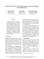
Báo cáo khoa học: "F-FDG PET/CT-based gross tumor volume definition for radiotherapy in head and neck Cancer: a correlation study between suitable uptake value threshold and tumor parameters" pdf
Bạn đang xem bản rút gọn của tài liệu. Xem và tải ngay bản đầy đủ của tài liệu tại đây (657.87 KB, 8 trang )
RESEA R C H Open Access
18
F-FDG PET/CT-based gross tumor volume
definition for radiotherapy in head and neck
Cancer: a correlation study between suitable
uptake value threshold and tumor parameters
Chia-Hung Kao
1,3
, Te-Chun Hsieh
1,5
, Chun-Yen Yu
2,5
, Kuo-Yang Yen
1,5
, Shih-Neng Yang
2,5
, Yao-Ching Wang
2
,
Ji-An Liang
2,3
, Chun-Ru Chien
2,3
, Shang-Wen Chen
2,3,4*
Abstract
Background: To define a suitable threshold setting for gross tumor volume (GTV) when using
18
Fluoro-
deoxyglucose positron emission tomography and computed tomogram (PET/CT) for radiotherapy planning in head
and neck cancer (HNC).
Methods: Fifteen HNC patients prospectively received PET/CT simulation for their radiation treatment planning.
Biological target volume (BTV) was derived from PET/CT-based GTV of the primary tumo r. The BTVs were defined as
the isodensity volumes when adjusting different percentage of the maximal standardized uptake value (SUVmax),
excluding any artifact from surrounding normal tissues. CT-based primary GTV (C-pGTV) that had been previously
defined by radiation oncologists was compared with the BTV. Suitable threshold level (sTL) could be determined
when BTV value and its morphology using a certain threshold level was observed to be the best fitness of the
C-pGTV. Suitable standardized uptake value (sSUV) was calculated as the sTL multiplied by the SUVmax.
Results: Our result demonstrated no single sTL or sSUV method could achieve an optimized volumetric match
with the C-pGTV. The sTL was 13% to 27% (mean, 19%), whereas the sSUV was 1.64 to 3.98 (mean, 2.46). The sTL
was inversely correlated with the SUVmax [sTL = -0.1004 Ln (SUVmax) + 0.4464; R
2
= 0.81]. The sSUV showed a
linear correlation with the SUVmax (sSUV = 0.0842 SUVmax + 1.248; R
2
= 0.89). The sTL was not associated with
the value of C-pGTVs.
Conclusion: In PET/CT-based BTV for HNC, a suitable threshold or SUV level can be established by correlating with
SUVmax rather than using a fixed threshold.
Introduction
18
Fluoro-deoxyglucose positron emission tomography
(
18
F-FDG PET) has been shown to improve the staging
of head and neck cancer (HNC) [1-5].
18
F-FDG PET
after definitive radiotherapy (RT) has also been shown
to have a good negative predictive value in patients with
HNC [6,7]. The use of
18
F-FDG PET in RT represents
an expansion of this already interdisciplinary process to
include information on the biologic status of tumors,
which is complementary to conventional computed
tomogram (CT) images and may result in target
volumes that contain proliferating tumor burden. Sev-
eral institutions have investigated the value of
18
F-FDG
PET in tumor target delineation for HNC [8-12]. While
CT remains the gold standard for delineation of tumor
volumes for RT planning, these studies reported PET
overlay on CT has shown to have some impact the
gross target volume (GTV), decrease inter-observer
variability and change the treatment planning. However,
when a radiation oncologist contours the GTVs on
fused PET and CT images at the radiation treatment
planning (RTP) workstation, a problem is emerged in
* Correspondence:
2
Department of Radiation Oncology, China Medical University Hospital,
Taichung Taiwan
Full list of author information is available at the end of the article
Kao et al. Radiation Oncology 2010, 5:76
/>© 2010 Kao et al; licensee BioMed Central Ltd. This is an Open Access article distributed under the term s of the Creative Commons
Attribution License ( which permits unr estricted use, distribution, and reproductio n in
any medium, provided the origina l work is prop erly cited.
setting the threshold for the PET images. The volume of
the GTVs on the PET images can be easily altered by
simply adjusting the threshold setting. Despites several
investigations declared PET-based target delineation
results in a change in the gross tumor volume (GTV)
compared to CT-based GTV [13-17], some standards
should be followed for
18
F-FDG-based delineation of
tumor boundaries when c omparing PET-based target
volume with conventional CT-based tumor volume [18].
One study used phantoms of a known size in an attempt
to define a standard threshold cutoff in
18
F-FDG PET
voxel values [19]. This study suggested that the thresh-
old can be set at 42% of the maximum uptake, though
the study considered only lesions in the size range of
0.4 to 5.5 mL, a range in which threshold levels are
extremely sensitive.
The published methods based on a threshold deter-
mined as a percentage of the maximal standardized
uptake value (SUVmax) have used values ranging from
15% to 50% for lung cancer [13-17,20-23]. In HNC ser-
ies, there was a great variation of validated standardized
methods for setting this threshold [5,8-12]; these include
using the absolute standardized uptake value (SUV) (i.e.,
GTV = SUV of > 2.5), using percentages of the SUVmax
(i.e., GTV = volume encompassed by > 50% the SUV-
max), or ignoring the threshold setting and simply con-
touring the CT volume corresponding to the visually
identified lesion. Three studies have investigated the
optimal threshold by different method in target delinea-
tion [24-26], but their results were not consistent. To
reduce intra-observer or inter-observer variability in
GTV delineation using PET, there is a need to conduct
another study to clarify this issue.
We hypothesized that a suitable threshold level of
18
F-
FDG PET can be obtained by certain tumor-related
parameters when defining GTV for HNC. Thus, this
study was conducted to evaluate the appropriateness of
the percentage threshold method or other approaches
by using PET/CT simulation in determining the suitable
threshold level for the best volumetric match for GTV.
The PET data of the PET/CT image was only used for
CT-based GTV comparison but not for seeking meta-
static disease or for changing the radiation treatment
strategy.
Methods
Patient population
After approval by local institutional review board (num-
ber: DMR98-IRB-067), a cohort of 15 fresh HNC patients
with a histological proof of squamous cell carcinoma,
who would undergo definitive concurrent chemora-
diotherapy with an intensity-mod ulated radiotherapy
technique (IMRT) at China Medical University Hospital,
were enrolled in this prospective study. The median age
was 46 years (range, 36-70 years). Thirteen patients were
men and two were women. They received a pretreatment
PET/CT for RT planning. No patient was known to have
a history of diabetes and all had a normal serum glucose
level before taking the PET/CT image. The characteris-
tics of the 15 patients are listed in Table 1.
PET-CT image acquisition
All patients were asked to fast for at least 4 hours before
18
F-FDG PET/CT imaging. Approximately 60 minutes
after the administration of 370 MBq of
18
F-FDG, simu-
lation images were taken by PET/CT scanner (PET/CT-
16 slice, Discovery STE, GE Medical System, Milwaukee,
Wisconsin USA). During the uptake period, patients
seated in a comfortable chair and were asked to rest.
WholebodyPET/CTimagesweretakenfirst.Thepro-
cedure did not required immobilization device and take
approximately 30 minutes to position the patient and to
acquire both the CT and PET data in total. CT images
were obtained at 120 kVp and variable mA (AutomA
technique) with 3.75-mm slice. The PET data were
reconstructed by application of the CT-based attenua-
tion correction and iterative reconstruction algorithm.
Immediately after whole body PET/CT images, patients
were simulated in a RT set-up position on the PET/CT
scanner table with a h ead and neck immobilization
device. An allocated PET/CT imaging field was taken
from the base of the skull to upper thorax. The images
were electronically transferred from t he PET/CT work-
station via DICOM3 to the RTP (Eclipse version 8.1,
Varian Medical System Inc, CA, USA) in the depart-
ment of radiatio n oncology. The workstation provided
the quantification of FDG uptake in terms of SUV.
Nuclear medicine physicians identified the locations and
values of SUVmax for all the primary tumors. This pro-
cedure is routinely used on the PET/CT workstatio n for
diagnostic readings, and it allows for definition of
threshold level and reproducible contouring of hyperme-
tabolic areas.
Delineation of CT-based tumor volume
On the basis of axial CT images, contouring of the
tumor volume and normal and critical structures was
performed without knowledge of the PET results in an
effort to reduce bias. Radiation oncologists then deli-
neated the primary gross tumor volume (pGTV) and the
metastatic lymph node volume (nGTV). Neck lymph
nodes were consider ed pathological when their small est
axis diameter was > 1 cm. The volumes of all tumors
were measured by outlining the lesion on each image if
it was visible. No attempts were made to differentiate
the tumors from any related edema. The tumor volumes
were contoured and the volumes calculated using t he
same planning system. To reduce inter-observer
Kao et al. Radiation Oncology 2010, 5:76
/>Page 2 of 8
variations, at least 2 different radiation oncologists car-
ried out the contouring of the tumors for each patient.
When the calculated values for any volume varied by
more than 10%, an average of the readings was used as
the measured volume. When the variation exceeded
10%, contouring and measurement were repeated by 3
rd
radiation oncologist to correct any bias. In brief, the
CT-based primary gross tumor volume w ould be finally
confirmed by at least two radiation oncologists, and
abbreviated as C -pGTV. This procedure was addressed
in our previous report [27].
Volumetric match between PET-CT-based GTV and
CT-based GTV
After the completion of the C-pGTV contouring in RTP
system, the radiation oncologists reviewed the consis-
tency of PET/CT images with nuclear medicine physi-
cians. They also reconfirmed the allocated point of the
SUVmax within the tumors.
Biological target volume (BTV) was derived from PET/
CT-based GTV of the primary tumor. The BTVs were
defined as the isodensity volumes when adjusting differ-
ent percentage of the maximal threshold levels, exclud-
ing any noise o r artifact from surrounding normal
tissues, including brain, extracting teeth pocket, or phar-
yngeal constrictors. The percentage threshold was
adjusted from 10% to 50% with interval of 5%, and the
BTVs were determined for each threshold. The interval
of threshold change could be further reduced to 1% for
achieving the best fitness of the defined C-pGTV from
both the tumor volume and the morphology. To sim-
plify the volume analysis, only signals overlying the
pGTV were chosen. The volumetric data of the different
BTVs were automatically measured by the RTP, and this
volume excluded any nGTVs. By this way, a suitable
threshold level (sTL) could be defined when the mor-
phology and the calculated BTV value using a certain
threshold level was observed to be the best fitness of the
volumetric data from the C-pGTV (Figure 1, 2, 3). In
addition, a suitable SUV (sSUV) values were calculated
as the sTL multiplied by individual SUVmax values.
Table 1 Patient’s characteristics and their volumetric and PET/CT data
Patient Tumor type (AJCC
stage)
C-pGTV
(mL)
SUVmax BTV (mL)
10% TL
BTV (mL)
20% TL
BTV (mL)
30% TL
BTV (mL)
40% TL
BTV (mL)
50% TL
sTL sSUV
1 HPC (T2N2) 43.3 30.6 47.9 35.5 30 25.4 19.2 13% 3.98
2 OPC (T4N1) 75.4 17.2 92.1 42.8 31.6 24.8 18.2 16% 2.75
3 NPC (T4N2) 110.2 17 139.2 77.3 59.3 47 37.5 15% 2.55
4 NPC (T3N1) 30.4 14.6 40.7 22.7 14.6 8.3 2.8 15% 2.19
5 NPC (T1N1) 14.8 15.7 36.6 12 7.2 5.4 4.2 17% 2.21
6 OPC (T3N1) 47.7 24.1 75.8 33.3 21.1 14.1 9.9 14% 3.37
7 NPC (T4N2) 38.7 8.0 - 60 30.8 17.4 12.3 27% 2.16
8 NPC (T3N2) 44.6 17.0 64.7 33.6 24.5 17.5 13.6 15% 2.55
9 NPC (T2N1) 12.8 7.8 39.6 13 5.6 4 2.4 21% 1.64
10 NPC (T4N1) 35.5 8.2 80.7 44 21.9 10 5.6 23% 1.89
11 NPC (T1N1) 9.6 9.0 37.6 13.8 4.9 2.2 1.2 23% 2.07
12 NPC (T3N3) 27.8 17.5 49.9 13.7 9.2 5.3 3.2 14% 2.45
13 NPC (T3N2) 37.1 12.9 67.4 37.9 20.6 12 9.5 20% 2.57
14 NPC (T2N2) 22.4 8.1 - 35.6 14.8 9.2 5.9 27% 2.19
15 HPC (T2N1) 14.3 12.2 46.8 14.5 5.2 3.1 2.3 20% 2.44
Abbreviation: NPC: nasopharyngeal cancer; OPC: oropharyngeal cancer; HPC: hypopharyngeal cancer; C-pGTV: CT-base primary gross tumor volume; BTV:
biological target volume from PET/CT-base primary gross tumor volume; TL: threshold level; sTL: suitable threshold; sSUV: suitable SUV.
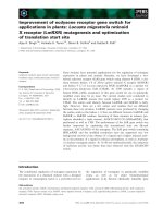
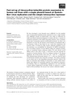
Figure 1 The biological target volume (BTV) of the primary
tumor was determined when using 10% isodensity volumes
(yellow line). CT-based GTV was outlined by red line.
Kao et al. Radiation Oncology 2010, 5:76
/>Page 3 of 8
Results
Volumetric and SUVmax data
Volumetric and SUVmax data for the 15 primary
tumors are listed in Table 1. The volumetric data and
related SUV information for the nGTVs were excluded
for simplification of the study. The mean C-pGTV was
36.9 ± 26.4 mL, and the range was 9.6 to 110.2 mL,
whereas the mean maximum tumor diameter in any
direction on CT was 4.33 ± 1.01 cm, and the rang e was
3.2 to 6.3 cm. The mean SUVmax was 13.98 ± 6.4 with
the range of 7.8 to 30.6. As listed in Table 1, the BTV
values at different threshold level showed an inverse
correlation with increasing threshold level. In addition,
there was no obvious association between the SUVmax
and the C-pGTV values in our patient cohort (Figure 4).
Also, there was no correlation between the maximum
tumor diameter and the SUVmax.
Correlation of sTL with C-pGTV and SUVmax
Table 1 also showed there was no demonstrated single
sTL or sSUV method for achieving optimized volu-
metric match with C-pGTV. For all patients, the sTL
for the best match was 1 3% to 27% (mean, 19%; stan-
dard deviation, 4.7%). The sSUV was 1.64 to 3.98
(mean, 2.46; standard deviation, 0.58). The sSUV
method of applying an isodensity volume of SUV > 2.5
failed to p rovide successful delineation in 60% of cases.
The relation between the sTL and the SUVmax is illu-
strated in F igure 5. The plot illustrated an inverse
hyperbolic curve with increasing SUVmax [sTL =
-0.1004 Ln (SUVmax) + 0.4464; R
2
= 0.81]. Converse ly,
the sTLs were not associated with the C-pGTVs using
different correlation modelsasdepictedinFigure6.
Furthermore, the sSUVs showed a direct proportion to
the SUVmax (Figure 7, sSUV = 0.0842 SUVmax +
1.248; R
2
= 0.89).
When excluding 4 tumors with SUVmax < 10 or elim-
inating 4 cases with C-pGTV < 20 mL, both the sTLs
and the sSUVs were found to have a similar pattern of
correlation with the SUVmax. There was no apparent
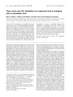
Figure 2 The BTV of the primary tumor was determined when
using 15% isodensity volumes (green line). CT-based GTV was
outlined by red line.
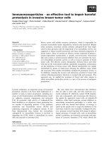
Figure 3 The BTV of the primary tumor was determined when
using 20% isodensity volumes (pink line). CT-based GTV was
outlined by red line.
Figure 4 TheassociationbetweentheSUVmaxandtheCT-
based pGTV.
Kao et al. Radiation Oncology 2010, 5:76
/>Page 4 of 8
association between the sTLs and the tumor volume
through stratification of different SUVmax or C-pGTV
levels in our studied cohort.
Mismatch analysis
Two direction mismatch analysi s was carried out as the
method described by El-Bassiouni et al. [25] . When the
BTVs were determined by using their sTL, the mean
valueforthemismatchBTVs/C-pGTVwas15.3±
10.3% (range, 2.4 ~ 37.5%). In contrast, the mean value
for the mismatch C-pGTV/BTV was 16.2 ± 14.3%
(range, 1.9 ~ 48.7%). There was no significant difference
between two mismatch comparison using paired t test
(p = 0.72).
Discussion
Rothschild et al. reported a matched-pair comparis on
study that P ET/CT staging followed by IMRT improved
treatment outcome of locally advanced pharyngeal carci-
noma [28]. While incorporating this biologic image,
there is also a great need for delineating tumor tissue
more precisely, particu larly in IMRT era. Various meth-
ods for incorporating PET into the RT plan have been
reported; including visual comparisons, image overlays,
fusion of PET and CT images, and PET/CT simulation.
Since there is less co-registration error between PET
and CT using the same DICOM coordinates, PET/CT
simulation is a promising modality to improve contour-
ing accuracy for reducing the risk of geographic misses
in RT planning [29,30]. However , care must be taken in
implementing this new technology as many physicians
concern the standard of threshold setting in
18
F-FDG
PET. This study provides an applicable way of volu-
metric match when selecting asuitablethresholdlevel
for CT-based GTVs which had been previously deli-
neated by radiation oncologists. Because these tumors
would be treated by RT rather than surgical resection,
our methods did not reflect a technique of determining
real tumor margin or volume. Although our patient
number was s mall, the result demonstrated a suitable
threshold levels can be derived from individual SUVmax
values, which might correspond to an intrinsic biological
nature of a tumor. Different from those investigators
that suggested using a fixed threshold for conto uring in
HNC [10,11,24], our results showed no distinctiv e value
for sSUV or sTL. In addition, no obvious correlation
between SUVmax and C-pGTV was found and this
might imply that a large tumor is not always associated
with an aggressive metabolic activity within a tumor.
There are many known factors responsible for SUV
measurements and therefore tumor contours: the meta-
bolic activity, tumor heterogeneity, and tumor motion
[21]. Despite the effect of tumor motion can be
neglected in RT set-up for HNC patients, Poisson distri-
bution of pixel intensity does make the use of SUVmax
a less reliable starting point for tumor delineation [31].
Nonetheless, SUVmax is important biologic parameter
and can be easily obtained from routine
18
F-FDG PET
Figure 5 The correlation curve between the suitable threshold
level and the SUVmax.
Figure 6 The association between the suitable threshold level
and the CT-based GTV.
Figure 7 The correlation curve between the suitable SUV and
the SUVmax.
Kao et al. Radiation Oncology 2010, 5:76
/>Page 5 of 8
image. On the other hand, the only investigati on pub-
lished to date on the use of a source-to-background
algorithm in patients focused on larynx tumors [32]. In
the chest, mean
18
F-FDG uptake in normal tissues may
vary between a SUV of < 1 (lung) up to a SUV of > 3
(liver) [20]. In the he ad and neck region, higher SUV
area can be ob served in adjacent brain, Waldeyer’sring,
extracted teeth pocket, pharyngeal con strictors, and
vocal cord region. Thus, it is required to carefully sub-
tract any tumor -unrelated artifact s from these areas
when delineating the BTV.
Black et al. reported the results of a phantom experi-
ment designed to evalua te the role of mean target SUVs
in conditions of various target-to background
18
F-FDG
activities [31]. They showed that the threshold SUV was
linearly correlated with the mean target SUV [threshold
SUV = 0.307 × ( mean target SUV + 0.588)]. Theoreti-
cally, it might be more ideal to use mean target SUV
instead of SUVmax for threshold analysis since mean
target SUV could characterize an average uptake value
of certain tumors. However, the volume of the GTV
must be identified first to obtain a mean target SUV.
This method may be feasible for a known-sized phan-
tom but not for real tumors whose contours are suscep-
tible to the inter-observer variances.
El-Bassiouni et al. repor ted a pilot study to define th e
best threshold of
18
F-FDG uptake for tumor volume deli-
neation of HNC [25]. By using the background-sub-
tracted tumor maximum (THR) uptake for PET signal
segmentation, they found an inverse correlation between
the threshold of THR and the tumor maximum uptake
(S), but no cor relation between the threshold of THR
and the ratio of tumor maximum uptake to the back-
ground uptake (S/G). They also suggested a threshold of
THR of 20% in tumors with S > 30% kBq/ml and 40%
with S < 30% kBq/ml. The correlation between the
threshold of THR and the S was a novel finding; however,
for those PET centers using SUV for counting FDG-avid
tumor uptake, direct measurement of the maximum
uptake values might be not always practicable.
Schinagl et al. compared five methods for determining
the BTV using coregistered CT and FDG-PET in HNC
patients [26], including visual GTV, 40% and 50% of
SUVmax, an absolute SUV of 2.5, and an adaptive
threshold based on the signal-to-background ratio. The
clinical implications from their studies were two folds.
First, an isodensity volume of SUV > 2.5 failed to pro-
vide delineation in 45% of cases, which was similar with
our finding. Second, PET frequen tly detected substantial
tumor extension outside the CT-based GTV (15-34% of
PET volume). The rate was also comparable with our
result that the mean value for the mismatch BTV/C-
pGTV was 15.3 ± 10.3%. Theoretically, the mismatch is
somewhat attributed to the limitation of voxel density
or a partial volume effect. In practice, it is hard to
exactly define the real tumor volume outside CT-based
GTV from PET image without surgical intervention.
However, contouring accuracy can be improved further
if radiation oncologists evaluate accordingly the change
of BTV by adjusting different threshold levels during
contouring.
Our study failed to show an inverse cor relation
between sTLs and C-pGTV sasthethresholdstudy
reported by Biehl et al. in lung cancer [21]. Using the
similar method, they found optimal threshold was inver-
sely correlated with CT-based GTV (R
2
= 0.79). The
optimal threshold level in their study was 24 ± 13%,
compared to that of 19 ± 4.7% in our study. This discre-
pancy might be attributed to two explanations. First, the
SUVmax in their data was in direct proportion to the
increase of maximum tumor diameter, which was not
observed in our result. Probably, reduction of optimal
threshold could be anticipated following the increase of
tumor volume or Smax. Second, the measured tumor
volumes in their study were far l arger than those of our
data (mean tumor volume: 198 ± 277 mL vs. 36.9 ±
26.4mL).Thedifferencemightnotonlyrepresentthe
dissimilar clinical situation when irradiating two types of
cancers, but perhaps contribute to the diverse experi-
mental findings. Of course, more investigations are
required to elucidate the biological difference of the two
cancers in
18
F-FDG PET/CT image.
In another study described by Nestle et al., they ana-
lyzed various modalities for determining the BTV for
lung cancer, includ ing vis ual GTV, 40% of SUVmax, an
absolute SUV of 2.5, and tumor-to-background ratio
[20]. They found substantial differences of up to 41%
among these 4 different methods. They concluded that
the 40% threshold m ethod was not suita ble for target
volume delineation. Based on the results o f our study
and other reports [20,21,24,25], a fixed thres hold model
is questionable in tumor volume delineation because it
relies mainly on the uniformity of SUVs within the
tumor. Theoretically, a unique threshold setting may fail
to adequately model the lack of uniformity of
18
F-FDG
uptake because of factors such as hypoxia and necrosis,
which are more likely to occur in large tumors or tumor
with a higher SUVmax. For other BTVs with higher
threshold than sTL, these metabolically active areas
might be useful in assigning dose intensification during
IMRT. Of course, the medical significance of including
these additional data in the original treatment plan on
final patient outcome is yet to be determined.
There are several limitations in our study. First, there
was no reason that the metabolic activity should be defi-
nitely related to the real tumor volume. Undoubtedly, a
surgicalstudymustbedonetoanswerthequestion.
Also, the C-pGTV, used as reference image in the
Kao et al. Radiation Oncology 2010, 5:76
/>Page 6 of 8
present study, could identify areas not strict ly related to
tumor tissue. Third, it is imperative to clarify whet her
the results could be reproducible when the same
patients were scanned at different time even if their
serum glucose levels were normal before images. Finally,
the results have to be tested on anoth er cohort of HNC
patients to see how well the correlation equations were
working. Certainly, a validation study is ongoing to
reconfirm our preliminary finding.
In conclusion, a suitable threshold or SUV level can
be established by an adaptive approach by correlating
with SUVmax rather than using a fixed value. It will be
a subject of our future work to correlate the threshold
with more tumor-related factors, such as hypoxia, prolif-
eration and histological difference. In PET-based RT
planning for HNC, careful selection of a suitable thresh-
old is imperative because this value is required to ade-
quately encompass tumor without compromising
adjacent normal tissues.
Acknowledgements
We want to thank the grant support (CMU98-C-13) in China Medical
University and the grant support (DOH99-TD-C-111-005) from department of
health in Taiwan.
Author details
1
Department of Nuclear Medicine and PET Center, China Medical University
Hospital, Taichung, Taiwan.
2
Department of Radiation Oncology, China
Medical University Hospital, Taichung Taiwan.
3
College of Medicine School,
China Medical University, Taichung, Taiwan.
4
College of Medicine School,
Taipei Medical University, Taipei, Taiwan.
5
Department of Biomedical Imaging
and Radiological Science, China Medical University, Taichung, Taiwan.
Authors’ contributions
CHK and SWC are responsible for the study design, coordination and drafted
the manuscript. TCH, YCY and KYY collected the PET/CT data and performed
analysis. SWC, SNY, YCW and JAL were responsible for the evaluation of the
patients and the collection of clinical data. CRC provided some intellectual
recommendation and reviewed the manuscript. CHK and SWC wrote the
final version of the manuscript. All authors read and approved the final
manuscript.
Competing interests
The authors declare that they have no competing interests.
Received: 14 June 2010 Accepted: 2 September 2010
Published: 2 September 2010
References
1. Laubenbacher C, Saumweber D, Wagner-Manslau C, Kau RJ, Herz M, Avril N,
Ziegler S, Kruschke C, Arnold W, Schwaiger M: Comparison of fluorine-18-
fluorodeoxyglucose PET, MRI and endoscopy for staging head and neck
squamous-cell carcinomas. J Nucl Med 1995, 36:1747-1757.
2. Veit-Haibach P, Luczak C, Wanke I, Fischer M, Egelhof T, Beyer T, Dahmen G,
Bockisch A, Rosenbaum S, Antoch G: TNM staging with FDG-PET/CT in
patients with primary head and neck cancer. Eur J Nucl Med Mol Imaging
2007, 34:1953-1962.
3. Kao CH, ChangLai SP, Chieng PU, Yen RF, Yen TC: Detection of recurrent
or persistent nasopharyngeal carcinomas after radiotherapy with 18-
fluoro-2-deoxyglucose positron emission tomography and comparison
with computed tomography. J Clin Oncol 1998, 16:3550-3555.
4. Wong RJ, Lin DT, Schoder H, Patel SG, Gonen M, Wolden S, Pfister DG,
Shah JP, Larson SM, Kraus DH: Diagnostic and prognostic value of [(18)F]
fluorodeoxyglucose positron emission tomography for recurrent head
and neck squamous cell carcinoma. J Clin Oncol 2002, 20:4199-4208.
5. Deantonio L, Beldì D, Gambaro G, Loi G, Brambilla M, Inglese E, Krengl M:
FDG-PET/CT imaging for staging and radiotherapy treatment planning
of head and neck carcinoma. Radiat Oncol 2008, 3:29.
6. Moeller BJ, Rana V, Cannon BA, Williams MD, Sturgis EM, Ginsberg LE,
Macapinlac HA, Lee JJ, Ang KK, Chao KS, Chronowski GM, Frank SJ,
Morrison WH, Rosenthal DI, Weber RS, Garden AS, Lippman SM,
Schwartz DL: Prospective risk-adjusted [18F]Fluorodeoxyglucose positron
emission tomography and computed tomography assessment of
radiation response in head and neck cancer. J Clin Oncol 2009,
27:2509-2515.
7. Yao M, Smith RB, Hoffman HT, Funk GF, Lu M, Menda Y, Graham MM,
Buatti JM: Clinical significance of postradiotherapy [18F]-
fluorodeoxyglucose positron emission tomography imaging in
management of head-and-neck cancer: a long-term outcome report. Int
J Radiat Oncol Biol Phys 2009, 74:9-14.
8. Ciernik IF, Dizendorf E, Baumert BG, Reiner B, Burger C, Davis JB, Lutolf UM,
Steinert HC, Von Schulthess GK: Radiation treatment planning with an
integrated positron emission and computer tomography (PET/CT): a
feasibility study. Int J Radiat Oncol Biol Phys 2003, 57:853-863.
9. Heron DE, Andrade RS, Flickinger J, Johnson J, Agarwala SS, Wu A,
Kalnicki S, Avril N: Hybrid PET-CT simulation for radiation treatment
planning in head-and-neck cancers: a brief technical report. Int J Radiat
Oncol Biol Phys 2004, 60:1419-1424.
10. Paulino AC, Koshy M, Howell R, Schuster D Davis LW: Comparison of CT-
and FDG-PET-defined gross tumor volume in intensity-modulated
radiotherapy for head-and-neck cancer. Int J Radiat Oncol Biol Phys 2005,
61:1385-1392.
11. Wang D, Schultz CJ, Jursinic PA, Bialkowski M, Zhu XR, Brown WD, Rand SD,
Michel MA, Campbell BH, Wong S, Li XA, Wilson JF: Initial experience of
FDG-PET/CT guided IMRT of head-and-neck carcinoma. Int J Radiat Oncol
Biol Phys 2006, 65:143-151.
12. Guido A, Fuccio L, Rombi B, Castellucci P, Cecconi A, Bunkheila F, Fuccio C,
Spezi E, Angelini AL, Barbieri E: Combined 18F-FDG-PET/CT imaging in
radiotherapy target delineation for head-and-neck cancer. Int J Radiat
Oncol Biol Phys 2009, 73:759-763.
13. Bradley J, Thorstad WL, Mutic S, Miller TR, Dehdashti F, Siegel BA, Bosch W,
Bertrand RJ: Impact of FDG-PET on radiation therapy volume delineation
in non-small-cell lung cancer.
Int J Radiat Oncol Biol Phys 2004, 59:78-86.
14. Erdi YE, Rosenzweig K, Erdi AK, Macapinlac HA, Hu YC, Braban LE, Humm JL,
Squire OD, Chui CS, Larson SM, Yorke EDL: Radiotherapy treatment
planning for patients with non-small cell lung cancer using positron
emission tomography (PET). Radiother Oncol 2002, 62:51-60.
15. Kalff V, Hicks RJ, MacManus MP, Binns DS, McKenzie AF, Ware RE, Hogg A,
Ball DL: Clinical impact of (18)F fluorodeoxyglucose positron emission
tomography in patients with non-small-cell lung cancer: a prospective
study. J Clin Oncol 2001, 19:111-118.
16. Mah K, Caldwell CB, Ung YC, Danjoux CE, Balogh JM, Ganguli SN, Ehrlich LE,
Tirona R: The impact of (18)FDG-PET on target and critical organs in CT-
based treatment planning of patients with poorly defined non-small-cell
lung carcinoma: a prospective study. Int J Radiat Oncol Biol Phys 2002,
52:339-350.
17. Vanuytsel LJ, Vansteenkiste JF, Stroobants SG, De Leyn PR, De Wever W,
Verbeken EK, Gatti GG, Huyskens DP, Kutcher GJ: The impact of (18)F-
fluoro-2-deoxy-D-glucose positron emission tomography (FDG-PET)
lymph node staging on the radiation treatment volumes in patients
with non-small cell lung cancer. Radiother Oncol 2000, 55:317-324.
18. Ford EC, Herman J, Yorke E, Wahl RL: 18F-FDG PET/CT for image-guided
and intensity-modulated radiotherapy. J Nucl Med 2009, 50:1655-1665.
19. Erdi YE, Mawlawi O, Larson SM, Imbriaco M, Yeung H, Finn R, Humm JL:
Segmentation of lung lesion volume by adaptive positron emission
tomography image thresholding. Cancer 1997, 80:2505-2509.
20. Nestle U, Kremp S, Schaefer-Schuler A, Sebastian-Welsch C, Hellwig D,
Rube C, Kirsch CM: Comparison of different methods for delineation of
18F-FDG PET-positive tissue for target volume definition in radiotherapy
of patients with non-Small cell lung cancer. J Nucl Med 2005,
46:1342-1348.
21. Biehl KJ, Kong FM, Dehdashti F, Jin JY, Mutic S, El Naqa I, Siegel BA,
Bradley JD: 18F-FDG PET definition of gross tumor volume for
radiotherapy of non-small cell lung cancer: is a single standardized
Kao et al. Radiation Oncology 2010, 5:76
/>Page 7 of 8
uptake value threshold approach appropriate? J Nucl Med 2006,
47:1808-1812.
22. Ashamalla H, Rafla S, Parikh K, Mokhtar B, Goswami G, Kambam S, Abdel-
Dayem H, Guirguis A, Ross P, Evola A: The contribution of integrated PET/
CT to the evolving definition of treatment volumes in radiation
treatment planning in lung cancer. Int J Radiat Oncol Biol Phys 2005,
63:1016-1023.
23. Brianzoni E, Rossi G, Ancidei S, Berbellini A, Capoccetti F, Cidda C,
D’Avenia P, Fattori S, Montini GC, Valentini G, Proietti A, Algranati C:
Radiotherapy planning: PET/CT scanner performances in the definition
of gross tumour volume and clinical target volume. Eur J Nucl Med Mol
Imaging 2005, 32:1392-1399.
24. Baek CH, Chung MK, Son YI, Choi JY, Kim HJ, Yim YJ, Ko YH, Choi J, Cho K,
Jeong HS: Tumor volume assessment by 18F-FDG PET/CT in patients
with oral cavity cancer with dental artifacts on CT or MR images. J Nucl
Med 2008, 49:1422-1428.
25. El-Bassiouni M, Ciernik IF, Davis JB, El-Attar I, Reiner B, Burger C, Goerres GW,
Studer GM: [18FDG] PET-CT-based intensity-modulated radiotherapy
treatment planning of head and neck cancer. Int J Radiat Oncol Biol Phys
2007, 69:286-293.
26. Schinagl DA, Vogel WV, Hoffmann AL, van Dalen JA, Oyen WJ, Kaanders JH:
Comparison of five segmentation tools for 18F-fluoro-deoxy-glucose-
positron emission tomography-based target volume definition in head
and neck cancer. Int J Radiat Oncol Biol Phys 2007, 69:1282-1289.
27. Chen SW, Yang SN, Liang JA, Lin FJ, Tsai MH: Prognostic impact of tumor
volume in patients with stage III-IVA hypopharyngeal cancer without
bulky lymph nodes treated with definitive concurrent
chemoradiotherapy. Head Neck 2009, 31:709-716.
28. Rothschild S, Studer G, Seifert B, Huguenin P, Glanzmann C, Davis JB,
Lütolf UM, Hany TF, Ciernik IF: PET/CT staging followed by Intensity-
Modulated Radiotherapy (IMRT) improves treatment outcome of locally
advanced pharyngeal carcinoma: a matched-pair comparison. Radiat
Oncol 2007, 2:22.
29. Breen SL, Publicover J, De Silva S, Pond G, Brock K, O’Sullivan B,
Cummings B, Dawson L, Keller A, Kim J, Ringash J, Yu E, Hendler A,
Waldron J: Intraobserver and interobserver variability in GTV delineation
on FDG-PET-CT images of head and neck cancers. Int J Radiat Oncol Biol
Phys 2007, 68:763-770.
30. Riegel AC, Berson AM, Destian S, Ng T, Tena LB, Mitnick RJ, Wong PS:
Variability of gross tumor volume delineation in head-and-neck cancer
using CT and PET/CT fusion. Int J Radiat Oncol Biol Phys 2006, 65:726-732.
31. Black QC, Grills IS, Kestin LL, Wong CY, Wong JW, Martinez AA: Defining a
radiotherapy target with positron emission tomography. Int J Radiat
Oncol Biol Phys 2004, 60:1272-1282.
32. Geets X, Daisne JF, Gregoire V, Hamoir M, Lonneux M: Role of 11-C-
methionine positron emission tomography for the delineation of the
tumor volume in pharyngo-laryngeal squamous cell carcinoma:
comparison with FDG-PET and CT. Radiother Oncol 2004, 71:267-273.
doi:10.1186/1748-717X-5-76
Cite this article as: Kao et al.:
18
F-FDG PET/CT-based gross tumor
volume definition for radiotherapy in head and neck Cancer: a
correlation study between suitable uptake value threshold and tumor
parameters. Radiation Oncology 2010 5:76.
Submit your next manuscript to BioMed Central
and take full advantage of:
• Convenient online submission
• Thorough peer review
• No space constraints or color figure charges
• Immediate publication on acceptance
• Inclusion in PubMed, CAS, Scopus and Google Scholar
• Research which is freely available for redistribution
Submit your manuscript at
www.biomedcentral.com/submit
Kao et al. Radiation Oncology 2010, 5:76
/>Page 8 of 8