Báo cáo khoa học: "Use of serum squamous cell carcinoma antigen for follow-up monitoring of cervical cancer patients who were treated by concurrent chemoradiotherapy" pot
Bạn đang xem bản rút gọn của tài liệu. Xem và tải ngay bản đầy đủ của tài liệu tại đây (332.81 KB, 6 trang )
RESEARC H Open Access
Use of serum squamous cell carcinoma antigen
for follow-up monitoring of cervical cancer
patients who were treated by concurrent
chemoradiotherapy
Sang Min Yoon
1,2
, Kyung Hwan Shin
1*
, Joo-Young Kim
1
, Sang Soo Seo
1
, Sang-Yoon Park
1
, Sung Ho Moon
1
,
Kwan Ho Cho
1
Abstract
Background: To investigate the significance of monitoring the levels of the serum squamous cell carcinoma
antigen (SCC-Ag) for the detection of recurrent disease in patients with cervical cancer treated by concurrent
chemoradiotherapy.
Methods: The records of 112 patients with cervical cancer were reviewed. Serum SCC-Ag levels were measured at
regular follow-up visits. A SCC-Ag level of 2 ng/mL was considered the upper limit of normal. Biochemical failure
was defined as two consecutively increasing SCC-Ag values above normal. Recurrent disease was confirmed by
histologic and radiographic studies.
Results: Eighteen patients (16%) developed recurrent disease. Sixteen patients had initially elevated SCC-Ag, post-
treatment normalization of SCC-Ag, and tumor recurrence. The SCC-Ag difference (ΔSCC-Ag), defined as the
difference between the last value after two consecutively increases above normal and the value immediately
before the elevation, had good clinical performance in predicting cancer recurrence. The cutoff value of ΔSCC-Ag
was 0.95 ng/mL.
Conclusions: SCC-Ag is a relatively good method for the detection of disease recurrence in patients with cervical
cancer who were treate d by concurrent chemoradiotherapy.
Background
Radiotherapy has maintained its place as the cornerstone
of therapy f or many decades for uteri ne cervical cancer.
Recently, the results of several randomized trials have
recommended the concomitant administration of che-
motherapy and radiotherapy as a standard treatment for
patients with locally advanced cervical cancer [1-3].
Although this combination treatment plays a role in
improving disease control, many patients suffer from
tumor recurrence during the follow-up period. Therefore,
the identification of prognostic factors associated with dis-
ease course and outcome following chemoradiotherapy
may help to guide the development of more effective treat-
ments and prevent tumor recurrence.
Over the past decade, several serum markers have been
investigated to search for additional prognostic para-
meters that could be used, to monitor the tr eatment
response, and detect the recurrence in patients with cer-
vical cancer. In particular, the squamous cell carcinoma
antigen (SCC-Ag) is the most commonly used tumor
marker for cervical cancer. SCC-Ag is a sub-fraction of
the tumor antigen TA-4, a 48 kDa glycoprotein first
isolated by Kato and Torigoe [4]. This antigen is present
in normal cervical epithelium, but has higher expression
in cervical neoplasms [5]. With the development of a sen-
sitive radioimmunoassay, this marker can be readily
detected in the serum and is now considered a valuable
tool for monitoring cervical cancer. Serum SCC-Ag level s
* Correspondence:
1
Research Institute and Hospital, National Cancer Center, 809 Madu 1-dong,
Ilsandong-gu, Goyang-si, Gyeonggi-do, 410-769, Republic of Korea
Full list of author information is available at the end of the article
Yoon et al. Radiation Oncology 2010, 5:78
/>© 2010 Yoon et al; licensee BioMed Central Ltd. This is an Open Access article distributed under the terms of the Crea tive Commons
Attribution License ( which perm its unrestricted use, distribut ion, and reproduction in
any mediu m, provided the original work is properly cited.
correlate with the extent of disease [6-8], response to
radiotherapy [9], response to chemotherapy [10,11], and
can be used to predict survival and tumor recurrence
during follow-up [12-16]. Several studies have reported
the use of serial SCC-Ag data for post-therapeutic moni-
toring. In these reports, 70-86% of cervical cancer
patients with recurrent disease had elevated SCC-Ag
levels at some time during follow-up [7,8,11,14,16]. How-
ever, few studies have focused on cervical cancer patients
treated by concurrent chemoradiotherapy (CCRT), which
is now considered the standard treatment for locally
advanced cervical cancer.
In the present study, we investigated the potential use
of SCC-Ag as a marker for predicting tumor recurrence
in uterine cervical cancer patients treated with CCRT.
Methods
Between July 2001 and February 2004, 124 previously
untreated women with the International Federation of
Gynecology and Obstetrics (FIGO) stage IB-IV uterine
cervical cancer were entered in a CCRT protocol at the
National Cancer Center (Goyang, Gyeonggi, Republic of
Korea). Twelve cases that lacked regular SCC-Ag deter-
minations in the follow-up period were excluded from
the study. The pre-treatment evaluation consisted of a
complete medical history, physical examination, full
bloo d counts, biochemical profile, serum SCC-Ag, chest
radiography, intravenous pyelogram, cystoscopy, recto-
sigmoidoscopy, magnetic resonance imaging (MRI) or
computed tomography (CT) scan, and/or [18F] -flouro-
2-deoxy-D-glucose positron emission tomography
(FDG-PET). Written informed consent was obtained
from all the patients.
All patients were given external beam radiotherapy
(EBRT) with 15 MV X-rays from a linear accelerator
(Varian Clinac 2100CD; Varian, Palo Alto, CA, USA).
EBRT was administered to the whole pelvic region using
a four-field box technique or parallel opposed anterior-
posterior beams. The r adiation field included the pri-
mary tumor, uterus, paracervical, parametrial and utero-
sacral regions, and the pelvic lymph nodes. The para-
aortic lymph nodes were also included if metastasis was
diagnosed during pre-treatment imaging study. The
radiation dose administered to the whole pelvis was
41.4-45 Gy (median 45 Gy), given in daily dose s of 1.8
Gy, five fractions per week. An additional boost of 5 .4-
21.4 Gy (median 10 Gy) was give n to the gross residu al
tumor and involved the parametrium without any mid-
line shielding. The dose to the para-aortic area was 45
Gy, with or without an additional booster dose of 10
Gy. Intracavitary brachytherapy was administered twice
weekly. Fletcher-Suit afterloading applicators were used
for high-dose-rate brachytherapy with an Iridium-192
source (Microselectron®; Nucletron, Veenendaal, The
Netherlands). The brachytherapy dose for each insertion
was 4 or 5 Gy at point A, and the total dose of
brachytherapy was 24-35 Gy (median 24 Gy). All
patients also were given concurrent chemotherapy,
which consisted of weekly doses of 40 mg/m
2
i.v. cispla-
tin for four to six cycles.
After completion of CCRT, all patients were given
clinical examinations and assayed for serum SCC-Ag
during the follow-up visits. A radiation oncologist and a
gynecologist followed the patients for one month after
treatment, then every three months for the first two
years, and every 3-6 months thereafter. The follow-up
intervals varied for patients suspected of having recur-
rent disease, based on individual situations. Patients
with suspicious symptoms, such a s signs at the physical
examination or an elevated serum SCC-Ag level during
follow-up periods, were given additional tests (histologi-
cal examination, abdomino-pelvic CT, pelvic MRI, FDG-
PET, etc.) to confirm the presence of recurrent diseases.
Serum SCC-Ag levels were measured using an i mmu-
noradiometric assay with a commercially available kit
(SCC-RIABEAD; SRL Inc., Tokyo, Japan). A measure-
ment of 2 ng/mL was c onsidered the upper limit of nor-
mal. Biochemical failure was defined as two consecutively
increasing S CC-Ag values above the normal limits. For
further stat istical analysis, after two consecutive elevated
SCC-Ag readings, the delta (Δ)SCC-Agwasdefinedas
the difference between the final value and the value
immediately before the elevation.
Logistic regression analysis was used to estimate the
probability of tumor recurrence from ΔSCC-Ag.
P values less than 0.05 were c onsidered significant.
A receiver operating characteristic (ROC) curve was
used to define the optimal cutoff point for the SCC-Ag
values relative to the probability of a recurrence.
Results
Table 1 shows the demographic and clinical characteris-
tics of the 112 enrolled patients. At diagnosis, 77 patients
had elevated serum SCC-Ag, corresponding to an overall
pre-treatment sensitivity of 68.8% (77/112). Most patients
(96.1%) had normalized SCC-Ag levels at one month
after c ompletion of CCRT. The median follow-up dura-
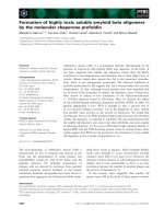
tion was 39 months (range, 16 - 55 months). Figure 1
shows the follow-up results.
After completion of CCRT, 18 patients (16%) experi-
enced tumor recurrence during the follow-up period.
Among these 18 patients, 11 had post-treatment SCC-
Ag elevations (biochemical failure) before the a ppear-
ance of clinically evident disease with a median
lead-time of 2 months (range, 1 - 15 months). An analy-
sis of all patients (n = 112) indicated that the sensitivity
and specificity of elevated post-treatme nt SCC-Ag levels
in association with recurrent disease were 61.1% and
Yoon et al. Radiation Oncology 2010, 5:78
/>Page 2 of 6
97.9%, respectively. For patients with elevated pre-treat-
ment SCC-Ag only (n = 77), these values were similar
(Table 2).
Next, we performed logistic regression of all patients
(n = 112) to estimate the probability of tumor recur-
rence based on ΔSCC-Ag measurements. Our results
indicate that ΔSC C-Ag was a significant predi ctor of
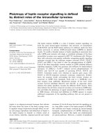
tumor recurrence (p = 0.001). Then, we performed
ROC analysis of ΔSCC-Ag to deter mine the optimal
levels for predicting the probability of tumor recur-
rence (Figure 2). The area under the ROC curve of
ΔSCC-Ag, which is considered to indicate the accuracy
ofthetest,was0.78.Thepredictabilityofrecurrence
was analyzed for patients who had an increase in the
level of pre-treatment SCC-Ag (n = 77). Logistic
regression analysis also indicated that ΔSCC-Ag was a
significant predictor of tumor recurrence (p = 0.002).
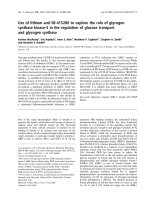
Figure 3 shows the ROC curve for ΔSCC-Ag in
patients with elevated pre-treatment SCC-Ag (n = 77).
TheseresultsindicatetheareaundertheROCcurve
of ΔSCC-Ag was 0.83 and t hat a ΔSCC-Ag val ue of
0.95 ng/mL was the optimal cutof f level f or prediction
oftumorrecurrence.Thesemeanthatthetruepositive
and false positive rates of tumor recurrence were 75%
and 11% (respectively) when the difference between
the last value after two consecutive increases above
normal and the value immediately before the elevation
was 0 .95 ng/mL.
Table 1 Patient characteristics
Variables
Age – years
Median 55
Range 22-78
FIGO stage – No. (%)
IB 11 ( 9.8)
IIA 13 (11.6)
IIB 66 (58.9)
III/IV 22 (19.7)
Histology – No. (%)
Squamous cell carcinoma 102 (91.1)
Adenocarcinoma 7 ( 6.2)
Adenosquamous cell carcinoma 3 ( 2.7)
Pelvic lymph node* – No. (%)
Positive 61 (54.5)
Negative 51 (45.5)
Tumor diameter – No. (%)
Range (cm) 1-10
≤4 cm 65 (58.0)
>4 cm 47 (42.0)
Abbreviations: FIGO = International Federation of Gynecology and Obstetrics.
* Pelvic lymph node status was diagnosed by imaging study.
Figure 1 A diagram of the follow-up results and the change in the SCC-Ag values in all patients.* Elevated serum SCC-Ag level means a
biochemical failure that is defined as two consecutively increasing tumor marker values above the normal limits, † No evidence of disease.
Table 2 Sensitivity, specificity, positive predictive value,
and negative predictive value of the tumor maker for
predicting a recurrence in all patients* and in patients
with elevated pretreatment tumor marker
†
SCC-Ag (n = 112)* SCC-Ag (n = 77)
†
Sensitivity 11/18 (61.1%) 11/16 (68.8%)
Specificity 92/94 (97.9%) 59/61 (96.7%)
PPV 11/13 (84.6%) 11/13 (84.6%)
NPV 92/99 (92.9%) 59/64 (92.2%)
Abbreviations: SCC-Ag = squamous cell carcinoma antigen; PPV = positive
predictive value; NPV = negative predictive value.
Yoon et al. Radiation Oncology 2010, 5:78
/>Page 3 of 6
Figure 2 Receiver operating characteristic curve for ΔSCC-Ag in predicting the probability of a recurrence in all patients.
Figure 3 Receiver operating characteristic curve for ΔSCC-Ag in predicting the probability of a recurrence in patients with elevated
pre-treatment tumor markers.
Yoon et al. Radiation Oncology 2010, 5:78
/>Page 4 of 6
Discussion
Our analysis of cervical cancer patients treated with
CCRT indicated that the sensitivity and specificity of
two consecutive increases in serum SCC-Ag for predict-
ing tumor recurrence were 61.1% and 97.9%, r espec-
tively. These results a re comparable to previously
repo rted result s. For exampl e, sev eral studies of cervical
cancer patients showed that an elevated serum SCC-Ag
level was associated with 70-92% rate of recurrent
tumors [ 8,11,14-19]; in addition, the specificity o f SCC-
Ag during the f ollow-up period was quite high, varying
from 95 to 98% [7,16,20]. According to our ROC analy-
sis,theareaundertheROCcurveindicatedtheΔSCC-
Ag was 0.83 for patients who had elevated pre-treatment
SCC-Ag. Therefore, our results indicate that ΔSCC-Ag
had good clinical performance in detection of recurrent
disease.
As described above, we defined biochemical failure as
two consecutive SCC-Ag values above normal. There
are no standard criteria used to define biochemical fail-
ure in cervical cancer, and many previous studies have
simply defined failure as an increase of tumor markers
above normal. However, Duk et al. [20] repor ted that
the predictive value of a single elevation of serum SCC-
Ag level for early detection of recurrence was only
48.6%, but that the predictive value was 76% when two
consecutive elevated serum SCC-Ag levels were used.
Furthermore, they reported that serum SCC-Ag in the
second sample was considerably higher in patients who
had tumor relapse than in those with no evidence of
relapse. Similarly, 10 of our p atients had only once
instance of elevated serum SCC-Ag, with normalization
at subsequent follow-ups. None of these 10 patients had
any evidence of disease at the final follow-up. Therefore,
we suggest use of additional measurement of SCC-Ag in
patients who have a single SCC-Ag elevation above the
normal limits. The use of sequential serum determina-
tions appears to increase the sensitivity and specificity of
serum SCC-Ag in predicting tumor recurrence.
The results reported here and those of others indica te
that routine sur veillance of serum SCC-Ag during fol-
low-up can be used to predict tumor recurrence, but it
is unclear if the early detection of recurrence will lead
to better survival. The major aim of follow-up surveil-
lance in cervical cancer is the early recognition of a
recurrence, based on the presumption that early detec-
tion of tumor r ecurrence may allow effective salvage
therapy [18]. However, the clinical relevance of detecting
early recurrence of cervical cancer is controversial. Esa-
jas et al. [18] showed that routine assessment of serum
SCC-Ag in follow-up resulted in the earlier detection of
a recurrence in a small proportion of patients, but did
not appear to contribute to better survival. Chan et al.
[17] reported similar results, in that the addition of
SCC-Ag monitoring provided no advant age over a regu-
lar follow-up with a clinical examination. They con-
cluded that posttreatment SCC-Ag monitoring was not
cost-effective because there was no curative treatment
for distant spread of disease. Other studies, however,
have suggested that follow-up SCC-Ag measurements
may improve survival. Chou et al. [21] studied the cli ni-
cal features of isolated para-aortic lymph node recur-
rence after definitive radiotherapy and reported a good
correlation between lower SCC-Ag levels (SCC-Ag ≤4
ng/mL) at recurrence and disease-free survival. They
concluded that periodic surveillance with this tumor
marker and imaging studies allowed the early detection
and implementation of salvage therapy. Forni et al. [22]
reported that a simplified diagnostic approach that
included the SCC-Ag assay and a gynecologic examina-
tion allowed early detection of cervical cancer recur-
rence with a very favorable cost-effective profile.
Moreover, o ther recent studies reported that the use of
FDG-PET in patients with asymptomatic SCC-Ag eleva-
tion allowed an earlier diagnosis of recurrence, with pro-
mising effects on survival [23,24]. There fore, further
study will be needed to confirm the value of routine
surveillance of SCC-Ag in improving survival r ate from
recurrent cervical cancer after earlier detection.
Conclusions
In summary, measurement of ΔSCC-Ag provided good
clinical performance in the detection of recurrent uter-
ine cervical cancer following CCRT. We suggest that
clinicians consider performing routine measurement of
SCC-Ag during the follow-up of such patient. However,
our results should be interpreted with some caution,
because our overall recurrence rate was very low and
the follow-up period was relatively short. Therefore, a
more comprehensive, large-scale study should be
performed to confirm our results.
List of abbreviations
SCC-Ag: squamo us cell carcinoma antigen; CCRT: con-
current chemoradiotherapy; FIGO: International Federa-
tion of Gynecology and Obstetrics; MRI: magnetic
resonance imaging; CT: computed tomography; FDG-
PET: [18F]-flouro-2-deoxy-D-glucose positron emission
tomography; EBRT: external beam radiotherapy; ROC:
receiver operating characteristic.
Author details
1
Research Institute and Hospital, National Cancer Center, 809 Madu 1-dong,
Ilsandong-gu, Goyang-si, Gyeonggi-do, 410-769, Republic of Korea.
2
Department of Radiation Oncology, Asan Medical Center, University of
Ulsan College of Medicine, Seoul, Republic of Korea.
Yoon et al. Radiation Oncology 2010, 5:78
/>Page 5 of 6
Authors’ contributions
Each author had participated sufficiently in the work to take public
responsibility for appropriate portions of the study. SMY participated in
research design, coded the patient database, conducted the analysis and
wrote the manuscript draft. KHS designed the project, analyzed the data and
revised the manuscript. KHC contributed to study conception and design.
SHM and JYK helped with the database and data analysis. SSS and SYP
provided additional guidance and support for this research. All authors read
and approved the final version of the manuscript.
Competing interests
The authors declare that they have no competing interests.
Received: 3 May 2010 Accepted: 15 September 2010
Published: 15 September 2010
References
1. Keys HM, Bundy BN, Stehman FB, Muderspach LI, Chafe WE, Suggs CL,
Walker JL, Gersell D: Cisplatin, radiation, and adjuvant hysterectomy
compared with radiation and adjuvant hysterectomy for bulky stage IB
cervical carcinoma. N Engl J Med 1999, 340:1154-1161.
2. Morris M, Eifel PJ, Lu J, Grigsby PW, Levenback C, Stevens RE, Rotman M,
Gershenson DM, Mutch DG: Pelvic radiation with concurrent
chemotherapy compared with pelvic and para-aortic radiation for high-
risk cervical cancer. N Engl J Med 1999, 340:1137-1143.
3. Rose PG, Bundy BN, Watkins EB, Thigpen JT, Deppe G, Maiman MA, Clarke-
Pearson DL, Insalaco S: Concurrent cisplatin-based radiotherapy and
chemotherapy for locally advanced cervical cancer. N Engl J Med 1999,
340:1144-1153.
4. Kato H, Torigoe T: Radioimmunoassay for tumor antigen of human
cervical squamous cell carcinoma. Cancer 1977, 40:1621-1628.
5. Maruo T, Shibata K, Kimura A, Hoshina M, Mochizuki M: Tumor-associated
antigen, TA-4, in the monitoring of the effects of therapy for squamous
cell carcinoma of the uterine cervix. Serial determinations and tissue
localization. Cancer 1985, 56:302-308.
6. Bolger BS, Dabbas M, Lopes A, Monaghan JM: Prognostic value of
preoperative squamous cell carcinoma antigen level in patients
surgically treated for cervical carcinoma. Gynecol Oncol 1997, 65:309-313.
7. Bolli JA, Doering DL, Bosscher JR, Day TG Jr, Rao CV, Owens K, Kelly B,
Goldsmith J: Squamous cell carcinoma antigen: clinical utility in
squamous cell carcinoma of the uterine cervix. Gynecol Oncol 1994,
55:169-173.
8. Strauss HG, Laban C, Lautenschlager C, Buchmann J, Schneider I, Koelbl H:
SCC antigen in the serum as an independent prognostic factor in
operable squamous cell carcinoma of the cervix. Eur J Cancer 2002,
38:1987-1991.
9. Ngan HY, Chan SY, Wong LC, Choy DT, Ma HK: Serum squamous cell
carcinoma antigen in the monitoring of radiotherapy treatment
response in carcinoma of the cervix. Gynecol Oncol 1990, 37:260-263.
10. Meier W, Eiermann W, Stieber P, Fateh-Moghadam A, Schneider A, Hepp H:
Squamous cell carcinoma antigen and carcinoembryonic antigen levels
as prognostic factors for the response of cervical carcinoma to
chemotherapy. Gynecol Oncol 1990, 38:6-11.
11. Scambia G, Benedetti Panici P, Foti E, Amoroso M, Salerno G, Ferrandina G,
Battaglia F, Greggi S, De Gaetano A, Puglia G, et al: Squamous cell
carcinoma antigen: prognostic significance and role in the monitoring
of neoadjuvant chemotherapy response in cervical cancer. J Clin Oncol
1994, 12:2309-2316.
12. Hong JH, Tsai CS, Chang JT, Wang CC, Lai CH, Lee SP, Tseng CJ, Chang TC,
Tang SG: The prognostic significance of pre- and posttreatment SCC
levels in patients with squamous cell carcinoma of the cervix treated by
radiotherapy. Int J Radiat Oncol Biol Phys 1998, 41:823-830.
13. Kato H, Miyauchi F, Morioka H, Fujino T, Torigoe T: Tumor antigen of
human cervical squamous cell carcinoma: correlation of circulating
levels with disease progress. Cancer 1979, 43:585-590.
14. Micke O, Bruns F, Schafer U, Prott FJ, Willich N: The impact of squamous
cell carcinoma (SCC) antigen in patients with advanced cancer of
uterine cervix treated with (chemo-)radiotherapy. Anticancer Res 2005,
25:1663-1666.
15. Ngan HY, Cheng GT, Yeung WS, Wong LC, Ma HK: The prognostic value of
TPA and SCC in squamous cell carcinoma of the cervix. Gynecol Oncol
1994, 52:63-68.
16. Pras E, Willemse PH, Canrinus AA, de Bruijn HW, Sluiter WJ, ten Hoor KA,
Aalders JG, Szabo BG, de Vries EG: Serum squamous cell carcinoma
antigen and CYFRA 21-1 in cervical cancer treatment. Int J Radiat Oncol
Biol Phys 2002, 52:23-32.
17. Chan YM, Ng TY, Ngan HY, Wong LC: Monitoring of serum squamous cell
carcinoma antigen levels in invasive cervical cancer: is it cost-effective?
Gynecol Oncol 2002, 84:7-11.
18. Esajas MD, Duk JM, de Bruijn HW, Aalders JG, Willemse PH, Sluiter W, Pras B,
ten Hoor K, Hollema H, van der Zee AG: Clinical value of routine serum
squamous cell carcinoma antigen in follow-up of patients with early-
stage cervical cancer. J Clin Oncol 2001, 19:3960-3966.
19. Pectasides D, Economides N, Bourazanis J, Pozadzizou P, Gogou L,
Koutsiouba P, Athanassiou A: Squamous cell carcinoma antigen, tumor-
associated trypsin inhibitor, and carcinoembryonic antigen for
monitoring cervical cancer. Am J Clin Oncol 1994, 17:307-312.
20. Duk JM, de Bruijn HW, Groenier KH, Hollema H, ten Hoor KA, Krans M,
Aalders JG: Cancer of the uterine cervix: sensitivity and specificity of
serum squamous cell carcinoma antigen determinations. Gynecol Oncol
1990, 39:186-194.
21. Chou HH, Wang CC, Lai CH, Hong JH, Ng KK, Chang TC, Tseng CJ, Tsai CS,
Chang JT: Isolated paraaortic lymph node recurrence after definitive
irradiation for cervical carcinoma. Int J Radiat Oncol Biol Phys 2001,
51:442-448.
22. Forni F, Ferrandina G, Deodato F, Macchia G, Morganti AG, Smaniotto D,
Luzi S, D’Agostino G, Valentini V, Cellini N, et al: Squamous cell carcinoma
antigen in follow-up of cervical cancer treated with radiotherapy:
evaluation of cost-effectiveness. Int J Radiat Oncol Biol Phys 2007,
69:1145-1149.
23. Chang TC, Law KS, Hong JH, Lai CH, Ng KK, Hsueh S, See LC, Chang YC,
Tsai CS, Chou HH, et al: Positron emission tomography for unexplained
elevation of serum squamous cell carcinoma antigen levels during
follow-up for patients with cervical malignancies: a phase II study.
Cancer 2004, 101:164-171.
24. Lai CH, Yen TC, Chang TC: Positron emission tomography imaging for
gynecologic malignancy. Curr Opin Obstet Gynecol 2007, 19:37-41.
doi:10.1186/1748-717X-5-78
Cite this article as: Yoon et al.: Use of serum squamous cell carcinoma
antigen for follow-up monitoring of cervical cancer patients who were
treated by concurrent chemoradiotherapy. Radiation Oncology 2010 5:78.
Submit your next manuscript to BioMed Central
and take full advantage of:
• Convenient online submission
• Thorough peer review
• No space constraints or color figure charges
• Immediate publication on acceptance
• Inclusion in PubMed, CAS, Scopus and Google Scholar
• Research which is freely available for redistribution
Submit your manuscript at
www.biomedcentral.com/submit
Yoon et al. Radiation Oncology 2010, 5:78
/>Page 6 of 6


![Báo cáo khoa học: Epoxidation of benzo[a]pyrene-7,8-dihydrodiol by human CYP1A1 in reconstituted membranes Effects of charge and nonbilayer phase propensity of the membrane pot](https://media.store123doc.com/images/document/14/rc/ld/medium_ldo1394248806.jpg)

