Báo cáo khoa học: " Effects of osteopontin inhibition on radiosensitivityof MDA-MB-231 breast cancer cells" doc
Bạn đang xem bản rút gọn của tài liệu. Xem và tải ngay bản đầy đủ của tài liệu tại đây (767.24 KB, 10 trang )
RESEARC H Open Access
Effects of osteopontin inhibition on
radiosensitivityof MDA-MB-231 breast cancer cells
Antje Hahnel
1*
, Henri Wichmann
1
, Matthias Kappler
1
, Matthias Kotzsch
3
, Dirk Vordermark
1
, Helge Taubert
2
,
Matthias Bache
1
Abstract
Background: Osteopontin (OPN) is a secreted glycopho sphoprotein that is overexpressed in various tumors, and
high levels of OPN hav e been associated with poor prognosis of cancer patients. In patients with head and neck
cancer, high OPN plasma levels have been associated with poor prognosis following radiotherapy. Since little is
known about the relationship between OPN expression and radiosensitivity, we investigated the cellular and
radiation induced ef fects of OPN siRNA in human MDA-MB-231 breast cancer cells.
Methods: MDA-MB-231 cells were transfected with OPN-specific siRNAs and irradiated after 24 h. To verify the OPN
knockdown, we measured the OPN mRNA and protein levels using qRT-PCR and Western blot analysis.
Furthermore, the functional effects of OPN siRNAs were studied by assays to assess clonogenic survival, migration
and induction of apoptosis.
Results: Treatment of MDA-MB-231 cells with OPN siRNAs resulted in an 80% decrease in the OPN mRNA level
and in a decrease in extracellular OPN protein level. Transfection reduced clonogenic survival to 42% (p = 0.008),
decreased the migration rate to 60% (p = 0.15) and increased apoptosis from 0.3% to 1.7% (p = 0.04). Combination
of OPN siRNA and irradiation at 2 Gy resulted in a further reduction of clonogenic survival to 27% (p < 0.001),
decreased the migration rate to 40% (p = 0.03) and increased apoptosis to 4% (p < 0.005). Furthermore, OPN
knockdown caused a weak radiosensitization with an enhancement factor of 1.5 at 6 Gy (p = 0.09) and a dose
modifying factor (DMF
10
) of 1.1.
Conclusion: Our results suggest that an OPN knockdown improves radiobiological effects in MDA-MB-231 cells.
Therefore, OPN seems to be an attractive target to improve the effectiveness of radiotherapy.
Background
OPN is a secreted phosphoglycopro tein (SSP1) expressed
by osteoclasts and osteoblasts, epithelial cells, activated
immune cells and tumor cells. OPN is a member of the
SIBLING (Small integrin-binding ligand N-linked glyco-
proteins) protein family and c ontains a characteristic
RGD-motif that mediates the binding to a
ν
b-integrin
receptors and a thrombin cleavage side, which releases a
CD44-binding domain. Several signaling cascades such as
the NF-kB/IkBa/IKK pathway, PI3’-kinase/Akt pathway
and the MAPK-dependent pathway are activated by the
interaction between OPN and membrane receptors and
take part in a variety of normal and pathologic processes.
Therefore, the OPN protein influences processes that are
important for tumor progression and metastasis (e.g.,
proliferation, cell motility, migration, invasion and apop-
tosis; reviewed in [1,2]).
In various studies, OPN overexpression has been
linked to high invasive and metastatic potential, recur-
rent disease and poor prognosis for cancer patients
[3-6]. Moreover, a recent immunohistochemical study of
prostate cancer tissues demonstrated that OPN protein
expression is not increased after radiotherapy. However,
patients with aggressive prostate cancer had significantly
higher OPN protein expression, which was associated
with decreased freedom from biochemical failure [7].
Furthermore, a study of rectal cancer showed that
patients who received successful therapy had much
lower pre-therapy OPN levels compared to patients who
later developed metastases [8]. OPN has been discussed
* Correspondence:
1
Department of Radiotherapy, Martin-Luther-University Halle-Wittenberg,
Dryanderstr.4, 06110 Halle, Germany
Full list of author information is available at the end of the article
Hahnel et al. Radiation Oncology 2010, 5:82
/>© 2010 Hahnel et al; licensee BioMed Central Ltd. This is an Open Acce ss article distributed under the terms of the Creative Commons
Attribution License ( which permits unrestricted use, distribution, and reproduction in
any medium, provided the or iginal work is properly cited.
not only as tumor marker but also as a marker of
hypoxia [9,10]. In a previous report from our group,
immunohistochemical OPN expression was found to be
associated with low tumor oxygenation in advanced
head and neck cancer treated with radiotherapy or che-
moradiation [11]. Similarly, Le and co-workers reported
that high OPN plasma levels are associated with tumor
hypoxia in head and neck squamous cell carcinomas
and correlate with poor clinical outcome [12]. In addi-
tion, a cl inical study by Overgaard and co-workers [13]
found that high OPN plasma concentrations are asso-
ciated with a poor prognosis after radiotherapy for
patients with head and neck cancer. However, prognosis
of patients with high OPN plasma levels could be
improved after treatment with the hypoxic radiosensiti-
zer nimorazole [13]. It is known that tumor hypoxia is a
major determinant of radiores istance. However, little is
known regarding the relationship between OPN expres-
sion levels in tumor cells and their radiosensitivity.
Therefore, it is important to investigate OPN and its
role in cancer progression to improve the opportunities
of cancer therapy, especially the effectiveness of
radiotherapy.
It is well known that OPN plays an important role in
breast cancer. Several studies prove that OPN is overex-
pressed in breast cancer and that this correlates with
high malignancy, poor prognosis and survival [3-5,14,15].
Accordingly, we chose the MDA-MB-231 cell line to
investigate the effect of an OPN knockdown and irradia-
tion on migration, apoptosis and clonogenic survival. Pri-
marytestsshowedthattheMDA-MB-231celllineisa
radiation insensitive cell line (dose response curve is not
shown). We determined an SF
2
-value of 0.60. Other
groups described similar SF
2
-values with an average of
0.65 (SF
2
= 0.82 [16]; SF
2
= 0.63 [17]; SF
2
= 0.5 [18]).
To determine the influence of OPN on migration,
apoptosis, clonogenic survival and radiosensitivity, we
reduced the OPN mRNA level in MDA-MB-231 breast
cancer cells by transfection with OPN specific siRNA.
Methods
Cell culture conditions
The human breast cancer cell line MDA-MB-231 was
grown as a monolayer in RPMI 1640 containing 25 mM
HEPES and L-glutamine (Lonza, Walkersville, USA).
The medium was supplemented with 10% fetal calf
serum (FCS) (PAA, Cölbe, Germany), 1% pyruvate (Invi-
trogen, Karlsruhe, Germany), 185 units/ml penicillin
(Invitrogen), and 185 μg/ml streptomycin (Invitrogen),
and cells were cultured in a humidified atmosphere of
3% CO
2
at 37°C. All experiments were performed with
cells in logarithmic growth phase.
Treatment with OPN siRNAs and irradiation
Two double-stranded OPN siRNA oligonucleotides
(Mix, OpnS) and a nonsense siRNA (negative control)
were transfected using INTERFERin™ reagent as reco m-
mended by the manufacturer (Polyplus Transfection Ill-
kirch, France). The cells (4-5*10
5
cells) were plated
overnightat37°C,3%CO
2
and then transfected with
100 nM of either nonsense non-targeting siRNA or tar-
get-specific siRNAs to knockdown OPN for 24 h and
72 h. The siRNA oligonucleotide sequences are shown
in Table 1.
Furthermore, the cells were irradiated in tissue culture
flasks (Greiner, Frickenhausen, Germany) at 2, 4 or
6Gy24hafterOPNsiRNAtransfection.Irradiationat
0 to 6 Gy was accomplished in logarithmically growing
cultures with 6 MV photons and adequate bolus mate-
rial on a SIEMENS ONCOR (Erlangen, Germany) linear
accelerator at a dose rate of 2 Gy/min. Referring to the
fractionated daily dose in therapy treatment and
DMF
10
-value of the MDA-MB-231 cell line, we have
chosen a radiation dose of 2 Gy and 6 Gy, respectively.
At 1 h and 48 h after irradiation, cells were processed
for RNA and protein extraction, clono genic assa ys (1 h)
and migration and apoptosis assays (48 h).
Quantitative real-time RT-PCR (qRT-PCR)
Total RNA was isolated using the RNeasy® Mini Kit as
recommended by the manufacturer (Qiagen, Hilden,
Germany). For hybridization, 1 μgofRNAwasincu-
bated with random primers (150 ng/μL) at 70°C for
10 min followed by addition of 5× first strand buffer,
0.1 M DTT, 2.5 mM dNTPs and SuperScript™ II rever se
transcriptase (200 U/μl ) (Invitrogen). The reaction con-
ditions were: 20°C for 10 min, 42°C for 80 min and
95°C for 10 min.
Table 1 siRNAs
target-mRNA siRNA sequence 5’!3’ localization source
nonsense Lu GL2 5’-CGTACGCGGAATACTTCGA-3’
osteopontin Mix (SMART pool) 5’-CAUCUUCUGAGGUCAAUUA-3’
5’-UGAACGCGCCUUCUGAUUG-3’
5’-CCGAUGUGAUUGAUAGUCA-3’
5’-GGACUGAGGUCAAAAUCUA-3’
1091-2009
797-814
938-956
661-679
Dharmacon Inc. (Chicago, IL, USA)
osteopontin OpnS 5’-GAACGACUCUGAUGAUGUA-3’ 480-498 [32]
Sequences and localization of siRNAs used in this study that correspond to mRNA sequences of OPN [GenBank: NM_001040058]
Hahnel et al. Radiation Oncology 2010, 5:82
/>Page 2 of 10
All qRT-PCR reactions were performed on a Rotor-
gene RG-6000 (LTF, Wasserburg, Germany) using the
QuantiTect SYBRGreen PCR Ki t (Qiagen). For each
PCR reaction, 1 μl of cDNA was added to SYBRGreen
Quantitect 2×, PCR primers (20 μM) and aqua bidest in
a total volume of 15 μl. As a negative control, we used a
no-template reaction. The primers used are cited in
Table 2. HPRT (hypoxanthineguanine phosphoribosyl-
transferase) served as a housekeeping gene and for con-
trol of cDNA integrity. PCR conditions were: 95°C for
15 min followed by 40 cycles of denaturation for 30 s at
95°C, hybridization for 30 s at 60°C, extension for 30 s
at 72°C, a final step for 30 s at 60°C and a melting curve
program (65-95°C with a heating rate of 0.2°C/s). RNA
was isolated as well as cDNA was generated and quanti-
fied from three independent experiments.
Western blot hybridization
The cells were lysed in RIPA buffer (50 mM Tris-HCl
pH 7.4, 200 mM NaCl, 1 mM EDTA, 1 mM EGTA, 1%
Triton X-100, 0.25% desoxycholate, 1:100 phosphatase
inhibitor, 1:100 proteinase inhibitor) followed by ultraso-
nic homogenization. The conditioned medium ( serum-
free RPMI) was harvested after 24 h and 48 h and spun
at 1,300 rpm for 10 min to remove cell debris. The
supernatant was concentrated using Amicon® Ultra Cen-
trifugal Filters (Millipore, Billerica, MA, USA) with a 3
kDa cut-off.
Equal amounts of protein (15-20 μg/lane) were elec-
trophoresed on 4-12% Bis-Tris gradient gels (Invitrogen)
under reducing conditions and transferred to PDVF
membrane (Millipore GmbH, Schwalbach, Germany).
The membrane was blocked with 10% non-fat milk in
TBST (50 mM NaCl, 30 mM Tris-HCl pH 8.0, 0.1%
Tween) for 1 h and probed with polyclonal rabbit anti-
human OPN (1:2,000, 0-17, IBL, Hamburg, Germany),
rabbit anti-human cleaved PARP (poly-(ADP-ribose)-
polymerase) (Asp214) (1:2,000, Cell Signaling, Danvers,
MA, USA) and mouse anti-b-actin (1:5,000, Sigma,
Steinheim, Germany) at 4°C overnight. The membrane
was washed three times with TBST buffer for 7 min fol-
lowed by incubation with HRP-conjugated secondary
antibodies (DAKO, Hamburg, G ermany) diluted 1:5,000
in TBST containing 10% non-fat milk for 1 h at room
temperature. After further washing steps (three times
with TBST buffer and one time with TBS), the immuno-
complexes were visualized by ECL or ECL Plus Blotting
Detection System (Amersham , Freiburg, Germany). We
analyzed the conditioned medium of two independent
experiments and the protein data of three independent
experiments.
Clonogenic survival assay and radiosensitivity
The cells were trypsinized 1 h afte r irradia tion , and dif-
ferent numbers of cells (100-10, 000), depending on
treatment and irradiation dose, were seeded into 25-cm
2
cell culture flasks. The cells were cultured in RPMI sup-
plemented with 10% FCS in a humidified atmosphere of
3% CO
2
at 37°C. The cells were incubated for two
weeks and then fixed with paraformaldehyde (Sigma),
and colony formation (colonies of ≥50 cells) was visua-
lized by staining with 10% Giemsa solution (Sigma). The
number of colonies was counted to determine the survi-
val fraction (SF), determined as the ratio of number of
colonies formed by irradiated cells to the number of
colonies formed by non-irradiated cells. The enhance-
ment factor was determined as the ratio of the survival
fraction of OPN siRNA-treated cells to nonsense
siRNA-treated control cells. The DMF
10
is the radiation
dose that characterizes an effect at the survival level of
10% of the colonies. The data represent at least three
independent experiments.
Migration assays
Cell migration was assessed using modified Boyden
chambers [19]. Cells (2.0*10
4
) were suspended in 300 μl
ofRPMIwithoutFCSandwereaddedtotheupper
chamber (membrane filter with 8 μm pore size ), and the
bottom chamber was filled with 1 ml of RPMI supple-
mented with 20% FCS as chemoattractant. The assay
was incubated at 37°C in a humidified atmosphere con-
taining 3% CO
2
for at least 16 h. Non-migrating cells on
the upper side of the transwell inserts were removed.
The migrated cells on the bottom side of the membrane
filter were trypsinized and counted with CASY® DT
(Schärfe System GmbH, Reutlingen, Germany). The data
represent at least three independent experiments.
Further more, we used a wound scratch assay to deter-
mine the migration of MDA-MB-231 cells after trans-
fection with OPN siRNA. Cells were grown in 6-well
Table 2 Primers for quantitative real-time RT-PCR
gene primer sequence 5’!3’ localization
HPRT HPRT fw 5’-TTGCTGACCTGCTGGATTAC-3’ sense 309-328
HPRT rev 5’-CTTGCGACCTTGACCATCTT-3’ antisense 551-570
OPN OPN fw 5’-TGGCCGAGGTGATAGTGTG-3’ sense 555-573
OPN rev 5’-CGGGGATGGCCTTGTATG-3’ antisense 686-703
Primer sequences and the localization of the primer binding side in the corresponding mRNA transcript
Hahnel et al. Radiation Oncology 2010, 5:82
/>Page 3 of 10
culture plates [19] in RPMI culture medium containing
10%FCSandculturedto100%confluence.Auniform
cell-free area was created by scratching a confluent
monolayer with a 200 μl pipette tip. To determine the
migration of MDA-MB-231 cells, the wound closure
wasobservedatdifferenttimepoints.Thewound
scratch assay was also performed in three independent
experiments.
Apoptosis
For quantitative determination of the rate of apoptosis,
we analyzed suspended cells and the corresponding
supernatant. T he cells were fixe d with 80% ethanol
(Merck, Darmstadt, Germany) and centrifuged on
microscope slides at 1000 g for 5 min. After staining
with DAPI solution (4,6-diamidino-2- phenylindole dihy-
drochloride) (Serva, Heidelberg, Germany) and washing
with PBS, the cells were covered with ProLong® Gold
antifade reagent (Invitrogen). The rate of apoptosis was
quantified with a fluorescent microscope at 200× magni-
fication (MC 100 Spot, Zeiss universal microscope, Jena,
Germany) by counting 500 cells in separate visual fields
(described in [20]). The data represent the results of at
least three independent experiments.
Statistical analysis
The experimental results were checked for normal dis-
tribution and therefore analyzed by unpaired Student’s
t-test, where p < 0.05 was considered as an indicator of
a significant difference between mean values.
Results
Effects of OPN siRNA constructs on mRNA and protein
levels with or without irradiation
At 24 h and 72 h after transfection, the OPN mRNA level
in cells treated with OPN-specific siRNAs (Mi x, OpnS)
was approximately 20% compared to that in cells treated
with control siRNA (nonsense siRNA) (Fig. 1A.). We
further studied the OPN mRNA level after treatment
with OPN-specific siRNAs and additional irradiation. We
found that irradiation alone had no effect on OPN
mRNA levels. However, a fter irradiation at 2 Gy in both
Mix and OpnS tra nsfect ed cells, OPN mRNA levels were
found to be reduced to 30% compared to cells treated
with control siRNA (Fig. 1A.). These effects could be
seen at 24 h as well as 72 h after transfection in combina-
tion with irradiation at 2, 4 or 6 Gy (data not shown).
Western blot analysis was used to determine the effects
of OPN knockd own on the O PN protein level. Transfec-
tion with either Mix or OpnS resulted in a clear decrease
in the extracell ular OPN protein level (Fig. 1B.). How-
ever, a decreased intrace llular OPN protein level after
siRNA transfection was only partially detectable (Fig.
1C.). Furthermore, our experiments demonstrated that
the OPN protein level is reduced in control cells trans-
fected w ith nonsense siRNA after irradiation a t 2 Gy
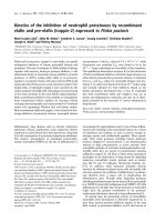
Figure 1 OPN mRNA and protein levels of either non-irradiated or irradiated MDA-MB-231 cells after siRNA transfection. A.
Quantitative real-time PCR: OPN mRNA levels of untreated cells and cells treated with siRNA targeting OPN or nonsense siRNA. Representative
values of OPN mRNA levels (72 h after transfection) treated with OPN-specific siRNAs were normalized to those treated with nonsense siRNA.
The value of the OPN mRNA level of cells that were treated with nonsense siRNA at 0 Gy was arbitrarily established as 100%. Data represent the
average values (± SD) of three independent experiments (* p < 0.05, ** p < 0.001). B./C. Western blot: Western blot analyses of OPN with OPN
specific antibody 0-17 (IBL). B. MDA-MB-231 cells were transfected with siRNA Mix as well as OpnS or with nonsense siRNA (non) for 24 h.
Thereafter, MDA-MB-231 cells were incubated with serum-free culture media for another 24 h and 48 h. The Western blot shows the extracellular
OPN protein levels (50 kDa) of MDA-MB-231 cells 48 h and 72 h after transfection with OPN specific siRNA Mix and OpnS, with nonsense siRNA
(non) and untreated MDA-MB-231 control cells (UT). The Western blot shows one representative result out of two independent experiments. C.
Intracellular OPN protein levels (64 kDa) of MDA-MB-321 cells 24 h after transfection. Cells were either untreated (UT) or treated with OPN
specific siRNA Mix and OpnS or with nonsense siRNA (non) with and without irradiation at 2 Gy. The Western blot shows one representative
result out of three independent experiments. Actin served as an internal loading control.
Hahnel et al. Radiation Oncology 2010, 5:82
/>Page 4 of 10
compared to non-irradiated cells. The irradiation-
induced inhibition of OPN protein expression was also
detected in cells transfected with OPN siRNAs (Fig. 1C.).
Effects of OPN siRNA constructs on migration and
induction of apoptosis with or without irradiation
We determined the effects of OPN siRNA and irradia-
tion on the migration rate of MDA-MB-231 cells with
theBoydenchamberassayandscratchassay.Cells
transfected with siRNA targeting OPN showed reduced
migration rates compared to control cells (control and
nonsense siRNA). Transfection with Mix resulted in a
decreased migration rate to 40% (p = 0.09), whereas
the migration rate of cells transfected with OpnS was
less than 62% (p = 0.15) compared to the migration
rate of cells treated with control siRNA (Fig. 2B.).
Similarly, we found a reduced migration rate after
transfection with OPN siRNA using the scratch assay
(Fig. 2A.). Furthermore, we demonstrated that irradia-
tion at 2 Gy to 6 Gy had no e ffect on the migration
rate (data not shown). However, combination of OPN
siRNA transfection and irradiation at 2 Gy resulted in
a significant inhibition of migration. After incubation
with Mix and 2 Gy irradiation, migration was reduced
to 32% (p = 0.03). Ad ditionally, transfection with OpnS
and irradiation at 2 Gy attenuated the migration rate
to 40% (p = 0.03). Using Western blot analysis, we
examined PARP cleavage as an indicator for the induc-
tion of apoptosis. However, 24 h after incubation with
OPN siRNA, we could not detect any PARP cleavage
products using Mix or OpnS. Moreover, Fig. 3A.
shows a distinctive accumulation of the PARP cleavage
product (89 kDa) 72 h after transfection with siRNA
OpnS. However, only OpnS, not M ix, induced apopto-
sis(Fig.3A.and3B.).Inaddition,weexaminedthe
morphology of the cell nuclei to quantify the rate of
apoptosisbytheuseofDAPIstaining.Theresults
observed in Western blot analyses were supported by
the findings of the quantitative assay. After incubation
with OpnS, the apoptosis rate increased from 0.3% to
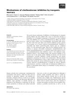
Figure 2 Migration behavior of either non-irradiated or irradiated (2 Gy) MDA-MB-231 cells after siRNA transfect ion. A. Scratch assay:
Wound scratch assay of MDA-MB-231 cells 24 h after transfection. Untreated cells and cells that were treated with nonsense siRNA were able to
close the wound scratch by migration. Cells treated with Mix as well as OpnS did not migrate and were unable to close the wound scratch. B.
Boyden chamber assay: The migration rate of cells treated with OPN-specific siRNAs was normalized to migration rate of cells treated with
nonsense siRNA. Treatment with siRNAs targeting OPN reduced the migration rate in non-irradiated cells as well as in cells irradiated at 2 Gy.
The migration rate of cells transfected with nonsense siRNA at 0 Gy was arbitrarily established as 100%. Data represent the average values (± SD)
of three independent experiments (* p < 0.05).
Hahnel et al. Radiation Oncology 2010, 5:82
/>Page 5 of 10
1.7% (p = 0.04), whereas transfection with Mix had no
effect on apoptosis. We found that irradiation alone at
2 Gy did not signi ficantly increase apoptosis in MDA-
MB-231 cells (Fig. 3B.). Nevertheless, the combination
of OpnS and irradiation at 2 Gy resulted in a signifi-
cant increase in apoptosis rate to 4% (p = 0.0001). In
contrast to that, incubation with Mix and irradiation at
2 Gy had no effect on ap optosis.
Effects of OPN siRNA on clonogenic survival and
radiosensitivity
We demonstrated that incubation with siRNA OpnS is
more effective to reduce the clonogenic survival of
MDA-MB-231 cells than incubation with siRNA Mix.
In particular, we found that transfection with OpnS
significantly decreased the clonogenic survival to 42%
(p = 0.008) (Fig. 4 A.). In contrast, transfection with
Mix was ineffective at reducing the clonogenic survival
(82%) (p = 0.4).
Irradiation of MDA-MB-231 cells at 2 Gy reduced the
clonogenic survival to 60% (SF
2
= 0.60) (data not
shown). The combinat ion of treatment with OpnS
siRNA and irradiation also reduced the clonogenic sur-
vival as compared to single siRNA treatment. Incubation
with OpnS, and additional irradiation at 2 Gy signifi-
cantly decreased the clonogenic survival to 30% (p <
0.001). Furthermore, with higher irradiation dose trans-
fection with OpnS resulted in a weak radiosensitization
with a DMF
10
of 1.1 and an enhancement factor of 1.5
at 6 Gy (p = 0.09) (Fig. 4B.).
Discussion
It is well known that intratumoral and plasma levels of
the phosphoprotein OPN are increased in many tumors
such as lung cancer [21], esophageal cancer [22], pros-
tate cancer [23], glioma [24], soft tissue sarcoma [25]
and breast cancer [5,14]. Furthermore, it has been
shown that an elevated OPN level is associated with
poor prognosis for cancer patients [5,6,12,14,15]. In
addition, different studies have found that high OPN
levels are associated with poor response to conventional
treatment modalities including radiotherapy (reviewed in
[9]). However, little is known about the relationship
between OPN expression and radiosensitivity.
Our analyses demonstrate that both Mix and OpnS
siRNAs (Table 1) are suitable to clearly reduce mRNA
levels of OPN (Fig. 1A.). Furthermore, we detected a
clear decrease of extracellular OPN protein levels after
transfection with OPN siRNA (Fig. 1B.). In contrast , the
intracellular OPN protein level was only partially
decreased after transfection with OPN siRNA. However,
intracellular OPN was detected at a higher molecular
weight range (64 kDa) as compared with extracellular
OPN that was detected at 50 kDa. The molecular weight
difference may represent post-translational modifications
such as glycosylation, phosphorylation and sulfatization
[4,26,27]. In addition, there is evidence from the litera-
ture that two forms of OPN exist: a secreted form
(sOPN) and an intracellular form (iOPN). Shinohara
and co-workers [28] proposed that sOPN and iOPN
represent alternative translational products of a single
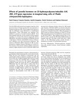
Figure 3 PARP protein levels and apoptosis rate of either non-irradiated or irradiated cells after siRNA transfection. A.Westernblot
analysis of PARP with rabbit anti-human cleaved PARP (Asp214) antibody [1] in MDA-MB-321 cells 24 h and 72 h after transfection. The cells
were untreated (UT), transfected with 100 nM of either nonsense siRNA (non) or target-specific siRNAs to knockdown OPN (Mix and OpnS). The
Western blot shows one representative result out of three independent experiments. Actin served as an internal loading control. B. The
morphology of DAPI stained cell nuclei was analyzed to quantify the apoptosis rate of MDA-MB-231 cells 72 h after transfection. The diagram
shows the apoptosis rate of the cells as a function of treatment and irradiation. A fluorescence microscope was used and 500 cells in several
fields of view were counted for each experiment. Data represent the average values (± SD) of three independent experiments (* p < 0.05, ** p <
0.001).
Hahnel et al. Radiation Oncology 2010, 5:82
/>Page 6 of 10
full-length OPN mRNA that have a molecular weight
difference of 5 kDa. In contrast to sOPN, the iOPN pro-
tein lacks a signal peptide, which allows the iOPN pro-
tein to localize to the cytoplasm but not to the Golgi
apparatus [28]. Furthermore, it has been shown that
extracellular OPN is important for bone marrow cell
activation and the subsequent outgrowth of distant
tumors [19], and it al so affects the cellular response and
increases lung metastasis in mice that have received
cells preincubated with OPN [29].
The siRNA transfection showed clear effects on dif-
ferent cellular parameters. Treatment with OpnS
resulted in a clear reduction of clonogenic survival,
inhibition of migration and increased rate of apoptosis
(Fig. 2, 3, 4A.), whereas treatment with the siRNA con-
struct Mix caused an obvious reduction in the rate of
migration. However, no differential effects were found
with respect to apoptosis and clonogenic survival. The
different effects of OpnS and Mix on clonogenic survi-
val and apoptosis frequency are possibly caused b y the
different sequences that are recognized by the siRNAs.
Possibly, OPN RNA sequences are not assessable in
thesamewaybythedifferentsiRNAs.Mixisapoolof
four siRNAs and might cause more off-target effects
than OpnS which could reverse the original effects.
We chose the siRNA technology for transient inhibi-
tion of OPN expression in MDA-MB-231 cells. A dis-
advantage of the siRNA technology is that it is not
possible to reach a permanent reduction of OPN
expression. However, in vitro it is an efficient method
to knockdown OPN.
Taken together the effects of OPN inhibition are in
agreement with previous findings that the knockdown of
OPN reduces the clonogenic survi val, migration and
invasion rate, and proliferation in different breast cancer
cell lines [30-32]. Furthermore, various studies have
demonstrated the effects of OPN silencing or OPN
overexpression on several downstream elements of OPN
in Western blot analysis. In particular, Tuck and co-
workers [33,34] found an induction of uPA expression
in response to OPN treatment and an association of
uPA expression with OPN-induced invasion and migra-
tion in human breast cancer cells. These findings are
consistent with our data analyzing the protein expres-
sion levels of the migration marker uPA with ELISA in
cell lysates of MDA-MB-231 cells that showed a clear,
albeit not significant, reduction of uPA protein levels
after transfection with OPN siRNAs and irradiation
(data not shown). Other investigators have demonstrated
that knockdown of OPN decreases the expression of
PI3’-kinase, JNK1/2, Src and Akt, uPA, MMP-2 and -9
in various tumor cell lines [35-39].
In the present study, for the first time we were able to
demonstrate that OPN silencing affects the radiobiologi-
cal behavior of hu man cancer cells. Moreover, we found
that OPN knoc kdown by OPN siRNA could very effec-
tively decrease OPN mRNA and p rotein levels after
additional irradiation (Fig. 1). Furthermore, an additional
Figure 4 Clonogenic survival of either non-irradiated or irradiated MDA-MB-231 cells after siRNA transfection. A. Clonogenic survival of
MDA-MB-231 cells after transfection. Treatment with just OpnS had a strong effect on clonogenic survival at 0 Gy. The relative clonogenic
survival of cells that were transfected with nonsense siRNA was arbitrarily established as 100%. Data represent the average values (± SD) of three
independent experiments (* p < 0.05, ** p < 0.001). B. Clonogenic survival after transfection with OPN-specific siRNA (Mix, OpnS) in combination
with irradiation at 2, 4 or 6 Gy. To examine the additional effects of irradiation all values of clonogenic survival at 0 Gy were set arbitrarily at
100%. Cells transfected with OpnS showed an increased radiosensitivity. After irradiation at 6 Gy, a dose modifying factor (DMF
10
) of 1.1 and an
enhancement factor of 1.5 (p = 0.09) were calculated for the siRNA construct OpnS. Data represent the average values (± SD) of three
independent experiments.
Hahnel et al. Radiation Oncology 2010, 5:82
/>Page 7 of 10
decrease in the intracellular OPN protein level was
detected in Western blot analyses after irradiation (Fig.
1C.). However, anothe r study analyzed the effect of
radiation on OPN lev els in osteoblastic cells and found
a slightly elevated expression of OPN on days 14 and
21 after irradiation [40].
Moreover, the a dditional irradiation at 2 Gy caused a
significant reduction in the rate of migration (Fig. 2B.).
We demonstrated that treatment with OpnS resulted in
a significant increase in irradiation-induced apoptosis
(Fig. 3B.). This is in agreement with Lee and co-workers
[41], who showed that treatment with recombinant
OPN confers an increased resistance to UV-induced
apoptosis in HT29 cells [41]. However, OPN siRNA
transfection alone and in combination with irradiation
showed only minor effects on apoptosis compared with
effects on clonogenic survival. Possibly the MAA (meth-
oxyacetic acid) assay can reflect a better correlation
because besides apoptosis this assay determines other
modes of cell death such as micronucleation or multinu-
cleated cells [42].
To our knowledge, this is the first study demonstrat-
ing that kn ockdown of OPN influences the radiosensi-
tivity of cancer cells. OPN knockdown even caused a
weak radiosensitization with a higher irradiation dose
(Fig. 4B.). Considering the non-significant effects on
radiosensitivity in vitro it appears that OPN siRNA
treatment predom inantly affects clonogenicity and
migration rate. However, in vivo we cannot be sure that
siRNAs would find their target molecules and concen-
trate as it would be appropriate i n solid tumors. There-
fore, a combined treatment of siRNAs with irradiation
might be necessary. Another study which analyzed the
influence of OPN silencing confirmed the impact of
OPN expression on the efficacy of irradiation. Solberg
and co-workers [43] found that irradiation of xenograft
tumors in mice induces the expression of mouse VEGF
(mVEGF) and mouse OPN (mOPN), which are both
closely associated with angiogenesis. Moreover, the
expression of mOPN was d irectly proportional to the
mVEGF levels in tumors which indicates that mOPN
can serve as an alternative marker of tumor recovery
after radiotherapy. Furthermore, clinical studies have
found that elevated OPN levels are associated with poor
prognosis in head and ne ck cancer [9,12,13,44-47] and
breast cancer [3,48].
Conclusions
In summary, in the present study we were able to
demonstrate for the first time that an OPN knockdown
comb ined with irradiation has additive effects on clono-
genic survival, migration and the induction of apoptosis.
Furthermore, we showed that silencing of OPN with
siRNA causes a weak radiosensitization of MDA-MB-
231 cells. This suggests that OPN is an attractive target
to improve the efficacy of radiotherapy. Additional
radiobiological studies are necessary to investigate the
role of OPN and its association with radiosensitivity of
other tumor cell lines.
Acknowledgements
We would like to thank our colleagues from the Department of
Radiotherapy for contributing to this study and for their continuous support.
We would also like to thank Kathrin Spröte, Gabriele Thomas and Antje
Zobjack for their excellent technical assistance. This work was supported by
the Wilhelm Sander Stiftung (grant number: 2007.123.1).
Author details
1
Department of Radiotherapy, Martin-Luther-University Halle-Wittenberg,
Dryanderstr.4, 06110 Halle, Germany.
2
Department of Oral and Maxillofacial
Plastic Surgery, Martin-Luther-University Halle-Wittenberg, Ernst-Grube-Str.40,
06120 Halle, Germany.
3
Institute of Pathology, Dresden University of
Technology, Fetscherstr.74, 01307 Dresden, Germany.
Authors’ contributions
AH designed the study, performed experimental procedures, analyzed the
data and drafted the manuscript. HW, MKa, HT and DV aided in study
design, analyzed the data and reviewed the manuscript. MKo performed
experimental procedures, analyzed the data and reviewed the manuscript.
MB designed the study, analyzed the data and drafted the manuscript. All
authors read and approved the final manuscript.
Competing interests
The authors declare that they have no competing interests.
Received: 6 July 2010 Accepted: 17 September 2010
Published: 17 Septemb er 2010
References
1. Rangaswami H, Bulbule A, Kundu GC: Osteopontin: role in cell signaling
and cancer progression. Trends Cell Biol 2006, 16:79-87.
2. Wai PY, Kuo PC: Osteopontin: regulation in tumor metastasis. Cancer
Metastasis Rev 2008, 27:103-118.
3. Bramwell VH, Doig GS, Tuck AB, Wilson SM, Tonkin KS, Tomiak A, Perera F,
Vandenberg TA, Chambers AF: Serial plasma osteopontin levels have
prognostic value in metastatic breast cancer. Clin Cancer Res 2006,
12:3337-3343.
4. Kazanecki CC, Kowalski AJ, Ding T, Rittling SR, Denhardt DT:
Characterization of anti-osteopontin monoclonal antibodies: Binding
sensitivity to post-translational modifications. J Cell Biochem 2007,
102:925-935.
5. Singhal H, Bautista DS, Tonkin KS, O’Malley FP, Tuck AB, Chambers AF,
Harris JF: Elevated plasma osteopontin in metastatic breast cancer
associated with increased tumor burden and decreased survival. Clin
Cancer Res 1997, 3:605-611.
6. Wu CY, Wu MS, Chiang EP, Wu CC, Chen YJ, Chen CJ, Chi NH, Chen GH,
Lin JT: Elevated plasma osteopontin associated with gastric cancer
development, invasion and survival. Gut 2007, 56:782-789.
7. Vergis R, Corbishley CM, Norman AR, Bartlett J, Jhavar S, Borre M, Heeboll S,
Horwich A, Huddart R, Khoo V, Eeles R, Cooper C, Sydes M, Dearnaley D,
Parker C: Intrinsic markers of tumour hypoxia and angiogenesis in
localised prostate cancer and outcome of radical treatment: a
retrospective analysis of two randomised radiotherapy trials and one
surgical cohort study. Lancet Oncol 2008, 9:342-351.
8. Debucquoy A, Goethals L, Geboes K, Roels S, Mc Bride WH, Haustermans K:
Molecular responses of rectal cancer to preoperative chemoradiation.
Radiother Oncol 2006, 80:172-177.
9. Bache M, Kappler M, Said HM, Staab A, Vordermark D: Detection and
specific targeting of hypoxic regions within solid tumors: current
preclinical and clinical strategies. Curr Med Chem 2008, 15:322-338.
10. Vordermark D, Said HM, Katzer A, Kuhnt T, Hansgen G, Dunst J, Flentje M,
Bache M: Plasma osteopontin levels in patients with head and neck
Hahnel et al. Radiation Oncology 2010, 5:82
/>Page 8 of 10
cancer and cervix cancer are critically dependent on the choice of ELISA
system. BMC Cancer 2006, 6:207.
11. Bache M, Reddemann R, Said HM, Holzhausen HJ, Taubert H, Becker A,
Kuhnt T, Hansgen G, Dunst J, Vordermark D: Immunohistochemical
detection of osteopontin in advanced head-and-neck cancer: prognostic
role and correlation with oxygen electrode measurements, hypoxia-
inducible-factor-1alpha-related markers, and hemoglobin levels. Int J
Radiat Oncol Biol Phys 2006, 66:1481-1487.
12. Le QT, Sutphin PD, Raychaudhuri S, Yu SC, Terris DJ, Lin HS, Lum B,
Pinto HA, Koong AC, Giaccia AJ: Identification of osteopontin as a
prognostic plasma marker for head and neck squamous cell carcinomas.
Clin Cancer Res 2003, 9:59-67.
13. Overgaard J, Eriksen JG, Nordsmark M, Alsner J, Horsman MR: Plasma
osteopontin, hypoxia, and response to the hypoxia sensitiser nimorazole
in radiotherapy of head and neck cancer: results from the DAHANCA 5
randomised double-blind placebo-controlled trial. Lancet Oncol 2005,
6:757-764.
14. Tuck AB, O’Malley FP, Singhal H, Harris JF, Tonkin KS, Kerkvliet N, Saad Z,
Doig GS, Chambers AF: Osteopontin expression in a group of lymph
node negative breast cancer patients. Int J Cancer 1998, 79:502-508.
15. Rudland PS, Platt-Higgins A, El Tanani M, De Silva RS, Barraclough R,
Winstanley JH, Howitt R, West CR: Prognostic significance of the
metastasis-associated protein osteopontin in human breast cancer.
Cancer Res 2002, 62:3417-3427.
16. Siles E, Villalobos M, Valenzuela MT, Nunez MI, Gordon A, McMillan TJ,
Pedraza V, Ruiz de Almodovar JM: Relationship between p53 status and
radiosensitivity in human tumour cell lines. Br J Cancer 1996, 73:581-588.
17. Torres-Roca JF, Eschrich S, Zhao H, Bloom G, Sung J, McCarthy S, Cantor AB,
Scuto A, Li C, Zhang S, Jove R, Yeatman T: Prediction of radiation
sensitivity using a gene expression classifier. Cancer Res 2005,
65:7169-7176.
18. Phillips TM, McBride WH, Pajonk F: The response of CD24(-/low)/CD44+
breast cancer-initiating cells to radiation. J Natl Cancer Inst 2006,
98:1777-1785.
19. McAllister SS, Gifford AM, Greiner AL, Kelleher SP, Saelzler MP, Ince TA,
Reinhardt F, Harris LN, Hylander BL, Repasky EA, Weinberg RA: Systemic
endocrine instigation of indolent tumor growth requires osteopontin.
Cell 2008, 133:994-1005.
20. Bache M, Würl P, Dietzel M, Meye A, Fröde D, Schmidt H, Rath FW,
Wohlrab W, Dralle H, Dunst J, Taubert H: Two human sarcoma cell lines
with different p53 gene status in their response on radiation. Int J Oncol
1997, 11:993-997.
21. Chambers AF, Wilson SM, Kerkvliet N, O’Malley FP, Harris JF, Casson AG:
Osteopontin expression in lung cancer. Lung Cancer 1996, 15:311-323.
22. Casson AG, Wilson SM, McCart JA, O’Malley FP, Ozcelik H, Tsao MS,
Chambers AF: ras mutation and expression of the ras-regulated genes
osteopontin and cathepsin L in human esophageal cancer. Int J Cancer
1997, 72:739-745.
23. Thalmann GN, Sikes RA, Devoll RE, Kiefer JA, Markwalder R, Klima I, Farach-
Carson CM, Studer UE, Chung LW: Osteopontin: possible role in prostate
cancer progression. Clin Cancer Res 1999, 5:2271-2277.
24. Saitoh Y, Kuratsu J, Takeshima H, Yamamoto S, Ushio Y: Expression of
osteopontin in human glioma. Its correlation with the malignancy. Lab
Invest 1995, 72:55-63.
25. Bache M, Kappler M, Wichmann H, Rot S, Hahnel A, Greither T, Said HM,
Kotzsch M, Wurl P, Taubert H, Vordermark D: Elevated tumor and serum
levels of the hypoxia-associated protein osteopontin are associated with
prognosis for soft tissue sarcoma patients. BMC Cancer 2010, 10:132.
26. Christensen B, Nielsen MS, Haselmann KF, Petersen TE, Sorensen ES: Post-
translationally modified residues of native human osteopontin are
located in clusters: identification of 36 phosphorylation and five O-
glycosylation sites and their biological implications. Biochem J 2005,
390:285-292.
27. Sorensen ES, Hojrup P, Petersen TE: Posttranslational modifications of
bovine osteopontin: identification of twenty-eight phosphorylation and
three O-glycosylation sites. Protein Sci 1995, 4:2040-2049.
28. Shinohara ML, Kim HJ, Kim JH, Garcia VA, Cantor H: Alternative translation
of osteopontin generates intracellular and secreted isoforms that
mediate distinct biological activities in dendritic cells. Proc Natl Acad Sci
USA 2008, 105:7235-7239.
29. Mandelin J, Lin EC, Hu DD, Knowles SK, Do KA, Wang X, Sage EH, Smith JW,
Arap W, Pasqualini R: Extracellular and intracellular mechanisms that
mediate the metastatic activity of exogenous osteopontin. Cancer 2009,
115:1753-1764.
30. Adwan H, Bauerle T, Najajreh Y, Elazer V, Golomb G, Berger MR: Decreased
levels of osteopontin and bone sialoprotein II are correlated with
reduced proliferation, colony formation, and migration of GFP-MDA-MB-
231 cells. Int J Oncol 2004, 24:1235-1244.
31. Chakraborty G, Jain S, Patil TV, Kundu GC: Down-regulation of osteopontin
attenuates breast tumour progression in vivo. J Cell Mol Med 2008,
12:2305-2318.
32. Shevde LA, Samant RS, Paik JC, Metge BJ, Chambers AF, Casey G, Frost AR,
Welch DR: Osteopontin knockdown suppresses tumorigenicity of human
metastatic breast carcinoma, MDA-MB-435. Clin Exp Metastasis 2006,
23:123-133.
33. Tuck AB, Arsenault DM, O’Malley FP, Hota C, Ling MC, Wilson SM,
Chambers AF: Osteopontin induces increased invasiveness and
plasminogen activator expression of human mammary epithelial cells.
Oncogene 1999, 18:4237-4246.
34. Tuck AB, Hota C, Chambers AF: Osteopontin(OPN)-induced increase in
human mammary epithelial cell invasiveness is urokinase (uPA)-
dependent. Breast Cancer Res Treat 2001, 70:197-204.
35. Chakraborty G, Jain S, Kundu GC:
Osteopontin promotes vascular
endothelial growth factor-dependent breast tumor growth and
angiogenesis via autocrine and paracrine mechanisms. Cancer Res 2008,
68:152-161.
36. Cheng J, Huo DH, Kuang DM, Yang J, Zheng L, Zhuang SM: Human
macrophages promote the motility and invasiveness of osteopontin-
knockdown tumor cells. Cancer Res 2007, 67:5141-5147.
37. Desai B, Rogers MJ, Chellaiah MA: Mechanisms of osteopontin and CD44
as metastatic principles in prostate cancer cells. Mol Cancer 2007, 6:18.
38. Ito T, Hashimoto Y, Tanaka E, Kan T, Tsunoda S, Sato F, Higashiyama M,
Okumura T, Shimada Y: An inducible short-hairpin RNA vector against
osteopontin reduces metastatic potential of human esophageal
squamous cell carcinoma in vitro and in vivo. Clin Cancer Res 2006,
12:1308-1316.
39. Mi Z, Guo H, Russell MB, Liu Y, Sullenger BA, Kuo PC: RNA aptamer
blockade of osteopontin inhibits growth and metastasis of MDA-MB231
breast cancer cells. Mol Ther 2009, 17:153-161.
40. Gevorgyan A, Sukhu B, Alman BA, Bristow RG, Pang CY, Forrest CR:
Radiation effects and radioprotection in MC3T3-E1 mouse calvarial
osteoblastic cells. Plast Reconstr Surg 2008, 122:1025-1035.
41. Lee JL, Wang MJ, Sudhir PR, Chen GD, Chi CW, Chen JY: Osteopontin
promotes integrin activation through outside-in and inside-out
mechanisms: OPN-CD44V interaction enhances survival in
gastrointestinal cancer cells. Cancer Res 2007, 67:2089-2097.
42. Abend M, Kehe K, Kehe K, Riedel M, Van Beuningen D: Correlation of
micronucleus and apoptosis assays with reproductive cell death can be
improved by considering other modes of death. Int J Radiat Biol 2000,
76:249-259.
43. Solberg TD, Nearman J, Mullins J, Li S, Baranowska-Kortylewicz J:
Correlation between tumor growth delay and expression of cancer and
host VEGF, VEGFR2, and osteopontin in response to radiotherapy. Int J
Radiat Oncol Biol Phys 2008, 72:918-926.
44. Celetti A, Testa D, Staibano S, Merolla F, Guarino V, Castellone MD, Iovine R,
Mansueto G, Somma P, De Rosa G, Galli V, Melillo RM, Santoro M:
Overexpression of the cytokine osteopontin identifies aggressive
laryngeal squamous cell carcinomas and enhances carcinoma cell
proliferation and invasiveness. Clin Cancer Res 2005, 11:8019-8027.
45. Hui EP, Sung FL, Yu BK, Wong CS, Ma BB, Lin X, Chan A, Wong WL,
Chan AT: Plasma osteopontin, hypoxia, and response to radiotherapy in
nasopharyngeal cancer. Clin Cancer Res 2008, 14:7080-7087.
46. Nordsmark M, Eriksen JG, Gebski V, Alsner J, Horsman MR, Overgaard J:
Differential risk assessments from five hypoxia specific assays: The basis
for biologically adapted individualized radiotherapy in advanced head
and neck cancer patients. Radiother Oncol 2007, 83:389-397.
47. Petrik D, Lavori PW, Cao H, Zhu Y, Wong P, Christofferson E, Kaplan MJ,
Pinto HA, Sutphin P, Koong AC, Giaccia AJ, Le QT: Plasma osteopontin is
an independent prognostic marker for head and neck cancers. J Clin
Oncol 2006, 24:5291-5297.
Hahnel et al. Radiation Oncology 2010, 5:82
/>Page 9 of 10
48. De Silva RS, Martin L, Roshanlall C, Winstanley J, Leinster S, Platt-Higgins A,
Carroll J, West C, Barraclough R, Rudland P: Association of S100A4 and
osteopontin with specific prognostic factors and survival of patients
with minimally invasive breast cancer. Clin Cancer Res 2006, 12:1192-1200.
doi:10.1186/1748-717X-5-82
Cite this article as: Hahnel et al.: Effects of osteopontin inhibition on
radiosensitivityof MDA-MB-231 breast cancer cells. Radiation Oncology
2010 5:82.
Submit your next manuscript to BioMed Central
and take full advantage of:
• Convenient online submission
• Thorough peer review
• No space constraints or color figure charges
• Immediate publication on acceptance
• Inclusion in PubMed, CAS, Scopus and Google Scholar
• Research which is freely available for redistribution
Submit your manuscript at
www.biomedcentral.com/submit
Hahnel et al. Radiation Oncology 2010, 5:82
/>Page 10 of 10