Báo cáo khoa học: "A biologically competitive 21 days hypofractionation scheme with weekly concomitant boost in breast cancer radiotherapy feasibility acute sub-acute and short term late effects" pot
Bạn đang xem bản rút gọn của tài liệu. Xem và tải ngay bản đầy đủ của tài liệu tại đây (387.54 KB, 7 trang )
RESEARC H Open Access
A biologically competitive 21 days hypofractionation
scheme with weekly concomitant boost in breast
cancer radiotherapy feasibility acute sub-acute and
short term late effects
Marina Guenzi
1†
, Stefano Vagge
1*†
, Ngwa Che Azinwi
1†
, Alessia D’Alonzo
1
, Liliana Belgioia
1
, Stefania Garelli
2
,
Marco Gusinu
2
, Renzo Corvò
1,2,3
Abstract
Background: Radiation therapy after lumpectomy is a standard part of breast conserving therapy for invasive
breast carcinoma. The most frequently used schedule worldwide is 60 Gy in 30 fractions in 6 weeks, a time
commitment that sporadically may dissuade some otherwise eligible women from undertaking treatment. The
purpose and primary endpoint of this perspective study is to evaluate feasibility and short-term late toxicity in a
hypofractionated whole breast irradiation schedule.
Methods: Between February and October 2008 we treated 65 consecutive patients with operable invasive early-
stage breast cancer with a hypofractionated schedule of external beam radiation therapy. All pa tients were
assigned to 39 Gy in 13 fractions in 3 weeks to the whole breast plus a concomitant weekly boost dose to the
lumpectomy cavity of 3 Gy in 3 fractions.
Results: All the patients had achieved a median follow up of 24 months (range 21-29 months). At the end of
treatment 52% presented grade 0 acute toxicity 39% had grade 1 and 9% had grade 2. At 6 months with all the
patients assessed there were 34% case of grade 1 subacute toxicity and 6% of grade 2. At 12 months 43% and 3%
of pat ients presented with clinical grade 1 and grade 2 fibrosis respectively and 5% presented grade 1
hyperpigmentation. The remaining patients were free of side effects. At 24 months, with 56 assessed, just 2
patients (3%) showed grade 2 of late fibrosis.
Conclusions: The clinical results observ ed showed a reasonably good feasibility of the accelerated
hypofractionated schedule in terms of acute, subacute and short-term late toxicity. This useful 13 fractions with a
concomitant boost schedule seems, in selected patients, a biologically acceptable alternative to the traditional 30
days regime.
Background
Radiation therapy after lumpectomy is a standard part of
breast conserving therapy for invasive breast cancer as it
has been shown that besides significantly reducing the
risk of local recurrence, it impacts favorably on patient
survival [1,2]. The generally recognized standard and the
most frequently used schedule worldwide is 60 Gy,
delivered in 30 fractions of 2 Gy over 6 weeks, a time
commitment that otherwise may generate discomfort in
some women eligible for Breast Conserving Therapy
(BCT). The possibility of delivering postoperative radia-
tion therapy in a shorter period of time could circum-
vent this problem and result in a dramatic reduction of
the nuisance factor for these patients. It would also con-
tribute to a far more judicious use of resources and
time in some busy Radi ation Oncology department. The
results of retrospective studies of hypofractionated
radiotherapy in early breast cancer suggest satisfactory
* Correspondence:
† Contributed equally
1
Department of Radiation Oncology, Istituto Nazionale per la Ricerca sul
Cancro, Genoa, Italy
Full list of author information is available at the end of the article
Guenzi et al. Radiation Oncology 2010, 5:111
/>© 2010 Guenzi et al; licensee BioMed Central Ltd. This is a n Open Ac cess article distributed under the term s of the Cr eative Commons
Attribution License (http://creati vecommons.org/licenses/by/2.0), which permits unrestr icted use, distribution, and reproduction in
any medium, provided the original work is properly cited.
outcomes in terms of tumor control and late adverse
effects [3-5]. Recent randomized trials have confirmed
that hypofractioned whole-breast irradiation is equiva-
lent to more conventional whole-breast irradiation with
respect to local recurrence and cosmet ic outcome [6-8].
In order to intensify treatment, a simultaneous boost
dose, concomitant or integrated, has been introduced in
clinics by using 3-D conformal radiotherapy or inten-
sity-modulated radiotherapy [9,10]. Preliminary results
from experiences where a boost dose was delivered
either daily after w hole-breast irradiation (WBI) [7] or
weekly appear interesting, with reas onably good feasibil-
ity in terms of acute toxicity [11,12]. The purpose and
primary endpoint of this study was to evaluate the feasi-
bility and the acute, subacu te and short term late toxi-
city of a hypofractionated three weeks whole breast
irradiation schedule with the addition of a concomitant
boost dose delivered to the tumor bed once-a-week in
patients with early breast cancer submitted to lumpect-
omy and sentinel node dissection.
Methods
Patients
Sixty-five consecutive patients with operable invasive
early-stage breast cancer were treated at the National
Institute for Cancer Research at Genoa with hypofrac-
tionated External Beam Radiation Therapy (EBRT) as
part of their BCT between Februarys to October 2008.
All eligible patients had stage I-II breast carcinoma as
defined by the international Union Against Cancer (fifth
edition) and had gone through macroscopic total resec-
tion of the primary tumor a nd sentinel node biopsy.
Three patients had positive or close margins because
they refused to undergo re-excision, that we usually
require, where possible, to obtain margins of at least 2
mm. They were nonetheless included in the protocol
after due risk cautioning. Patient demographics, disease
characteristics and therapy are displayed in the table 1.
Patients were excluded from the study if they presented
any of the following conditions: evidence of distant
metastasis, presence of serious co-morbidities that could
preclude radiotherapy such as cardiovascular or psychia-
tric disorders, tumor greater than 5 c m in its largest
dimension, presence of more than 3 positive nodes,
macroscopically positive margins, age less than 55 years
initially, the presence of ac tive con nective tissue disease
and a history of previous irradiation to the c hest wall.
Patients with large breasts (as defined by a cup size
separation of greater than 25 cm, that is, the breast
measured more than 25 cm left to right at its widest
part) were also excluded [8,9]. All patients duly provided
written informed consent before being assigned to treat-
ment. Therapy was planned immediately afte r Breast
Conserving Surgery (BCS) in low-risk patients or
sequentially after systemic chemotherapy (CT) in those
at higher risk of failure. Prognostic classes were assigned
according t o the St. Gallen Consensus Conference [13].
This protocol have be en submitted and approved by our
institutional ethics committee.
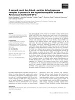
Radiation fractionation and treatment
The basic scheme of treatment consisted in the deliv ery
of 39 Gy in 13 fractions 4 times a week to the whole
breast plus a once weekly concomitant boost dose of 1
Gy to the lumpectomy area immediately after whole
breast irradiation (WBI) (thus a total boost dose of 3 Gy
in 3 fractions once a week). Doses were prescribed to
international reference points. T otal treatment time was
3 weeks plus 1 day, and the total nominal dose to the
lumpectomy area (considering the cumulative dose to
the whole breast and to the surgical bed) was 42 Gy.
Generally, weekly treatment would start on Monday and
end on Friday with a pause planned for Wednesday.
The boost dose was added on Monday (Figure 1). Portal
films of the whole breast were taken at least once during
the first day of irradiation and compared with Digitally
Reconstructed Radiographs (DRR) for matching. The
ethic committee of our institution approved the final
protocol.
Table 1 Patient demographics, disease characteristics and
therapy
Number of patients N=65 Surgical
margins
Mean age (range) in yrs 69(53 -
86)
Negative 62
(95%)
Tumour class(AJCC) Positive 1 (2%)
pTis 3 (5%) Close 2 (3%)
pT1a 4 (6%) Hormonal status
pT1b 10 (15%) HR positive 60
(92%)
pT1c 34 (52%) HR negative 5 (8%)
pT2 14 (22%) Hormone
therapy
Max tumour diam. (range)
mm
3 - 30 Yes 57
(88%)
Grading No 5 (13%)
G1 12 (18%) Chemotherapy 9 (14%)
G2 39 (60%)
G3 14 (22%)
Proliferative index (Ki67) %
≤ 15 39 (60%)
> 15 26 (40%)
Nodal status
pN0 58 (89%)
pN1(a) 7 (11%)
Guenzi et al. Radiation Oncology 2010, 5:111
/>Page 2 of 7
Radiobiological equivalent dose
Using the Linear-quadratic cell survival model [equation
1, appendix] we calculated Biologically Equivalent Doses
(BEDs) for the breast and boost volumes [14]. For this
calculation w e assumed an a/b ratio of 4 Gy for tum or
response [15], 10 Gy for acute responding normal ti s-
sues [16], 1.7 Gy for late-responding tissues (fibrosis)
[17] and 2.5 Gy for vascular damage [18]. The biological
comparison betw een the standard and the explored RT
schedule is shown in table 2. Although the BED for can-
cer clonogens was equivalent for the 42 Gy in 13 frac-
tions schedule, we hypothesized that this similar dose
equivalence could be advantageous for our schedule by
the g reater microvascular dysfunction on the boost site
that the higher dose per fra ction could achieve. It may
be worth noting that this factor of tumor kill is normally
not included in mathematical models for BED
calculation.
Volumes of interest and treatment planning
A planning CT scan was carried out for each patient
with the patient positioned supine on a “ wing-board”
with both arms raised above the head. Radiopaque wires
and markers were used to locate palpable breast tissue
and visible surgical scars. Three tattoos were made on
thethoracicskintoenablepatientrepositioningduring
treatment. The CT scans went fr om the level of the lar-
ynx to the upper abdomen with both lungs included.
Scan thickness was 10 mm. The Whole Breast Clinical
Target Volume (WB-CTV) included glandular breast tis-
sue and did not extend to cover the pectorals major, the
ribs or the skin. The Whole Breast Planning Target
Volume (WB-PTV) was generated by the a ddition of a
3-D 3 -5 mm margin ar ound the WB-CTV where possi-
ble considering the presence of nearby organs at risk
(OARs) while for the c ranial and caudal directions a 10
mm margin was used. The definition of the lumpectomy
cavity was guided by the presence of surgical clips,
hematoma, seroma or other surgery-induced changes
considered to be part of the cavity. The boost CTV was
gene rated by adding at lea st a 2 mm ma rgin around the
lumpectomy cavity and the corresponding PTV created
by adding a further 2 mm 3 D margin. The heart and
ipsilateral lung were considered OARs. The heart was
contoured from the pulmonary trunks superiorly to its
base and included the pericardium. The major blood
vessels were excluded. The ipsilateral lung was con-
toured in all its extension. Three Dimensional Confor-
mal Radiotherapy (3DCRT) plans were generated using
either of two TPS systems (CMS Xio or Varian Eclipse).
Treatment plans for the whole breast were generate d
using two opposed tangential beams. Beam weighting,
gantry angles, wedges, multi leaf collimator (MLC)
shielding and beam energies were determined to achieve
optimal dose conformity and distribution as well as
maximal avoidance of the heart and ipsilateral lung. The
boost plan consisted of two or more photon beams sui-
tably angled and optimized by the use of wedges and
Figure 1 Fractionation scheme. m: monday; t: tuesday; w:
wednesday; t: thursday; f: Friday. WBI: whole breast irradiation. cc.
boost: concomitant boost
Table 2 BED comparison between standard and explored RT schedule
RT schedule BED tumor control a/b
4
BED acute effects a/b
10
BED fibrosis a/b
1.7
BED vascular damage a/b
2.5
W.B. = whole breast
B.S. = tumor bed side
W.B. B.S. W.B. B.S. W.B. B.S. W.B. B.S.
60 Gy/30 F/6 W
(50 Gy + 10 Gy seq.boost)
75 90 60 72 109 131 90 108
50 Gy/25 F/5 W
(no boost)
75 75 60 60 109 109 90 90
42 Gy/13 F/3W + 1 day
(39 Gy + 3 Gy cc.boost)
68 77 51 56 108 123 86 97
52 Gy/20/F/5 W
(46 Gy + 6 Gy cc.boost)
72 87 57 66 108 135 88 108
UK START TRIAL A
41.6 Gy/13 F/5 W
75 75 55 55 120 120 95 95
UK START TRIAL A
39 Gy/13 F/5W
68 68 51 51 108 108 86 86
boost = concomitant boost; seq.boost = sequential boost; F = fractions; W = weeks
Guenzi et al. Radiation Oncology 2010, 5:111
/>Page 3 of 7
selective MLC shielding. Both plans (Whole breast and
Boost) a imed for a 95% isodose level encompassing the
PTVs and plan evaluation was enhanced by the use of
Dose Volume Histograms (DVHs) and a chosen Confor-
mity Index (CI). An example of a sum plan and DVH
are displayed in figure 2.
Follow up
Clinical checks w ere carried out halfway through treat-
ment. Follow up for acute toxicity was arranged at treat-
ment end and at 3 months. Baseline mammography was
planned at 8 months after completion of treatment and
yearly thereafter. Acute toxicities were graded based on
the RTOG acute toxicity scale [19] (table 3). Subacute
and late toxicities were graded using the Modified
LENT SOMA scoring system [20] (table 4) and was
assessed at 6 months, at 12 months and thereafter
planned every six months. The toxicity paramet ers
examined included the following: erythema, breast
edema, desquamation, ulceration, fibrosis, telangiectasia,
hyperpigmentation, retraction and atrophy.
Results
At the time of reporting, 65 patients had achieved a
minimum follow up o f 21 months (median FU 24
months, range 21-29 months). All accrued patients were
included in this analysis. The mea n PTV of the whole
breast volume was 642 cc (range 319-1198 cc), the
mean PTV of the boost volume was 57 cc (range 21-
148) and the mean ratio between the whole breast and
boost volume in percentage was 9% (range 3-20 cc). At
the end of treatment and until the first 3 months the
majority of patients were free of noteworthy acute toxi-
city, just the 9% of them presented bright erythema
(table 5). The evaluation of subacute toxicity at 6 months
showed a grade 2 barely in 4 patients (6%). Mild hyper -
pigmetation have been detected in 22 (34%) patients,
therest,39(60%)weretoxicityfree(table6).At12
months, with all patients assessed, 28 (43%) and 2
patients (3%) presented with clinical grade 1 and grade
2 fibrosis respectively while 3 patients (5%) presented
grade 1 hyperpigmentation (table 6). At 24 months
grade 2 late fibrosis was present just in 2 patients (3%)
o 56 evaluable (table 6).
Discussion
Radiotherapy after lumpectomy improves local control
and overall survival [2] and it is considered part of the
conservative treatment. Standard radiation requires daily
treatment for 6 to 7 weeks and this may be a serious
inconvenience for many patients, e specially for the
elderly. Delivering postoperative radiation therapy in a
shorter period of time could result in a significant
reduction of this problem for patients. Shorter radiation
schedules ba sed on radiobiological models offer the pro-
mise of equivalent local control to standard radiation
therapybygivinglargerdosesperfractioninshorter
periods of time [21]. Several e xperiences and results of
randomized trials have been reported and offer encoura-
ging outcomes. Recently Whelan et al examined whether
a 22-day radiation therapy fractionation schedule was as
effective as the more traditional 35-day schedule in
reducing recurrence in 1234 women with invasive breast
cancer who underwent BCS with pathologically clear
resection margins and negative axillary lymph nodes.
The patients were randomly assigned to receive whole
breast irradiation of 42.5 Gy in 16 fractions over 22 days
(short arm - 622 pts) or whole breast irradiation of 50
Gy in 25 fractions over 35 days (long arm - 612 pts).
With a m edian follow-up of 12 years no difference i n
local recurrence, disease-free or overall survival rates
and cosmetic outcome was detected between study
arms. They conclude that the more convenient 22-day
fraction ation schedule appears to be an acceptable alter-
native to the 35-day schedule [8]. The START A (Stan-
dardization of Breast Radiotherapy) from the UK trial
Figure 2 An example of a sum plan and Dose Volume
Histogram. A: whole breast; B: boost; C: plan sum
Table 3 RTOG Acute Skin Score
Grade
0
No change over baseline
Grade
1
Follicular, faint or dull erythema/epilation/dry desquamation/
decreased sweating
Grade
2
Tender or bright erythema, patchy moist desquamation/
moderate edema
Grade
3
Confluent, moist desquamation other than skin folds, pitting
edema
Grade
4
Ulceration, haemorrhage, necrosis
Guenzi et al. Radiation Oncology 2010, 5:111
/>Page 4 of 7
[6] has shown that 41.6 Gy/13 fractions or 39 Gy/13
fractions are simila r to the control regimen of 50 Gy/25
fractions in terms of local-regional tumor control and
late normal tissue effects, a result consistent with the
results of START trial B [7], which has shown that a
radiation schedule of 40 Gy/15 fractions offers equiva-
lent results to the standard schedule of 50 Gy/25 frac-
tions. Fujii et al. [22], from Kawasaki Medical School in
Japan, in a prospe ctive study have reported early toxicity
and treatment results of a total of 248 patients (251
breasts) treated with a shorter fractionation regimen.
The whole breast was irradiated with a total dose of
42.5-47.8 Gy in 16-20 fractions. Patients with positive
margins received an additional boost irradiation to the
tumor bed of 10-13.3 Gy in 4-5 fractions using 4-11
MeV electrons. With a median follow-up time of 26
months radiation dermatitis was observed in 221
patients (207 patients with grade 1, 14 with grade 2):
they c onclude that that shorter fractionation of RT fol-
lowing BCS has acceptable acute morbidity and can
obtain a reasonably good cosmetic outcome. Livi et al
[23] evaluated the incidence of locoregional recurrence
and the cosmetic results in a group of 539 patients with
breast cancer treated with a hy pofractionated schedule
of adjuvant radiotherapy after conservative surger y. The
dose delivered was 44 Gy (2.75 Gy daily fraction). The
tumor bed boost (10 Gy) was administered by the use of
electrons. They obtai n a low local relapse rate and good
tolerance (late toxicity: 76.4% pts or grade 0-1, 20.9%
pts grade 2, 2.5% pts grade 3. No patients developed
grade 4 toxicity). They conclude that this approach
resulted in an effective treatment in terms of local con-
trol in patients with negative or one to three positive
axillary nodes and negative surgical margins. Patients
treated with a hypofractionated schedule showed very
good cosmesis. Through empiric observation, it has
become clear that the therapeutic rati o, the balance
between tumor cell kill and normal tissue damage, is
affected not only by fraction size but also the total dose
of radiation and in some instances o verall treatment
time and the volume of tissue irradiated. Radiobiological
models have been developed i n an attempt to predict
improvement in the therapeutic ratio through manipula-
tion of these different variables. The most commonly
used model is the linear-quadratic equation; it predicts
that the biological effect of radiation will be directly pro-
portional to total dose and fraction size. Based on the
results of some important randomized trial s [6-8], from
February 2007 we began treating early stage breast can-
cer patients using a hypofractionated schedule of 46 Gy
prescribed to the ICRU 50 reference point dose and
delivered in 20 fractions, 4 times a week for 5 weeks.
Once a week, immediately after whole breast irradiation,
a concomitant photon boost o f 1,2 Gy was delivered to
the lumpectomy area. Corvò et al. [12] already published
their experience and found this schedule to be well tol-
erated, without important acute toxicity. On this basis,
in an attempt to intensify treatment using a more hy po-
fractionated radiotherapy scheme and a weekly simulta-
neous boost, we began a phase two study. The basic
course consisted of 39 Gy prescribed to the ICRU 50
reference point dose and delivered in 13 fractions, 4
times a week for 3.1 weeks. Once a week, immediately
after whole breast irradiation, a concomitant photon
boost of 1 Gy was delivered to the lumpectomy area.
Table 4 Modified LENT SOMA Scale
Grade1 Grade 2 Grade 3 Grade 4
Fibrosis Barely palpable increased
density
Definite increased density and
firmness
Very marked density, retraction and
fixation
Telangiectasia < 1cm
2
1cm
2
- 4cm
2
> 4cm
2
Hyperpigmentation Mild Moderate Severe
Retraction/Atrophy 10 - 25% > 25 - 40% > 40 - 75% Whole breast
Ulcer Epidermal only, ≤ 1cm
2
Dermal, > 1cm
2
Subcutaneous Bone exposed,
necrosis
Table 5 Acute toxicity assessment (based on RTOG acute
skin scoring)
G0 G1 G2 G3 N
0
of patients
Treatment end 34 (52%) 25 (39%) 6 (9%) 0 65
3 months 40 (62%) 19 (29%) 6 (9%) 0 65
Table 6 Late toxicity assessment (based on Modified
LENT SOMA)
G1 G2 G3 G4 N
0
of patients
At 6 months (subacute)
Hyperpigmentation 22 (34%) 4 (6%) 0 0 65
At 12 months
Fibrosis 28 (43%) 2 (3%) 0 0 65
Hyperpigmentation 3 (5%) 0 0 0 65
At 24 months*
Fibrosis 25 (45%) 2 (3%) 0 0 56
Hyperpigmentation 0 0 0056
* A total of 56 patients seen at 24 months or more with 29 (52%) free of side
effects.
Guenzi et al. Radiation Oncology 2010, 5:111
/>Page 5 of 7
Using the classic linear-quadratic cell s urvival model
[equation 1, appendix] we calculated the Biologica l
Equivalent Doses (BED) for the standard radiotherapy
and hypofractionated schedules. We then atte mpted a
BED comparison between the schemes. Based on recent
investigations, an a/b value of 4 Gy was assumed for
tumor control, which is quite close to that estimated for
late responding tissues [15]. To compare the effective-
ness of schedules consisting of different total doses and
doses per fraction we convert each schedule into an
equivalent schedule of 2 Gy fractions that would give
the same biological effect [equation 2, appendix][14].
The values calculated are reported in table 3. Our
shorter fractionation regiment (42 Gy/13fx/21 days)
came out as equivalent to 77 Gy, on the tumor b ed,
given by way of the standard schedule. None of the
comparisons assessed the influence of the time factor on
the value of the equivalent doses. Calculating BED
[equation 3, appendix] were time is taken into account
as an independent variable [21], our more hypofractio-
nated schedule again turns out to be similar or actually
comp ares favorably, in terms of acute effects and tumor
control, with the standard regimen as well as with the
UK START TRIAL A schemes (table 7). The vascular
damage was calculated on the basis o f the a/b ratio of
capillary component [18] with the hypothesis that the
microvascular dysfunction induced by radiation [24]
should be advantageous for clonogenic cell control on
the tumor bed.
Conclusions
The purpose and primary endpoint of this st udy was to
determine the acute toxicity and feasibility of a course
of radiation administered in hypofractionation. The clin-
ical results observed in 65 consecutive patients with a
median follow-up 24 months (range 21 - 29 months)
demonstrated a reasonably good feasibility of the sche-
dule in terms of acute and subacute toxicity as well as
in terms of compliance to treatment. The initi al analysis
of late effects appears equally promising. At the moment
this more convenient 13 fraction schedule seems an
acceptable alternative to the traditional 30 day regime.
Longer follow-up is being arranged to confirm these
results and to evaluate whether this schedule assures
excellent local-regional disease control besides good tol-
erability. If that turns out to be the case, our results
would be in line with the results of other important stu-
dies in the literature which indicate a significant
improvement in patient quality of life through the
reduction of total treatment time while guaranteeing
acceptable late effects and local control endpoints.
Furthermore, a reduction of such magnitude in treat-
ment duration would possibly allow for a far more effi-
cient use of healthcare resources.
Appendix
Equation 1
BED D
d
=+
⎛
⎝
⎜
⎞
⎠
⎟
1
/
where:
D: total dose delivered in Gy
d: the size of fractions in Gy
Equation 2
LQED D
d
2
2
=
+
+
⎛
⎝
⎜
⎜
⎜
⎜
⎞
⎠
⎟
⎟
⎟
⎟
where:
Table 7 BED comparison considering total treatment time for different schedules
RT schedule BED tumor control a/b
4
BED acute effects a/b
10
BED fibrosis a/b
1.7
BED vascular damage a/b
2.5
W.B. = whole breast
B.S. = tumor bed side
W.B. B.S. W.B. B.S. W.B. B.S. W.B. B.S.
60 Gy/30 F/6 W
(50 Gy + 10 Gy seq.boost)
68 78 53 60 109 131 90 108
50 Gy/25 F/5 W
(no boost)
68 68 53 53 109 109 90 90
42 Gy/13 F/3W + 1 day
(39 Gy + 3 Gy cc.boost)
68 77 51 56 108 123 86 97
52 Gy/20/F/5 W
(46 Gy + 6 Gy cc.boost)
65 75 49 54 108 135 88 108
UK START TRIAL A
41.6 Gy/13 F/5 W
68 68 48 48 120 120 95 95
UK START TRIAL A
39 Gy/13 F/5W
61 61 43 43 108 108 86 86
boost = concomitant boost; seq.boost = sequential boost; F = fractions; W = weeks
Guenzi et al. Radiation Oncology 2010, 5:111
/>Page 6 of 7
LQED
2
: is the biologic equivalent of a total dose in
2Gy fractions.
d: is the size of fractions in Gy
Equation 3
BED D
d
Tp
TTk=+
⎛
⎝
⎜
⎞
⎠
⎟
−−
()
1
2
/
ln
.
.
where:
T: overall time of radiotherapy (days, with first day
counted as Day 0)
Tk: onset (Kick-off) time of repopulation in the tissue
of interest: 21 days
a: radiosensitivity coefficient of non-recoverable
damage: 0.27 Gy
Tp: potential doubling time of cancer repopulating
cells = 3 days
Author details
1
Department of Radiation Oncology, Istituto Nazionale per la Ricerca sul
Cancro, Genoa, Italy.
2
Department of Medical Physics, Istituto Nazionale per
la Ricerca sul Cancro, Genoa, Italy.
3
Università degli Studi di Genova, Italy.
Authors’ contributions
RC, MG* carried study design. MG*, NCA, SV collected the data and
performed statistical analysis and drafted the manuscript. AD, LB, SV, NCA
took care of the patients and helped to draft the manuscript. SG, MG:
performed treatment plans and gave advice on the work. All authors have
read and approved the final manuscript.
MG*: Marina Guenzi
Competing interests
The authors declare that they have no competing interests.
Received: 8 September 2010 Accepted: 22 November 2010
Published: 22 November 2010
References
1. Freedman GM, Andersson PR, Goldstein LJ, Ma CM, Li J, Swaby RF, Litwin S,
Watkins-Bruner D, Sigurdson ER, Morrow M: Four-week course of radiation
for breast cancer using hypofractionated intensity modulated radiation
therapy with an incorporated boost. Int J Radiat Oncol Biol Phys 2007,
68(2):347-53.
2. Clarke M, Collins R, Darby S, Davies C, Elphinstone P, Evans E, Godwin J,
Gray R, Hicks C, James S, MacKinnon E, McGale P, McHugh T, Peto R,
Taylor C, Wang Y, Early Breast Cancer Trialists’ Collaborative Group
(EBCTCG): Effects of radiotherapy and of differences in the extent of
surgery for early breast cancer on local recurrence and 15-year survival:
An overview of the randomised trials. Lancet 2005, 366:2087-2106.
3. Ash DV, Benson EA, Sainsbury JR, Round C, Head C: Seven-year follow-up
on 334 patients treated by breast conserving surgery and short course
radical postoperative radiotherapy: a report of the Yorkshire Breast
Cancer Group. Clin Oncol (R Coll Radiol) 1995, 7(2):93-6.
4. Olivotto IA, Weir LM, Kim-Sing C, Bajdik CD, Trevisan CH, Doll CM, Lam WY,
Basco VE, Jackson SM: Late cosmetic results of short fractionation for
breast conservation. Radiother Oncol 1996, 41(1):7-13.
5. Shelley W, Brundage M, Hayter C, Paszat L, Zhou S, Mackillop W: A shorter
fractionation schedule for postlumpectomy breast cancer patients. Int J
Radiat Oncol Biol Phys 2000, 47(5):1219-28.
6. START Trialists’ Group, Bentzen SM, Agrawal RK, Aird EG, Barrett JM, Barrett-
Lee PJ, Bliss JM, Brown J, Dewar JA, Dobbs HJ, Haviland JS, Hoskin PJ,
Hopwood P, Lawton PA, Magee BJ, Mills J, Morgan DA, Owen JR,
Simmons S, Sumo G, Sydenham MA, Venables K, Yarnold JR: The UK
Standardisation of Breast Radiotherapy (START) Trial A of radiotherapy
hypofractionation for treatment of early breast cancer: a randomised
trial. Lancet Oncol 2008, 9(4):331-41.
7. START Trialists’ Group, Bentzen SM, Agrawal RK, Aird EG, Barrett JM, Barrett-
Lee PJ, Bentzen SM, Bliss JM, Brown J, Dewar JA, Dobbs HJ, Haviland JS,
Hoskin PJ, Hopwood P, Lawton PA, Magee BJ, Mills J, Morgan DA, Owen JR,
Simmons S, Sumo G, Sydenham MA, Venables K, Yarnold JR: The UK
Standardisation of Breast Radiotherapy (START) Trial B of radiotherapy
hypofractionation for treatment of early breast cancer: a randomised
trial. Lancet 2008, 371(9618):1098-107.
8. Whelan T, Pignol J, Levine M, Julian J, MacKenzie R, Parpia S, Shelley W,
Grimard L, Bowen J, Lukka H, Perera F, Fyles A, Schneider K, Gulavita S,
Freeman C: Long-Term Results of Hypofractionated Radiation Therapy
for Breast Cancer. N Engl J Med 2010, 362 :513-20.
9. Guerrero M, Li XA, Earl MA, Sarfaraz M, Kiggundu E: Simultaneous
integrated boost for breast cancer using IMRT: a radiobiological and
treatment planning study. Int J Radiat Oncol Biol Phys 2004, 59(5):1513-22.
10. Van der Laan HP, Dolsma WV, Maduro JH, Korevaar EW, Hollander M,
Langendijk JA: Three-dimensional conformal simultaneously integrated
boost technique for breast-conserving radiotherapy. Int J Radiat Oncol
Biol Phys 2007, 68(4):1018-23.
11. Jalali R, Malde R, Bhutani R, Budrukkar A, Badwe R, Sarin R: Prospective
evaluation of concomitant tumour bed boost with whole breast
irradiation in patients with locally advanced breast cancer undergoing
breast-conserving therapy. Breast 2008, 17(1):64-70.
12. Corvò R, Giudici S, Maggio F, Bevegni M, Sampietro C, Lucido MR, Orsatti M:
Weekly concomitant boost in adjuvant radiotherapy for patients with
early breast cancer: preliminary results on feasibility. Tumori 2008,
94(5):706-11.
13. Primary Therapy of Early Breast Cancer; 9th International Conference,
January, 26-29, 2005. In Breast. Volume 14. St Gallen, Switzerland;
2005:(suppl 1):S1-56.
14. Joiner MC, van der Kogel AJ: The linear-quadratic approach to
fractionation and calculation of isoeffect relationships. In Basic Clinical
Radiobiology. 2nd edition. Edited by: Steel GG. Arnold, London;
1997:106-122.
15. Owen JR, Ashton A, Bliss , Homewood J, Harper C, Hanson J, Haviland J,
Bentzen SM, Yarnold JR: Effect of radiotherapy fraction size on tumour
control in patients with early stage breast cancer after local tumour
excision. Long-term results of a randomized trial. Lancet Oncol 2006,
7:467-471.
16. Herrmann T, Baumann M, Dörr W: Klinische stahlenbiologie - kurz und
bündig, 4th end Munich: Elsevier; 2006.
17. Bentzen SM, Overgaard M: Relationship between early and late normal-
tissue injury after postmastectomy radiotherapy. Radiother Oncol 1991,
20(3):159-65.
18. Gaya AM, Ashford RFU: Cardiac Complications of Radiation Therapy.
Clinical Oncology 2005, 17:153-159.
19. Radiation Therapy Oncology Group: Acute Radiation Morbidity Scoring
Criteria.[ />20. LENT/Soma Tables Radiat Oncol 1995, 35:17-60.
21. Fowler JF: The linear-quadratic formula and progress in fractionated
radiotherapy. Br j Radiol 1989, 62:679-694.
22. Fujii O, Hiratsuka J, Nagase N, Tokiya R, Yoden E, Sonoo H, Murashima N,
Iha S, Imajyo Y: Whole-breast radiotherapy with shorter fractionation
schedules following breast-conserving surgery: short-term morbidity and
preliminary outcomes. Breast Cancer 2008, 15(1):86-92.
23. Livi L, Stefanacci M, Scoccianti S, Dicosmo D, Borghesi S, Nosi F,
Simontacchi G, Mangoni M, Paiar F, Ponticelli P, Nori J, Chiavacci A, Biti GP:
Adjuvant hypofractionated radiation therapy for breast cancer after
conserving surgery. Clin Oncol (R Coll Radiol) 2007, 19(2) :120-4.
24. Fajardo LF, Berthrong M, Anderson RE: Radiation pathology. New York:
Oxford University Press; 2001.
doi:10.1186/1748-717X-5-111
Cite this article as: Guenzi et al.: A biologically competitive 21 days
hypofractionation scheme with w eekly concomitant boost in breast cancer
radiotherapy feasibility acute sub-acute and short term late effects.
Radiation Oncology 2010 5:111.
Guenzi et al. Radiation Oncology 2010, 5:111
/>Page 7 of 7