Báo cáo khoa học: "Intra-fraction setup variability: IR optical localization vs. X-ray imaging in a hypofractionated patient population" doc
Bạn đang xem bản rút gọn của tài liệu. Xem và tải ngay bản đầy đủ của tài liệu tại đây (2.04 MB, 8 trang )
RESEARCH Open Access
Intra-fraction setup variability: IR optical
localization vs. X-ray imaging in a
hypofractionated patient population
Maria Francesca Spadea
1,2*
, Barbara Tagaste
3
, Marco Riboldi
2,3
, Eleonora Preve
4
, Daniela Alterio
5
, Gaia Piperno
5
,
Cristina Garibaldi
4
, Roberto Orecchia
3,5
, Antonio Pedotti
2
and Guido Baroni
2,3
Abstract
Background: The purpose of this study is to investigate intra-fraction setup variability in hypo-fractionated cranial
and body radiotherapy; this is achieved by means of integrated infrared optical localization and stereoscopic kV X-
ray imaging.
Method and Materials: We analyzed data coming from 87 patients treated with hypo-fractionated radiothe rapy at
cranial and extra-cranial sites. Patient setup was realized through the ExacTrac X-ray 6D system (BrainLAB,
Germany), consisting of 2 infrared TV cameras for external fiducial localization and X-ray imaging in double
projection for image registration. Before irradiation, patients were pre-aligned relying on optical marker localization.
Patient position was refined through the automatic matching of X-ray images to digitally reconstructed
radiographs, providing 6 corrective parameters that were automatically applied using a robotic couch. Infrared
patient localization and X-ray imaging were performed at the end of treatment, thus providing independent
measures of intra-fraction motion.
Results: According to optical measurements, the size of intra-fraction motion was (median ± quartile) 0.3 ± 0.3
mm, 0.6 ± 0.6 mm, 0.7 ± 0.6 mm for cranial, abdominal and lung patients, respectively. X-ray image registration
estimated larger intra-fraction motion, equal to 0.9 ± 0.8 mm, 1.3 ± 1.2 mm, 1.8 ± 2.2 mm, correspondingly.
Conclusion: Optical tracking highlighted negligible intra-fraction motion at both cranial and extra-cranial sites. The
larger motion detected by X-ray image registration showed significant inter-patient variability, in contrast to
infrared optical tracking measurement. Infrared localization is put forward as the optimal strategy to monitor in tra-
fraction motion, featuring robustness, flexibility and less invasivity with respect to X-ray based techniques.
1. Background
Over the l ast few years, the development of Image
Guided Radiation Therapy (IGRT) technologies has
resulted in the design and realization of s ystems allow-
ing precise patient setup and monitoring at each therapy
fraction [1-3]. The rationale is related to do se escalation
and hypo-fractionated protocols, which require the pre-
cise localization of the target throughout the treatment.
Morphological changes, tumor shrinkage and organ
motion effects lead to inter-fraction variations that
potentially jeopardize the dose delivered to the target
volume, as defined on the treatment planning CT.
Recently, different in room imaging modalities (stereo-
scopic X-rays, Kilo-Voltage and Mega-Voltage cone-
beam CT, megavoltage CT, CT on rail, ultrasonography)
have been made available for the implementation of
IGRT protocols relying on bony anatomy and/or soft
tissue contrast [4-9]. The availability of these technolo-
gies provides the minimization of patient setup errors
and the capabilities to evaluate the need for re-planning,
in the framework of and Adaptive Radiotherapy (ART)
approach [10]. Along with inter-fraction variations,
intra-fraction uncertainties due to physiological (respira-
tion, swallowing, heartbeat and peristalsis) and/or
* Correspondence:
1
Department of Experimental and Clinical Medicine, Università degli Studi
Magna Græcia, Catanzaro, Italy
Full list of author information is available at the end of the article
Spadea et al. Radiation Oncology 2011, 6:38
/>© 2011 Spadea et al; licensee BioMed Central Ltd. This is an Open Access article distributed under the terms of the Creative Commons
Attribution License (http://creativecomm ons.org/licenses/by/2.0), which permits unrestricted use, distribution, and reproduction in
any medium, provided the or iginal work is properly cited.
random movements of the patient may also influence
the treatment qual ity, especially for extra-crani al sites.
This requires the definition of specific procedures for
the verification of intra-fractional patient motion as part
of IGRT treatment protocols.
When imaging techniques are used, the assessment of
intra-fraction uncertainties in most cases is measured
off-line at the end of irradiation. Actual real-time patient
monitoring is u sually achieved by tracking external sur-
rogates, like Infra-Red (IR) markers [11,12] or the entire
skin surface [13,14] or by acquiring the position of
implanted seeds. These latter can either be radio-opaque
markers, to be detected by fluoroscopy, or electromag-
netic transponders, which can be localized continuously
with non ionizing radiation [15-18]. The main draw-
backs of implanted fiducials are related to the fact that
theprocedureisinvasiveandmayimplynon-negligible
risks for the patient [19,20]. Moreover, inter-fraction
seed migration can compromise the accuracy of using
implanted fiducials as surrogates [21]. On the other
hand, IR markers or surface detection represent non
invasive techniques but they provide information related
to distant surrogates from the target. For this reason,
their application needs to be supported by studies aim-
ing at understanding their reliability with respect to
image-based procedures.
In 2006 Linhout et al. [22] investigated the capabilities
of the ExacTrac X-ray 6D system (BrainLab, Germany)
in detecting intra-fraction motion in 13 head and neck
patients treated with IMRT. The system from BrainLab
consists of 2 infrared (IR) TV cameras for the 3-D loca-
lization of 5-7 surface markers, and stereoscopic X-ray
imaging for the automatic matching of daily images and
digitally reconstructed radiographs (DRR). The authors
found significant discrepancies between the corrective
parameters suggested by the two sub-systems for intra-
fraction measurement. Their conclusion was that in the
cranial district, where a large percentage of bony struc-
tures is c learly visible, X-ray registration is more accu-
rate and reliable to detect intra-fraction movements of
the head within the immobilization mask.
In this work, we extend the analysis to frame-based
and frameless hypo-fractionated (1-to-4 sessions) radia-
tion therapy including cranial and extra-cranial treat-
ment sites. An off-line analysis was performed on the
log files storing the position of markers before and after
treatment to measure 3D displacements. Stereoscopic
X-ray images were acquired and matched before and
after treatment to measure bony anatomy shifts. The
specific aim of our study was the multimodal measure-
ment of intra-fraction variations and the exploration of
optimal strategies for monitoring the intra-fraction
setup variability in high precision radiation therapy.
2. Materials and methods
Patients selection
We randomly selected 87 patients treated between May
2007 and March 2009 with hypo-fractionated stereotac-
tic radiotherapy. The number of analyzed therapy ses-
sions was 151 out the total of 231. Time limitations in
the clinical routine and the absence of dedicated person-
nel on a regular basis did not allow us to acquire data at
every fraction. Details about the patient population ar e
presented in Table 1.
Target definition and irradiation technique
The treatment plan was calculated on a planning CT
image set acquired with 3 mm slice thickness, using the
BrainScan software (BrainLab, Germany). In cranial
patients, isotropic margins ranging between 3 mm and 5
mm were added to the CTV (Clinical Target Volume)
to define the PTV (Planned target volume). For extra-
cra nial treatm ents, anisotropic margins were defined on
the basis of a breath hold CT scan acquisition around
the target region, thus taking into account the tumor
excursion from exhale to inhale (Internal Margin). A
slow CT scan was also acquired to ensure that tumor
motion, during normal breathing, was inc luded in the
PTV. Additional 3 mm were added, in order to take
into acco unt setup uncertainties. The dose was normal-
ized at the ICRU (International Commission on Radia-
tion Units and Measurements) reference point in order
to obtain that the 95% of PTV was covered by t he 95%
isodose. The treatment was delivered with the support
of a 3 mm multileaf collimator from Brainlab.
Patient setup
The clinical protocol was designed and approved to
monitor intra-fraction setup variability in selected
patients. Head and neck patients (see Figure 1, left panel)
were immobilized with a personal thermoplastic mask
(the Head and Neck Frameless SRS from BrainLab) fitted
with 6-7 IR markers for stereotactic localization. For
extra-cranial treatments ( see Figure 1, right panel), a
vacuum cushion (Vac-Lok Cushions from CIVCO) was
modeled on the body and arm/leg supports were used for
lung/abdomen patients, respectively. Markers were placed
on the patient skin without the use of any stereotactic
frame, as described by Baroni et al. [12].
Patient setup was driven by the ExacTrac X-Ray sys-
tem, an IGRT device featuring two sub-components; 1)
an Infra-Red (IR) optoelectronic localizer and 2) a radio-
graphic kV X-ray imaging device in double oblique pro-
jection. The IR localization features real time detection
(30 Hz) of passive spherical markers (10 mm of diameter)
with a ± 0.3 mm localization error . The field of view of
kV images is 20.4 × 20.4 cm
2
, sampled in 512 × 512
Spadea et al. Radiation Oncology 2011, 6:38
/>Page 2 of 8
pixel units. In our protocol, image registration is per-
formed on the basis of bony anatomy matching (skull or
spine). The user can manually exclude up to 70% of the
image in order to remove ambiguous structures (like ribs,
external marker proje ctions, organs shadows etc.) from
the registration process. The outcomes of image fusion
are 6 corrective parameters that are applied through the
robotic couch (ExacTrac Remote couch by Brainlab). A
comprehensive technical description of the system can be
found in Jin et al. [23].
At each therapy fraction, automatic patient alignment
was perfom ed by the optical system along the three lin-
ear directions (Left-Right, LR, Cranio-Caudal, CC,
Antero-Posterior, AP). After that, t wo orthogonal kV
images were acquired and automatically matched to
DRR for computing setup corrections in 6 degrees of
freedom (Dof, 3 translati ons and 3 rotations) relying on
bony anatomy. The correction was then performed
through the 6 Dof robotic couch. A second X-ray acqui-
sition was performed to measure the residual errors
acco rding to the imaging system . If residual translations
and rotations were found below 1 mm and 1° respec-
tively, the patient position was considered acceptable for
treatment; otherwise the procedure was repeated itera-
tively to improve patient setup.
Intra-fraction variation monitoring and data analysis
Following patient setup procedures an d before irradia-
tion started, the 3D location of external markers (PreIR)
was acquired and averaged over at least 2 breathing
cycles (8-10 seconds). The PreIR configuration repre-
sents the reference position for monitoring intra-fraction
variations in our analysis, including the position of the
target, which was automatically estimated by the Exac-
Trac software from the current arrangement of markers.
In Figure 2, the workflow for the assessment of intra-
fraction motion is depicted. The time interval between
start and end of treatment ranged between 5 and 10 min-
utes. As soon as irradiation ended, IR markers were again
localized and stored, for the definition of the post-irradia-
tion configuration (PostI R), that was averaged over the
same time duration (8-10 seconds) that was used for
PreIR. A post irradiation set of X-ray images was also
acquired and registered to DRRs, for the estimation of
post-irradiation 6 Dof roto-translation parameters (Ω)
describing image-based intra-fraction motion. Off-line
analysis of intra-fraction motion was expressed in terms
of positional variations between pre and post irradiation
and was performed following two approaches:
1. Optical measurement: 3D displacements between
PreIR and PostIR.
2. X-ray measurement: for consistency sake intra-
fraction motion was quantified in terms of displace-
ments of surface control points, accounting for
information provided by pre-irradiation and post-
irradiation image registration. This was achieved as
follows:
Table 1 Patient population
Number of patients Number of treatment fractions Number of analyzed fractions Dose per fraction (min-max) [Gy]
Cranial 18 33 31 15-21
Abdomen 26 77 52 8-15
Lung 43 121 68 8-18
Figure 1 Patient set up and immobilization. Panel A, patient setup for cranial treatment. The thermoplastic mask is fitted with 7 IR markers
for stereotactic localization. Panel B, patient setup for body treatments. A vacuum cushion is modeled on the subject who lies aided by an arm
support. For body treatments a leg support device is also used for immobilization purposes. In both cases, markers are placed on patient’s skin
with a biocompatible tape.
Spadea et al. Radiation Oncology 2011, 6:38
/>Page 3 of 8
• Roto-translation of the PreIR configuration,
according to the residual corrective parameters
provided by image matching before irradiation;
this resulted in the PreIR* configuration of con-
trol points, accounting for residual patient setup
errors as detected by X-ray imaging
• Roto-translation of the PostIR configuration
according to post-irradiation image registration
(Ω correction vector), leading to PostXRay
configuration.
• Calculation of 3D displacements between
PreIR* and PostXRay.
A further analysis was performed on the target loca-
tion. The center of mass of the tumor was estimated by
applying the weighted strategy algorithm proposed by
Riboldi et al.[24]. The Euclidean distance between post-
irradiation and reference target positions was calculated
for both PostIR and PostXray configuration.
3. Results
The normality test r ejected the hypothesis of normal
distribution in the population of 3D fiducial displace-
ments. For this reason, data were analyzed following a
non-parametric statistical approach. Due to statistically
significant differences (Kruskal-Wallis test followed by
post hoc Siegel-Tukey test [25], p < 10
-6
) results for cra-
nial, abdomen and lung patients are reported separately.
In Figure 3, results relative to the IR-based and X-ray-
based intra-fraction motion measurement s are repor ted.
Pre-versus post-irra diation 3D displacements of external
fiducials (median ± quartile - 95
th
percentile) were 0.3 ±
0.3 mm - 1.0 mm, 0.6 ± 0.6 mm - 2.1 mm, 0.7 ±
0.6 mm - 1.4 mm (cranial, abdomen and lung patients
respectively) for optical mea surements. Conversely, X-
ray detected values measured 0.9 ± 0.8 mm - 2.9 mm,
1.3 ± 1.2 mm - 3.9 mm, 1.8 ± 2.2 mm - 7.1 mm.
The Wilcoxon matched pair test demonstrated statisti-
cal difference between optical and X-ray systems in each
Ω
Ω
!
Figure 2 Workflow of data acquisition and analysis. The 3D position of external surrogates was acquired before and after the irradiation.
Patient was also imaged trough X-ray imaging before and after the treatment. Data were analyzed off line to measure the intra-fraction motion
according to the two subsystem.
Spadea et al. Radiation Oncology 2011, 6:38
/>Page 4 of 8
patient population (p < 10
-6
). As reported in Table 2 the
most relevant difference between optical and X-ray mea-
surements was found in the Left-Right direction for cra-
nial patients, and in the Cranio-Caudal direction for
extra-cranial patients.
Figure 4 shows the Euclidean distance between the
esti mated position of the target before and after irradia-
tion. Median ± quartile - 95th percentile values were 0.1
± 0.1 mm - 0.5 mm, 0.4 ± 0.4 mm - 1.1 mm, 0.4 ± 0.3
mm - 1.3 mm for optical measurements, vs. 0.3 ± 0.4
mm - 1.2 mm, 0.6 ± 0.6 mm - 1.6 mm, 0.7 ± 0.7 mm -
2.5 mm, for X-ray measurements, in cranial, abdomen
and lung patients respectively. Also in this case a statis-
tical difference was found between the two monitoring
systems (p < 10
-3
).
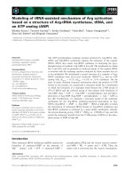
Figure 5 reports the frequency-histograms of the 6
verification parameters (Ω) for all patients, as detected
by image registration after treatment. In 58 out of 151
analyzed fractions, one or more parameters were larger
than the threshold of clinical acceptability established in
our clinical protocol (1 mm and 1° for linear and angu-
lar deviations respectively).
One outlier, which is not displayed in the plots,
showed 12.8 mm translation along the left-right direc-
tion, with acceptable values for the other directions (up
to 2.2 mm translation in AP-direction and up to 0.6°
yaw rotation). The optical system did not detect relevant
shifts for this case.
Discussion
In this work, we measured intra-fraction motion in
hypo-fractionated radiotherapy using a multimodal
approach. Our main goal was to assess the quality of
patient immobilization during treatment and t o high-
light the optimal measurement strategy (IR localization
vs. X-ray imaging). It is important to underline three
relevant aspects of the implemented methodology:
1. since extra-cranial treatments are performed in
free breathing conditions, IR data were collected and
averaged over at least two respiratory cycles to com-
pensate possible respiration motion effects in a short
time window. The effect of respiration movements
was furthermore evaluated by measuring the stan-
dard deviation (std) of marker positions over each
acquisition. The mean standard deviation ranged
between 0.3 and 0.5 mm in the extra-cranial patient
population. These values are due to the fact that
most of the IR markers (4-5 over 7) were placed in
correspondence of stable landmarks, like upper
thorax or pelvis, thus leading to a robust measure-
ment of patient position.
2. The cranial patient population potentially repre-
sents an ideal situation, as intra-fraction motion is
less relevant. How ever, the presence of the thermo-
plastic mask may repre sent a limitation because
markers are typically placed onto the mask in our
protocol. Therefore, the discrepancies found between
Figure 3 Intra-fraction error on external markers. 3D mismat ches on control points before and after the irradiation according to the two
different measurement approaches.
Table 2 Mean and standard deviation [mm] errors along
left-right (LR), cranio-caudal (CC) and antero-posterion
(AP) direction resulted after optical and X-ray
measurement.
Optical X-Ray
LR CC AP LR CC AP
Cranial 0.07
(0.40)
0.06
(0.28)
-0.11
(0.10)
0.00
(1.25)
-0.01
(0.45)
0.00
(0.49)
Abdomen -0.10
(0.46)
-0.13
(0.59)
-0.14
(0.67)
-0.25
(1.40)
-0.23
(1.06)
0.49
(1.13)
Lung 0.01
(0.50)
0.00
(0.63)
-0.15
(0.57)
0.25
(1.73)
-0.18
(2.24)
-0.04
(1.61)
Spadea et al. Radiation Oncology 2011, 6:38
/>Page 5 of 8
the two measurements approaches can be due in
part to movements of the patient within the mask,
as suggested by Linthout et al. [22].
3. The X-ray measurements were depurated from
setup residuals, computed before treatment by
means of image registration. This gave us more
robustness in understanding and analyzing an X-ray
based quantitative measurement of intra-fraction
variations.
The analysis was performed off-line, by analyzing both
the log files of markers position and t he X-ray images
stored immediately before and after irradiation. Compared
to the methodology proposed by Linthout et al., the main
differences in our data analysis were the following:
1.IntheworkbyLinthoutet al. the intra-fraction
motion monitored by the optical localizer was evalu-
ated in terms of the 6 Dof corrective parameters
estimated by the Brainlab software. Here, we
assessed the residual displacements on each externa l
marker after opt ical measurements and then we esti-
mated t he isocenter position from the configuration
of fiducials. This allowed us also to explore potential
deformations in the configuration of markers, in
order to test its reliability in patient setup control.
Figure 4 Estimation of intra-fraction error on target. 3D estimated intra-fraction motion of the target accordin g to the two different
measurement approaches.
Figure 5 6 dof corrective parameters. Frequency distribution plots of the linear (Tx, Ty, Tz) and angular deviations (Ax, Ay, Az) resulting from
kV X-ray images and DRR matching after irradiation. Bars are centered on labels and ranges over a 0.5 mm interval.
Spadea et al. Radiation Oncology 2011, 6:38
/>Page 6 of 8
2. In Linthout et al. the comparison between the two
sub-systems was performed by evaluating the correc-
tive parameters coming from external point registra-
tion and image fusion. This kind of analysis has a
conceptual flaw since an indeterminate number of
roto-translations are able to match 2 different con-
figurations in space at the same uncertainty level.
Here, we roto-translated the external configuration
of marker points according to image fusion and then
we compared the 2 fiducial sets, point by point, to
preciselyexaminethedifferencebetweenthe2
approaches.
Measurements performed by the optical localizer
showed on average sub-millimetric intra-fraction motion
for both extra-cranial and cranial treatments. These
results were confirmed when looking at target position,
as estimated according to the external marker configura-
tion under a rigid body assumption. Target position
resulted essentially stable, with average intra-fraction
motion within 1 mm. On the basis of these results, we
can assume that immobilization devices and the auto-
mation of setup procedures help the patient to be com-
fortable and stable, thus leading to small intra-fraction
variations.
When comparing optical versus X-ray measurements,
differences were on average 1-1.5 mm, with worst
results in lung cases. It should be noted from Figures 3
and 4 that X-ray imaging resulted in larger intra-frac-
tion motion compared to IR localization, with
increased i nter-patient variability. Such discrepancies
should be judged against the intrinsic accuracy of the
two systems (around 0.3 mm for optical localization
[23] and half CT slice thickness for image matching,
1.5 mm in our case). Digital image noise and image
artifacts might occasionally originate considerable
errors in registration as testified by the outlier case
that we reported in the results section ( 12.8 mm linear
shift). T he influence of image quality on the reliability
of image registration was also demonstrated during
internal commissioning studies on an anthropomorphic
radio-equivalent phantom. In Figure 6, we report a
Figure 6 x-ray image quality . Upper panels: X-ray images acquired on an anthropomorphic radio-equivalent phantom. Lower panels:X-ray
images acquired on a patient after treatment.
Spadea et al. Radiation Oncology 2011, 6:38
/>Page 7 of 8
comparison between images acqui red on phantom and
patients. Phantom studies showed no appreciable dif-
ference between the optical localizer and X-ray image
registration in 10 repeated measurements. In the
patient case, the image is clearly more blurred and
noisy and image registration led to a discrepancy of
about 2 mm in target lo calization compared to optical
measurements. Our conclusion is that the quality of X-
ray images must be accurately verified when using
image registration for intra-session monitoring, as the
sensitivity is extremely case specific.
Conclusions
Patient setup veri fication should rely on multimodal
monitoring systems (X-ray and IR optical) for the high-
est reliability in detecting and correcting geometric
uncertainties. The reported analysis shows that optical
tracking is able to provide robust measurement for the
real-time detection of intra-fraction variations.
List of abbreviations
AP: Antero-Posterior; ART: Adaptive Radiation Therapy; CBCT: Cone Beam
Computed Tomography; CC: Cranio-Caudal; CT Computed Tomography; Dof;
degrees of freedom; IGRT: Image Guided Radiation Therapy; IMRT: Intensity
Modulated Radiation Therapy; IR: Infra-Red; kV: kilo Voltage; LR: Left-Right;
MV: Mega Voltage; PostIR: 3D Marker position detected by the IR localizer
after treatment; PostXRay: PostIR roto-translated according to the corrective
parameters (Ω) estimated by image registration after treatment; PreIR: 3D
Marker position detected by the IR localizer before treatment; PreIR*: PreIR
roto-translated according to the verification parameters estimated by image
registration before treatment
Author details
1
Department of Experimental and Clinical Medicine, Università degli Studi
Magna Græcia, Catanzaro, Italy.
2
Department of Bioengineering, Politecnico
di Milano University, Milano, Italy.
3
Centro Nazionale di Adroterapia
Oncologica, Pavia, Italy.
4
Medical Physics Department, Istituto Europeo di
Oncologia, Milano, Italy.
5
Radiotherapy Division, Istituto Europeo di
Oncologia, Milano, Italy.
Authors’ contributions
MFS had primary role in study design, data analysis, results interpretation
and manuscript editing; BT and EP participated to data acquisition; MR and
GB gave important contributions in data analysis, results interpretation,
manuscript editing and final approval; CG was the medical physicist in
charge of computing the dose and running the ExacTrac System; DA, GP
were the physicians in charge of treatments; AP and RO gave final approval
to conceptual study and manuscript.
All authors read and approved the final manuscript.
Authors declare that no competing interest exist
Authors declare that written informed consent was obtained from the
patient for publication of this case report and accompanying images. A
copy of the written consent is available for review by the Editor-in-Chief of
this journal.
Received: 10 December 2010 Accepted: 15 April 2011
Published: 15 April 2011
References
1. Xing L, Thorndyke B, Schreibmann E, et al: Overview of image-guided
radiation therapy. Med Dosim 31(2):91-112.
2. Verellen D, Ridder MD, Linthout N, et al: Innovations in image-guided
radiotherapy. Nat Rev Cancer 2007, 7(12):949-60.
3. White E, Kane G: Radiation medicine practice in the image-guided
radiation therapy era: new roles and new opportunities.
SeminRadiatOncol 2007, 17(4):298-305.
4. Yan H, Yin FF, Kim JH: A phantom study on the positioning accuracy of
the Novalis Body system. Med Phys 2003, 30(12):3052-3060.
5. Tenn SE, Solberg TD, Medin PM: Targeting accuracy of an image guided
gating system for stereotactic body radiotherapy. Phys Med Biol 2005,
50:5443-5462.
6. Purdie TG, Bissonnette JP, Franks K, et al: Cone-beam computed
tomography for on-line image guidance of lung stereotactic
radiotherapy: localization, verification, and intrafraction tumor position.
Int J RadiatOncolBiol Phys 2007, 68(1):243-52.
7. Huntzinger C, Munro P, Johnson S, et al: Dynamic targeting Image-Guided
radiotherapy. Med Dos 2006, 31(2):113-125.
8. deCrevoisier R, Melancon AD, Kuban DA, Lee AK, Cheung RM, Tucker SL,
Kudchadker RJ, Newhauser WD, Zhang L, Mohan R, Dong L: Changes in
the pelvic anatomy after an IMRT treatment fraction of prostate cancer.
Int J RadiatOncolBiol Phys 2007, 68(5):1529-36.
9. Evans PM: Anatomical imaging for radiotherapy. Phys Med Biol 2008, 53:
R151-R191.
10. Yan D: Developing quality assurance processes for image-guided
adaptive radiation therapy. Int J RadiatOncolBiol Phys 2008, 71(1 Suppl):
S28-32.
11. Wagner TH, Meeks SL, Bova FJ, et al: Optical tracking technology in
stereotactic radiation therapy. Med Dosim 2007, 32(2):111-20,.
12. Baroni G, Garibaldi C, Riboldi M, et al: 3D optoelectronic analysis of
interfractional patient setup variability in frameless extracranial
stereotactic radiotherapy. Int J RadiatOncolBiol Phys 2006, 64(2):635-42.
13. Gierga DP, Riboldi M, Turcotte JC, et al: Comparison of target registration
errors for multiple image-guided techniques in accelerated partial breast
irradiation. Int J RadiatOncolBiol Phys 2008, 70(4):1239-46.
14. Riboldi M, Gierga DP, Chen GT, Baroni G: Accuracy in breast shape
alignment with 3D surface fitting algorithms. Med Phys 2009, 36(4):1193-8.
15. Tang X, Sharp GC, Jiang SB: Fluoroscopic tracking of multiple implanted
fiducial markers using multiple object tracking. Phys Med Biol 2007,
52(14):4081-98.
16. Lin T, Cerviño LI, Tang X, Vasconcelos N, Jiang SB: Fluoroscopic tumor
tracking for image-guided lung cancer radiotherapy. Phys Med Biol 2009,
54(4):981-92.
17. Bittner N, Butler WM, Reed JL, Murray BC, Kurko BS, Wallner KE, Merrick GS:
Electromagnetic tracking of intrafraction prostate displacement in
patients externally immobilized in the prone position. Int J RadiatOncol
Biol Phys 2010, 77(2):490-5.
18. Krauss A, Nill S, Tacke M, Oelfke U: Electromagnetic Real-Time Tumor
Position Monitoring and Dynamic Multileaf Collimator Tracking Using a
Siemens 160 MLC: Geometric and Dosimetric Accuracy of an Integrated
System. Int J Radiat Oncol Biol Phys 2011, 79(2):579-87.
19. Arslan S, Yilmaz A, Bayramgurler B, Uzman O, Nver E, Akkaya E: CT-guided
transthoracic fine needle aspiration of pulmonary lesions accuracy and
complications in 294 patients. Med Sci Monit 2002, 8:CR493-7.
20. Al-Qaisieh B, Carey B, Ash D, Bottomley D: The use of linked seeds
eliminates lung embolization following permanent seed implantation for
prostate cancer. Int J RadiatOncolBiol Phys 2004, 59(2):397-9.
21. Kitamura K, Shirato H, Shimizu S, et al: Registration accuracy and possible
migration of internal fiducial gold marker implanted in prostate and
liver treated with real-time tumor-tracking radiation therapy (RTRT).
RadiotherOncol 2002, 62(3):275-81.
22. Linthout N, Verellen D, Tournel K, Storme G: Six dimensional analysis with
daily stereoscopic x-ray imaging of intrafraction patient motion in head
and neck treatments using five points fixation masks. Med Phys 2006,
33(2):504-13.
23. Jin JY, Yin FF, Tenn SE, et al: Use of the BrainLABExacTrac X-Ray 6D
system in image-guided radiotherapy. Med Dosim 2008, 33(2):124-34.
24. Riboldi M, Baroni G, Spadea MF, et al: Robust frameless stereotactic
localization in extra-cranial radiotherapy. Med Phys 2006, 33(4):1141-1152.
25. Erich L: Nonparametrics: Statistical Methods Based on Ranks, Springer; 2006.
doi:10.1186/1748-717X-6-38
Cite this article as: Spadea et al.: Intra-fraction setup variability: IR
optical localization vs. X-ray imaging in a hypofractionated patient
population. Radiation Oncology 2011 6:38.
Spadea et al. Radiation Oncology 2011, 6:38
/>Page 8 of 8