Báo cáo khoa học: "Value of diffusion weighted MR imaging as an early surrogate parameter for evaluation of tumor response to high-dose-rate brachytherapy of colorectal liver metastase" docx
Bạn đang xem bản rút gọn của tài liệu. Xem và tải ngay bản đầy đủ của tài liệu tại đây (1.74 MB, 8 trang )
RESEARC H Open Access
Value of diffusion weighted MR imaging as an
early surrogate parameter for evaluation of tumor
response to high-dose-rate brachytherapy of
colorectal liver metastases
Christian Wybranski
1
, Martin Zeile
1
, David Löwenthal
1
, Frank Fischbach
1
, Maciej Pech
1
, Friedrich-Wilhelm Röhl
2
,
Günther Gademann
3
, Jens Ricke
1
and Oliver Dudeck
1*
Abstract
Background: To assess the value of diffusion weighted imaging (DWI) as an early surrogate parameter for
treatment response of colorectal liver metastases to image-guided single-fraction
192
Ir-high-dose-rate brachytherapy
(HDR-BT).
Methods: Thirty patients with a total of 43 metastases underwent CT- or MRI-guided HDR-BT. In 13 of these
patients a total of 15 additional lesions were identified, which were not treated at the initial session and served for
comparison. Magnetic resonance imaging (MRI) including breathhold echoplanar DWI sequences was performed
prior to therapy (baseline MRI), 2 days after HDR-BT (early MRI) as well as after 3 months (follow-up MRI). Tumor
volume (TV) and intratumoral apparent diffusion coefficient (ADC) were measured independently by two
radiologists. Statistical analysis was performed using univariate comparison, ANOVA and paired t test as well as
Pearson’s correlation.
Results: At early MRI no changes of TV and ADC were found for non-treated colorectal liver metastases. In
contrast, mean TV of liver lesions treated with HDR-BT increased by 8.8% (p = 0.054) while mean tumor ADC
decreased significantly by 11.4% (p < 0.001). At follow-up MRI mean TV of non-treated metastases increased by
50.8% (p = 0.027) without significant change of mean ADC values. In contrast, mean TV of treated lesions
decreased by 47.0% (p = 0.026) while the mean ADC increased inversely by 28.6% compared to baseline values
(p < 0.001; Pearson’s correlation coefficient of r = -0.257; p < 0.001).
Conclusions: DWI is a promising imaging biomarker for early prediction of tumor response in patients with
colorectal liver metastases treated with HDR-BT, yet the optimal interval between therapy and early follow-up
needs to be elucidated.
Background
The liver with its dual blood supply is a predisposed
organ for me tastatic disease [1]. Colorectal carcinoma
(CRC) represents the most frequent malignancy with
isolated hepatic metastases [2]. Hepatic resection has
become the standard of care and has been shown to
lead to a significant improvement of long-term survival,
however curative resection is possible in less than 25%
of the patients with isolated hepatic metastases [3]. For
unresectable metastases selective internal radiation ther-
apy (SIRT) and radiofrequency ablation (RFA) have
been shown to be efficient treatment alternatives [4,5].
Image-guided single-fraction
192
Ir-high-dose-rate bra-
chytherapy (HDR-BT) is a high precision percutaneous
ablation technique which has been shown to yield pro-
mising results with regards to safety and efficacy in the
treatment of unresectable colorectal liver metastases
[6-8]. Precise application of high irradiation doses to
* Correspondence: .de
1
Department of Radiology and Nuclear Medicine, Otto-von-Guericke
University Magdeburg, Germany
Full list of author information is available at the end of the article
Wybranski et al. Radiation Oncology 2011, 6:43
/>© 2011 Wybranski et al; licensee BioMed Central Ltd. This is an Open Access article distributed under the ter ms of the Creative
Commons Attribution License ( which permits unrestricted use, distribution, and
reproduction in any medium, provided the original work is properly cited.
tumor tissue with steep dose gradients resulting in
sparing of adjacent liver parenchyma allows this techni-
que to be applied repeatedly for treatment of recurrent
hepatic metastases [9,10]. Nonetheless, it would be of
great benefit to be able to evaluate treatment response
as early as possible. This would be particularly impor-
tant in individual cases in which irradiation doses have
to be reduced because of diminished functional hepatic
reserve or adjacent organs at r isk such as stomach or
intestine [11]. Early response evaluation in such patients
would be of major clinical significance to allow for
prompt modification of anticancer treatment, e.g.
repeated HDR-BT or additional radiofrequency ablation
in underdosed regions, and avoid unnecessary treatment
delays.
Diffusion-weighted imaging (DWI) supplies informa-
tion of water proton mobility [12,13]. This can be
employed to assess the microstructural organization of a
tissue like cell density , cell membrane integrit y and ulti-
mately cell viability which affect water diffusion proper-
ties in the extracellular space [14]. Liver DW MR
imaging has in the past been hampered by technical
challenges, mostly related to motion sens itivity and eddy
currents [15]. However, owing to improvement, the
technique has also successfull y been used in the liver to
predict and monitor a variety of anticancer therapies
[16-21]. The purpose of this study was to test the
hypothesis that DWI can predict tumor response in
patients with colorectal liver metastases as early as
2 days after interstitial HDR-BT.
Methods
Patient population
The study was approved by the local institutional review
boardandwritteninformedconsentwasobtainedfrom
each patient. 30 patients (14 women and 16 men; mean
age 65.6 years; range: 43 - 84 years) with a total of 43
unresectable colorectal metastases underwent HDR-BT
in a total of 37 sessions. Sixteen patients were found
surgically unresectable due to unfavourable anatomic
localization (bilobar metastases, infiltration of liver ves-
sels), 10 patients had limited extrah epatic disease, and 4
patients presented with comorbidities which excluded
resection.
Seven patients under went previous liver surgery, 25
patients were previously treated with chemotherapy, and
two patients received adjuv ant chemotherapy within the
follow-up period. The follow-up MRI data of these two
patients was excluded from analysis. In 13 o f these
patients, who presented with more than one colorectal
liver metastasis, a total of additional 15 lesions were
identified which were not treated at the initial session
(mean time interval between HDR-BT sessions: 40 days;
range: 26 - 66 days). In order to minimize the risk of
hepatic toxicity patients with multiple metastases were
treated in sequential HDR-BT sessions. These 15 lesions
served as control in order to compare changes in tumor
volume (TV) and apparent diffusion coefficient (ADC)
between treated and non-treated colorectal liver metas-
tases. Patients with tumor diameters less than 1 cm, or
poor image quality, e.g. respiratory motion or pulsation
artifacts, in which valid quantification of the mean ADC
was questionable were excluded from the study.
Image-Guided Interstitial HDR Brachytherapy
Brachytherapy catheters were positioned in analgoseda-
tion using either CT fluoroscopy (n = 2 0; Aqilion 16,
Toshiba medical systems, Otawara, Tochigi, Japan) or
high field open MRI guidance (n = 23; Panorama, Philips
Healthcare, Best, the N etherlands) based on conspicuity
of the metastases in eit her imaging modality. Patients
received 0.1 ml/kg body weight of a 0.25 mol/L solution
of Gd-EOB-DTPA (Primovist, BayerSchering, Berlin,
Germany) prior to MRI guided catheter placement to
improve tumor visualization, for which a T
1
-weighted
gradient echo sequence (T1 FFE; TR = 11 ms, TE = 6 ms,
flip angle = 35°, section thick ness of 8 mm, image acqui-
sition every 1.1 s) was used. For adequate coverage of the
target volume one catheter was placed per 1 - 2 cm
tumor diameter which resulted in a mean of 2.5 ± 1.8
catheters (range: 1 - 6 catheters) utilized per intervention
depending on tumor size and configu ration. After cathe-
ter positioning, either contrast enhanced multi-slice CT
(collimation: 16 × 0 .5 mm, slice thickness: 1 mm; table
feed: 5.5 mm/rotation; 90 ml Imeron 300; flow, 3 ml/s;
start delay 70 s) or T
1
-weighted fat signal saturated 3D
high resolution isotropic volume examination (THRIVE;
TR = 5.4 ms, T E = 2.6 ms, flip angle = 12°, section thick-
ness of 3 mm) were acquired to depict the exact position
of brachytherapy catheters in relation to tumor extension
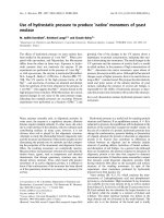
for treatment planning (Figure 1a). T his was performed
with the Oncentra-MasterPlan, BrachyModul planning
Figure 1 Illustration of MR-guided HDR-BT and 3D dosimetry.
77-year-old man with colorectal liver metastasis in segment VII
scheduled for high-dose-rate brachytherapy (HDR-BT). The
implantation of one brachytherapy catheter was performed under
MRI guidance (A). The tumor enclosing dose (D
100
) was 21.8 Gy (B).
Wybranski et al. Radiation Oncology 2011, 6:43
/>Page 2 of 8
software package ( Nucleotron, Veenendaal, the Nether-
lands; Figure 1b). The HDR afterloading system (micro-
Selectron Digital V3, Nucleotron, Veenendaal, the
Nethe rlands) employed a
192
Ir point source of 10 Ci (370
GBq). The minimal targe t dose prescribed f or colorectal
metastases was 19.4 ± 3.1 Gy (range: 10.3 - 24.0 Gy).
MR Imaging Protocol
Magnetic resonance imaging was performed with a 1.5
T MR system (Gyroscan, Intera, Phillips Medical S ys-
tems, Best, The Netherlands) emp loying a SENSE torso
surface coil. Imaging was performed at three time
points: Baseline MRI was performed at a mean of 5 days
(range: 0 - 36 days) prior to CT- or MRI-guided HDR-
BT. All but one patient received early MRI one to three
days after HDR-BT. Another patient was scanned five
days after treatment. Follow-up MRI was performed a
mean of 79 days (range: 36 - 120 days) after HDR-BT.
Unenhanced T
1
-weighted gradient echo (TR = 211
ms, TE = 5 ms, 350-mm FOV, 256 × 144 matrix,
SENSE factor 2, section thickness 8 mm) and T
2
-
weighted fast spin echo (TR = 1,600 ms, TE = 100 ms,
flip angle = 80°, 350-mm FOV, 384 × 196 matrix,
SENSE factor 2, section thickness 8 mm) axial imaging
were performed before DWI and Gd-EOB-DTPA con-
trast medium administration.
Breath-hold axial single shot echo planar (EPI) DWI
was acquired using the following parameters: TR = 1850
ms; TE = 68 ms; b factors 0 and 500 s/mm²; 112 × 111
matrix size, 350-mm FOV; section thickness 8 mm;
NSA 2; half-scan factor 0.608. Twelve sections through
the liver were acquired in each 20-s breath-hold, and
the entire liver (from the level of the diaphragm to the
inferior edge of the liver) was typically evaluated in two
to three breath-holds (Figure 2a). ADC maps were cal-
culated on a voxel-by-voxel b asis with an implemented
algorithm according to the following equation:
ADC
(
mm
2
s
−1
)
=[ln
(
S
0
S
b
)
]/
b
in which S
0
and S
b
represent the signal intensities of
the images with different gradient b factors, and b is the
difference between gradient b factors (Figure 2b).
Then, 0.1 mmol/kg body weight of Gd-EOB-DTPA
was administered with an infusion rate of 1.5 ml/s fol-
lowed by a 30-ml saline flush. THRIVE images were
acquired with the following parameters: TR = 3.9 ms,
TE = 1.9 ms, flip angle = 10°, 350-mm FOV, 192 × 136
matrix, SENSE factor 2, section thickness 6 mm, spectral
adiabatic inversion recovery (SPAIR). In order to mini-
mise differences in contrast media circulation time, the
first post-contrast (arterial phase) sequence was started
manually by using the bolus tracking technique at the
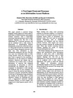
Figure 2 Baseline MRI preceding HDR-BT. Pre-treatment
diffusion-weighted image (DWI) with b = 500 s/mm
2
(A),
corresponding apparent diffusion coefficient (ADC) map (B) and
T1w Gd-EOB-DTPA enhanced MR image in hepatocyte-selective
(hepatobiliary) phase (C) of the same patient as in Figure 1 depict
the colorectal metastasis in liver segment VII with a mean ADC of
1.29 × 10
-3
mm
2
s
-1
and a mean volume of 23.3 cm
3
(arrow).
Wybranski et al. Radiation Oncology 2011, 6:43
/>Page 3 of 8
time when contrast agent reached the ascending aorta,
typically 14-17 s after the start of injection. For subse-
quent acquisitions, intervals allowing patient’ sfree
breathing were placed between the arterial and portal
venous phase (20 s) and the portal venous and equili-
brium (i.e. interstitial) phase (40 s), respectively.
THRIVE as well as T
1
-weighted2Dgradientechowith
selective water excitation (WATS) images (TR = 131
msec, TE = 5 msec, flip angle = 70°, 350-mm FOV, 256
× 135 matrix, SENSE factor 2, section thickness 8 mm)
were acquired 20 min after contrast material administra-
tion at the hepatocyte-selective (hepatobiliary) phase
(Figure 2c).
Tumor Volume Assessment and ADC Calculation
Assessment of tumor areas was performed with the
OsiriX imaging software version 3.6.1. Tumor borders
were segmented manually on transversal Gd-EOB-
DTPA enhanced THRIVE images by two i ndependent
investigators. The mean of the volumetric measurements
was t aken as representative TV for each lesion. TV was
expressed by OsiriX in cubic centimeters (cm
3
).
For ADC calculation up to three slices of the ADC
map depicting the largest tumor diameter were selected,
depending on the volume of the tumor. In each slice a
region of interest (ROI) was delineated according to the
tumor geometry. The border of the ROI w as placed in
the tumor periphery close to the tumor margin, so that
the ROI encompassed almost the whole tumor area
(Figure 3). The measurements were performed indepen-
dently by two experienced investigators and the mean of
the measurements was recorded as representative ADC
value for each lesion. Initial and follow-up images were
matched and ADC calculations were perform ed on cor-
responding sections on follow-up MRI (Figure 4).
Statistical Analysis
SPSS, version 17.0 (Chicago, IL) was used for statistical
analysis. Interobserver agreement was assessed with
Cohen’ sKappa( ≤ 0.40 poor agreement, =0.41-
075 good agreement, ≥ 0.76 excellent agreement).
To discuss the treatment effect, we performed a univari-
ate comparison between treated and non-treated colorec-
tal metastases with regards to changes in mean ADC and
TV at early and follow-up MRI compared to baseline MRI
using the t test (Welch test, Satterthwaite’s approximation
to compute the degrees of freedom).
After that we performed an ANOVA with the adjusted
F-Test by Greenhouse-Geisser to get a global test for
time effects in each of the two groups. Paired t test with
Bonferroni correction for multiple testing was applied to
test the significance of the differences of treatment
induced changes of ADC values and TV between early
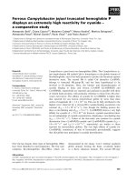
Figure 3 Early MRI 3 days after HDR-BT. Early DWI (A) and
corresponding ADC map (B) performed 3 days after HDR-BT (same
patient as in Figure 1) reveal a decrease in mean ADC by 27.1% to
0.94 × 10
-3
mm
2
s
-1
. The ROI within the lesion indicates an ADC
value of 1.09 × 10
-3
mm
2
s
-1
in this slice of the ADC map (arrow).
T1w Gd-EOB-DTPA enhanced MR image in hepatobiliary phase (C)
indicates no relevant change in size of the treated lesion (24.1 cm
3
).
Wybranski et al. Radiation Oncology 2011, 6:43
/>Page 4 of 8
and follow-up MRI compared to baseline MRI. The
correlation between the change of the mean ADC and
TV was expressed with the Pearson’s correlation coeffi-
cient r. A two-taile d p-value of 0.05 was set to be the
level of statistical significance.
Results
There was an excellent interobserver agreement between
the two readers with a kappa coef ficient of 0.93 for the
assessment of TV and 0.89 for ADC values.
At baseline, mean TV of treated colorectal liver
metastases was 62.2 c m
3
(range: 0.5 - 786.2 cm
3
)while
mean tumor ADC was 1.75 × 10
-3
mm
2
s
-1
(range: 0.65 -
3.22 × 10
-3
mm
2
s
-1
). In non-treated lesions mean TV
was 50.0 cm
3
(range: 2 .3 - 136.9 cm
3
) with a mean
tumor ADC of 1.88 × 10
-3
mm
2
s
-1
(range: 1.40 - 2.67 ×
10
-3
mm
2
s
-1
). The difference between treated and non-
treated lesions with regards to mean TV and mean
tumor ADC at baseline was non significant (p> 0.25).
The change in mean TV (p = 0.007) and mean tumor
ADC (p < 0.001) differed significantly between treated
and non-treated colorectal liver metastases at early MRI.
No changes of TV (50.2 cm
3
; range: 2.3 - 140.6 cm
3
)as
well as mean tumor ADC (1.90 × 10
-3
mm
2
s
-1
;range:
1.41 - 2.64 × 10
-3
mm
2
s
-1
) were found for the non-trea-
ted lesions (Figure 5 and 6). In contrast, mean TV of
colorectal liver metastases treated with HDR-BT
increased by 8.8% to 67.7 cm
3
(range: 0.5 - 886.0 cm
3
),
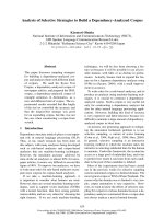
Figure 4 Follow-up MRI. DWI (A) and ADC map (B) performed 105
days post intervention (same patient as in Figure 1) show a rise of
mean tumor ADC of 75.2% to 2.26 × 10
-3
mm
2
s
-1
(arrow). This
finding correlates with a decrease in tumor volume by 90.6% (2.2
cm
3
), depicted in T1w Gd-EOB-DTPA enhanced MR image in
hepatobiliary phase (C). The circular hypointense region around the
treated lesion in (C) indicates the area of irradiation induced
reversible hepatocyte dysfunction.
Figure 5 Boxplot depicting changes of mean volume of non-
treated and treated tumors at early and follow-up MRI
compared to baseline MRI. Boxplot shows changes of mean tumor
volume (TV) of non-treated (*: p = 0.027) and treated colorectal liver
metastases (*: p = 0.026) 2 days (early MRI) as well as 3 months
(follow-up MRI) after HDR-BT as compared to baseline MRI.
Wybranski et al. Radiation Oncology 2011, 6:43
/>Page 5 of 8
but only a trend towards a statistically significant differ -
ence was observed (p = 0.054; Figure 5). Remarkably,
mean tumor ADC of treated colorectal liver metastases
decreased significantly by 11.4% to 1.55 × 10
-3
mm
2
s
-1
(range: 0.64 - 2.60 × 10
-3
mm
2
s
-1
; p < 0.001; Figure 6).
The change between mean TV and mean tumor A DC
of the treated lesions did not differ significantly between
one and three days (p = 0.708 and p = 0.945).
The change in mean TV (p = 0.002) and mean tumor
ADC (p < 0.001) differed significantly between treated
and non-treated colorectal liver metastases at follow-up
MRI. At follow-up MRI mean TV of non-treated color-
ectal liver metastases increased significantly by 50.8% to
75.4 cm
3
(range: 10.2 - 170.3 cm
3
) as compared to base-
line (p = 0.027; Figure 5). Mean tumor ADC at the time
of follow-up MRI was 1.92 × 10
-3
mm
2
s
-1
(range: 1.32 -
3.23 × 10
-3
mm
2
s
-1
), which resembled a non significant
change of only 1.0% (p>0.9;Figure6).Incontrast,
mean TV at follow-up MRI of colorectal liver metastases
treated with HDR-BT decreased by 47 .0% to 33.0 cm
3
(range: 0.5 - 397.8 cm
3
) as compared to baseline (p =
0.026; Figure 5). This reflected a local tumor control
rate of 97.7% with absence of progression in 40 of 41
treated lesion s. The mean tumor ADC increased signifi-
cantly by 28.6% to 2.25 × 10
-3
mm
2
s
-1
(range: 0.72 -
3.31 × 10
-3
mm
2
s
-1
)ascomparedtobaseline(p < 0.001;
Figure 6). Pearson’s correlation indicated a weak but sta-
tistically significant linear relationship between the
change of mean TV and mean tumor ADC of r = -0.257
( p < 0.001; Figure 7). Hence, differences in ADC were
inversely correlated with morphological changes.
Discussion
Our study demonstrated HDR-BT to be highly efficient
for the treatment of unresectable colorectal liver metas-
tases [8,10,22,23]. Furthermore, tumor size reduction
was inversely correlated with a significant increase in
mean tumor ADC values after 3 months. These results
are well in agreement with the current understanding of
therapy induced changes assessed by DWI: effectiv e
anticancer treatment results in tumor lysis, loss of cell
membrane integrity, increased extracellular space, and,
therefore, an increase in water diffusion [24,25]. Our
results were also in accordance with results of previous
studies of primary and secondary liver tumors, which all
haveshownanincreaseinADCafteranumberof
different therapeutic modalities [16-21].
On early MRI performed in mean 2 days after HDR-
BT, DWI was able to depict tumor response as only in
treated lesions mean tumor ADC values decreased sig-
nificantly. A slight increase in TV accompanied the
decrease in ADC ( compare Figures 5 and 6). How may
this decrease in mean tumor ADC and increase in TV
be explained ? Current models of tumor response postu-
late cell swelling to occur soon after initiation of antic-
ancer therapy. This can lead to a transient decrease in
Figure 6 Boxplot depicting changes of mean ADC of non-
treated and treated tumors at early and follow-up MRI
compared to baseline MRI. Boxplot illustrates changes of mean
ADC of non-treated and treated colorectal liver metastases 2 days
(early MRI) as well as 3 months (follow-up MRI) following HDR-BT as
compared to baseline MRI (*: p < 0.001).
Figure 7 Scatter plot depicting the relationship between
changes of mean tumor volume and mean tumor ADC at
follow-up MRI compared to baseline MRI. Scatter plot depicts
the relationship between changes of mean tumor volumes and
mean ADC values of colorectal liver metastases 3 months after
treatment with HDR-BT as compared to baseline MRI. A decrease in
tumor size is inversely associated with an increase in ADC. Pearson’s
correlation indicated a weak but statistically significant linear
relationship of r = -0.257 (p < 0.001).
Wybranski et al. Radiation Oncology 2011, 6:43
/>Page 6 of 8
tumor ADC [14,24,26]. Such cellular changes have been
recognized as an early hallmark of cellular necrosis
[27-29]. In H DR-BT applied doses in next proximity to
the brachytherapy catheters can exceed 100 Gy inducing
even immediate cell lysis [30,31]. Additionally, irradiation
compromises tumor microvasculature by causing
endothelial damage at an early stage [32]. Endothelial
damage may lead to increased transient vascular perme-
ability to macromolecules like albumin, which can
become insoluble in the interstitium [33-36]. Consecutive
restriction of extracellular microcirculation leads to a
decrease in ADC. Restriction of the extracellular micro-
circulation in turn may compromise microperfusion
through compression of capillaries and terminal lymph
vessels [34]. As DWI provides simultaneous information
on diffusion as well as microperfusion this effect may
also have contributed to this early decrease in mean
tumor ADC [37-39]. Cell swelling and transudation of
plasma components into the extravascular-extracellular
space of the tumor are also the most likely mechanisms
responsible for the transient increase in TV.
Obviously, the timing of the evaluation of tumor
response after the start of treatment is a key issue. For
the present stu dy, we chose to perform MRI including
DWI very early at a median of 2 days following HDR-
BT. Thus, we were enabled to obtain first inform ation
on the treatment response before the patient was dis-
charged, which is routinely 2 to 3 days after HDR-BT at
our instituti on. Although decrease in mean tumor ADC
of treated colorectal liver metastases at early MRI was
significant, the observed range of ADC values was rela-
tively wide. Thus, at this early interval after HDR-BT
this difference was not distinct enough t o base clinical
decisions in individuals exclusiv ely on these findings.
Perhaps a larger time interval of 1-2 weeks would have
been superior, but we did not want to prolong hospitali-
zation of these advanced cancer patients. Hence, larger
clinical studies have to confirm the ability of DWI to
identify treatment response to anticancer therapy and
identify the best time point to p erform early MRI,
before inferences can be drawn that influence the thera-
peutic strategy.
Conclusions
In conclusion, DWI is a promising imaging biomarker
for early prediction of tumor response in patients with
colorectal liver meta stases treated with HDR-BT, yet the
optimal interval between therapy and early follow-up
needs to be elucidated.
Author details
1
Department of Radiology and Nuclear Medicine, Otto-von-Guericke
University Magdeburg, Germany.
2
Institute of Biometry and Medical
Informatics, Otto-von-Guericke University Magdeburg, Germany.
3
Department
of Radiotherapy, Otto-von-Guericke University Magdeburg, Germany.
Authors’ contributions
CW participated in the design and coordination of the study, data
acquisition and analysis and drafted the manuscript. MZ and DL participated
in data acquisition and analysis as well as literature review. MP, FF and JR
participated in the design of the study and carried out the interventions.
FWR performed the statistical analysis. GG participated in the design of the
study and the treatment planning procedures. OD conceived of the study
and participated in its design and coordination. All authors have read and
approved the final manuscript.
Competing interests
The authors declare that they have no competing interests.
Received: 14 December 2010 Accepted: 27 April 2011
Published: 27 April 2011
References
1. Rappaport AM: Hepatic blood flow: morphologic aspects and physiologic
regulation. Int Rev Physiol 1980, 21:1-63.
2. Kasper HU, Drebber U, Dries V, Dienes HP: [Liver metastases: incidence
and histogenesis]. Z Gastroenterol 2005, 43:1149-1157.
3. Khatri VP, Petrelli NJ, Belghiti J: Extending the frontiers of surgical therapy
for hepatic colorectal metastases: is there a limit? J Clin Oncol 2005,
23:8490-8499.
4. Guenette JP, Dupuy DE: Radiofrequency ablation of colorectal hepatic
metastases. J Surg Oncol 2010, 102:978-987.
5. Welsh JS, Kennedy AS, Thomadsen B: Selective Internal Radiation Therapy
(SIRT) for liver metastases secondary to colorectal adenocarcinoma. Int J
Radiat Oncol Biol Phys 2006, 66:S62-S73.
6. Seidensticker M, Wust P, Ruhl R, Mohnike K, Pech M, Wieners G,
Gademann G, Ricke J: Safety margin in irradiation of colorectal liver
metastases: assessment of the control dose of micrometastases. Radiat
Oncol 2010, 5:24.
7. Ricke J, Wust P, Stohlmann A, Beck A, Cho CH, Pech M, Wieners G, Spors B,
Werk M, Rosner C, et al: CT-guided interstitial brachytherapy of liver
malignancies alone or in combination with thermal ablation: phase I-II
results of a novel technique. Int J Radiat Oncol Biol Phys 2004,
58:1496-1505.
8. Ricke J, Mohnike K, Pech M, Seidensticker M, Ruhl R, Wieners G, Gaffke G,
Kropf S, Felix R, Wust P: Local response and impact on survival after local
ablation of liver metastases from colorectal carcinoma by computed
tomography-guided high-dose-rate brachytherapy. Int J Radiat Oncol Biol
Phys 2010, 78:479-485.
9. Ruhl R, Ludemann L, Czarnecka A, Streitparth F, Seidensticker M, Mohnike K,
Pech M, Wust P, Ricke J: Radiobiological restrictions and tolerance doses
of repeated single-fraction hdr-irradiation of intersecting small liver
volumes for recurrent hepatic metastases. Radiat Oncol 2010, 5:44.
10. Ricke J, Seidensticker M, Ludemann L, Pech M, Wieners G, Hengst S,
Mohnike K, Cho CH, Lopez HE, Al-Abadi H, et al: In vivo assessment of the
tolerance dose of small liver volumes after single-fraction HDR
irradiation. Int J Radiat Oncol Biol Phys 2005, 62:776-784.
11. Streitparth F, Pech M, Bohmig M, Ruehl R, Peters N, Wieners G, Steinberg J,
Lopez-Haenninen E, Felix R, Wust P, et al: In vivo assessment of the gastric
mucosal tolerance dose after single fraction, small volume irradiation of
liver malignancies by computed tomography-guided, high-dose-rate
brachytherapy. Int J Radiat Oncol Biol Phys 2006, 65:1479-1486.
12. Le Bihan D, Breton E, Lallemand D, Grenier P, Cabanis E, Laval-Jeantet M:
MR imaging of intravoxel incoherent motions: application to diffusion
and perfusion in neurologic disorders. Radiology 1986, 161:401-407.
13. Le Bihan D, Turner R, Douek P, Patronas N: Diffusion MR imaging: clinical
applications. AJR
Am J Roentgenol 1992, 159:591-599.
14. Koh DM, Collins DJ: Diffusion-weighted MRI in the body: applications and
challenges in oncology. AJR Am J Roentgenol 2007, 188:1622-1635.
15. Taouli B, Koh DM: Diffusion-weighted MR imaging of the liver. Radiology
2010, 254:47-66.
16. Cui Y, Zhang XP, Sun YS, Tang L, Shen L: Apparent diffusion coefficient:
potential imaging biomarker for prediction and early detection of
Wybranski et al. Radiation Oncology 2011, 6:43
/>Page 7 of 8
response to chemotherapy in hepatic metastases. Radiology 2008,
248:894-900.
17. Dudeck O, Zeile M, Wybranski C, Schulmeister A, Fischbach F, Pech M,
Wieners G, Ruhl R, Grosser O, Amthauer H, et al: Early prediction of
anticancer effects with diffusion-weighted MR imaging in patients with
colorectal liver metastases following selective internal radiotherapy. Eur
Radiol 2010, 20:2699-2706.
18. Eccles CL, Haider EA, Haider MA, Fung S, Lockwood G, Dawson LA: Change
in diffusion weighted MRI during liver cancer radiotherapy: Preliminary
observations. Acta Oncol 2009, 1-10.
19. Koh DM, Scurr E, Collins D, Kanber B, Norman A, Leach MO, Husband JE:
Predicting response of colorectal hepatic metastasis: value of
pretreatment apparent diffusion coefficients. AJR Am J Roentgenol 2007,
188:1001-1008.
20. Marugami N, Tanaka T, Kitano S, Hirohashi S, Nishiofuku H, Takahashi A,
Sakaguchi H, Matsuoka M, Otsuji T, Takahama J, et al: Early detection of
therapeutic response to hepatic arterial infusion chemotherapy of liver
metastases from colorectal cancer using diffusion-weighted MR imaging.
Cardiovasc Intervent Radiol 2009, 32:638-646.
21. Schraml C, Schwenzer NF, Clasen S, Rempp HJ, Martirosian P, Claussen CD,
Pereira PL: Navigator respiratory-triggered diffusion-weighted imaging in
the follow-up after hepatic radiofrequency ablation-initial results. J Magn
Reson Imaging 2009, 29:1308-1316.
22. Ricke J, Wust P, Wieners G, Beck A, Cho CH, Seidensticker M, Pech M,
Werk M, Rosner C, Hanninen EL, et al: Liver malignancies: CT-guided
interstitial brachytherapy in patients with unfavorable lesions for
thermal ablation. J Vasc Interv Radiol 2004, 15:1279-1286.
23. Wieners G, Pech M, Hildebrandt B, Peters N, Nicolaou A, Mohnike K,
Seidensticker M, Sawicki M, Wust P, Ricke J: Phase II Feasibility Study on
the Combination of Two Different Regional Treatment Approaches in
Patients with Colorectal “Liver-Only” Metastases: Hepatic Interstitial
Brachytherapy Plus Regional Chemotherapy. Cardiovasc Intervent Radiol
2009, 32:937-945.
24. Moffat BA, Chenevert TL, Lawrence TS, Meyer CR, Johnson TD, Dong Q,
Tsien C, Mukherji S, Quint DJ, Gebarski SS, et al: Functional diffusion map:
a noninvasive MRI biomarker for early stratification of clinical brain
tumor response. Proc Natl Acad Sci USA 2005, 102:5524-5529.
25. Moffat BA, Hall DE, Stojanovska J, McConville PJ, Moody JB, Chenevert TL,
Rehemtulla A, Ross BD: Diffusion imaging for evaluation of tumor
therapies in preclinical animal models. MAGMA 2004, 17:249-259.
26. Thoeny HC, De Keyzer F, Chen F, Ni Y, Landuyt W, Verbeken EK, Bosmans H,
Marchal G, Hermans R: Diffusion-weighted MR imaging in monitoring the
effect of a vascular targeting agent on rhabdomyosarcoma in rats.
Radiology 2005, 234:756-764.
27. Denecker G, Vercammen D, Declercq W, Vandenabeele P: Apoptotic and
necrotic cell death induced by death domain receptors. Cell Mol Life Sci
2001, 58:356-370.
28. Galluzzi L, Maiuri MC, Vitale I, Zischka H, Castedo M, Zitvogel L, Kroemer G:
Cell death modalities: classification and pathophysiological implications.
Cell Death Differ 2007, 14:1237-1243.
29. Taatjes DJ, Sobel BE, Budd RC: Morphological and cytochemical
determination of cell death by apoptosis. Histochem Cell Biol 2008,
129:33-43.
30. Gromoll C, Karg A: Determination of the dose characteristics in the near
area of a new type of 192Ir-HDR afterloading source with a pinpoint
ionization chamber. Phys Med Biol 2002, 47:875-887.
31. Nath R, Anderson LL, Luxton G, Weaver KA, Williamson JF, Meigooni AS:
Dosimetry of interstitial brachytherapy sources: recommendations of the
AAPM Radiation Therapy Committee Task Group No. 43. American
Association of Physicists in Medicine. Med Phys 1995, 22:209-234.
32. Denham JW, Hauer-Jensen M: The radiotherapeutic injury–a complex
‘wound’. Radiother Oncol 2002, 63:129-145.
33. Krishnan L, Krishnan EC, Jewell WR: Immediate effect of irradiation on
microvasculature. Int J Radiat Oncol Biol Phys 1988, 15:147-150.
34. Baker DG, Krochak RJ: The response of the microvascular system to
radiation: a review. Cancer Invest 1989, 7:287-294.
35. Potchen EJ, Kinzie J, Curtis C, Siegel BA, Studer RK: Effect of irradiation on
tumor microvascular permeability to macromolecules. Cancer 1972,
30:639-643.
36. Kobayashi H, Reijnders K, English S, Yordanov AT, Milenic DE, Sowers AL,
Citrin D, Krishna MC, Waldmann TA, Mitchell JB, et al: Application of a
macromolecular contrast agent for detection of alterations of tumor
vessel permeability induced by radiation. Clin Cancer Res 2004,
10:7712-7720.
37. Latour LL, Svoboda K, Mitra PP, Sotak CH: Time-dependent diffusion of
water in a biological model system. Proc Natl Acad Sci USA 1994,
91:1229-1233.
38. Thoeny HC, De Keyzer F, Vandecaveye V, Chen F, Sun X, Bosmans H,
Hermans R, Verbeken EK, Boesch C, Marchal G, et al: Effect of vascular
targeting agent in rat tumor model: dynamic contrast-enhanced versus
diffusion-weighted MR imaging. Radiology 2005, 237:492-499.
39. Jordan BF, Runquist M, Raghunand N, Baker A, Williams R, Kirkpatrick L,
Powis G, Gillies RJ: Dynamic contrast-enhanced and diffusion MRI show
rapid and dramatic changes in tumor microenvironment in response to
inhibition of HIF-1alpha using PX-478. Neoplasia 2005, 7:475-485.
doi:10.1186/1748-717X-6-43
Cite this article as: Wybranski et al.: Value of diffusion weighted MR
imaging as an early surrogate parameter for evaluation of tumor
response to high-dose-rate brachytherapy of colorectal liver metastases.
Radiation Oncology 2011 6:43.
Submit your next manuscript to BioMed Central
and take full advantage of:
• Convenient online submission
• Thorough peer review
• No space constraints or color figure charges
• Immediate publication on acceptance
• Inclusion in PubMed, CAS, Scopus and Google Scholar
• Research which is freely available for redistribution
Submit your manuscript at
www.biomedcentral.com/submit
Wybranski et al. Radiation Oncology 2011, 6:43
/>Page 8 of 8