Báo cáo khoa học: "Density of CD4(+) and CD8(+) T lymphocytes in biopsy samples can be a predictor of pathological response to chemoradiotherapy (CRT) for rectal cancer" ppt
Bạn đang xem bản rút gọn của tài liệu. Xem và tải ngay bản đầy đủ của tài liệu tại đây (865.33 KB, 6 trang )
RESEARCH Open Access
Density of CD4(+) and CD8(+) T lymphocytes in
biopsy samples can be a predictor of
pathological response to chemoradiotherapy
(CRT) for rectal cancer
Koji Yasuda, Takako Nirei, Eiji Sunami, Hirokazu Nagawa and Joji Kitayama
*
Abstract
Background: Although preoperative radiotherapy (RT) is widely used as the initial treatment for locally advanced
rectal cancer (RC) in the neoadjuvant setting, factors determining clinical response have not been adequately
defined. Radiosensitivity has recently been shown to be greatly affected by immune function of the host.
Methods: In 48 cases of advanced RC, we retrospectively examined the density of tumor infiltrating CD4(+) and
CD8(+) T cells using immunohistochemical staining of biopsy samples before CRT, and examined the correlation
with tumor response.
Results: The numbers of both CD4(+) and CD8(+) tumor-infiltrating lymphocytes (TIL) in pre-CRT biopsy samples
were strongly correlated with tumor reduction ratio evaluated by barium enema. Moreover, the densities of CD4(+)
and CD8(+) TIL were significantly associated with histological grade after CRT. The density of CD8(+) TIL was an
independent prognostic factor for achieving complete response after CRT.
Conclusions: In RC patients, T lymphocyte-mediated immune reactions play an important role in tumor response
to CRT, and the quantitative measurement of TIL in biopsy samples befo re CRT can be used as a predictor of the
clinical effectiveness of CRT for advanced RC.
Introduction
Previous studies have demon strated that preoperative
radiotherapy (RT) can produce down-staging in
advanced rectal cancer (RC), resulting in longer survi-
val, a reduced rate of postoperative local recurrence.
Recently, adding chemotherapy to RT (CRT) has
achieved even more favorable results [1-3]. Thus, pre-
operative RT in the neoadjuvant setting is currently
recognized as the standard treatment for locally
advanced RC. However, in unresponsive cases, it may
have disadvantages such as delaying surgery or
immune suppression. Although many clinical factors
[4,5], radiologic findings [6,7] and molecular markers
[7-10] have been suggested to be related to therapeu-
tic response, the clinical usefulness of these markers
remains controversial, and thus identifying factors
predicting the efficacy of neoadjuvant CRT is essential
for decision-making in the management of patients
with RC.
Recent studies have demonstrated that radiosensitiv-
ity is greatly affected by immune function of the host
[11,12]. In fact, we recently showed that the circulat-
ing lymphocyte count is an important parameter
determining the clinical outcome of RC patients who
undergoCRT[13].Thisfactinspiredustoevaluate
the relation betwe en the response and the characteris-
tics of tumor-infiltrating lymphocytes (TIL) in rectal
tumors. In this study, we used immunohistochemical
staining and examined the distribution and cell den-
sity of CD4(+) and CD8(+) TIL in biopsy samples
before the start of CRT.
* Correspondence:
Department of Surgery, Division of Surgical Oncology, University of Tokyo,
Japan
Yasuda et al. Radiation Oncology 2011, 6:49
/>© 2011 Yasuda et al; licensee BioMed Central Ltd. This is an Open Access article distributed under the terms of the Creative Common s
Attribution Lice nse ( http://c reativecommons.or g/licenses/by/2.0), which permits unrestricted use, distribution, and re production in
any medium, provided the original work is properly cited.
Materials and methods
Patients
Forty eight consecutive patients with rectal adenocarci-
noma who received preoperative chemoradiotherapy
(CRT) between November 2005 and August 200 9 and
following surgery in Tokyo University Hospital were
included in this study. All the patients received a total
dose of 50.4Gy radiation and concomitant 5-Fu-based
chemotherapy. Among the 48 cases, 46 underwent total
mesorectal excision at 6~8 week s after the end of CRT
in the Department of Surgical Oncology. In 6 cases, no
tumor cells were detected at either the primary site or
in regional lymph n odes on pa thological examination,
confirming pathological complete response (pCR). Two
other patients showed a clinical CR (cCR) after CRT,
with no detectable cancer cells in multiple biopsy speci-
mens, and were thus followed without surgery and
showed no evidence of recurrence for more than 16
months. In all cases, a barium enema (BE) was per-
formed before and after CRT, the longitudinal dimen-
sion of the rectal tumor was measured on BE images
before (A) and after (B) CRT, and the reduction rate
was calculated as (A-B)/A.
Biopsy samples were obtained at 3-17 days before the
start of CRT, and serial-step sections of the biopsy sam-
ples were cut with 3 μm width, fixed in 10% formalin
solution, then embedded in paraffin, stained with hema-
toxylin-eosin, and the grade of tumor response was eval-
uated by pathologists according to the definitions in the
Japanese Classification of Colorectal Carcinoma [14]:
Grade0,noremarkablechanges;Grade1,swellingof
cells, enlarged vesicles, pyknosis of nuclei and vacuo-
lated cytoplasm (< 2/3 of tumor cells); Grade 2, cell
nests consisting of markedly damaged cells, of ten exhi-
biting a moth-eaten appearance and simplified granular
structures in more than 2/3 tumor cells; and Grade 3,
extensive degenerative changes and replaced by granulo-
matous or fibrous tissue. This study was performed with
the approval by t he Ethics Committee of the University
of Tokyo, and written informed consent was obtained
from the patient for publication of this case report and
accompanying images. A copy of the written consent is
available for review by t he Editor-in-Chief of this
journal.
Immunohistochemical study of human samples
The distribution and density of CD4(+) and CD8(+)
lymphocytes in biopsy samples of primary rectal tumor
were evaluated by immunohistochemical staining using
affinity purified mouse monoclonal antibodies against
CD4 (1F6, mIgG1) and CD8 (4B11, mIgG1) (Novocastra,
CA). The specificities of these mAbs i n immunohisto-
chemistry on paraffin embedded samples were con-
firmed with human tonsil tissue sections (data not
shown). Sections (3 μm thick) at the center of the the
biopsy specimens were deparaffinized in xylene,
hydrated through a graded series of ethano l, and heated
in a microwave oven for two 7-minute cycles (500
wat ts). After rinsing in phosphate buffered saline (PBS),
endogenous peroxidase activity was inhibite d by incuba-
tion with 0.3% hydrogen peroxide in 100% methanol for
30 minutes. After 3 washes in PBS, nonspecific reaction
was blocked b y incubation with PBS containing 5%
skimmed milk for 30 minut es at room temperature, and
then the sections were incubated with normal rabbit or
goat serum for 30 min. The sections were incubated
overnight at 4°C in humid chambers with the primary
antibodies to CD4 and CD8 at a dilution of 1/50. After
three washes with PBS, the sections were incubated with
biotinylated rabbit anti-goat or rabbit immunoglobulin
for 30 min. After washing again with PBS, the slides
were treated with peroxidase-conjugated streptavidin for
30 min, and developed by immersion in 0.01% H
2
O
2
and 0.05% diaminobenzidine tetrahydrochloride for 3
min. Light counterstaining with Mayer’ s hematoxyl in
was performed. The number of immunoreactive lym-
phocytes was counted under light microscope in a r an-
domly selected field at the magnification of 400× in
three different sections. Analysis was performed blind
with respect to clinical outcome by two pathologists.
Statistical Analysis
The associations of CR with blood cell counts and var-
ious other clinical parameters were examined using Wil-
coxon’ s test and chi-squared test, respectively.
Multivariate stepwise logistic regression analysis was
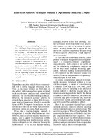
C
D
AB
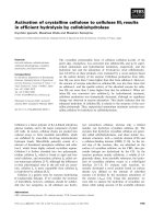
Figure 1 Immunohistochemical detection of CD4(+)and CD8(+)
T cells in biopsy samples of rectal cancer before CRT. Tissue
sections of biopsy samples were immunostained with anti-CD4 (A,C)
or anti-CD8 (B,D) mAb. ×100 (A,B), ×400 (C,D) Arrow: 100 μm.
Yasuda et al. Radiation Oncology 2011, 6:49
/>Page 2 of 6
performed to determine the independence of all vari-
ables identified as possibly significan t. All analyses wer e
performed with JMP8.0 software, and p-values less than
0.05 were considered to be statistically significant.
Results
The number and distribution of T cells in biopsy
samples before CRT were evaluated with immunostainig
withm Abs against CD4 and CD8. As shown in
Figure 1, both CD4 and CD8 were clearly stained in the
cell membrane of interstitial infiltrates. In most cases,
CD4(+) or CD8(+) T cells were evenly distributed in the
whole tissue sections, while many cells clustered in spe-
cific fields in some cases. When the numbers were
counted in each case, the densities of CD4(+) and CD8
(+) T cells showed a strong association (data not
shown).
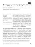
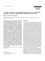
More importantly, the density of CD4(+) as well as
CD8(+) T cells was highly correlated with tumor
response to CRT. As shown in Figure 2, the density of
both CD4(+) and CD8(+) T cells showed a strong corre-
lation with the rate of decrease of tumor size evaluated
by barium enema study (P = 0.0013, 0.0020). The corre -
lation was also observed at the histological level. As
showninFigure2,whenthecasesweredividedbythe
histological response grade according to the definitions
in the Japanese Classification of Colorectal Carcinoma,
the density of CD4(+) T cells was 68.7 ± 27.3/field in 27
0
20
40
60
80
100
120
140
123
0
20
40
60
80
100
120
123
Grade 1 Grade 2 Grade3
Histological response
Grade 1 Grade 2 Grade3
Histological response
0.000
0.100
0.200
0.300
0.400
0.500
0.600
0.700
0.800
0 50 100 150 200 250
P=0.0013
0.000
0.100
0.200
0.300
0.400
0.500
0.600
0.700
0.800
0 50 100 150 200 250
P=0.0020
Density of CD4(+) T cells (/field)
Densit
y
of CD8(+) T cells (/field)
Ratio of tumor reduction
Rat
i
o o
f
tumor re
d
uct
i
on
Density of CD4(+) T cells (/field)Density of CD8(+) T cells (/field)
P=
0
.
009
P=0.004
Figure 2 Density of CD4(+) and CD8(+) T cells and ratio o f tumor reduction an d histologica l response to CRT. The densit y of
immunoreactive T cells was determined in three different biopsy samples, and mean value was calculated in each case. The longitudinal length
of the rectal tumor was measured by barium enema study before and after CRT, and the ratio of tumor reduction was calculated. Histological
response grade was evaluated by pathologists according to the definitions in the Japanese Classification of Colorectal Carcinoma.
Yasuda et al. Radiation Oncology 2011, 6:49
/>Page 3 of 6
cases of grade 1 and 89.6 ± 34.0/field in 13 ca ses of
grade 2. Moreover, 8 cases of grade 3 contained a much
higher number of CD4(+) T cells (109.5 ± 48.2/field).
This trend was statistically significant (p = 0.009). Simi -
larly, the density of CD8(+) T cells was 46.7 ± 20.7, 71.9
± 31.9, and 95.1 ± 48.6/field in cases of grade 1, 2 and
3, respectively. (p = 0.0004).
Then, we evaluated the association between TIL den-
sity and CR. As shown in Table 1 CR cases were
achieved more frequently in cases with circumferential
extent less than 60% and those with size less than 4 cm
on CT image. However, it was not correlated with T, N,
M stage or CEA level as well as age and sex. As
expected, the densities of CD4(+) and CD8(+) T cells
werehigherinthe8CRcasesascomparedwiththe
other non-CR cases. When the correlation was evaluated
by multivariate analysis using continuous valuables, CD8
(+) T cell density, but not tumor size, was an indepen-
dent factor related to CR.
Discussion
In this study, we found that the density of CD4(+) and
CD8(+) T cells in biopsy samples of rectal cancer
showed a strong correlation with tumor response to
CRT, indicating that tumors attracting T cells are more
liable to respond to CRT. Many previous reports have
suggested that a high number of TIL in colorectal can-
cer is strongly associated with a favorable outcome in
the patients with colorectal c ancer [15-18]. Among
them, the density of TIL was shown to be positively
associated with response to 5-Fu chemotherapy [17].
However, in our literature search, there are no report to
evaluate the correlation between TIL and radiosensitiv-
ity, and this i s the first one to show the direct link
between the density of T cells infiltrating in solid tumor
and response to CRT.
On the other hand, Grabenbauer et al previously
reported that tumor-infiltrating CD3(+) T cells, espe-
cially granzymeB(+) CD8(+) T cells, were an adverse
prognostic marker for chemoradiation for anal squa-
maous cell carcinoma, which is totally inconsistent
with our results [19]. The same negative prognostic
effect of activated cytotoxic TIL accumulation was
reported for Epstein-Barr (EB) virus-related nasophar-
yngeal tumor and Hodgkin lymphoma [20,21], which is
contrary to the general findings in other tumors
[16,22,23]. Since anal carcinoma is usually associated
with human papilloma virus, specific viral proteins
processed in tumor cells may critic ally affect the histo-
logical characteristics of the tumor stroma, which may
account for the discrepancy between their study and
ours on rectal tumors.
Table 1 Correlation between clinical and pathological factors and CR
Histological response Non-CR CR Univariate Multivariate
p-value p-value
Age 63.6 ± 10.6 60.8 ± 9.6 NS
Sex M 25 4 NS
F15 4
Tumor Size ≥4 cm 21 1 0.043 0.127
<4 cm 19 7
Circumferential tumor extent ≥60% 21 1 0.043
<60% 19 7
Distance from anal verge ≥5cm 15 5 NS
<5 cm 25 3
T stage 2 12 1 NS
≥328 7
N stage 0 26 7 NS
114 1
M stage 0 38 8 NS
12 0
Serum level of CEA ≥5 ng/ml 20 5 NS
<5 ng/ml 20 3
CD4 density ≥78 17 7 0.015
<78 23 1
CD8 density ≥54 17 7 0.015 0.0072
<54 23 1
NS: not significant
Size was determined by CT scan, and circumferential extent and distance from anal verge by colonoscopy. Histological grade was determined by Japanese
criteria for colorectal carcinoma.
Yasuda et al. Radiation Oncology 2011, 6:49
/>Page 4 of 6
Although RT is widely used for the treatment of solid
tumors in clinical settings, the detailed mechanisms of the
antitumor effects have not been fully elucidated. Since the
first report in 1979 [11], it has been proposed that tu mor
shrinkage is not simply dependent on direct damage to
irradiated tumor cells, but is also greatly affected by the
host immune response [24]. In fact, in vivo studies have
suggested that cancer cells, dead or dying due to RT and/
or chemotherapy, can present tumor-associated antigens
to host immune cells and thereby evoke anti-tumor
immune responses [25,2 6]. Since tumors with a higher
number of tumor infiltrating T lymphocytes (TIL) are sug-
gested to be originally immunogenic, it is speculated that
CRT can further enhance the expression of so-called
tumor associated antigens from those tumors, causing a
better response t o CRT. Our results provide further evi-
dence suggesting a mechanical linkage between host
immunity and tumor response to CRT.
The tumor response may be caused by destruction of
the tumor microenvironment by CRT, which facilitates
the recruitment of circulating T cells. In fact, Lugade et
al have suggested that radiation-induced IFNg pro duc-
tion in the tumor microenvironment [27], and Matsu-
mura el al have suggested t hat CXCL16 release from
irradiated tumor attracts T cells [28]. In a previous
study, we fo und that the circulating lymphocyte count is
correlated with tumor response to CRT [13]. This find-
ing is in line with the data in this study, and suggested
that the maintenance of circulating lymphocytes number
can recruit many anti-tumor lymphocytes into the irra-
diated tumor during CRT, which may lead to improve-
ment of the clinical efficacy of RT in rectal cancer.
Taken together, our results indicate that the quantita-
tive measurement of TIL in biopsy samples before CRT
showed an independent c orrelation with histological as
well as macroscopic tumor response to CRT, and thus
can be used as a predictor of the clinical effe ctiveness of
CRT for advanced rectal cancer. For the cases with low
TIL, other ant i-cancer drugs may be useful instead of 5-
Fu based drugs combined with RT. Also, addition of
biological response modifiers to enhance the recruit-
ment of T cells into tumor may be critically important
to improve the effectiveness of CRT.
Acknowledgements
This study was funded by the Ministry of Education, Culture, Sports, Science
and Technology of Japan, and the Ministry of Health, Labor and Welfare of
Japan.
Authors’ contributions
JK participated in the study design and data retrieval and analysis. KY, KK, ES
participated in immunostaining and data analysis. HN participated in the
management of this study. All authors read and approved the final
manuscript.
Competing interests
The authors declare that they have no competing interests.
Received: 31 January 2011 Accepted: 16 May 2011
Published: 16 May 2011
References
1. Sauer R, Becker H, Hohenberger W, Rodel C, Wittekind C, Fietkau R,
Martus P, Tschmelitsch J, Hager E, Hess CF, et al: Preoperative versus
postoperative chemoradiotherapy for rectal cancer. N Engl J Med 2004,
351(17):1731-1740.
2. Bosset JF, Collette L, Calais G, Mineur L, Maingon P, Radosevic-Jelic L,
Daban A, Bardet E, Beny A, Ollier JC: Chemotherapy with preoperative
radiotherapy in rectal cancer. N Engl J Med 2006, 355(11):1114-1123.
3. Ortholan C, Francois E, Thomas O, Benchimol D, Baulieux J, Bosset JF,
Gerard JP: Role of radiotherapy with surgery for T3 and resectable T4
rectal cancer: evidence from randomized trials. Dis Colon Rectum 2006,
49(3):302-310.
4. Das P, Skibber JM, Rodriguez-Bigas MA, Feig BW, Chang GJ, Wolff RA,
Eng C, Krishnan S, Janjan NA, Crane CH: Predictors of tumor response and
downstaging in patients who receive preoperative chemoradiation for
rectal cancer. Cancer 2007, 109(9):1750-1755.
5. Park HC, Janjan NA, Mendoza TR, Lin EH, Vadhan-Raj S, Hundal M, Zhang Y,
Delclos ME, Crane CH, Das P, et al: Temporal Patterns of Fatigue Predict
Pathologic Response in Patients Treated with Preoperative
Chemoradiation Therapy for Rectal Cancer. Int J Radiat Oncol Biol Phys
2009, 75(3):775-81.
6. Kremser C, Trieb T, Rudisch A, Judmaier W, de Vries A: Dynamic T(1)
mapping predicts outcome of chemoradiation therapy in primary rectal
carcinoma: sequence implementation and data analysis. J Magn Reson
Imaging 2007, 26(3):662-671.
7. Konski A, Li T, Sigurdson E, Cohen SJ, Small W, Spies S, Yu JQ, Wahl A,
Stryker S, Meropol NJ: Use of molecular imaging to predict clinical
outcome in patients with rectal cancer after preoperative chemotherapy
and radiation. Int J Radiat Oncol Biol Phys 2009, 74(1):55-59.
8. Jiang SM, Wang RB, Yu JM, Zhu KL, Mu DB, Xu ZF: Correlation of VEGF and
Ki67 expression with sensitivity to neoadjuvant chemoradiation in rectal
adenocarcinoma. Zhonghua Zhong Liu Za Zhi 2008, 30(8):602-605.
9. Kikuchi M, Mikami T, Sato T, Tokuyama W, Araki K, Watanabe M, Saigenji K,
Okayasu I: High Ki67, Bax, and thymidylate synthase expression well
correlates with response to chemoradiation therapy in locally advanced
rectal cancers: proposal of a logistic model for prediction. Br J Cancer
2009, 101(1):116-123.
10. Kuremsky JG, Tepper JE, McLeod HL: Biomarkers for response to
neoadjuvant chemoradiation for rectal cancer. Int J Radiat Oncol Biol Phys
2009, 74(3):673-688.
11. Stone HB, Peters LJ, Milas L: Effect of host immune capability on
radiocurability and subsequent transplantability of a murine
fibrosarcoma. J Natl Cancer Inst 1979, 63(5):1229-1235.
12. Formenti SC, Demaria S: Systemic effects of local radiotherapy. Lancet
Oncol 2009, 10(7):718-726.
13. Kitayama J, Yasuda K, Kawai K, Sunami E, Nagawa H: Circulating
lymphocyte number has a positive association with tumor response in
neoadjuvant
chemoradiotherapy for advanced rectal cancer. Radiat
Oncol 5:47.
14. Japanese Classification of Colorectal Carcinoma. Tokyo: Kanehara; 2009.
15. Koch M, Beckhove P, Op den Winkel J, Autenrieth D, Wagner P, Nummer D,
Specht S, Antolovic D, Galindo L, Schmitz-Winnenthal FH, et al: Tumor
infiltrating T lymphocytes in colorectal cancer: Tumor-selective
activation and cytotoxic activity in situ. Ann Surg 2006, 244(6):986-992,
discussion 992-983.
16. Galon J, Costes A, Sanchez-Cabo F, Kirilovsky A, Mlecnik B, Lagorce-Pages C,
Tosolini M, Camus M, Berger A, Wind P, et al: Type, density, and location
of immune cells within human colorectal tumors predict clinical
outcome. Science 2006, 313(5795):1960-1964.
17. Morris M, Platell C, Iacopetta B: Tumor-infiltrating lymphocytes and
perforation in colon cancer predict positive response to 5-fluorouracil
chemotherapy. Clin Cancer Res 2008, 14(5):1413-1417.
18. Laghi L, Bianchi P, Miranda E, Balladore E, Pacetti V, Grizzi F, Allavena P,
Torri V, Repici A, Santoro A, et al: CD3+ cells at the invasive margin of
Yasuda et al. Radiation Oncology 2011, 6:49
/>Page 5 of 6
deeply invading (pT3-T4) colorectal cancer and risk of post-surgical
metastasis: a longitudinal study. Lancet Oncol 2009, 10(9):877-884.
19. Grabenbauer GG, Lahmer G, Distel L, Niedobitek G: Tumor-infiltrating
cytotoxic T cells but not regulatory T cells predict outcome in anal
squamous cell carcinoma. Clin Cancer Res 2006, 12(11 Pt 1):3355-3360.
20. Oudejans JJ, Jiwa NM, Kummer JA, Ossenkoppele GJ, van Heerde P,
Baars JW, Kluin PM, Kluin-Nelemans JC, van Diest PJ, Middeldorp JM, et al:
Activated cytotoxic T cells as prognostic marker in Hodgkin’s disease.
Blood 1997, 89(4):1376-1382.
21. Oudejans JJ, Harijadi H, Kummer JA, Tan IB, Bloemena E, Middeldorp JM,
Bladergroen B, Dukers DF, Vos W, Meijer CJ: High numbers of granzyme B/
CD8-positive tumour-infiltrating lymphocytes in nasopharyngeal
carcinoma biopsies predict rapid fatal outcome in patients treated with
curative intent. J Pathol 2002, 198(4):468-475.
22. Zhang L, Conejo-Garcia JR, Katsaros D, Gimotty PA, Massobrio M,
Regnani G, Makrigiannakis A, Gray H, Schlienger K, Liebman MN, et al:
Intratumoral T cells, recurrence, and survival in epithelial ovarian cancer.
N Engl J Med 2003, 348(3):203-213.
23. Zou W: Immunosuppressive networks in the tumour environment and
their therapeutic relevance. Nat Rev Cancer 2005, 5(4):263-274.
24. Demaria S, Formenti SC: Sensors of ionizing radiation effects on the
immunological microenvironment of cancer. Int J Radiat Biol 2007, 83(11-
12):819-825.
25. Lorimore SA, Coates PJ, Scobie GE, Milne G, Wright EG: Inflammatory-type
responses after exposure to ionizing radiation in vivo: a mechanism for
radiation-induced bystander effects? Oncogene 2001, 20(48):7085-7095.
26. Apetoh L, Ghiringhelli F, Tesniere A, Obeid M, Ortiz C, Criollo A, Mignot G,
Maiuri MC, Ullrich E, Saulnier P, et al: Toll-like receptor 4-dependent
contribution of the immune system to anticancer chemotherapy and
radiotherapy. Nat Med 2007, 13(9):1050-1059.
27. Lugade AA, Sorensen EW, Gerber SA, Moran JP, Frelinger JG, Lord EM:
Radiation-induced IFN-gamma production within the tumor
microenvironment influences antitumor immunity. J Immunol 2008,
180(5):3132-3139.
28. Matsumura S, Wang B, Kawashima N, Braunstein S, Badura M, Cameron TO,
Babb JS, Schneider RJ, Formenti SC, Dustin ML, et al: Radiation-induced
CXCL16 release by breast cancer cells attracts effector T cells. J Immunol
2008, 181(5):3099-3107.
doi:10.1186/1748-717X-6-49
Cite this article as: Yasuda et al.: Density of CD4(+) and CD8(+) T
lymphocytes in biopsy samples can be a predictor of pathological
response to chemoradiotherapy (CRT) for rectal cancer. Radiation
Oncology 2011 6:49.
Submit your next manuscript to BioMed Central
and take full advantage of:
• Convenient online submission
• Thorough peer review
• No space constraints or color figure charges
• Immediate publication on acceptance
• Inclusion in PubMed, CAS, Scopus and Google Scholar
• Research which is freely available for redistribution
Submit your manuscript at
www.biomedcentral.com/submit
Yasuda et al. Radiation Oncology 2011, 6:49
/>Page 6 of 6