Báo cáo khoa học: " Impact and relationship of anterior commissure and time-dose factor on the local control of T1N0 glottic cancer treated by 6 MV photon" doc
Bạn đang xem bản rút gọn của tài liệu. Xem và tải ngay bản đầy đủ của tài liệu tại đây (551.87 KB, 9 trang )
RESEARCH Open Access
Impact and relationship of anterior commissure
and time-dose factor on the local control of T1N0
glottic cancer treated by 6 MV photons
Chi-Chung Tong
*
, Kwok-Hung Au, Roger KC Ngan, Sin-Ming Chow, Foon-Yiu Cheung, Yiu-Tung Fu, Joseph SK Au
and Stephen CK Law
Abstract
Background: To evaluate prognostic factors that may influence local control (LC) of T1N0 glottic cancer treated by
primary radiotherapy (RT) with 6 MV photons.
Methods: We retrospectively reviewed the medical records of 433 consecutive patients with T1N0 glottic cancer
treated between 1983 and 2005 by RT in our institution. All patients were treated with 6 MV photons. One
hundred and seventy seven (41%) patients received 52.5 Gy in 23 fractions with 2.5 Gy/fraction, and 256 (59%)
patients received 66 Gy in 33 fractions with 2 Gy/fraction.
Results: The median follow-up time was 10.5 years. The 10-year LC rates were 91% and 87% for T1a and T1b
respectively. Multivariate analysis showed LC rate was adversely affected by poorly differentiated histology (Hazard
Ratio [HR]: 7.5, p = 0.035); involvement of anterior commissure (HR: 2.34, p = 0.011); fraction size of 2.0 Gy (HR: 2.17,
p = 0.035) and tumor biologically effective dose (BED) < 65 Gy
15
(HR: 3.38, p = 0.017).
Conclusions: The negative impact of anterior commissure involvement could be overcome by delivering a higher
tumor BED through using fraction size of > 2.0 Gy. We recommend that fraction size > 2.0 Gy should be utilized,
for radiation schedules with five daily fractions each week.
Keywords: T1N0 glottic cancer, radiotherapy, 6 MV, anterior commissure, Biologically effective dose
Background
Laryngeal cancer is the third most common head and
neck (H&N) cancer in Hong Kong. The age-standar-
dized incidence rate was 2.3 per 100,000 [1] and is com-
parable to those of other developed countries like USA,
the Netherlands and Japan. In Hong Kong, around 95%
of early glot tic cancer (GC) patients were treated by pri-
mary radiotherapy (RT) alone [2].
There is extensive published data regarding manage-
ment of early GC treated by RT with Cobalt-60 or 2-4
megavoltage (MV) photons beam, with local control
(LC) rates ranging from appr oxim ately 85-94% in T1N0
disease [3-5]. The reported treatment outcome of e arly
GC by primary irradiation with 6 MV photons is limited
and conflicting. Some authors reported comparable
results with lower energies [6,7] whereas others raised
concern about a poorer outcome [8,9]. We present our
institution’s experience in this report.
Methods
Patient characteristics
In mid 2010, we conducted a retrospective analysis of
laryngeal cancer patients referred to our center for radi-
cal treatment over a 26 year period between January
1983 to December 2005. A tota l of 1256 consecutive
patients were identified. This retrospective study was
approved by our Institutional Review Board and Ethics
committee. According to the Hong Kong Cancer Regis-
try, about a quarter of all laryngeal cancer cases diag-
nosedinHongKongoverthatperiodweretreatedin
our institution. Out of the 1256 patients, there were 433
previously untreated patients with T1N0 GC.
* Correspondence:
Department of Clinical Oncology, Queen Elizabeth Hospital, 30 Gascoigne
Road, Kowloon, Hong Kong
Tong et al. Radiation Oncology 2011, 6:53
/>© 2011 Tong et al; licensee BioMed Central Ltd. This is an Open Access article distribute d under the terms of the Creative Commons
Attribution License ( s/by/2.0), which permits unrestricted use, distribution, and reproduction in
any medium, provided the original work is properly cited.
Staging
All patients had full physical examination, routine blood
counts, renal and liver function tests , ches t x ray, endo-
scopic examination and biopsy for histology diagnosis.
Computed tomography (CT) scan of larynx and neck
was performed in 412 (95%) patients. Patients were
restaged according to UICC TNM 2002 classification
[10]. Table 1 summarized the various patient, tumor
and treatment parameters.
Radiotherapy Treatment
All patients were treated exclusively with 6-MV photons
from linear accelerator (LA). They were treated in a
supine position, immobilized with a customized cobex
H&N cast. All patients received a continuous course of
RT with once-daily fractionation, 5 fractions per week.
All fields were equally weighted and treated in each
fraction.
Field size and set up
All patients were treated with parallel-opposed fields, to
cover the glottic larynx with 1-2 cm margins. The field size
was obtained by m ultiplying the field length by the field
width. It ranged from 22-38.5 cm
2
(median: 27.5 cm
2
).
Typically, the superior border was put at around the top of
the thyroid cartilage, the inferior border at around the bot-
tom of the cricoid cartilage; the anterior border extended
beyond the skin surface and the posterior border placed at
the anterior edge of vertebral body of the cervical verteb-
rae. Elective nodal irradiation was not given. Optimized
wedge filters were used to improve the dose homogeneity.
0.5 cm thickness wax up bolus was used for diseases invol-
ving or close to the anterior commissure (AC). From Feb-
ruary 1990, doses were prescribed to the 100% isodose line
on a 2- dimensional plan derived from the plane of the
patient contour at the l evel of the isocenter.
Dose and fractionation
RT dose was prescribed at the midline along the central
axis or recalculated at the ICRU reference point. Between
the period of 1983-1988 and 1996-2005, patients were
treated with a fraction size of 2.0 Gy whereas during
1989-1995, a fraction size of 2.5 Gy was utilized because
of constraints in LA machine in our hospital.
We o pted to compute the tumor biologically effective
dose (BED) by using the standard linear quadratic for-
mula (LQ) with time factors corrected: [11]
T
umor BED = nd(1 + d/[α/β]) − log
e
2
(
T − Tk
)
/αT
p
where n fractions of d Gy are g iven in an overall time
of T days and kick off time (Tk) for tumor repopulation.
Table 1 Patient, tumor and treatment parameters
Parameters Patients no (%)
Sex
Male 413 (95.3%)
Female 20 (4.6%)
T stage
T1a 324 (74.8%)
T1b 109 (25.1%)
Grade
Well differentiated 154 (35.5%)
Mod differentiated 273 (63.0%)
Poorly differentiated 6 (1.3%)
AC involvement
Yes 197 (45.4%)
No 236 (54.1%)
Hemoglobin level
≤ 13 g/dL 45 (10.4%)
> 13 g/dL 388 (89.6%)
Field size (cm2)
< 30.5 215
30.5-35.5 165
≥ 35.5 53
A. Dose fraction size
2.5 Gy 177 (40.8)
Total dose (Gy)
55 30 (6.9)
57.5 134 (30.9)
60 13 (3.0)
Tx duration (days)
≤ 30 25 (5.7)
31-33 141 (32.5)
≥ 34 11 (2.5)
BEDcGy
15
(cGy)
Median 6520
range 6058-6820
B. Dose fraction size
2.0 Gy 256 (59.1)
Total dose (Gy)
64 52 (12.0)
66 202 (46.6)
68 2 (0.46)
Tx duration (days)
≤ 45 48 (11.0)
46-50 203 (46.8)
≥ 51 5 (1.5)
BEDcGy
15
(cGy)
Median 6340
range 6040-6700
Abbreviations: AC = Anterior Commissure, Tx: treatment,
BEDcGy
15
: Tumor biologically effective dose
Tong et al. Radiation Oncology 2011, 6:53
/>Page 2 of 9
We assume a/b = 15 for laryngeal cancer [12], Tk=28
for tumor[13], Tp = average cell number doubling time
during continuing radiation, 3 days for tumor[14]. Alpha
(a) = 0.35 Gy
-1
[14][coefficient of non-repai rable injury,
log cell kill (exponentially-based logs) per gray of dose].
One hundred and seventy-seven (40.8%) were treated
with a dose fraction size of 2.5 Gy, with total dose of
55-60 Gy (median: 57.5 Gy), within a treatment duration
of 30-38 days (median 31 days). The most commonly
used dose-fractionation schedule was 57.5 Gy in 23 frac-
tions. Tumor BEDGy
15
ranged from 6 0.5 to 68.2 Gy
15
(median = 65.2 Gy
15
).
Two hundred and fifty- six (59.1%) patients were trea-
ted with a dose fraction size of 2.0 Gy, with a total dose
of 64-68 Gy (med ian: 66 Gy), within a treatm ent dura-
tion of 44-58 days (median: 46). The most commonly
used dose-fractionation schedule was 66 Gy in 33 frac-
tions. Tumor BEDGy
15
ranged from 6 0.4 to 67.0 Gy
15
(median = 63.4 Gy
15
).
Follow up and assessment
All patients underwent evaluation of response to treat-
ment by endoscopy examination at 6 to 8 weeks after
completion of RT treatment. Patients were regularly
seen once eve ry two or three months during the initial
2 years and then six-monthly up to 5 years and then
yearly thereafter.
Complications
Acute and chronic complications were scored according
to the Common Terminology Criteria for Adverse
Events version 3.0 [15].
Statistical analysis
Local and neck failure was defined as clinically/radiolo-
gical detectable disease in larynx and cervical lymph
node (LN) respectively. Distant metastasis (DM) was
defined as clinically or radiologically detectable disease
outside the larynx and cervical LN. Clinicopathologic
parameters that were analyzed included age (<61 vs.
61-70 vs. >71), gender (male vs. female), pre-treatment
hemoglobin (Hb) level (<13.0 vs. ≥13.0 g/dl), T sub-
stage (T1a vs. T1b), tumor grading (well vs. moderate
vs. poorly differentiated squamous cell carcinoma),
involvement of AC (yes vs. no). Treatment parame ters
included dose fraction size (2.0 Gy vs. 2.5 Gy),
BEDGy
15
given (< 65.0 Gy
15
vs. ≥ 65.0 Gy
15
), treatment
field size in cm
2
(< 30.5 vs. 30.5 - 35.5 vs. > 35.5), and
treatment period (1983-1990 vs. 1991-2000 vs. 2001-
2005).
All time-related e vents were measured from date of
the first RT treatment. The actuarial local/neck failure
rate and ultimate local/neck failure rate were calculated
by the Kaplan-Meier method. Difference of the
endpoints stratified by the various prognostic factors
were evaluated by the Log- rank test.
Cox proportional hazard model was used for both uni-
variate and multivariate analysis to determine the hazard
ratios and significance of potential risk factors for local
control (LC). All statistical tests were two-sided and per-
formed at the 0.05 level of significance (p value). Only
factors with a level of significance less than 0.05 in uni-
variate analysis would be further analyzed in the multi-
variate analysis. We us ed SPSS, version 15.0, (SPSS Inc.,
Chicago, IL) for all statistical analyses.
Results
Local and Neck control
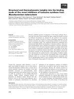
The median follow-up time was 10.5 years (range 3.3 -
26.6 years). The clinical course of this patient cohort is
shown in figure 1. The 5-year and 10-year LC rates for
T1a group were 92% and 91% respectively whereas
those for T1b group were 89% and 87% respectively
(figure 2a).
Complete response (CR) was achieved in 430 (99.3%)
patients, while 3 (0.7%) patients had residual disease/dis-
ease progression at vocal cord(s) at 8 weeks after com-
pletion of RT. Thirty-six (8.3%) among the 430 patients
who achieved CR had their first relapse observed at a
median interval of 15 months after completion of R T
treatment. All first relapses occurred in the laryngeal
Figure 1 Clinical Course. Abbreviations: pts = pati ents; RT =
radiotherapy.
Tong et al. Radiation Oncology 2011, 6:53
/>Page 3 of 9
glottis and none of them occurred in neck LNs or dis-
tant sites.
Salvage surgery after recurrence/residual disease
Of the 39 patients who developed local recurrence or
persistent disease, 36 were salvaged by total laryngect-
omy. Three patients refused or were not considered
medical ly fit for salvage treatment. Seven patients devel-
oped second relapse or progression as regional or dis-
tant metastasis despite total laryngectomy, resulting in
overall ultimate disease failure in 10 patients. This
resulted in an ultimate 10 year LC of 97%. Larynx pre-
servation was achieved in 394 (91%) patients.
Complications
RT was well tolerated by all patients. No patient had grade
III or IV toxicity that necessitated treatment interruption
>3 days, nasogastric tube feeding, intravenous fluid supple-
ment or tracheostomy. There is no clinical or radiological
chondroradionecrosis that warranted laryngectomy.
Factors affecting Local Control
On multivariate analysis, LC was adversely affected by
poorly differentiated histology (Hazard Ratio [HR]: 7.5,
p = 0.035); involvement of AC (HR: 2.34, p = 0.011);
fraction dose size of 2.0 Gy (HR: 2.17, p =0.035)
and tumor BEDGy
15
<65Gy
15
(HR: 3.38, p = 0.017)
[table 2] .
Figure 2b depicts LC rate according to presence of AC
involvement. There was a significant difference in LC
between those with presence of AC involvement and
without AC involvement (86% vs. 95% at 5 years, 85%
vs. 94% at 10 years (p = 0.011). Figure 2c depicts LC
rate according to fraction size. There was a significa nt
difference between the 2.0 Gy group and the 2.5 Gy
group (89% vs. 95% at 5 years; 87% vs. 95% at 10 year, p
= 0.035). Figure 2d depicts LC rate according to tumor
BEDGy
15
. There was a significant difference between the
group with tumor BED < 65 Gy
15
vs. the group with
tumor BED ≥ 65 Gy
15
(90% vs. 96% at 5 years; 88% vs.
96% at 10 years, p = 0.017).
Figure 2 Local contr ol rate according to T sub-stage; AC involvement; Fraction size; tumor BEDGy
15
. a. T sub stage (T1a vs T1b). b. AC
involvement (AC - vs AC +). c. fraction size (2.5 Gy vs 2.0 Gy). d. Tumor BEDGy
15
(<65 Gy
15
vs ≧ 65 Gy
15
). Abbreviations: AC: anterior commissure;
AC–: absence of AC involvement; AC+: presence of AC involvement; BED: biologically effective dose.
Tong et al. Radiation Oncology 2011, 6:53
/>Page 4 of 9
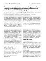
We further categorized patients into 4 groups (A1-A4)
according to involvement of AC and fraction size (cate-
gory- A) or another 4 groups (B1-B4) according to
involvement of AC and tumor BED (category-B), i.e.
(A1) no AC involvement with fraction size of 2.5 Gy,
(A2) no AC involvement with fraction size of 2.0 Gy,
(A3) presence of AC involvement with fraction size of
2.5 Gy, (A4) presence of AC involvement with fraction
size of 2.0 Gy [table 3]; (B1) no AC involvement and
BED Gy
15
≥ 65 Gy
15
,(B2)noACinvolvementandBED
Gy
15
<65Gy
15
, (B3) presence of AC involvement and
BED Gy
15
≧65 Gy
15
, (B4) presence of AC involvement
and BED Gy15 <65 Gy
15
[table 4].
There was a statistically significant difference in LC
rates among 4 groups in category-A: 96% vs. 93% vs.
91% vs. 82% respectively at 5 years; 96% vs. 92% vs. 91%
vs.79% respectively at 10 year (p = 0.002) [figure 3 a].
Again, similar statistically significant difference in LC
rates was also observed among 4 groups in category-B:
96% vs. 92% vs. 89% vs.82% at 5 years; 96% vs. 92% vs.
89% vs. 80% respectively at 10 year p= 0.003 [figure 3b].
Discussion
In western countries, both definitive RT a nd conserva-
tive surgery (endoscopic laser surgery/open organ pre-
serving surgery) are accepted standar d treatment
modalities for stage one GC [16,17]. A survey conducted
in eleven regions/countries in Asia reve aled that in
regions following the ‘ British school’ like Hong Kong
and Singapore, RT alone has remained the primary
treatment modality for early laryngeal cancers [2]. As
laser surgery has become more popular since Stener’s
landmark report [18], it is expected that it will be
increasingly employed in local institutions.
Focusing on primary irradia tion, there is extensive lit-
erature regarding the efficacy and prognostic factors for
RT in early GC [ 3-5,19-23]. All data except one series
[24] was retrospective series. Broadly, prognostic factors
can be divided into patient/tumor- as well as treatment-
related factors. Apart from stage, other patient or tumor
prognostic factors have been reported, including tumor
bulk [4,19,25], bilaterality [4,5], AC involvement (see
below), tumor grade [3,26] and hemoglobin level
Table 2 Univariate and multivariate analysis of factors
affecting local control
Parameters Events/
patients
Uni-variate
analysis
Multivariate
analysis
P value HR (95%
CI)
P
value
Age
<61 18/142
61-70 15/153 0.302 _ _
>70 9/138
Sex
Male 41/413 0.445 _ _
Female 1/20
Sub-stage
T1A 28/324 0.24 _ _
T1B 14/109
Grade
Well diff 9/154 1
Mod diff 29/273 0.0001* 1.91 (1.2-
3.85)
0.035*
Poorly diff 4/6 7.5 (3.42-
15.24)
Hb
< 13.0 6/45 0.367 _ _
≥ 13.0 36/388
AC
No 14/236 0.004* 1 0.011*
Yes 28/197 2.34 (1.21-
4.52)
Field size
(cm
2
)
<30.5 35/215
30.5-35.5 7/165 0.534 _ _
> 35.5 0/53
Dose size
2.0 Gy 32/256 0.021* 2.17 (1.28-
4.18)
0.035*
2.5 Gy 10/177 1
Tumor BED
< 65 (Gy
15
) 29/239 0.025* 3.38 (1.29-
7.83)
0.017*
≥ 65 (Gy
15
) 13/194 1
Tx period
1983-1990 10/115
1991-2000 25/224 0.643 _ _
2001-2005 7/94
Abbreviations: HR = Hazard ratio; CI = confidence interval; Gy = Gray ;
diff = differentiated; AC = anterior commissure; BED = Biologically Effective
Dose;
* = statistically significant
Table 3 Category- A: grouping according to AC
involvement and fraction size
AC- AC+
2.5 Gy/fraction 94 (A1) 83 (A3)
2.0 Gy/fraction 142 (A2) 114 (A4)
Table 4 Category- B: grouping according to AC
involvement and BED
AC- AC+
BED ≥ 65 Gy
15
94 (B1) 100 (B3)
BED < 65 Gy
15
142 (B2) 97 (B4)
Abbreviations: AC = anterior commissure; BED = Biologically Effective Dose
Tong et al. Radiation Oncology 2011, 6:53
/>Page 5 of 9
[5,26,27]. Radiation treatment- related factors included
dose fraction size, total dose, overall treatment time
(OTT) [see below].
The majority of these published data were derived
from patients treated by Cobalt-60 machine or LA gen-
erating 2-4 MV photons [3-5,21,26]. In many RT cen-
ters, these therapy units have been decommissioned.
With a general shift from the use of Cobalt-60 to LA
treatment units, it is anticipated that 6 MV photon
beams generated by LA will become the prevailing
workhorse for treatment in clinical practice [28].
Table 5 showed published results for T1N0 GC treated
with 6 MV photons in the recent two decades.
The impact of AC involvement on the RT treatment
outcome of early GC is still controversial. The so called
AC or Broyle’s tendon is the insertion of vocalis tendon
into thyroid cartilage in the area o f AC. This is consid-
ered as a weak point for tumor spread because in this
area, there is no thyroid cartilage perichondrium to
resist tumor spread. Although some data suggested that
AC involvement portended a worse prognosis, it has not
been included in the staging system.
In the recent two decades, many authors identified AC
involvement as one of the independent poor prognostic
factors in LC for T1N0 GC treated by prima ry RT
[4,21,29].InarecentreportbySmeeetal.[30],itwas
found that AC involvemen t was one of the independent
poor prognostic factors for LC as well as cause specific
survival. One explanation is related to the possibility of
‘ understaging’ without CT scan staging, as patients
might have a larger tumor burden anteriorly, and in
some cases unrecognized subglottic extension [31]. In
$%
Figure 3 Local control rate a ccordin g fraction size, tu mor BED 15, AC involvement. a. fraction size, together with AC involvement. b .
tumor BED G15, together with AC involvement. Abbreviations: AC: anterior commissure tumor BED Gy
15
: tumor biologically effective dose N:
patients numbers AC– : absence of AC involvement AC+: presence of AC involvement .
Table 5 Reports in literature on results of T1N0 glottic cancer treated with 6 MV photons
Author year [ref] Patients no Total Dose (Gy) Dose size (Gy) Local Control (5 year)%
Akine et al. 1991 [7] 151 62.5-67.5 2.0-2.4 89
Fein et al. 1996 [27] 43 66 2 95
Foote et al. 1996 [6] 27 63 2.25 100
Lee et al. 2001 [28] 86 66 2 T1a: 82
T1b: 76
Gowda et al. 2003 [36] 100 50-52.5 3.12-3.28 T1a: 93
T1b: 89
Franchin et al. 2003 [20] 323 63-65.2 2.25 T1: 90
Sjögren et al. 2009 [37] 59 60 2.0-2.8 T1a: 87
T1b: 85
current study 433 57.5-66 2.0-2.5 T1a: 92
T1b: 89
Tong et al. Radiation Oncology 2011, 6:53
/>Page 6 of 9
our patient cohort, since 95% of patients had evaluation
by CT scan, the issue of under-staging should be
minimal.
Another probable reason is the theoretical risk of
under-dosage at the air- tissue interface with the depth-
dose characteristics of 6 MV photons compared with
those of Cobalt-60 beam. This is related to inadequate
tissuepresentattheareaofACwheretheneckisthin,
as well as lack of electronic equilibrium at the air-tissue
interface which might be more pronounced with high-
energy photons treated with small field size [32,33].
Hence, poorer coverage of the prescribed dose t o the
tumor may occur in early glottic tumors with AC invol-
vement, particularly whe n treated with 6 MV photons.
Sombeck et al. [34] performed a dosimetric evaluation
comparing 6MV photons with Cobalt-60 beam. They
revealed that there was no significant difference in the
dose received at any point along the voca l cords. On the
other hand, a recent study by Spirydovich [35] demon-
strated a significant under- dosage occurring at the a ir-
tissue interface of larynx trea ted by 6 MV photons. The
authors performed Monte Carlo dose calculation to CT-
based mathematical neck. They identified that at least
5% of a hypothetical tumor of 3.5 cm
3
received less than
86% o f the maximum tumor dose in neck that contains
air cavities in comparison to 91% of the maximum
tumor dose in the homogeneous neck.
However, some other major reports did not reveal the
impact of AC on LC of early glottic cancer [3,5,36,37].
With regard to the impact of dose fraction size for
early glottic disease, there is little controversy that infer-
ior LC is associated with fraction size < 2.0 Gy when
patients are treated once daily, 5 days per week [38,39].
Among the reports published in the literature, the
common contemporary irradiation schedules for T1N0
GC included: 66 Gy in 33 fractions in 6.5 weeks, 63 Gy
in 28 fractions in 5.5 weeks, and 60 Gy in 25 fractions
in 5 weeks [17,40]. In fact, a prospective randomized
study from Yamazaki et al. [24] demonstrated a statisti-
call y superior 5-year LC rate of 92% for patients treated
with fraction size of 2.25 Gy compared with 77% for
those treated with 2.0 Gy.
Besides, many reports have shown that prolonging
OTT in T1N0 GC has an adverse impact on LC and
dose compensation is needed to maintain the tumor
control probability. Indeed, several authors have high-
lighted the complex inter- relationship among the vari-
ables of total dose, fraction size and OTT [41,42].
Fowler [43] commented that according to radiobiolo-
gical principles, ev en if there would be a positive effect
of increasing total dose or fraction size on LC, and a
strong negative effect of treatment prolongation, these
effects become minimal where the LC was already at a
very high level, because of the plateau of the slope o f
the sigmoid- shaped dose-response curve above 70 or
80%. This theoretical postulation has also been verified
by observations reported. Fein et al.[27] and Le et al.
[21] did not observe a relationship between fraction size
and LC. Although there was a trend for higher LC in
patients treated with fraction size of ~2.25 Gy when
compared to smaller fraction size, the difference did not
reach statistical significance. The authors attributed the
lackofdifferencetothelowrecurrencerateinT1
lesions, thus under- powering the studies to demon-
strate a significant relationship between fraction size
and LC.
The debate over these discrepancies was rebuffed after
the impact of shortening of OTT in LC of H&N cancers
was confirmed in randomized trials with accelerated
schedules. Both the Danish Head and Neck Cancer
Study Group study (DAHANCA 6 & 7) [44] and the
International A tomic Energy Agency (IAEA- ACC) trial
[45] delivered s ix fractions per week but keeping same
tot al dose, enabled a treatment of 66 Gy in 33 fractions
to be given in 8 days less than the conventional sche-
dule. They revealed a 10-12% improvement in LC of
H&N cancers (especially for early laryngeal cancer sub-
set) upon shortened OTT. It appeared that by shorten-
ing the OTT, treatment outcome is improved as
accelerated repopulation of tumor clonogens would be
reduced. But these accelerated schedules are also shown
to have more acute radiation toxicity in terms of severe
skin reactions, confluent mucositis necessitating tube
feeding.
In evaluating the efficacy of various fractionation sche-
dules, we opted to test the impact of tumor BEDGy
15
which incorporates the components of fraction size,
OTT and total dose. Our analysis shows that tumor
BED ≥ 65 Gy
15
is associated with better LC. Table 6
illustrated the common radiation schedules in which
fraction size is > 2.0 Gy, the resulting tumor BEDGy
15
would be > 65 Gy
15
but the BEDs for both early mucosa
and late normal tissues are well below the correspond-
ing dose constraints for complications [aim at 59-63
Gy
10
for acute mucosa; < 117 Gy
3
for late normal tissue
respectively] [46].
Since the treatment field size for T1N0 GC is small, it
permits slight hypofractionated schedule without caus-
ing excessive acute radiation toxicity. Shortened OTT
overcomes the accelerated repopulation of tumor
clonogens.
This also supports the cu rrent contemporary practice
of fraction dose size > 2.0 Gy (i.e. 2.25 Gy) for treatment
of T1N0 GC by other centers [3,6,20,21,24,37]
To the best of our knowledge, our report is the largest
study on RT outcomes in T1N0 GC primarily treated
with 6 MV photons. As the treatment of choice for
early GC in our institution or Hong Kong at large has
Tong et al. Radiation Oncology 2011, 6:53
/>Page 7 of 9
been and in the near future will still be RT alone [2],
this represents a relatively unselected cohort of patients.
While this study spans a considerable period of time,
the clinical evaluation and tr eatment techniques have
been consistent over the years, thus allowing a valid
analysis to be performed. Our results demonstrate that
the LC rate with primary RT with 6 MV photons is
comparable and agrees with other reports of “unremark-
able” treatment outcome difference when comparing
Cobalt-60 beam and 6 MV photons [3,5-7,27].
However, we observe that AC involvement is associated
with a poor LC rate
We suspect that the issue of ‘cold spot’ is more apparent
at the AC region, especially when treated with 6MV
photons. Certainly, further dosimetric evaluation is
needed to validate this suspicion. While involvement of
AC is an adverse progn ostic factor, we have shown that
its negative impact can be overcome by delivering a
higher tumor BED (≧ 65 Gy
15
). In order to achieve this
tumor BED level in conventional schedule of five daily
fractionation each week, we rec ommend that fraction
size > 2.0 Gy should be utilized. In fact, modest hypo-
fractionati on is safe and effective for T1N0 GC in terms
of both LC and morbidity. Having a shorter OTT is
more convenient for patients and is also more cost-
effective for RT facility implication.
Nevertheless, the results need to be interpreted with
caution, because the current report was a retrospective,
single institution study and therefore subjected to biases.
For example, we did not have volume measurements on
tumor, which has been shown in other reports as one of
the important prognostic factors in LC [4,19,25]. In fact,
AC involvement may reflect “tumor bulk” and thus may
represent a surrogate marker for tumor volume . We
suggest the degree of AC involvement should be further
defined to better evaluate and confirm its significance in
outcome prognostication. We also agree with some
authors that the degree of AC involvement should be
incorporated into the new UICC staging system for bet-
ter comparison of results among various studies [ 47].
Besides, modification of the RT treatment technique like
adding anterior field/anterior oblique field can be con-
sidered to combat under-dosage at AC [3,20].
Conclusions
Our data concur with other published result about the
efficacy of RT with 6 MV photons for T1N0 GC. While
involvement of AC is associated with poor LC rate, its
negative impact could be overcome by delivering a
higher tumor BED through using fraction size of >2.0
Gy. We recommend that fraction size > 2.0 Gy should
be utilized, for radiation schedules with five daily frac-
tions each week.
Authors’ contributions
CCT participated in the study’s design and coordination, performed
acquisition of data and drafted the manuscript. KHA and FYC participated in
data analysis and revised the manuscript. RKCN and SMC participated in
study’s design and revised the manuscript. JSKA, YTF and SCKL revised
manuscript critically for important intellectual content. All author s read and
approved the final manuscript.
Competing interests
The authors declare that they have no competing interests.
Received: 13 February 2011 Accepted: 21 May 2011
Published: 21 May 2011
References
1. Hospital Authority: Hong Kong Cancer Registry web site.[.
org.hk/cancereg/e_stat.asp].
2. Wei W: Management of early carcinoma of the larynx: the Asian
perspective. ENT News 2000, 9:18-19.
3. Mendenhall WM, Amdur RJ, Morris CG, et al: T1-T2N0 Squamous Cell
Carcinoma of the Glottic Larynx Treated With Radiation Therapy. J Clin
Oncol 2001, 19:4029-4036.
4. Cellai E, Frata P, Magrini SM, et al: Radical radiotherapy for early glottic
cancer: Results in a series of 1087 patients from two Italian radiation
oncology centers. I. The case of T1N0 disease. International Journal of
Radiation Oncology Biology Physics 2005, 63:1378-1386.
Table 6 Calculated tumor BED (Gy
15
), acute mucosal BED (Gy
10
) and late normal tissue BED (Gy
3
) for common
radiation schedules
Dose
size
(Gy)
Fraction
number
Total
dose
(Gy)
Overall treatment
time (OTT) in days
Tumor
BED
(Gy
15
)
Acute Mucosal BED (Gy
10
)
(aim 59-63 Gy
10
) [46]
Late normal BED (Gy
3
)
(aim <117 Gy
3
) [46]
references
2.0 33 66 45 64.60 49.1 110 [22,25,27] &
current study
2.25 28 63 38 66.45 52.62 110.2 [3,5,20,24,37]
2.5 23 57.5 31 65.28 52.87 105.4 current study
BED = biologically effective dose
= total dose (1 + fraction size/[a/b]) - log
e
2(OTT -Tk)/aTp [14]
Assume: 1. a/b = 15 for laryngeal cancer [12]
2. Tk: kick off time (Tk) for tumor repopulation = 28 days [13]
3. Tp = average cell number doubling time during continuing radiation, 3 days for tumor [14]
4. Alpha (a) = [coefficient of non-repairable injury, log cell kill (exponentially-based logs) per gray of dose]
= 0.35 Gy
-1
(14)]
Tong et al. Radiation Oncology 2011, 6:53
/>Page 8 of 9
5. Warde P, O’Sullivan B, Bristow RG, et al: T1/T2 Glottic Cancer Managed by
External Beam Radiotherapy: The Influence of Pretreatment Hemoglobin
on Local Control. International Journal of Radiation Oncology, Biology,
Physics 1998, 41:347-353.
6. Foote RL, Grado GL, Buskirk SJ, et al: Radiation therapy for glottic cancer
using 6-MV photons. Cancer 1996, 77:381-386.
7. Akine Y, Tokita N, Ogino T, et al: Radiotherapy of T1 glottic cancer with 6
MeV X rays. International Journal of Radiation Oncology Biology Physics 1991,
20:1215-1218.
8. Izuno I, Sone S, Oguchi M, et al: Treatment of early vocal cord carcinoma
with 60 Co gamma rays, 8/10 MV X-rays, or 4 MV X-rays – are the
results different ? Acta Oncol 1990, 29:637-639.
9. Devineni VR, King K, Perez C: Early glottic carcinoma treated with
radiotherapy: impact of treatment energy on sucess rate. International
Journal of Radiation Oncology Biology Physics 1992, 24:106.
10. Sobin LH, Wittekind CH: UICC TNM Classification of malignant tumours.
New York.: John Wiley & Sons;, 6 2002.
11. Fowler JF: The linear-quadratic formula and progress in fractionated
radiotherapy. British Journal of Radiology 1989, 62:679-694.
12. Robertson AG, Robertson C, Boyle P, et al: The effect of differing
radiotherapyeutic schedules on the response of glottic carcinoma of the
larynx. Eur J Cancer 1993, 29A:501-510.
13. Skladowski K, Law MG, Maciejewski B, et al: Planned and unplanned gaps
in radiotherapy: The importance of gap position and gap duration.
Radiotherapy and Oncology 1994, 30:109-120.
14. Fowler JF: 21 years of Biologically Effective Dose. Br J Radiol 2010,
83:554-568.
15. Trotti A, Colevas AD, Setser A, et al: CTCAE v3.0: development of a
comprehensive grading system for the adverse effects of cancer
treatment. Seminars in Radiation Oncology 2003, 13:176-181.
16. Pfister DG, Laurie SA, Weinstein GS, et al: American Society of Clinical
Oncology Clinical Practice Guideline for the Use of Larynx-Preservation
Strategies in the Treatment of Laryngeal Cancer. J Clin Oncol 2006,
24
:3693-3704.
17. Kaanders JH, Hordijk GJ: Carcinoma of the larynx: the Dutch national
guideline for diagnostics, treatment, supportive care and rehabilitation.
Radiotherapy and Oncology 2002, 63:299-307.
18. Steiner W: Results of curative laser microsurgery of laryngeal carcinomas.
American Journal of Otolaryngology 1993, 14:116-121.
19. Jing J, Zhongxing L, Li G, et al: Analysis of prognostic factors for T1N0M0
glottic cancer treated with definitive radiotherapy alone: experience of
the cancer hospital of Peking Union Medical College and the Chinese
Academy Of Medical Sciences. International Journal of Radiation Oncology,
Biology, Physics 2002, 54:471-478.
20. Franchin G, Minatel E, Gobitti C, et al: Radiotherapy for patients with
early-stage glottic carcinoma. Cancer 2003, 98:765-772.
21. Le Q-TX, Fu KK, Kroll S, et al: Influence of fraction size, total dose, and
overall time on local control of T1-T2 glottic carcinoma. International
Journal of Radiation Oncology Biology Physics 1997, 39:115-126.
22. Yu E, Shenouda G, Beaudet MP, et al: Impact of radiation therapy fraction
size on local control of early glottic carcinoma. International Journal of
Radiation Oncology Biology Physics 1997, 37:587-591.
23. Johansen LV, Grau C, Overgaard J: Glottic carcinoma - patterns of failure
and salvage treatment after curative radiotherapy in 861 consecutive
patients. Radiotherapy and Oncology 2002, 63:257-267.
24. Yamazaki H, Nishiyama K, Tanaka E, et al: Radiotherapy for early glottic
carcinoma (T1N0M0): Results of prospective randomized study of
radiation fraction size and overall treatment time. International Journal of
Radiation Oncology Biology Physics 2006, 64:77-82.
25. Reddy SP, Mohideen N, Marra S, et al: Effect of tumor bulk on local
control and survival of patients with T1 glottic cancer. Radiotherapy and
oncology 1998, 47:161-166.
26. Johansen LV, Grau C, Overgaard J: Laryngeal Carcinoma - multivariate
analysis of prognostic factors in 1252 consecutive patients treated with
primary radiotherapy. Acta Oncologica 2003, 42:771-778.
27. Fein DA, Lee WR, Hanlon AL, et al: Do overall treatment time, field size,
and treatment energy influence local control of T1-T2 squamous cell
carcinomas of the glottic larynx? International Journal of Radiation
Oncology Biology Physics 1996, 34:823-831.
28. Lee JH, Machtay M, McKenna MG, et al:
Radiotherapy with 6-megavolt
photons for early glottic carcinoma: Potential impact of extension to the
posterior vocal cord. American Journal of Otolaryngology 2001, 22:43-54.
29. Marshak G, Brenner B, Shvero J, et al: Prognostic factors for local control
of early glottic cancer: the Rabin Medical Center retrospective study on
207 patients. International Journal of Radiation Oncology Biology Physics
1999, 43:1009-1013.
30. Smee RI, Meagher NS, Williams JR, et al: Role of radiotherapy in early
glottic carcinoma. Head & Neck 2010, 32:850-859.
31. Sessions DG, Ogura JH, Fried MP: The anterior commissure in glottic
carcinoma. Laryngoscope 1975, 85:1624-1632.
32. Epp ER, Boyer AL, Doppke KP: Underdosing of lesions resulting from lack
of electronic equilibrium in upper respiratory air cavities irradiated by
10 MV X- ray beams. International Journal of Radiation Oncology Biology
Physics 1977, 2:613-619.
33. Klein EE, Chin LM, Rice RK, et al: The influence of air cavities on interface
doses for photon beams. International Journal of Radiation Oncology
Biology Physics 1993, 27:419-427.
34. Sombeck MD, Kalbaugh KJ, Mendenhall WM, et al: Radiotherapy for early
vocal cord cancer: A dosimetric analysis of CO-60 versus 6 MV photons.
Head & Neck 1996, 18:167-173.
35. Spirydovich S, Papiez L, Moskvin V, et al: Evaluation of underdosage in the
external photon beam radiotherapy of glottic carcinoma: Monte Carlo
study. Radiotherapy and Oncology 2006, 78:159-164.
36. Gowda RV, Henk JM, Mais KL, et al: Three weeks radiotherapy for T1
glottic cancer: the Christie and Royal Marsden Hospital Experience.
Radiotherapy and Oncology 2003, 68:105-111.
37. Sjögren EV, Wiggenraad RG, Le Cessie S, et al: Outcome of radiotherapy in
T1 glottic carcinoma: a population-based study. European Archives of Oto-
Rhino-Laryngology 2009, 266:735-744.
38. Kim RY, Marks ME, Salter MM: Early-stage glottic cancer: importance of
dose fractionation in radiation therapy. Radiology 1992, 182:273-275.
39. Mendenhall WM, Parsons JT, Million RR, et al: T1-T2 squamous cell
carcinoma of the glottic larynx treated with radiation therapy:
relationship of dose-fractionation factors to local control and
complications. International Journal of Radiation Oncology Biology Physics
1988, 15:1267-1273.
40. NCCN Practice Guidelines in Oncology- v.2.2010:[].
41. Skladowski K, Tarnawski R, Maciejewski B, et al: Clinical radiobiology of
glottic T1 squamous cell carcinoma. International Journal of Radiation
Oncology Biology Physics 1999, 43:101-106.
42. van der Voet JC, Keus RB, Hart AA, et al: The impact of treatment time
and smoking on local control and complications in T1 glottic cancer.
International Journal of Radiation Oncology Biology Physics 1998, 42:247-255.
43. Fowler JF: Fractionation and glottic carcinoma. International Journal of
Radiation Oncology Biology Physics 1997, 39:1-2.
44. Overgaard J, Hansen HS, Specht L, et al: Five compared with six fractions
per week of conventional radiotherapy of squamous-cell carcinoma of
head and neck: DAHANCA 6&7 randomised controlled trial. The Lancet
2003, 362:933-940.
45. Overgaard J, Mohanti BK, Begum N, et al: Five versus six fractions of
radiotherapy per week for squamous-cell carcinoma of the head and
neck (IAEA-ACC study): a randomised, multicentre trial. The Lancet
Oncology 2010, 11:553-560.
46. Fowler JF, Harari PM, Leborgne F, et al: Acute radiation reactions in oral
and pharyngeal mucosa: tolerable levels in altered fractionation
schedules. Radiotherapy and Oncology 2003, 69:161-168.
47. Rucci L, Gammarota L, Gallo O: Carcinoma of the anterior commissure of
the larynx. II. Proposal of a new staging system. The Annals of Otology
Rhinol Laryngol 1996, 105:391-396.
doi:10.1186/1748-717X-6-53
Cite this article as: Tong et al.: Impact and relationship of anterior
commissure and time-dose factor on the local control of T1N0 glottic
cancer treated by 6 MV photons. Radiation Oncology 2011 6:53.
Tong et al. Radiation Oncology 2011, 6:53
/>Page 9 of 9