Báo cáo khoa học: "Constitutive gene expression profile segregates toxicity in locally advanced breast cancer patients treated with high-dose hyperfractionated radical radiotherapy" pot
Bạn đang xem bản rút gọn của tài liệu. Xem và tải ngay bản đầy đủ của tài liệu tại đây (620.58 KB, 7 trang )
BioMed Central
Page 1 of 7
(page number not for citation purposes)
Radiation Oncology
Open Access
Short report
Constitutive gene expression profile segregates toxicity in locally
advanced breast cancer patients treated with high-dose
hyperfractionated radical radiotherapy
Luis Alberto Henríquez Hernández*
1,2,3
, Pedro Carlos Lara
2,4
,
Beatriz Pinar
2,4
, Elisa Bordón
2
, Carlos Rodríguez Gallego
2,5
, Cristina Bilbao
2
,
Leandro Fernández Pérez
2,3
and Amílcar Flores Morales
6
Address:
1
Canary Foundation of Investigation and Health (FUNCIS), Spain,
2
Canary Institute for Cancer Research (ICIC), Spain,
3
Clinic Sciences
Department of Las Palmas de Gran Canaria University (ULPGC), Spain,
4
Radiation Oncology Department, Hospital Universitario de Gran Canaria
Dr Negrín, Spain,
5
Inmunology Department, Hospital Universitario de Gran Canaria Dr Negrín, Spain and
6
Molecular Endocrinology Group,
Center for Molecular Medicine, Karolinska Intitute, Stockholm, Sweden
Email: Luis Alberto Henríquez Hernández* - ; Pedro Carlos Lara - ;
Beatriz Pinar - ; Elisa Bordón - ; Carlos Rodríguez Gallego - ;
Cristina Bilbao - ; Leandro Fernández Pérez - ; Amílcar Flores Morales -
* Corresponding author
Abstract
Breast cancer patients show a wide variation in normal tissue reactions after radiotherapy. The
individual sensitivity to x-rays limits the efficiency of the therapy. Prediction of individual sensitivity
to radiotherapy could help to select the radiation protocol and to improve treatment results. The
aim of this study was to assess the relationship between gene expression profiles of ex vivo un-
irradiated and irradiated lymphocytes and the development of toxicity due to high-dose
hyperfractionated radiotherapy in patients with locally advanced breast cancer. Raw data from
microarray experiments were uploaded to the Gene Expression Omnibus Database http://
www.ncbi.nlm.nih.gov/geo/ (GEO accession GSE15341). We obtained a small group of 81 genes
significantly regulated by radiotherapy, lumped in 50 relevant pathways. Using ANOVA and t-test
statistical tools we found 20 and 26 constitutive genes (0 Gy) that segregate patients with and
without acute and late toxicity, respectively. Non-supervised hierarchical clustering was used for
the visualization of results. Six and 9 pathways were significantly regulated respectively. Concerning
to irradiated lymphocytes (2 Gy), we founded 29 genes that separate patients with acute toxicity
and without it. Those genes were gathered in 4 significant pathways. We could not identify a set of
genes that segregates patients with and without late toxicity. In conclusion, we have found an
association between the constitutive gene expression profile of peripheral blood lymphocytes and
the development of acute and late toxicity in consecutive, unselected patients. These observations
suggest the possibility of predicting normal tissue response to irradiation in high-dose non-
conventional radiation therapy regimens. Prospective studies with higher number of patients are
needed to validate these preliminary results.
Published: 4 June 2009
Radiation Oncology 2009, 4:17 doi:10.1186/1748-717X-4-17
Received: 10 March 2009
Accepted: 4 June 2009
This article is available from: />© 2009 Henríquez Hernández et al; licensee BioMed Central Ltd.
This is an Open Access article distributed under the terms of the Creative Commons Attribution License ( />),
which permits unrestricted use, distribution, and reproduction in any medium, provided the original work is properly cited.
Radiation Oncology 2009, 4:17 />Page 2 of 7
(page number not for citation purposes)
Introduction
Radiation is an effective therapy in patients with local
advanced breast cancer (LABC) [1,2]. Tumor control by
radiotherapy (RT) requires the use of maximum dose that
can be delivered while maintaining a tolerance risk of nor-
mal tissue toxicity [3]. Better local control outcomes with
an acceptable toxicity have been obtained by using high
total doses radiation administered in two small fractions
per day compared with standard RT protocols [4]. Some
patients treated with RT will develop early or late reactions
limiting the efficacy of RT. Knowledge of individual varia-
tions of normal tissue toxicities determining tolerance
would be of great value in patients treated with high-dose
radiation protocol [5]. Microarray technology is a high
throughput method that allows large scale genomic stud-
ies. Because intrinsic radiosensitivity is genetically deter-
mined, different cells from the patient can be used to
measure sensitivity to radiation [6]. Few studies have been
published with regard to radiation induced toxicity and
microarrays [2,7-10]. Patients were previously selected
according to the clinical toxicity observed and only three
publications included breast cancer patients [see Addi-
tional file 1].
The aim of this study was to assess the relation of the gene
expression profile from un-irradiated and irradiated lym-
phocytes and the development of toxicity due to RT in
patients with LABC.
Patients and methods
Twelve consecutive patients treated between 1991 and
1997 by a hyperfractionated dose-escalation radiation
therapy schedule at the Hospital Dr. Negrín suffering from
LABC were prospectively recruited and inform consent
was given. The study was approved by the Research and
Ethics Committee of our institution. Blood samples were
extracted and tested during 2005 and follow up was
closed on December 2008. Characteristics of the patients
are shown in Table 1. Early toxicity was evaluated during
and at the end of RT, and late toxicity was evaluated at 6-
month follow-up examination. The RTOG morbidity
score system was used to classify the toxicity of patients
into three levels: grades 1, 2 and 3–4 (Table 2). All
patients were referred to recieve 60 Gy to the whole breast
over a period of 5 weeks in two daily fractions of 1.2 Gy
separated by at least 6 h on 5 days each week, and fol-
lowed by a boost of 21.6 Gy to a total dose of 81.6 Gy.
Culture of lymphocytes and radiation protocol details
were previously reported [11]. Twenty four independent
hybridizations were performed to compare lymphocytes
from twelve patients, before and after 2 Gy irradiation,
against a human RNA universal control. A microarray
containing 35.327 human 70-mer oligo probe sets, pro-
duced at the SweGene DNA Microarray Resource Center
(Lund University, Sweden) was used. Array scanning,
image analysis and data normalization were performed as
previously described [12,13]. Identification of differen-
tially-expressed genes was performed using the SAM (Sig-
nificance Analysis for Microarrays) statistical technique
[14]. A q value was assigned for each of the detectable
genes in the array measuring the lowest false discovery
rate (FDR). Genes with a FDR of less than 10% were con-
sidered to present significant differential expression. Thus,
we studied gene expression profile of lymphocytes treated
with 0 and 2 Gy separately. To explore genes modulated
by radiation, we also compared gene expression profiles
of lymphocytes treated with 0 versus 2 Gy. T-test and
ANOVA test [15,16] were used to compare the set of genes
significantly regulated, in un-irradiated and in 2 Gy-irradi-
ated lymphocytes, with toxicity. Non-supervised hierar-
chical clustering [17] was made using MultiExperiment
Viewer (The Institute for Genomic Research, http://
www.tigr.org/tdb/microarray/). A genetic signature that
could separate toxicity and non toxicity in a constitutive
and in a modulated-by-radiation way was performed (Fig-
ure 1). Functional classification and pathway analysis of
expressed genes were performed by using the web-based
tools Onto-Express (OE) and Pathway-Express (PE)
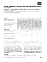
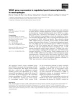
Experimental designFigure 1
Experimental design. RNA from lymphocytes treated
with 0 and 2 Gy dose of radiation were compared against a
human universal RNA. SAM analyses were performed to dis-
close significant regulated genes in these two ways. In order
to explore genes modulated by radiation, a two-class paired
test was performed using SAM. To discriminate genes that
could be significantly associated with RT toxicity, non-super-
vised hierarchical clustering, in MeV, was used to visualize
the whole set of significant genes modulated before and after
X-ray exposure in patients with and without acute/late toxic-
ity.
Radiation Oncology 2009, 4:17 />Page 3 of 7
(page number not for citation purposes)
(Intelligence Systems and Bioinformatics Laboratory,
Wayne University, Detroit, MI. http://vor
tex.cs.wayne.edu) [18,19]. OE classify genes in order to
biological process (BP), cellular component and molecu-
lar function, and it is able to estimate statistical differences
between different gene ontology terms [20]. PE is based
on a novel method that uses a system biology approach
that includes important biological factors that describes
how these genes interacts and the type of signaling inter-
actions between them [21,22].
Results
Comparison of gene expression profiles from 0 Gy and 2
Gy-treated lymphocytes, using two-class paired test in
SAM program, identified a total of 81 genes significantly
regulated by RT [see Additional file 2]. We could not clus-
ter these genes in order to segregate patients with acute or
late toxicity. PE was used to explore biological pathways
significantly regulated by radiation. Fifty seven genes were
mapped and PE identified 50 pathways significantly regu-
lated (p < 0.01). Among the RT modulated pathways there
were cell cycle, nucleotide excision repair, DNA replica-
tion, mismatch repair; MAPK, erbB, and VEGF signaling,
ubiquitination mediated proteolysis, notch and Wnt [see
Additional file 3]. A functional classification of 81 regu-
lated genes was made using OE. Forty-five genes were clas-
sified according to the BP and several processes were
modified by RT [see Additional file 4].
SAM analysis from un-irradiated lymphocytes revealed
7391 constitutive regulated genes. ANOVA test in MeV
identified 20 genes that segregated patients with grade 1,
from grade 2 and grade 3–4 acute toxicity (p < 0.01) (Fig-
ure 2). PE identified 6 pathways significantly regulated (p
< 0.01): protein export, regulation of autophagy, vibrio
cholerae infection, phosphatidylinositol signaling system,
focal adhesion and regulation of actin cytoskeleton [see
Additional file 5]. OE classified 14 genes according to the
BP. Processes as chromatin remodeling, regulation of
endothelial cell proliferation, oxidation reduction and
cellular respiration were constitutively modulated (p <
0.05) [see Additional file 6]. The same strategy was fol-
lowed for late toxicity. T-test identified 26 genes that con-
stitutively segregated patients who suffered severe late
toxicity from patients who did not (p < 0.01) (Figure 2).
PE identified 9 pathways significantly regulated (p <
0.01): regulation of actin cytoskeleton, MAPK signaling,
epithelial cell signaling in helicobacter, Erb B signaling
pathway, renal cell carcinoma, natural killer cell mediated
cytotoxicity, T cell receptor signaling, axon quidance and
focal adhesion [see Additional file 5]. The role of PAK1
(p21-Cdc42/Rac)-activated kinase 1 must be highlighted
since it was involved in all the 9 pathways. OE scored 13
genes according to the BP. Processes significantly regu-
lated (p < 0.05) were: lipid, cholesterol and sterol-biosyn-
thetic processes, cytoskeleton organization and
biogenesis, positive regulation of gene specific transcrip-
tion, hair follicle development, ER-nuclear signaling path-
way, positive regulation of JNK activity and others [see
Additional file 7].
Lymphocytes from patients were also irradiated at 2 Gy
dose. SAM identified 7393 genes significantly regulated.
ANOVA test (p < 0.01) identified 29 genes that separated
patients with grade 1, from grade 2 and grade 3–4 acute
toxicity (Figure 3). We did not observe common genes
between this set of genes and those corresponding to un-
Table 1: Characteristics of the patients included in the study.
Age, menopause status, characteristics of the tumor and
systemic treatment were added.
Characteristic n %
Age
<60 years 5 41.7
≥ 60 years 7 58.3
Menopause
Premenopausal 3 25.0
Postmenopausal 9 75.0
Tumor type
Inflammatory 5 41.7
Non-inflammatory 7 58.3
Tumor size (T)
T3 1 8.3
T4a 1 8.3
T4b 5 41.7
T4c 0 0
T4d 5 41.7
Nodes (N)
N0 7 58.3
N1 3 25.0
N2 2 16.7
Metastasis (M)
M0 12 100
M1 0 0
Systemic treatment
Chemotherapy 1 8.3
Hormonal therapy 1 8.3
Chemotherapy-hormonal therapy 10 83.4
Table 2: Grade of acute and late toxicity of patients included in
the study.
Patient Code Age Acute Toxicity Late Toxicity
1 01 34 Grade 1 Grade 2
2 02 60 Grade 1 Grade 3
3 03 47 Grade 1 Grade 2
4 04 68 Grade 2 Grade 2
5 05 67 Grade 2 Grade 3
6 06 70 Grade 2 Grade 2
7 07 63 Grade 2 Grade 3
8 08 72 Grade 2 Grade 3
9 09 57 Grade 3 Grade 3
10 10 64 Grade 3 Grade 3
11 11 47 Grade 3 Grade 2
12 12 53 Grade 4 Grade 4
Age is shown.
Radiation Oncology 2009, 4:17 />Page 4 of 7
(page number not for citation purposes)
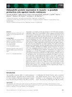
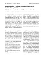
Non-supervised hierarchical clustering of constituve genes regulated in un-irradiated lymphocytesFigure 2
Non-supervised hierarchical clustering of constituve genes regulated in un-irradiated lymphocytes. Clustering
used Euclidean distance correlation and average linkage, and was processed and displayed with MultiExperiment Viewer http://
www.tigr.org/tdb/microarray/. Upper panel shows a 20 gene set that segregated patients with different grade of acute toxicity
(First three patients, grade 1; next five patients, grade 2; last four patients, grades 3–4) ANOVA test, p < 0.01. Lower panel
shows a 26 gene set that segregated patients with different grade of late toxicity (First five patients, grade 2; last seven patients,
grades 3–4) T-test, p < 0.01. The dendogram to the left of the heat map shows clustering of the genes. Accession number, gene
symbol, gene description and fold change were added. Colour boxes indicate the biological process of each gene.
Radiation Oncology 2009, 4:17 />Page 5 of 7
(page number not for citation purposes)
irradiated lymphocytes (constitutive genes). PE identified
4 significantly regulated pathways (p < 0.01): phosphati-
dylinositol signaling system, regulation of actin cytoskel-
eton, cell cycle and TGF-beta signaling pathway [see
Additional file 5]. OE scored 15 genes according to BP
with some processes also significantly regulated [see Addi-
tional file 8]. We could not obtain a consistent set of genes
able to separate patients with regard to late toxicity in irra-
diated lymphocytes (Table 3).
Discussion
Constitutive gene expression pattern from un-irradiated
lymphocytes can segregates LABC patients with acute and
late toxicity from patients without toxicity after hyperfrac-
tionated radiation therapy treatment. Using 2 Gy irradi-
ated lymphocytes from the same patients we could only
observe association related to acute toxicity. Few series
were published to explore the relation of radiation
induced toxicity and microarray, and only three were
referred to breast cancer [7,9,10]. The paper published by
Svensson et al. is similar to the present work in relation to
the experimental design, but was assessed in prostate can-
cer patients [2]. Recently, Rødningen et al. published two
relevant papers [10,23]. Our results were not similar
related to genes involved in late toxicity. Anyhow, we
coincided in relation to some BP. Differences in cell type,
microarray platform, experimental design, RT protocol
and statistical strategy could explain those differences.
Compared with previously available studies, this is the
first work in which: i) patients were consecutive and non-
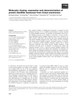
Non-supervised hierarchical clustering of genes regulated in irradiated lymphocytes (2 Gy)Figure 3
Non-supervised hierarchical clustering of genes regulated in irradiated lymphocytes (2 Gy). Clustering used Eucli-
dean distance correlation and average linkage, and was processed and displayed with MultiExperiment Viewer http://
www.tigr.org/tdb/microarray/. A 29 gene set segregated patients with different grades of acute toxicity (First three patients,
grade 1; next five patients, grade 2; last four patients, grades 3–4) ANOVA test, p < 0.01. The dendogram to the left of the heat
map shows clustering of the genes. Accession number, gene symbol, gene description and fold change were added. Colour
boxes indicate the biological process of each gene.
Radiation Oncology 2009, 4:17 />Page 6 of 7
(page number not for citation purposes)
previously selected, ii) patients were treated with high-
dose radiation protocol with altered fractionation, iii) the
complete human genome was analyzed and iv) compara-
tive studies of constitutive gene expression profiles of
LABC patients and toxicity were made.
Pak1 seems to have an important role in late toxicity in
our study. Pak1 overexpression is related to apoptosis-
resistance in normal and tumour cells [24]. An appropri-
ate apoptotic response seems to protect normal tissue
against radiation late toxicity [25]. Therefore, over-expres-
sion of Pak1 observed in our patients would be related to
resistance to late toxicity. The role of PAK1 in late toxicity
should be explored.
This long term study makes a novel contribution to shed
light to the relationship between the constitutive gene
expression profile of peripheral blood lymphocytes and
toxicity after RT. This analysis opens the possibility that
the different constitutive expression levels of a selected
group of genes would predict acute and late toxicity
caused by RT. The feasibility and cost effectiveness of this
assay would encourage clinical application in larger series
of patients. Further prospective experiments are needed to
validate those genomic profiles.
Abbreviations
LABC: Local Advanced Breast Cancer; BP: Biological Proc-
ess; FDR: False Discovery Rate; MeV: Multiexperiment
Viewer; OE: Onto-Express; PE: Pathway-Express; RT: Radi-
otherapy; SAM: Significant Analysis for Microarray.
Competing interests
The authors declare that they have no competing interests.
Authors' contributions
LAHH has made the microarray analysis as well as the
interpretation of the data, likewise the writing of the man-
uscript and the confection of tables and figures.
PCL has been involved in conception and design of the
study as well as in drafting the manuscript, and has given
final approval of the version to be published.
BP has made the selection of patients, the evaluation of
clinical variables and grade of toxicity as well as all the
aspects related with the patients selected.
EB and CRG have made the irradiation experiments with
lymphocytes and the obtaining of samples.
CB and LFP have been involved in revising the manuscript
critically for important intellectual content.
AFM has made the microchip experiments, sample prepa-
ration, images acquisition and initial processed of data.
Additional material
Additional file 1
Studies that have applied microarray analysis to compare gene expres-
sion profiles in patients with severe versus mild normal tissue damage
after radiotherapy. Brief summary of studies related to radiotherapy and
microarrays. The table includes the author's name and the year of publi-
cation, the cell type used the tumour type, some characteristics of the study
and the most relevant findings.
Click here for file
[ />717X-4-17-S1.doc]
Additional file 2
Genes significantly regulated by radiotherapy in human lymphocytes.
Eighty one genes regulated by radiation. The table contains gene symbol,
description, numerator, fold change, q value, gene id, transcript id, Ref-
Seq, description RefSeq and GeneBank Acc number.
Click here for file
[ />717X-4-17-S2.xls]
Additional file 3
Pathways significantly regulated by radiotherapy in human lym-
phocytes. Fifty pathways regulated by radiation. The table contains rank,
database name, pathway name, impact factor, genes in pathway, input
genes in pathway, pathway genes on chip and p value.
Click here for file
[ />717X-4-17-S3.xls]
Additional file 4
Functional Classification. Genes modulated by radiotherapy. The table
contains the functional classification in relation to biological process of 81
genes modulated by radiotherapy.
Click here for file
[ />717X-4-17-S4.xls]
Additional file 5
Canonical pathways that were significantly modulated in the differ-
ent set of genes. Pathways modulated and related to acute and late tox-
icity, 0 and 2 Gy. Pathway name, p-value, gene name and GeneBank
accession number were included.
Click here for file
[ />717X-4-17-S5.doc]
Table 3: Summary of results obtained after non-supervised
hierarchical clustering.
Group Association Gene Set N° of pathways
Acute 0 Gy Y20 6
Late 0 Gy Y26 9
Acute 2 Gy Y29 4
Late 2 Gy N11 -
"Y" indicates positive association between toxicity and gene
expression. "N" indicates no association. The number of genes
associated for each group were included, as well as the number of
significant pathways derived from the gene sets.
Radiation Oncology 2009, 4:17 />Page 7 of 7
(page number not for citation purposes)
Acknowledgements
This work was supported by a grant from Canary Institute for Cancer
Research, ICIC (ISCiii, RTICCC 10/2004). We appreciate the help and
guide in the use, comprehension and learning of Onto-tools of Dr. Sorin
Draghici and his team at The Intelligent Systems and Bioinformatics Labo-
ratory (ISBL), Wayne University (Detroit, MI).
References
1. Shenkier T, Weir L, Levine M, Olivotto I, Whelan T, Reyno L: Clini-
cal practice guidelines for the care and treatment of breast
cancer: 15. Treatment for women with stage III or locally
advanced breast cancer. Cmaj 2004, 170:983-994.
2. Svensson JP, Stalpers LJ, Esveldt-van Lange RE, Franken NA, Haveman
J, Klein B, Turesson I, Vrieling H, Giphart-Gassler M: Analysis of
gene expression using gene sets discriminates cancer
patients with and without late radiation toxicity. PLoS Med
2006, 3:e422.
3. Bedwinek J, Rao DV, Perez C, Lee J, Fineberg B: Stage III and local-
ized stage IV breast cancer: irradiation alone vs irradiation
plus surgery. Int J Radiat Oncol Biol Phys 1982, 8:31-36.
4. Budach W, Hehr T, Budach V, Belka C, Dietz K: A meta-analysis of
hyperfractionated and accelerated radiotherapy and com-
bined chemotherapy and radiotherapy regimens in unre-
sected locally advanced squamous cell carcinoma of the
head and neck. BMC Cancer 2006, 6:28.
5. Johansson S, Svensson H, Denekamp J: Timescale of evolution of
late radiation injury after postoperative radiotherapy of
breast cancer patients. Int J Radiat Oncol Biol Phys 2000,
48:745-750.
6. Gatti RA: The inherited basis of human radiosensitivity. Acta
Oncol 2001, 40:702-711.
7. Quarmby S, West C, Magee B, Stewart A, Hunter R, Kumar S: Dif-
ferential expression of cytokine genes in fibroblasts derived
from skin biopsies of patients who developed minimal or
severe normal tissue damage after radiotherapy. Radiat Res
2002, 157:243-248.
8. Sonis S, Haddad R, Posner M, Watkins B, Fey E, Morgan TV, Mookan-
amparambil L, Ramoni M: Gene expression changes in periph-
eral blood cells provide insight into the biological
mechanisms associated with regimen-related toxicities in
patients being treated for head and neck cancers. Oral Oncol
2007, 43:289-300.
9. Rieger KE, Hong WJ, Tusher VG, Tang J, Tibshirani R, Chu G: Tox-
icity from radiation therapy associated with abnormal tran-
scriptional responses to DNA damage. Proc Natl Acad Sci USA
2004, 101:6635-6640.
10. Rodningen OK, Borresen-Dale AL, Alsner J, Hastie T, Overgaard J:
Radiation-induced gene expression in human subcutaneous
fibroblasts is predictive of radiation-induced fibrosis. Radi-
other Oncol 2008,
86:314-320.
11. Pinar B, Lara PC, Lloret M, Bordon E, Nunez MI, Villalobos M, Guer-
rero R, Luna JD, Ruiz de Almodovar JM: Radiation-induced DNA
damage as a predictor of long-term toxicity in locally
advanced breast cancer patients treated with high-dose
hyperfractionated radical radiotherapy. Radiat Res 2007,
168:415-422.
12. Quackenbush J: Microarray data normalization and transfor-
mation. Nat Genet 2002, 32(Suppl):496-501.
13. Rico-Bautista E, Greenhalgh CJ, Tollet-Egnell P, Hilton DJ, Alexander
WS, Norstedt G, Flores-Morales A: Suppressor of cytokine sign-
aling-2 deficiency induces molecular and metabolic changes
that partially overlap with growth hormone-dependent
effects. Mol Endocrinol 2005, 19:781-793.
14. Tusher VG, Tibshirani R, Chu G: Significance analysis of micro-
arrays applied to the ionizing radiation response. Proc Natl
Acad Sci USA 2001, 98:5116-5121.
15. Pan W: A comparative review of statistical methods for dis-
covering differentially expressed genes in replicated micro-
array experiments. Bioinformatics 2002, 18:546-554.
16. Zar JH: Biostatistical analysis 4th edition. Upper Saddle River, N.J.:
Prentice Hall; 1999.
17. Eisen MB, Spellman PT, Brown PO, Botstein D: Cluster analysis
and display of genome-wide expression patterns. Proc Natl
Acad Sci USA 1998, 95:14863-14868.
18. Draghici S, Khatri P, Bhavsar P, Shah A, Krawetz SA, Tainsky MA:
Onto-Tools, the toolkit of the modern biologist: Onto-
Express, Onto-Compare, Onto-Design and Onto-Translate.
Nucleic Acids Res 2003, 31:3775-3781.
19. Draghici S, Khatri P, Tarca AL, Amin K, Done A, Voichita C, Geor-
gescu C, Romero R: A systems biology approach for pathway
level analysis. Genome Res 2007, 17:1537-1545.
20. Khatri P, Bhavsar P, Bawa G, Draghici S: Onto-Tools: an ensemble
of web-accessible, ontology-based tools for the functional
design and interpretation of high-throughput gene expres-
sion experiments. Nucleic Acids Res 2004, 32:W449-456.
21. Khatri P, Sellamuthu S, Malhotra P, Amin K, Done A, Draghici S:
Recent additions and improvements to the Onto-Tools.
Nucleic Acids Res 2005, 33:W762-765.
22. Tarca AL, Draghici S, Khatri P, Hassan SS, Mittal P, Kim JS, Kim CJ,
Kusanovic JP, Romero R: A novel signaling pathway impact anal-
ysis. Bioinformatics 2009, 25:75-82.
23. Alsner J, Rodningen OK, Overgaard J: Differential gene expres-
sion before and after ionizing radiation of subcutaneous
fibroblasts identifies breast cancer patients resistant to radi-
ation-induced fibrosis. Radiother Oncol 2007, 83:261-266.
24. Friedland JC, Lakins JN, Kazanietz MG, Chernoff J, Boettiger D,
Weaver VM: alpha6beta4 integrin activates Rac-dependent
p21-activated kinase 1 to drive NF-kappaB-dependent resist-
ance to apoptosis in 3D mammary acini. J Cell Sci 2007,
120:3700-3712.
25. Nuyten DS, Vijver MJ van de: Using microarray analysis as a
prognostic and predictive tool in oncology: focus on breast
cancer and normal tissue toxicity. Semin Radiat Oncol 2008,
18:105-114.
Additional file 6
Functional Classification. Acute toxicity, 0 Gy. The table contains the
functional classification in relation to biological process of genes regulated
in un-irradiated lymphocytes and involved in acute toxicity.
Click here for file
[ />717X-4-17-S6.xls]
Additional file 7
Functional Classification. Late toxicity, 0 Gy. The table contains the
functional classification in relation to biological process of genes regulated
in un-irradiated lymphocytes and involved in late toxicity.
Click here for file
[ />717X-4-17-S7.xls]
Additional file 8
Functional Classification. Acute toxicity, 2 Gy. The table contains the
functional classification in relation to biological process of genes regulated
in irradiated lymphocytes and involved in acute toxicity.
Click here for file
[ />717X-4-17-S8.xls]