Báo cáo khoa học: " 3D-conformal Accelerated Partial Breast Irradiation treatment planning: the value of surgical clips in the delineation of the lumpectomy cavity" doc
Bạn đang xem bản rút gọn của tài liệu. Xem và tải ngay bản đầy đủ của tài liệu tại đây (357.93 KB, 8 trang )
BioMed Central
Page 1 of 8
(page number not for citation purposes)
Radiation Oncology
Open Access
Research
3D-conformal Accelerated Partial Breast Irradiation treatment
planning: the value of surgical clips in the delineation of the
lumpectomy cavity
Maia Dzhugashvili
1
, Elodie Tournay
3
, Charlotte Pichenot
2
, Ariane Dunant
3
,
Eduardo Pessoa
1
, Adel Khallel
1
, Sébastien Gouy
4
, Catherine Uzan
4
, Jean-
Rémy Garbay
4
, Françoise Rimareix
4
, Marc Spielmann
5
, Philippe Vielh
6
,
Hugo Marsiglia
1,7
and Céline Bourgier*
1
Address:
1
Department of Radiation Oncology, Institut Gustave Roussy, Villejuif, France,
2
Department of Physics, Institut Gustave Roussy, Villejuif,
France,
3
Biostatistics, and Epidemiology Unit, Institut Gustave Roussy, Villejuif, France,
4
Department of Breast Surgery, Institut Gustave Roussy,
Villejuif, France,
5
Department of Breast Oncology, Institut Gustave Roussy, Villejuif, France,
6
Department of Pathology, Institut Gustave Roussy,
Villejuif, France and
7
University of Florence, Italy
Email: Maia Dzhugashvili - ; Elodie Tournay - ; Charlotte Pichenot - ;
Ariane Dunant - ; Eduardo Pessoa - ; Adel Khallel - ; Sébastien Gouy - ;
Catherine Uzan - ; Jean-Rémy Garbay - ; Françoise Rimareix - ; Marc Spielmann - ;
Philippe Vielh - ; Hugo Marsiglia - ; Céline Bourgier* -
* Corresponding author
Abstract
Background: Accurate localisation of the lumpectomy cavity (LC) volume is one of the most
critical points in 3D-conformal Partial breast irradiation (3D-APBI) treatment planning because the
irradiated volume is restricted to a small breast volume. Here, we studied the role of the placement
of surgical clips at the 4 cardinal points of the lumpectomy cavity in target delineation.
Methods: Forty CT-based 3D-APBI plans were retrieved on which a total of 4 radiation
oncologists, two trainee and two experienced physicians, outlined the lumpectomy cavity. The
inter-observer variability of LC contouring was assessed when the CTV was defined as the
delineation that encompassed both surgical clips and remodelled breast tissue.
Results: The conformity index of tumour bed delineation was significantly improved by the
placement of surgical clips within the LC (median at 0.65). Furthermore, a better conformity index
of LC was observed according to the experience of the physicians (median CI = 0.55 for trainee
physicians vs 0.65 for experienced physicians).
Conclusions: The placement of surgical clips improved the accuracy of lumpectomy cavity
delineation in 3D-APBI. However, a learning curve is needed to improve the conformity index of
the lumpectomy cavity.
Published: 31 December 2009
Radiation Oncology 2009, 4:70 doi:10.1186/1748-717X-4-70
Received: 3 October 2009
Accepted: 31 December 2009
This article is available from: />© 2009 Dzhugashvili et al; licensee BioMed Central Ltd.
This is an Open Access article distributed under the terms of the Creative Commons Attribution License ( />),
which permits unrestricted use, distribution, and reproduction in any medium, provided the original work is properly cited.
Radiation Oncology 2009, 4:70 />Page 2 of 8
(page number not for citation purposes)
Background
Accelerated Partial Breast Irradiation (APBI) is still under
investigation to demonstrate equivalence to whole breast
irradiation in terms of local control. Among the different
APBI techniques (invasive or non-invasive), 3D-confor-
mal APBI is widely used given its accessibility in radiother-
apy centres [1]. However, several issues related to this
technique still warrant investigation: e.g. the identifica-
tion and contouring of the lumpectomy cavity (LC), the
patient's set-up and optimal dose determination. The def-
inition of the lumpectomy cavity is an essential part of
3D-conformal APBI treatment planning as the irradiation
is confined to a limited volume of breast tissue adjacent to
the lumpectomy cavity. Unlike intra-operative partial
breast irradiation, LC determination is critical as treat-
ment delivery is delayed after breast surgery. In 3D-APBI,
the GTV (Gross Tumour Volume) and CTV (Clinical Tar-
get Volume) are generally defined as the contouring of a
seroma within the lumpectomy cavity, expanded by a 1
cm margin [2,3]. However, the delineation of the seroma
could vary among different observers and even among
experienced ones[4].
In France, breast tissues are usually remodelled after
lumpectomy. Consequently, none or a few lumpectomy
cavities with seroma are visible. In such cases, it is more
difficult to delineate the LC. In order to better visualize the
lumpectomy cavity after breast tissue remodelling at the
Institut Gustave Roussy (IGR), the surgical process in
breast cancer consists in systematically placing 4 surgical
clips within the lumpectomy cavity, in order to locate the
LC in the ongoing APBI trial [5]. In the IGR APBI trial, tar-
get delineation consists in outlining the surgical clips and
the visible lumpectomy cavity (surgically remodelled
breast tissue) instead of delineating seroma; and the CTV
is considered equivalent to the GTV. Then the PTV is uni-
formly expanded by 1.5 to 2.0 cm around the CTV.
In a previous study [5], we showed that surgical clips were
needed to locate the lumpectomy cavity. In the present
study, we investigated whether surgical clips would
improve the conformity index when a seroma is less or
not visible.
Methods and materials
Breast Surgery Procedures and Treatment Planning
From January 2008 to April 2008, 40 patients underwent
breast-conserving surgery which included a lumpectomy,
placement of surgical clips at the four cardinal points of
the LC and then breast tissue remodelling and an axillary
node biopsy or dissection. The breast tissue remodelling
technique consisted in mobilizing glandular tissues adja-
cent to the tumour bed after wide cutaneo-glandular
detachment, and suturing them together. This procedure
implies that none or few lumpectomy cavities with
seroma are visible. When breast tissue remodelling is per-
formed without surgical clip placement, visualization of
the lumpectomy cavity is really difficult and this jeopard-
izes adequate localisation of the tumour bed, which is
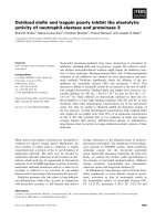
essential for APBI [5]. In the present study, 4 experienced
breast surgeons in a single Institution performed breast
lumpectomies and placed clips within the tumour bed
according to the surgical placement procedure, i.e. 4 clips
were placed at the upper, inner, outer and lower surgical
margins of the tumour bed (Figure 1).
All patients had a computed tomography (CT)-based sim-
ulation for postoperative breast irradiation in the treat-
ment position, i.e. in the supine position on the Med Tec
inclined breast board with arms up (Model MT-350-N).
The clinical breast borders, the LC scar and the post-surgi-
cal indurations were outlined with radio-opaque wires.
The CT images extended approximately from the upper-
clavicle to the upper abdomen in 2 or 4-mm thick slices
(Siemens SOMATOM Sensation Open/Siemens Naviga-
tor/SOMARIS/5 Syngo).
Two experienced radiation oncologists specialized in
breast cancer radiotherapy (Experts E1 and E2) and 2
trainee radiation oncologists (Juniors J1 and J2) blindly
delineated 40 consecutive lumpectomy cavities without
reviewing the contours of the other observers. Expertise
was defined as the delineation of more than 400 breast CT
scans per year for at least 2 years, and trainee physicians
either had no prior experience in outlining breast CT scans
or had less than 6 months experience. All volume deline-
ation was performed on axial slices and on sagittal and/or
coronal views if needed, on the Dosisoft/Isogray virtual
simulation software (version 4.0.05 gL). The 4 trainee and
expert radiation oncologists were allowed to choose how
they wished to outline the target volume (the CTV was
considered equivalent to the GTV). If they felt they should
only include the "seroma", they exclusively outlined the
"seroma". If on the other hand they felt they should out-
line both the seroma and the remodelled breast tissue,
they outlined both. In all cases, medical and surgical files
were available if needed.
Surgical procedure of clip placementFigure 1
Surgical procedure of clip placement.
Radiation Oncology 2009, 4:70 />Page 3 of 8
(page number not for citation purposes)
In this study, we exclusively assessed inter-observer varia-
bility of LC delineation, focusing (i) on the entire CTV
(CTV = surgical clips + remodelled breast tissue); (ii) only
on the study of the CTV when it was restricted to the CT
slices which contained clips; and finally (iii) on the
remodelled breast tissue.
Analysis of contoured volumes
Inter-observer variability was quantified according to the
3 assays described by Landis et al.: (i) Variability of CTV
volume (cm
3
); (ii) The Percent Volume Overlap; (iii) and
the Centre Of the Mass assay (COM) [4]. All of these
parameters were calculated by the virtual simulation soft-
ware. Then, inter-observer variability was assessed (1)
using the distance between each COM that was calculated
for each pairwise volume comparison; (2) using the
number of slices including a recorded contour; (3) and
using the percent volume overlap obtained between pair-
wise volumes (Microsoft/Excel 2003 software). Finally,
interobserver variability was evaluated using the conform-
ity index (CI) of two delineated structures (V1 and V2).
The CI was defined as the ratio of the overlapping volume
V1UV2 and the encompassing total delineated volume
V1UV2.
Statistical methods
Normality was tested using the Kolmogorov Smirnov test.
As variables did not follow a normal distribution, data
were summarized with medians (minimum-maximum).
Comparisons within studies 1 and 2 or between readers
were made using the Wilcoxon matched-pair test. All tests
were two-sided, and p-values below 0.01 were considered
to denote statistical significance because of multiple tests.
Statistical analyses were performed using SAS software,
version 9.1 (SAS Institute, Cary, NC).
Results
Patient demographics
Median age was 60 years (range, 44-94 years). Median
time from surgery to CT scan simulation was 51 days
(range, 14-252). The median surgical lumpectomy vol-
ume based on histological reports was 76 cm
3
(range, 13-
364 cm
3
). The median tumour size was 12 mm (range, 2-
35 mm) and most of the patients had a pT1N0 breast can-
cer (73%). Chemotherapy was administered to 13
patients before breast radiotherapy (33%) (Table 1).
CTV Volume (Table 2 and 3)
Contouring by juniors was much larger, even though
these volumes were restricted exclusively to slices contain-
ing clips (excess volume of 2 cm3, p = 0.03 and 0.002).
Indeed, remodelled breast tissue was not outlined by the
juniors but was almost exclusively outlined within the tar-
get volumes by both experienced radiation oncologists (p
< 0.0001) (Table 2). In the case of experts, the volumes
were more extensive in terms of the number of CT slices,
because they included remodelled breast tissue. Yet in
spite of incorporating remodelled breast tissue, their vol-
umes were still smaller than those of trainees. Discrepan-
cies were observed more particularly between experts than
between juniors (p = 0.004). They mostly concerned the
remodelled breast tissue, and only to a small extent the
part containing the surgical clips.
Number of CT slices used to delineate CTV
Experts outlined their CTV volume on more CT slices
(median of 11 CT slices) than junior radiation oncologists
(median of 9 CT slices). Besides a greater number of CT
slices, the upper and lower limits of the lumpectomy cav-
ity were not always concordant between the experienced
radiation oncologists (p < 0.0001) whereas they were con-
cordant when delineated by juniors as they only used CT
Table 1: Patient demographics and clinical data.
N = 40 Median Min - max N (%)
Age (years old) 60 44 - 94
Tumour size (mm) 12 2 - 35
Histological tumour volume (cm
3
)7613 - 364
Time from surgery to radiotherapy (days) 51 14 - 252
Adjuvant chemotherapy (number of patients) 13 (33%)
pTis pN0 1 (2.5%)
pT1 pN0-1 33 (82.5%)
pT2 pN0-1 6 (15%)
Radiation Oncology 2009, 4:70 />Page 4 of 8
(page number not for citation purposes)
slices which contained surgical clips (p = 0.19). In conclu-
sion, the experienced radiation oncologists outlined the
lumpectomy cavity on CT slices which contained both
surgical clips and remodelled breast tissue whereas jun-
iors restrained their CTV delineation to CT slices which
exclusively contained surgical clips.
Conformity index (CI) of CTV delineation (Figure 2a and
2b)
when the entire CTV was assessed, the CI was relatively
low, ranging from 0.48 (range = 0.10 - 0.77) to 0.58
(range = 0.27 - 0.8). The CI was more concordant between
juniors (CI = 0.58) than between experts (CI = 0.56), but
was clearly lower for juniors with a CI at 0.48 (range = 0.1-
0.77) compared to experts with a CI at 0.53 (range = 0.25-
0.73). When the assessment of the CTV was restricted to
CT slices containing surgical clips (Table 3), the CI was
improved, ranging from 0.55 to 0.65. The CI was more
concordant between juniors with a CI at 0.64 (range =
0.36 - 0.8) than between experts with a CI at 0.62 (range
= 0.38 - 0.73). Once again, the CI of the CTV delineated
by junior radiation oncologists was clearly lower with a
median CI at 0.55 (range = 0.07-0.88) than the CTV delin-
eated by experienced radiation oncologists with a median
at 0.65 (range = 0.13-0.82). Finally, a higher concordance
of CTV delineation was related to experience and to the
presence of surgical clips even though, given the limited
number of physicians involved in this study, the results
should exclusively be interpreted at the individual rather
than at the collective level.
Table 2: Discrepancies in lumpectomy cavity volumes between the 4 radiation oncologists (Juniors J1 and J2; Experts E1 and E2).
N = 40 Volume of lumpectomy cavity (cm
3
) Volume of lumpectomy cavity (cm
3
) when the assessment was
restricted to CT slices containing surgical clips
p
Median Min - max Median Min - max
J1 14 5 - 53 15 6 - 53 0.98
J2 16 5 - 61 16 5 - 60 0.39
E1 13 5 - 48 12 5 - 46 p < 0.0001
E2 11 5 - 96 11 4 - 50 p < 0.0001
Table 3: Discrepancies in lumpectomy cavity (LC) volumes; in conformity index and in distances between centres of gravity of each LC
volume when the study was restricted to CT slices containing surgical clips.
Volume of lumpectomy cavity - cm
3
(n = 40)
P* Conformity index Distance between gravity centres
(mm)
ΔJ1-J2 Med
(min; max)
- 0.9
(-17.9; 13.1)
0.02 0.64
(0.36; 0.80)
2.2
(0.0; 11.9)
ΔJ1-E1 Med
(min; max)
1.5
(-4.5; 17.7)
0.007 0.65
(0.13; 0.82)
1.8
(0.5; 26.3)
ΔJ1-E2 Med
(min; max)
3.5
(-20.9; 24.5)
< 0.0001 0.57
(0.09; 0.77)
2.4
(0.6; 30.9)
ΔJ2-E1 Med
(min; max)
3.5
(-4.9; 22.8)
< 0.0001 0.57
(0.24; 0.86)
2.4
(0.7; 20.4)
ΔJ2-E2 Med
(min; max)
4.2
(-7.5; 27.1)
< 0.0001 0.55
(0.07; 0.88)
3.1
(1.3; 24.7)
ΔE1-E2 Med
(min; max)
1.6
(-27.4; 10.2)
< 0.0002 0.62
(0.38; 0.73)
2.51
(0.2; 10.0)
* p value for testing equality of volumes between physicians ( < 0.01 is significant).
ΔJ1-J2 = between J1 and J2. ΔJ1-E1 = between J1 and E1. ΔJ1-E2 = between J1 and E2. ΔJ2-E1 = between J2 and E1. ΔJ2-E1 = between J2 and E2.
ΔE1-E2 = between E1 and E2.
Radiation Oncology 2009, 4:70 />Page 5 of 8
(page number not for citation purposes)
Surgical clips improved the Conformity IndexFigure 2
Surgical clips improved the Conformity Index. Figure 2a, CI without clips; Figure 2b, CI with clips. In each figure,
lumpectomy cavity delineation according to each physician (green: J1; red: J2; orange: E1 and blue: E2).
a
b
Radiation Oncology 2009, 4:70 />Page 6 of 8
(page number not for citation purposes)
Centre of the Mass (COM) of CTV (Table 4)
The Distance inter-COM (DICOM) between the different
observers ranged from +2.6 mm to +3.5 mm. The DICOM
was higher between juniors (DICOM = +3.4 mm) than
between experts (DICOM = +3.1 mm). When we restricted
the study of CTV delineation to the CT slices containing
surgical clips, the distances between the centres of gravity
of each LC volume were smaller, ranging from +1.8 mm
to +3.1 mm between the different observers.
Discussion
This study showed that the placement of surgical clips
improved the conformity index with less inter-observer
variability in the definition of the lumpectomy cavity after
breast-conserving surgery. Different surrogates have been
used to define the lumpectomy cavity for the tumour
boost: the scar on breast [6], placement of surgical clips
[7-9] or imaging by ultrasound or CT scan [10,11].
Regarding surgical clips, their use could vary in terms of
their number [from one to multiple clips] [12] and in
terms of their placement [prepectoral or within the LC]. In
3D-conformal APBI, no consensus has been reached con-
cerning surgical clip placement. In the NSABP B39/RTOG
0413 trial, the definition of the target volume is based
either on surgical clips, if present or on seroma if clearly
visible. Then the CTV is expanded by 15 mm around the
GTV [13]. In the APBI-MGH trial, the GTV and the CTV are
considered as equivalent [3]. Surgical clips were not
required in either protocol whereas clip placement is
mandatory in the ongoing IGR APBI trial. Nevertheless,
the target volume in our study was in the same range as
those described in the literature [2,14-16] (Table 5) even
though remodelled breast tissue was included in CTV
delineation.
The present study showed that surgical clips placed at the
4 cardinal points of the lumpectomy cavity improved the
conformity index between the different observers, from
49% to 65%. However, defining the lumpectomy cavity
using surgical clips within the lumpectomy cavity still
seems insufficient for adequately defining the GTV in 3D-
conformal APBI. Indeed, some issues regarding LC con-
touring still need to be more clearly characterized such as
the need to outline breast tissue changes, the expansion of
seroma margins etc. Thus, other imaging techniques are
needed, such as either ultrasound, positron emission tom-
ography/CT (PET-CT) that identifies inflammatory tissue
remodelling or magnetic resonance imaging (MRI).
Recently, Berrang et al. reported on the usefulness of com-
bining 3D-breast ultrasound (3D-US) with the CT scan in
treatment planning for APBI. Indeed, they showed that
less inter-observer variability was seen when contouring
seroma with 3D-US, with smaller seroma volumes when
they were outlined using 3D-US [15]. However, 3D-US
has never been evaluated in cases of remodelled breast tis-
sue alone, the most frequent type of breast-conserving sur-
gery in France, without the presence of seroma. PET-CT,
another type of functional imaging has also been assessed.
A recent study showed that the lumpectomy cavity was
well visualized with PET-CT but contouring volumes were
always larger than those outlined on CT scan [16]. How-
ever, PET-CT identified inflammatory tissue remodelling
but not residual disease. Magnetic resonance imaging may
be of interest for visualising and defining the postopera-
tive lumpectomy cavity because of its superiority in terms
of soft tissue contrast and better differentiation of normal
tissue from the lumpectomy cavity. Recently, Kirby et al
showed that MRI allowed a higher conformity index (CI
at 0.89) when compared with CT scan. Even though MRI
Table 4: Discrepancies in distances inter-Com (Centre Of the Mass of the target volume) when the CTV was entirely assessed (study
1) and when the study was restricted to CT slices containing surgical clips (study 2).
Study 1
Distance inter-Com (mm)
Study 2
Distance inter-Com (mm)
Median Min - max Median Min - max
ΔJ1-J2 3.4 0 - 11.9 2.2 0 - 11.9
ΔJ1-E1 2.6 0.9 - 34.2 1.8 0.5 - 26.3
ΔJ1-E2 3.3 1.1 - 36.5 2.4 0.6 - 30.9
ΔJ2-E1 3.3 0.9 - 24.4 2.4 0.7 - 20.4
ΔJ2-E2 3.5 1.3 - 26.2 3.1 1.3 - 24.7
ΔE1-E2 3.1 0.2 - 24.3 2.5 0.2 - 10.0
ΔJ1-J2 = between J1 and J2. ΔJ1-E1 = between J1 and E1. ΔJ1-E2 = between J1 and E2. ΔJ2-E1 = between J2 and E1. ΔJ2-E1 = between J2 and E2.
ΔE1-E2 = between E1 and E2.
Radiation Oncology 2009, 4:70 />Page 7 of 8
(page number not for citation purposes)
is of interest for CTV delineation, it is not readily accessi-
ble and when available, it is often performed in the prone
position whereas treatments are performed in the supine
position [17,18]. Further evaluations are needed to clearly
define the role of either 3D-US or PET-CT or MRI in con-
touring LC.
Besides surgical clip placement, the present study also
showed that the CI was higher with experience (CI for jun-
iors at 0.55 and for experts at 0.65) suggesting the need for
a learning curve. Indeed, a learning curve was highly rec-
ommended by Wong et al in the contouring of the
lumpectomy cavity as the CTV observed among trainee
radiation oncologists was always larger than those of
trained physicians [19,20]. In our study, the LC volumes
defined by the juniors were always larger than those of the
experts because of their uncertainty regarding volume
delineation. Juniors positioned their target volume close
to surgical clips without including remodelled breast tis-
sue. Although the experts outlined the LC volume with the
remodelled breast and surgical clips, their LC volumes
were still smaller than those of trainees.
Conclusion
The definition of the lumpectomy cavity (GTV and CTV)
is an essential part of 3D-APBI treatment planning. The
placement of surgical clips at the 4 cardinal points of the
lumpectomy cavity strongly improved the accuracy of tar-
get contouring. Therefore, the use of surgical clips in the
delineation of LC in 3D-conformal APBI is required.
List of abbreviations
LC: lumpectomy cavity; APBI: accelerated partial breast
irradiation; CI: confidence interval; COM: Centre of the
Mass assay; DICOM: Distance inter-COM; GTV: Gross
Tumour Volume; CTV: Clinical Tumour Volume; PTV:
Planning treatment volume; 3D-US: 3D-breast ultra-
sound; PET-CT: Positron emission tomography/CT; MRI:
magnetic resonance imaging.
Competing interests
The authors declare that they have no competing interests.
Authors' contributions
MD: acquisition, analysis and interpretation of data; CP,
ET, AD and HM: analysis and interpretation of data; EP,
AK, SG, CU, JRG, FR, MS, PV: data acquisition; CB: con-
ception, design. All the listed authors have been involved
in drafting or in revising the manuscript. All authors read
and approved the final manuscript.
Acknowledgements
The authors thank Lorna Saint Ange for editing.
References
1. Chen PY, Gustafson GS, Mitchell C, Wallace M, Hasan Y, Martinez
AA, Vicini F: Three-year Clinical Experience Utilizing 3D-Con-
formal Radiation Therapy to Deliver Accelerated Partial
Breast Irradiation (APBI). Int J Radiat Oncol Biol Phys 2008,
72:S3-S4.
2. Baglan KL, Sharpe MB, Jaffray D, Frazier RC, Fayad J, Kestin LL,
Remouchamps V, Martinez AA, Wong J, Vicini FA: Accelerated par-
tial breast irradiation using 3D conformal radiation therapy
(3D-CRT). Int J Radiat Oncol Biol Phys 2003, 55:302-11.
3. Taghian AG, Kozak KR, Doppke KP, Katz A, Smith BL, Gadd M,
Specht M, Hughes K, Braaten K, Kachnic LA, Recht A, Powell SN: Ini-
tial dosimetric experience using simple three-dimensional
conformal external-beam accelerated partial-breast irradia-
tion. Int J Radiat Oncol Biol Phys 2006, 64:1092-9.
4. Landis DM, Luo W, Song J, Bellon JR, Punglia RS, Wong JS, Killoran
JH, Gelman R, Harris JR: Variability among breast radiation
oncologists in delineation of the postsurgical lumpectomy
cavity. Int J Radiat Oncol Biol Phys 2007, 67:1299-308.
5. Dzhugashvili M, Pichenot C, Dunant A, Balleyguier C, Delaloge S,
Mathieu MC, Garbay JR, Marsiglia H, Bourgier C: Surgical Clips
Assist in the Visualization of the Lumpectomy Cavity in
Three-Dimensional Conformal Accelerated Partial-Breast
Irradiation. Int J Radiat Oncol Biol Phys 2009.
Table 5: Lumpectomy cavity (LC) volumes in APBI trials
Median volume of lumpectomy cavity (cc) Min - max (cc)
Berrang et al [13] Seroma visible on CT-scan 31.8 7.3 - 131.9
Seroma visible on 3D-US 23.2 5 - 93.6
Ford et al [14] LC on CT scan 15.4 5.2 - 133.5
LC on PET-CT 32.8 7 - 199.4
Vicini et al [1] LC 12 5 - 65
Formenti et al [12] LC 34 7 - 379
IGR study LC by juniors 14-16 5 - 61
LC by experts 11-13 5 - 96
Publish with BioMed Central and every
scientist can read your work free of charge
"BioMed Central will be the most significant development for
disseminating the results of biomedical research in our lifetime."
Sir Paul Nurse, Cancer Research UK
Your research papers will be:
available free of charge to the entire biomedical community
peer reviewed and published immediately upon acceptance
cited in PubMed and archived on PubMed Central
yours — you keep the copyright
Submit your manuscript here:
/>BioMedcentral
Radiation Oncology 2009, 4:70 />Page 8 of 8
(page number not for citation purposes)
6. Machtay M, Lanciano R, Hoffman J, Hanks GE: Inaccuracies in using
the lumpectomy scar for planning electron boosts in primary
breast carcinoma. Int J Radiat Oncol Biol Phys 1994, 30:43-8.
7. Harrington KJ, Harrison M, Bayle P, Evans K, Dunn PA, Lambert HE,
Saidan Z, Lynn J, Stewart JS: Surgical clips in planning the elec-
tron boost in breast cancer: a qualitative and quantitative
evaluation. Int J Radiat Oncol Biol Phys 1996, 34:579-84.
8. Fein DA, Fowble BL, Hanlon AL, Hoffman JP, Sigurdson ER, Eisenberg
BL: Does the placement of surgical clips within the excision
cavity influence local control for patients treated with
breast-conserving surgery and irradiation. Int J Radiat Oncol Biol
Phys 1996, 34:1009-17.
9. Hunter MA, McFall TA, Hehr KA: Breast-conserving surgery for
primary breast cancer: necessity for surgical clips to define
the tumor bed for radiation planning. Radiology 1996,
200:281-2.
10. Benda RK, Yasuda G, Sethi A, Gabram SG, Hinerman RW, Menden-
hall NP: Breast boost: are we missing the target? Cancer 2003,
97:905-9.
11. Smitt MC, Birdwell RL, Goffinet DR: Breast electron boost plan-
ning: comparison of CT and US. Radiology 2001, 219:203-6.
12. Goldberg H, Prosnitz RG, Olson JA, Marks LB: Definition of
postlumpectomy tumor bed for radiotherapy boost field
planning: CT versus surgical clips. Int J Radiat Oncol Biol Phys
2005, 63:209-13.
13. Vicini F: A Randomized Phase III Study of Conventional
Whole Breast Irradiation (WBI) Versus Partial Breast Irra-
diation (PBI) for Women with Stage 0, I, or II Breast Cancer.
Pittsburgh, PA: NSABP PROTOCOL B-39 - RTOG PROTOCOL 0413 2007.
14. Formenti SC, Truong MT, Goldberg JD, Mukhi V, Rosenstein B, Roses
D, Shapiro R, Guth A, Dewyngaert JK: Prone accelerated partial
breast irradiation after breast-conserving surgery: prelimi-
nary clinical results and dose-volume histogram analysis. Int
J Radiat Oncol Biol Phys 2004, 60:493-504.
15. Berrang TS, Truong PT, Popescu C, Drever L, Kader HA, Hilts ML,
Mitchell T, Soh SY, Sands L, Silver S, Olivotto IA: 3D Ultrasound
Can Contribute to Planning Ct to Define the Target for Par-
tial Breast Radiotherapy. Int J Radiat Oncol Biol Phys 2008.
16. Ford EC, Lavely WC, Frassica DA, Myers LT, Asrari F, Wahl RL, Zel-
lars RC: Comparison of FDG-PET/CT and CT for delineation
of lumpectomy cavity for partial breast irradiation. Int J Radiat
Oncol Biol Phys 2008, 71:595-602.
17. Kirby AM, Yarnold JR, Evans PM, Morgan VA, Schmidt MA, Scurr ED,
Desouza NM: Tumor bed delineation for partial breast and
breast boost radiotherapy planned in the prone position:
what does MRI add to X-ray CT localization of titanium clips
placed in the excision cavity wall? Int J Radiat Oncol Biol Phys 2009,
74:1276-82.
18. Whipp EC, Halliwell M: Magnetic resonance imaging appear-
ances in the postoperative breast: the clinical target volume-
tumor and its relationship to the chest wall. Int J Radiat Oncol
Biol Phys 2008, 72:49-57.
19. Wong EK, Truong PT, Kader HA, Nichol AM, Salter L, Petersen R,
Wai ES, Weir L, Olivotto IA: Consistency in seroma contouring
for partial breast radiotherapy: impact of guidelines. Int J
Radiat Oncol Biol Phys 2006, 66:372-6.
20. Petersen RP, Truong PT, Kader HA, Berthelet E, Lee JC, Hilts ML,
Kader AS, Beckham WA, Olivotto IA: Target volume delineation
for partial breast radiotherapy planning: clinical characteris-
tics associated with low interobserver concordance. Int J
Radiat Oncol Biol Phys 2007, 69:41-8.