Báo cáo y học: "Immunohistological assessment of the synovial tissue in small joints in rheumatoid arthritis: validation of a minimally invasive ultrasound-guided synovial biopsy procedure" potx
Bạn đang xem bản rút gọn của tài liệu. Xem và tải ngay bản đầy đủ của tài liệu tại đây (426.84 KB, 9 trang )
Open Access
Available online />Page 1 of 9
(page number not for citation purposes)
Vol 9 No 5
Research article
Immunohistological assessment of the synovial tissue in small
joints in rheumatoid arthritis: validation of a minimally invasive
ultrasound-guided synovial biopsy procedure
Carlo Alberto Scirè
1
, Oscar Epis
1
, Veronica Codullo
1
, Frances Humby
2
, Patrizia Morbini
3
,
Antonio Manzo
2
, Roberto Caporali
1
, Costantino Pitzalis
2
and Carlomaurizio Montecucco
1
1
Chair and Division of Rheumatology, University of Pavia, Fondazione IRCCS Policlinico San Matteo, Piazzale Golgi 12, I 27100 Pavia, Italy
2
Centre for Experimental Medicine and Rheumatology, 2nd floor John Vane Science Centre, William Harvey Research Institute, St Bartholomew's
and Royal London School of Medicine. Charter House Square, London, EC1M6BQ, UK
3
Department of Pathology, University of Pavia, Fondazione IRCCS Policlinico San Matteo, Piazzale Golgi 12, I 27100 Pavia, Italy
Corresponding author: Carlomaurizio Montecucco,
Received: 11 Jul 2007 Revisions requested: 23 Aug 2007 Revisions received: 4 Sep 2007 Accepted: 28 Sep 2007 Published: 28 Sep 2007
Arthritis Research & Therapy 2007, 9:R101 (doi:10.1186/ar2302)
This article is online at: />© 2007 Scirè et al., licensee BioMed Central Ltd.
This is an open access article distributed under the terms of the Creative Commons Attribution License ( />),
which permits unrestricted use, distribution, and reproduction in any medium, provided the original work is properly cited.
Abstract
The aim of the present study was to perform an
immunohistological assessment of the synovial tissue from
involved small joints in rheumatoid arthritis (RA) and to explore
the reliability of a mini-invasive ultrasound (US)-guided
technique of small joint synovial biopsy for the histopathological
assessment. Synovial tissue collected during arthrotomic
surgery of small joints in nine patients served as the gold
standard for the validation of the histological assessment. Small
hand-joint synovial biopsies from an additional nine patients with
erosive RA were obtained by a mini-invasive US-guided
procedure, performed percutaneously by the portal and rigid
forceps technique. Using digital image analysis, the area
fractions of synovial macrophages (CD68 cells), T cells (CD3
cells) and B cells (CD20 cells) were measured in all high-power
fields of every sample at different cutting levels. The
representative sample was defined as the minimal number of
high-power fields whose mean area fraction would reflect the
overall mean area fraction within a percentage mean difference
of 10%. For each patient, a range of three to five large samples
for surgical biopsies and a range of 8–12 samples for US-
guided biopsies were collected and analysed. In arthrotomic
samples, the analysis of a randomly selected tissue area of 2.5
mm
2
was representative of the overall value for CD68, CD3 and
CD20 cells. US-guided samples allowed histological evaluation
in 100% of cases, with a mean valid area of 18.56 mm
2
(range
7.29–38.28 mm
2
). The analysis of a cumulative area of 2.5 mm
2
from eight randomly selected sections (from different samples or
from different cutting levels) allowed to reduce the percentage
mean difference to less than 10% for CD68, CD3 and CD20
cells. In conclusion, US-guided synovial biopsy represents a
reliable tool for the assessment of the histopathological features
of RA patients with a mini-invasive approach.
Introduction
A number of approaches to the assessment of the synovial
membrane have been proposed in an attempt to establish the
degree of inflammation and the phenotypic characterization of
infiltrating cell subsets [1,2]. Of the cell types found in the syn-
ovium, the intensity of CD68-positive macrophage infiltration
at baseline has been associated with progressive joint dam-
age [3,4], and has been confirmed as an optimal biomarker of
clinical response in several randomized clinical trials of both
disease-modifying antirheumatic drugs and biologic agents [5-
8].
The importance of the evaluation of the synovial membrane,
particularly for clinical trials, has been reinforced by work dem-
onstrating that changes in the synovial membrane are more
reliable than clinical assessments, such as the disease activity
score, when determining response to treatment [9]. In addi-
tion, the development of techniques such as digital image
analysis has made the rapid reliable assessment of large areas
of tissue a realistic proposition [10,11].
There are several possible approaches to the acquisition of
synovial tissue, but arthroscopic biopsy is generally accepted
H & E = haematoxylin and eosin; HPF = high-power field; RA = rheumatoid arthritis; US = ultrasound.
Arthritis Research & Therapy Vol 9 No 5 Scirè et al.
Page 2 of 9
(page number not for citation purposes)
as the gold standard [12], allowing for good quality, sizeable
biopsy specimens. The knee joint has been the favourite
biopsy site owing to the ease of arthroscopic access and to
the knowledge that it appears representative microscopically
of other synovial joints [13]. Several validation studies demon-
strated the reliability of a multiple sampling of synovial mem-
brane for immunohistological studies, inferring that synovial
sampling from clinically involved knee joints might provide a
picture of the disease for each patient at every disease phase
[1,12,14-19].
The knee joint, however, is only involved in a subset of patients
with early arthritis, and knee involvement at onset would
appear to identify a cohort of patients with a more aggressive
disease course [20]. Studies based exclusively on knee
biopsy, at least in early arthritis, would therefore be inherently
biased towards recruitment of patients with a worse progno-
sis. In addition, as the small joints of the hands and feet are
most commonly involved in early arthritis and since associated
outcome measures of erosive burden are assessed here,
acquisition of the synovial membrane from these joints would
appear imperative for high-quality translational research.
For these reasons, different biopsy techniques have been
developed to acquire synovial tissue from small joints, both by
needle and by arthroscopic approach [21,22]. The recent
development of ultrasound (US)-guided synovial biopsy may
help to overcome the blindness of the needle biopsy and the
invasiveness of arthroscopic biopsy of small joints [23].
US-guided synovial biopsy of small joints is not, however,
presently a standard technique in clinical practice – predomi-
nantly because of several still unanswered important issues
regarding the pathologic variability in small joints and the valid-
ity of the technique to produce meaningful biological
specimens.
The present study aimed to address a number of these issues:
whether the analysis of randomly selected synovial tissue col-
lected from small joints in rheumatoid arthritis (RA) patients
could be representative of the overall inflammatory status of
the joint; whether adequate synovial tissue could be obtained
by US-guided synovial biopsy of small joints with regard to a
series of standard immune (CD3 cells, CD20 cells, CD68
cells) and histological parameters; and to determine the mini-
mum number of synovial biopsies under US guidance required
to achieve reliable measurements of the above immune histo-
logical features.
For this purpose, we first analysed RA patient synovial mem-
branes collected from surgical procedures to assess the min-
imum area of synovial tissue representative of the overall joint
status for a series of standardized immunohistological param-
eters. We next tested the availability of that area in our synovial
samples collected by the novel US-guided procedure, and
evaluated in US-guided biopsies the variability of the main
immune-phenotypic features to assess the number of samples
needed to achieve reliable measurements.
Materials and methods
Patients
To assess the immunohistological features of rheumatoid syn-
ovitis, surgical synovial tissue specimens were obtained dur-
ing arthroplasty or synovectomy from clinically involved
(swollen) small joints of nine patients with longstanding ero-
sive RA who fulfilled the 1987 American Rheumatism Associ-
ation classification criteria [24].
To validate US-guided synovial biopsy, the procedure was per-
formed in an additional nine patients with longstanding erosive
RA before starting biologic agent treatment.
All patients gave their informed consent for biopsy, and the
study was approved by the Local Ethical Committee. The main
demographic and clinical characteristics of the patients as
well as the biopsy sites are presented in Table 1.
Ultrasound-guided synovial biopsies
US-guided synovial biopsy was performed using the portal
and forceps technique [23]. Briefly, US examination was per-
formed with the Toshiba Nemio (Toshiba America Medical
Systems, Inc. Tustin, CA, USA) using a multifrequency linear
transducer (Hockey Stick Linear 8–14 MHz; Toshiba America
Medical Systems). The transducer was used for standard
Doppler-sonographic evaluation of the joint and for direct
assistance of the biopsy procedure. Effusion and synovitis
were identified and distinguished according to the following
definition: effusion was defined as hypoechoic or anechoic
compressible intra-articular material, within synovial recesses.
Synovitis was defined as echogenic noncompressible intra-
articular tissue, within synovial recesses. Power-Doppler vari-
ables were adjusted to the lowest permissible pulse repetition
frequency to maximize the sensitivity. Low wall filters were
used. The colour gain was set just below the level at which col-
our noise appeared underlying bone (no flow should be visual-
ized at the bony surface). [25]
Under sterile conditions, US dorsal longitudinal guidance was
used for metacarpophalangeal and proximal interphalangeal
biopsies. The skin and subcutaneous tissue and the synovial
space were infiltrated with 1–3 ml local anaesthetic (Xilonest
1%; Astrazeneca, Wedel, Germany) using a 25-gauge needle.
After 2 minutes a 14-gauge needle was inserted into the
planned area under direct US vision. A 6 F percutaneous
sheath introducer (Cordis Corporation, Miami, FL, USA) was
inserted into the joint under US guidance following a flexible
wire. The flexible wire was then removed, and a rigid Hart-
mann's ear forceps (Medicon, Tuttlingen, Germany) was used
for the biopsy procedures through the portal. Several inde-
pendent samples (at least eight) could be taken through the
Available online />Page 3 of 9
(page number not for citation purposes)
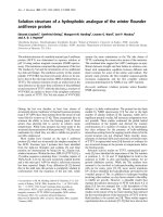
same portal (Figure 1) from the hypertrophic synovium includ-
ing power-Doppler-positive areas. The whole procedure was
completed within 30 minutes and it was generally well toler-
ated. No procedure-related adverse events were recorded in
our series.
Synovial samples
Biopsy specimens were immediately fixed in 4% formalin for
up to 24 hours. After paraffin embedding, 20 serial sections 5
μm thick (100 μm interval) from each sample at three different
cutting levels were mounted onto glass slides.
With regard to the synovial surgical biopsies, slides obtained
from all available specimens were analysed at three different
cutting levels. Given the higher homogeneity of the cellular
reaction in the upper subsynovial layer and the sampling
potential of the US-guided biopsy, the proximal 600 μm of
synovial sublining immediately adjacent to the lining layer was
considered for the purpose of histomorphometric analysis
[26].
Tissue sections from US-guided biopsy were considered valid
for histological analysis only if an intact lining was visible on H
& E-stained sections. All sections were coded and systemati-
cally examined throughout their entire area at three different
cutting levels by a single, blinded observer (CAS).
Immunohistochemistry
Formalin-fixed, paraffin-embedded tissue sections were
deparaffinized and rehydrated through graded ethanol solu-
tions, and were immunostained as previously reported [27].
Briefly, once hydrated the sections were heated for 35 min-
utes at 96°C in DAKO Target Retrieval Solution (S1699;
DAKO, Carpinteria, CA, USA). The sections were then
washed in Tris-buffered saline (pH 7.6) and incubated for 10
minutes with Protein Block Serum Free (X0909; DAKO). The
following primary antibodies were used (2 hours of incuba-
tion): rabbit anti-human CD3 polyclonal antibody (immu-
noglobulin, A0452; DAKO), mouse anti-human CD20
antibody (IgG2a, clone L26; DAKO), and mouse anti-human
CD68 antibody (IgG3, clone PG-M1; DAKO). Sections were
Table 1
Clinicopathological characteristics of synovial tissues obtained by surgical biopsy and ultrasound-guided biopsy
Biopsy Age (years) Sex Disease
duration
(years)
RF/aCCP Disease-modifying
antirheumatic drugs
Joint Valid
samples/
total
samples
Valid
sections
Cumulative
area (mm
2
)
S-SY1 65 Female 18 +/+ MTX Wrist 3/3 9 24.47
S-SY2 44 Female 12 -/- PDN I MTP 3/3 9 37.72
S-SY3 63 Female 14 +/+ MTX + HCQ II and III
MTPs
3/3 9 44.02
S-SY4 46 Female 34 -/+ HCQ Wrist 4/4 12 41.68
S-SY5 46 Female 17 -/- LFN + PDN Wrist 5/5 15 60.32
S-SY6 75 Female 7 -/- MTX + PDN MCPs 3/3 9 34.75
S-SY7 78 Female 8 -/- MTX + PDN MCPs 3/3 9 38.68
S-SY8 46 Male 2 -/+ MTX + PDN MCPs 3/3 9 22.42
S-SY9 68 Female 5 -/- MTX + HCQ + PDN I MTP 3/3 9 45.01
U-SY1 23 Female 5 +/+ CYA + PDN II MCP 4/8 12 16.86
U-SY2 59 Female 4 -/- MTX + PDN V MCP 5/8 15 17.64
U-SY3 64 Female 18 +/+ PDN II PIP 10/12 30 38.28
U-SY4 61 Female 11 -/+ MTX + PDN V MCP 3/12 9 7.29
U-SY5 69 Male 10 +/+ MTX + HCQ + PDN II MCP 8/12 24 20.16
U-SY6 75 Male 11 +/+ HCQ + SLZ + PDN V MCP 10/12 29 28.71
U-SY7 67 Female 7 -/+ MTX + HCQ + PDN II MCP 8/12 23 8.82
U-SY8 62 Female 8 -/- MTX + PDN II MCP 7/12 20 8.43
U-SY9 61 Female 11 +/+ MTX + HCQ + PDN II MCP 9/12 26 20.91
The good quality/overall number of collected sample rate and the number of valid sections obtained considering three different cutting levels are
provided. The cumulative area is computed on overall sections for each patient. S-SY, surgical biopsy synovial tissue; U-SY, ultrasound-guided
biopsy synovial tissue; RF/aCCP, rheumatoid factor/anticyclic citrullinated peptide antibodies; MTX, methotrexate: PDN, prednisone; HCQ,
hydroxychloroquine; LFN, leflunomide; CYA, cyclosporine A; SLZ, sulfasalazine; MCP, metacarpophalangeal; MTP, metatarsophalangeal; PIP,
proximal interphalangeal.
Arthritis Research & Therapy Vol 9 No 5 Scirè et al.
Page 4 of 9
(page number not for citation purposes)
then incubated with the appropriate biotinylated secondary
antibody (E0431 swine anti-rabbit immunoglobulin, or Z0259
rabbit anti-mouse immunoglobulin; DAKO) for 30 minutes, fol-
lowed by streptavidin biotin–alkaline phosphatase complex
(K0391; DAKO) for an additional 30 minutes. Reactions were
then developed using the New Fuchsin Substrate Kit (K0698;
DAKO) and sections were counterstained with Meyer's Hae-
matoxylin 0.1% (MHS-16; Sigma Diagnostics, St Louis, MO,
USA) and mounted with Aquamount mounting medium (BDH,
Poole, UK). Primary and secondary antibodies were diluted in
DAKO Antibody Diluent (S3022; DAKO).
Microscopic analysis
All high-power (× 40) microscopic fields (HPFs) were exam-
ined on an Olympus microscope (BX51; Olympus, Tokyo,
Japan), captured using a digital camera (Olympus) and trans-
ferred to a computer platform. The resultant colour images
were of dimension 2,048 × 1,536 pixels, RGB format, with a
24-bit-per-pixel resolution. For each acquisition session, the
microscope, camera and computer were calibrated according
to a standardized procedure. The images obtained were
stored in an uncompressed TIFF format and were examined
using the image-analysis system ImageJ 1.35 s (National Insti-
tutes of Health, Bethesda, MD, USA). Image segmentation
was performed by RGB colour discrimination using threshold
ranges such that a binary overlay was created covering only
the positively stained areas. This threshold was determined by
two distinct observers and was kept constant for all measure-
ments for the same marker. A separate binary mask was cre-
ated that identified the total tissue area in each image, so the
final parameter of analysis was the area fraction. To speed up
the image analysis process, all procedures were performed
using an ad hoc macro program for each marker.
Statistical analysis
The area fraction of immunoreactivity for each marker was
measured on multiple HPFs for each patient. The interfield var-
iability was determined as the percentage difference between
the mean area fraction when all HPFs from each patient were
considered and the mean area fraction calculated from ran-
domly selected individual HPFs. For surgical biopsies, this
analysis was performed with multiple sets of data (10 sets) of
an increasing number of randomly chosen HPFs from all avail-
able specimens of each patient.
For US-guided biopsies the same analysis was performed with
an increasing number of samples at different cutting levels. To
increase the sampling efficiency, the number of HPFs required
to reduce the percentage mean difference to less than 10% of
the total sample mean was used as the variability threshold for
the assay, as previously reported [26].
The variance of the measurements was reduced into its con-
tributing factors (patient, sample and cutting level) and was
analysed by analysis of variance using a general linear model
and nested design. All samples at three different cutting levels
were analysed for the computations. Both samples and cutting
levels were nested in the patient variable. The F values for
each analysis were provided. Differences were considered
significant at P < 0.05. This variance component analysis was
carried out using the STATISTICA data analysis software sys-
tem (version 7.1, 2005; StatSoft, Inc., Tulsa, OK, USA).
Results
Determination of maximum sampling efficiency on
surgical synovial samples
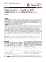
Figure 2 shows the effects of an increasing sample (HPF)
number on the estimate of the overall sample mean for each
immunostain. As the number of fields increases (and hence
the fraction of the total sampling population used to create the
area fraction calculation increases), the proximity of the sam-
ple mean to the overall mean area fraction is asymptotic. The
number of HPFs required to reduce the sample mean to within
10% of the overall sample mean was set as the efficiency
threshold for the analysis. Any further sampling analysis after
this point would lead to an insignificant gain in the estimate of
Figure 1
Ultrasound-guided synovial biopsy of a second metacarpophalangeal joint using the portal and forceps techniqueUltrasound-guided synovial biopsy of a second metacarpophalangeal joint using the portal and forceps technique. Arrow, open forceps
inside the joint; asterisk, metacarpal hand.
Available online />Page 5 of 9
(page number not for citation purposes)
the mean area fraction for the particular tissue marker being
examined.
It can be seen that the analysis of a cumulative area of 2.5 mm
2
(randomly selected from all available samples) is sufficient to
reduce the variability of the estimate to within 10% of the total
sample mean for all markers. Given the more homogeneous
distribution of CD68-positive cells within the sublining, a vari-
ation of less than 10% was typically obtained by evaluation of
only 1.2 mm
2
, while CD3 and CD20 cells needed a larger area
because of their focal distribution in the sublining layer. These
results provide essential baseline data for the comparative
evaluation of different biopsy regimes and methodologies, and
they represent a 'gold standard' for the development of a mor-
phometric protocol for the biopsy of synovial tissue from the
minor joints.
Efficacy of ultrasound-guided small joint biopsies
Qualitative histological examination of H & E-stained speci-
mens showed that good quality synovial tissue was available
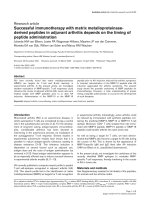
in all cases of US-guided biopsy. A representative synovial
sample obtained by US-guided biopsy from a small joint of a
RA patient is illustrated in Figure 3. The histological validity
and the amount of valuable synovial tissue are detailed in Table
1.
The success rate of the synovial sampling (that is, valid sam-
ples/total collected samples) ranged from 25% to 83%, with
a mean value of 63% (95% confidence interval, 52–75%). The
mean valuable area for each valid sample at a single cutting
level was 0.88 mm
2
(95% confidence interval, 0.80–0.97
mm
2
), ranging from 0.16 to 3.19 mm
2
. By analysing three dif-
ferent cutting levels for each valid sample, the minimum useful
Figure 2
Evaluation of the minimum area required for quantitative analyses of CD68, CD3 and CD20 cellsEvaluation of the minimum area required for quantitative analyses of CD68, CD3 and CD20 cells. For each of the tissue markers studied, a plot
was generated to show the effects of increasing sample size (that is, the number of high-power fields examined) and the proximity of this sample
mean from the overall determination of area fraction. Data represent the mean values of all cases. As the number of high-power fields examined
increases, the difference between the sampling mean and the overall mean reduces. A threshold value of 10% of the overall mean (arrows) was set
as providing a reasonable estimator of the true sample mean.
Arthritis Research & Therapy Vol 9 No 5 Scirè et al.
Page 6 of 9
(page number not for citation purposes)
area (2.5 mm
2
) for quantitative analysis was achieved in all
patients (Table 1).
Quantitative analysis in ultrasound-guided samples
The overall number of sections for each patient ranged from 9
to 30 (three cutting levels for each valid sample).
We first analysed the variability of each immunohistological
parameter between patients, and between samples and cut-
ting levels. Table 2 summarizes the results of the components
of variance analysis. The observed differences were mainly
due to interpatient differences, and secondarily to differences
between samples or cutting levels in a similar way.
To estimate the number of sections to analyse in each patient
to minimize intrapatient variability, we calculated the difference
between the mean values in all sections and those obtained
from a 2.5 mm
2
randomly selected area from an increasing
number of sections.
The outcome of such analysis is depicted in Figure 4. From
this figure it can be concluded that the percentage mean dif-
ference for the staining of a marker decreases below ± 10%
when a minimum of eight samples are considered in the
evaluation.
Discussion
The results presented here show that the analysis of a small
amount of synovial tissue is also representative of the joint sta-
tus in small joints of RA patients, show that US-guided synovial
biopsy at this site represents a reliable approach for good
quality tissue collection, and show that quantitative immuno-
histological studies are feasible through the examination of
multiple specimens obtained by US-guided biopsy of small
joints.
Several studies have addressed the heterogeneity of cellular
and molecular marker expression in RA synovial biopsies,
investigating the amount of tissue or the number of samples
needed to obtain reliable, reproducible results capable of
detecting small changes within the synovial membrane [15-
19,26]. One of the first studies that attempted to address this
question was performed on synovial tissue from RA patients
undergoing joint surgery [26], using a semiquantitative
approach to estimate the degree of cellularity. The study dem-
onstrated that the analysis of a cumulative area of 2.5 mm
2
from at least three biopsy specimens gave an accurate esti-
mate of the overall joint. Analogous results have been reported
on the synovium obtained from the knee joint either arthroplas-
tically, arthroscopically or using blind needle biopsies, when
considering T-cell infiltration, lining layer thickness or vascular-
ity scores [15]. Our analysis focusing on synovial tissue
obtained surgically from small joints reproduces these find-
ings, demonstrating that the analysis of a 2.5 mm
2
tissue sec-
tion allows an estimate of the number of macrophages (CD68-
positive cells), T cells (CD3-positive cells) and B cells (CD20-
positive cells) within 10% of the mean for the overall tissue.
Although we limited our study to the examination of only these
three cell subsets, it is widely recognized that the combination
of CD68 cells, CD3 cells and CD20 cells, which are highly
variable focal parameters, gives a biologically relevant assess-
ment of the overall cellular infiltration within the synovium.
Macrophage infiltration is currently regarded as the main his-
topathological marker of activity and severity in RA [6,28,29].
In addition, despite the rapidly growing interest in the patho-
genic role of B cells within the synovial membrane [30], the
present study is the first to specifically address the question of
what constitutes a representative synovial sample analysis for
B-cell infiltration.
There have been numerous previous studies attempting to
standardize the quantity of synovial tissue required to achieve
a representative measure of the overall joint, the majority using
an arthroscopic approach to obtain synovial tissue from knee
joints and hence able to determine the number of biopsies
from exact sites within the joint to allow accurate his-
topathologcial evaluation of the synovial membrane [12]. The
recent development of a novel minimally invasive technique of
US-guided synovial biopsy has been reported in the literature
[23,31], and includes assessment of the small joint biopsy
Figure 3
Microphotograph of an ultrasound-guided sampleMicrophotograph of an ultrasound-guided sample. H & E staining of
a metacarpophalangeal sample of a rheumatoid arthritis patient (patient
U-SY1), a result of multiple high-power fields (40× objective) merged
into a single image (montage).
Available online />Page 7 of 9
(page number not for citation purposes)
with success rates in acquisition of histologically reliable tis-
sue ranging from 89% to 93%. Among 120 US-guided biop-
sies, however, only one report is made of
metacarpophalangeal and metatarsophalangeal joint biopsy.
Our study therefore describes the largest case series of small
joint synovial US-guided biopsies in RA. The collection of
several independent samples (up to 12 samples) is feasible
and allows a high histological success rate (100% in our
series). Since the procedure is minimally invasive, repeated
biopsies could be planned to monitor the disease course and/
or the response to therapy.
A basic stereological rule for the analysis of all tissue states
that the degree of variation is greatest between individuals and
is least between sections from the same biopsy. This stereo-
logical rule was elegantly demonstrated for synovial tissue by
Dolhain and colleagues, who looked at the degree of T-cell
infiltration within and between multiple biopsy sites [14]. We
used a similar approach to address the problem of variability in
cellular infiltrates in biopsies obtained under US guidance
from small joints, and we came to similar conclusions demon-
strating that the main component of the variance was due to
the differences between patients rather than between samples
Table 2
Components of variance for each marker
Marker Patients Samples Cutting levels
CD68 cells 34.44, P < 0.001 10.29, P < 0.001 13.00, P < 0.001
CD3 cells 23.22, P < 0.001 2.58, P < 0.001 5.51, P < 0.001
CD20 cells 16.11, P < 0.001 4.05, P < 0.001 3.77, P < 0.001
F value and significance level reported for each marker in each component of variance.
Figure 4
Number of ultrasound-guided biopsy sections required for quantitative analysisNumber of ultrasound-guided biopsy sections required for quantitative analysis. Evaluation of the number of ultrasound-guided biopsy sec-
tions required for quantitative analyses of CD68, CD3 and CD20 cells. Reduction in the percentage mean difference can be obtained by studying
2.5 mm
2
from an increasing number of sections. Arrows, number of sections that allow one to achieve a percentage mean difference lower than
10%. x axis, number of sections studied; y axis, percentage mean difference.
Arthritis Research & Therapy Vol 9 No 5 Scirè et al.
Page 8 of 9
(page number not for citation purposes)
or cutting levels of the same sample. We concluded that eight
different sections obtained from either different samples or dif-
ferent cutting levels are required to reduce the sampling error
to less than 10% for a reliable analysis of CD68, CD3 and
CD20 cells. In our series, considering one cutting level in five
out of nine cases, two cutting levels in eight out of nine cases,
and three cutting levels in all cases produced a reliable result.
The limitation of the methodology used in this study mainly
results from the analysis of a heterogeneous group of RA
patients and from the application of results derived from surgi-
cal biopsies (from a different set of patients) to US-guided
biopsies. Performing US-guided biopsy and surgery on the
same patient, however, can be easily appreciated as far from
simple, from both a practical point of view and from an ethical
point of view. In addition, the benefit of our approach is that we
provide data applicable to synovial samples from patients with
different disease durations and different pharmacological
treatments, which maximizes differences between patients [1],
thus increasing the representativeness of our study. Moreover,
the bias in the evaluations of joint replacement synovial tissue
is limited in our series because, as in US-guided biopsies, syn-
oviectomy or arthroplasty in small joints were performed in
active diseases, differing from large joint surgery where it is
generally performed in end-stage disease [3].
Conclusion
In summary, the present study shows that US-guided biopsy
of synovial hand joints in RA patients is a reliable tool for his-
tological evaluation. If 12 different samples are taken, a valid
assessment at least for CD20 cells, CD3 cells and CD68 cells
is possible.
These findings are comparable with those obtained when syn-
ovial tissue from the knee is examined, and are the first attempt
to standardize the minimum requirements for analysis of the
small joints of the hands. The study of the synovial tissue from
small joints can be a valuable research tool, allowing for this
tissue to be incorporated into future trial designs, which is crit-
ical for further understanding of the pathogenesis of this dis-
ease and for assessing marker changes in the course of
disease or in response to targeted therapies.
Competing interests
The authors declare that they have no competing interests.
Authors' contributions
CAS substantially contributed to the conception of the study,
and the acquisition, analysis and interpretation of data. OE
substantially contributed to acquisition of tissue specimens.
VC contributed to the acquisition and interpretation of the
results. FH participated in drafting the manuscript. PM sub-
stantially participated in the methodological aspect of the
study. AM substantially contributed to interpretation of the
data. RC contributed to interpretation of the data and to criti-
cal review of the manuscript. CP substantially contributed to
the critical review of the manuscript. CM provided final
approval of the version to be published.
References
1. Rooney M, Condell D, Quinlan W, Daly L, Whelan A, Feighery C,
Bresnihan B: Analysis of the histologic variation of synovitis in
rheumatoid arthritis. Arthritis Rheum 1988, 31:956-963.
2. Koizumi F, Matsuno H, Wakaki K, Ishii Y, Kurashige Y, Nakamura
H: Synovitis in rheumatoid arthritis: scoring of characteristic
histopathological features. Pathol Int 1999, 49:298-304.
3. Tarner IH, Harle P, Muller-Ladner U, Gay RE, Gay S: The different
stages of synovitis: acute vs chronic, early vs late and non-ero-
sive vs erosive. Best Pract Res Clin Rheumatol 2005, 19:19-35.
4. Mulherin D, Fitzgerald O, Bresnihan B: Synovial tissue macro-
phage populations and articular damage in rheumatoid
arthritis. Arthritis Rheum 1996, 39:115-124.
5. Haringman JJ, Kraan MC, Smeets TJ, Zwinderman KH, Tak PP:
Chemokine blockade and chronic inflammatory disease: proof
of concept in patients with rheumatoid arthritis. Ann Rheum
Dis 2003, 62:715-721.
6. Haringman JJ, Gerlag DM, Zwinderman AH, Smeets TJ, Kraan MC,
Baeten D, McInnes IB, Bresnihan B, Tak PP: Synovial tissue mac-
rophages: a sensitive biomarker for response to treatment in
patients with rheumatoid arthritis. Ann Rheum Dis 2005,
64:834-838.
7. Smeets TJ, Barg EC, Kraan MC, Smith MD, Breedveld FC, Tak PP:
Analysis of the cell infiltrate and expression of proinflamma-
tory cytokines and matrix metalloproteinases in arthroscopic
synovial biopsies: comparison with synovial samples from
patients with end stage, destructive rheumatoid arthritis. Ann
Rheum Dis 2003, 62:635-638.
8. Gerlag DM, Haringman JJ, Smeets TJ, Zwinderman AH, Kraan MC,
Laud PJ, Morgan S, Nash AF, Tak PP: Effects of oral pred-
nisolone on biomarkers in synovial tissue and clinical
improvement in rheumatoid arthritis. Arthritis Rheum 2004,
50:3783-3791.
9. Baeten D, Houbiers J, Kruithof E, Vandooren B, Van den Bosch F,
Boots AM, Veys EM, Miltenburg AM, De Keyser F: Synovial
inflammation does not change in the absence of effective
treatment: implications for the use of synovial histopathology
as biomarker in early phase clinical trials in rheumatoid
arthritis. Ann Rheum Dis 2006, 65:990-997.
10. Haringman JJ, Vinkenoog M, Gerlag DM, Smeets TJ, Zwinderman
AH, Tak PP: Reliability of computerized image analysis for the
evaluation of serial synovial biopsies in randomized controlled
trials in rheumatoid arthritis. Arthritis Res Ther 2005,
7:
R862-R867.
11. Humby F, Manzo A, Kirkham B, Pitzalis C: The synovial mem-
brane as a prognostic tool in rheumatoid arthritis. Autoimmun
Rev 2007, 6:248-252.
12. Smith MD, Baeten D, Ulfgren AK, McInnes IB, Fitzgerald O, Bres-
nihan B, Tak PP, Veale D, OMERACT synovial special interests
group: Standardisation of synovial tissue infiltrate analysis:
how far have we come? How much further do we need to go?
Ann Rheum Dis 2006, 65:93-100.
13. Kraan MC, Reece RJ, Smeets TJ, Veale DJ, Emery P, Tak PP: Com-
parison of synovial tissues from the knee joints and the small
joints of rheumatoid arthritis patients: Implications for patho-
genesis and evaluation of treatment. Arthritis Rheum 2002,
46:2034-2038.
14. Dolhain RJ, Ter Haar NT, De Kuiper R, Nieuwenhuis IG, Zwinder-
man AH, Breedveld FC, Miltenburg AM: Distribution of T cells
and signs of T-cell activation in the rheumatoid joint: implica-
tions for semiquantitative comparative histology. Br J
Rheumatol 1998, 37:324-330.
15. Bresnihan B, Cunnane G, Youssef P, Yanni G, Fitzgerald O, Mul-
herin D: Microscopic measurement of synovial membrane
inflammation in rheumatoid arthritis: proposals for the evalu-
ation of tissue samples by quantitative analysis. Br J
Rheumatol 1998, 37:636-642.
16. Boyle DL, Rosengren S, Bugbee W, Kavanaugh A, Firestein GS:
Quantitative biomarker analysis of synovial gene expression
by real-time PCR. Arthritis Res Ther 2003, 5:R352-R360.
Available online />Page 9 of 9
(page number not for citation purposes)
17. Crotti TN, Ahern MJ, Lange K, Weedon H, Coleman M, Roberts-
Thomson PJ, Haynes DR, Smith MD: Variability of RANKL and
osteoprotegerin staining in synovial tissue from patients with
active rheumatoid arthritis: quantification using color video
image analysis. J Rheumatol 2003, 30:2319-2324.
18. Youssef PP, Triantafillou S, Parker A, Coleman M, Roberts-Thom-
son PJ, Ahern MJ, Smith MD: Variability in cytokine and cell
adhesion molecule staining in arthroscopic synovial biopsies:
quantification using color video image analysis. J Rheumatol
1997, 24:2291-2298.
19. Lindberg J, af Klint E, Ulfgren AK, Stark A, Andersson T, Nilsson P,
Klareskog L, Lundeberg J: Variability in synovial inflammation in
rheumatoid arthritis investigated by microarray technology.
Arthritis Res Ther 2006, 8:R47.
20. Linn-Rasker SP, Van der Helm-van Mil AH, Breedveld FC, Huizinga
TW: Arthritis of the large joints, in particular the knee, at first
presentation is predictive for a high level of radiological
destruction of the small joints in rheumatoid arthritis. Ann
Rheum Dis 2007, 66:646-50.
21. Arayssi TK, Schumacher HR Jr: Evaluation of a modified needle
for small joint biopsies. J Rheumatol 1998, 25:876-878.
22. Sekiya I, Kobayashi M, Taneda Y, Matsui N: Arthroscopy of the
proximal interphalangeal and metacarpophalangeal joints in
rheumatoid hands. Arthroscopy 2002, 18:292-297.
23. Koski JM, Helle M: Ultrasound guided synovial biopsy using
portal and forceps. Ann Rheum Dis 2005, 64:926-929.
24. Arnett FC, Edworthy SM, Bloch DA, McShane DJ, Fries JF, Cooper
NS, Healey LA, Kaplan SR, Liang MH, Luthra HS, et al.: The Amer-
ican Rheumatism Association 1987 revised criteria for the
classification of rheumatoid arthritis. Arthritis Rheum 1988,
31:315-324.
25. Taylor P: The value of sensitive imaging modalities in rheuma-
toid arthritis. Arthritis Res Ther 2003, 5:210-213.
26. Kennedy TD, Plater-Zyberk C, Partridge TA, Woodrow DF, Maini
RN: Representative sample of rheumatoid synovium: a mor-
phometric study. J Clin Pathol 1988,
41:841-846.
27. Bugatti S, Caporali R, Manzo A, Vitolo B, Pitzalis C, Montecucco
C: Involvement of subchondral bone marrow in rheumatoid
arthritis: lymphoid neogenesis and in situ relationship to
subchondral bone marrow osteoclast recruitment. Arthritis
Rheum 2005, 52:3448-3459.
28. Yanni G, Whelan A, Feighery C, Bresnihan B: Synovial tissue
macrophages and joint erosion in rheumatoid arthritis. Ann
Rheum Dis 1994, 53:39-44.
29. Jahangier ZN, Jacobs JW, Kraan MC, Wenting MJ, Smeets TJ,
Bijlsma JW, Lafeber FP, Tak PP: Pretreatment macrophage infil-
tration of the synovium predicts the clinical effect of both radi-
ation synovectomy and intra-articular glucocorticoids. Ann
Rheum Dis 2006, 65:1286-1292.
30. Vos K, Thurlings RM, Wijbrandts CA, van SD, Gerlag DM, Tak PP:
Early effects of rituximab on the synovial cell infiltrate in
patients with rheumatoid arthritis. Arthritis Rheum 2007,
56:772-778.
31. Marin F, Lasbleiz J, Albert JD, Askri A, Werner-Leyval S, Duval H,
Duvauferrier R: Synovial biopsy under US guidance: technical
considerations and results. J Radiol 2006, 87:561-565.