Báo cáo y học: "SOX9 transduction of a human chondrocytic cell line identifies novel genes regulated in primary human chondrocytes and in osteoarthritis" docx
Bạn đang xem bản rút gọn của tài liệu. Xem và tải ngay bản đầy đủ của tài liệu tại đây (441.35 KB, 10 trang )
Open Access
Available online />Page 1 of 10
(page number not for citation purposes)
Vol 9 No 5
Research article
SOX9 transduction of a human chondrocytic cell line identifies
novel genes regulated in primary human chondrocytes and in
osteoarthritis
Simon R Tew
1
, PeterDClegg
1,2
, Christopher J Brew
1
, Colette M Redmond
2
and
Timothy E Hardingham
1
1
UK Centre for Tissue Engineering, Wellcome Trust Centre for Cell-Matrix Research, Faculty of Life Sciences, University of Manchester, Michael Smith
Building, Oxford Road, Manchester M13 9PT, UK
2
Faculty of Veterinary Sciences, University of Liverpool, Leahurst, Neston, CH64 7TE, UK
Corresponding author: Timothy E Hardingham,
Received: 10 Aug 2007 Revisions requested: 30 Aug 2007 Revisions received: 26 Sep 2007 Accepted: 12 Oct 2007 Published: 12 Oct 2007
Arthritis Research & Therapy 2007, 9:R107 (doi:10.1186/ar2311)
This article is online at: />© 2007 Tew et al.; licensee BioMed Central Ltd.
This is an open access article distributed under the terms of the Creative Commons Attribution License ( />),
which permits unrestricted use, distribution, and reproduction in any medium, provided the original work is properly cited.
Abstract
The transcription factor SOX9 is important in maintaining the
chondrocyte phenotype. To identify novel genes regulated by
SOX9 we investigated changes in gene expression by
microarray analysis following retroviral transduction with SOX9
of a human chondrocytic cell line (SW1353). From the results
the expression of a group of genes (SRPX, S100A1, APOD,
RGC32, CRTL1, MYBPH, CRLF1 and SPINT1) was evaluated
further in human articular chondrocytes (HACs). First, the same
genes were investigated in primary cultures of HACs following
SOX9 transduction, and four were found to be similarly
regulated (SRPX, APOD, CRTL1 and S100A1). Second, during
dedifferentiation of HACs by passage in monolayer cell culture,
during which the expression of SOX9 progressively decreased,
four of the genes (S100A1, RGC32, CRTL1 and SPINT1) also
decreased in their expression. Third, in samples of osteoarthritic
(OA) cartilage, which had decreased SOX9 expression
compared with age-matched controls, there was decreased
expression of SRPX, APOD, RGC32, CRTL1 and SPINT1. The
results showed that a group of genes identified as being
upregulated by SOX9 in the initial SW1353 screen were also
regulated in expression in healthy and OA cartilage. Other
genes initially identified were differently expressed in isolated
OA chondrocytes and their expression was unrelated to
changes in SOX9. The results thus identified some genes
whose expression appeared to be linked to SOX9 expression in
isolated chondrocytes and were also altered during cartilage
degeneration in osteoarthritis.
Introduction
The chondrocytes within articular cartilage are responsible for
the maintenance of the specialized extracellular matrix (ECM)
of the tissue and for its biomechanical properties. The
chondrocyte phenotype is characterized by the expression of
specific genes, such as collagen type II and the transcription
factor SOX9 [1]. Collagen type II is an abundant component
in the cartilage ECM and is essential for its integrity. Damage
to collagen type II and loss of other cartilage ECM compo-
nents occur during degenerative joint diseases such as oste-
oarthritis (OA), which result in severe disability and present a
major health problem in the ageing population [2]. This may
arise from complex pathogenic mechanisms, which result in
decreased matrix synthesis and upregulated pathways of tis-
sue degradation [3]. Characteristic of cartilage in OA are
changes in the expression of ECM genes and the downregu-
lation of the key chondrogenic transcription factor SOX9 [4,5].
A large number of cartilage matrix genes have been shown to
come under the transcriptional control of SOX9. They include
COL2A1, COL9A1, COL11A2, aggrecan and cartilage link
protein (CRTL1) genes [6-9], all of which play important roles
in articular cartilage structure and function. Furthermore,
SOX9 is expressed in presumptive cartilage during embryo
development, and mutations in the human SOX9 gene, which
result in haploinsufficiency of SOX9, cause campomelic dys-
CLC = cardiotrophin-like cytokine; CNTFR = ciliary neurotrophic factor receptor; DMEM = Dulbecco's modified Eagle's medium; ECM = extracellular
matrix; FBS = foetal bovine serum; GFP = green fluorescent protein; LPC = lateral posterior condyle; MFC = medial femoral condyle; OA = osteoar-
thritis.
Arthritis Research & Therapy Vol 9 No 5 Tew et al.
Page 2 of 10
(page number not for citation purposes)
plasia with skeletal malformation and dwarfism [10]. Moreover,
mice chimaeras containing both wild-type and SOX9-null cells
develop normally, but there is no contribution by the SOX9-null
cells towards cartilage formation [11].
The expression of SOX9 declines rapidly in chondrocytes that
are isolated and cultured in monolayer [12], and this is accom-
panied by a reduction in the expression of cartilage matrix
genes such as COL2A1 [13]. Overexpression of SOX9 in
human chondrocytes passaged in culture increases COL2A1
expression and increases their capacity to reform a cartilage
ECM when placed in chondrogenic culture [14-16]. In view of
the importance of SOX9 in the development and maintenance
of the chondrocyte phenotype, its down regulation in OA is
clearly likely to contribute to cartilage pathology. We investi-
gated SOX9 transduction of a human chondrocytic cell line to
identify genes that are differentially expressed in the presence
by SOX9. We then investigated the expression of these genes
in both cDNA samples representative of normal or OA carti-
lage and in primary human articular chondrocytes during cul-
ture and dedifferentiation to establish whether they were
similarly regulated in vitro and in cartilage pathology.
Materials and methods
Tissue collection
Osteoarthritic cartilage was obtained from patients undergo-
ing total knee arthroplasty for clinically and radiologically diag-
nosed OA [17]. Patients were excluded if there was any
history of inflammatory arthropathies, or infection within the
knee. Normal articular cartilage was obtained from patients
undergoing above-knee amputation who had no history of joint
disease. All tissue was obtained with fully informed consent
and ethical approval. For tissue culture, cartilage from intact
regions of joints with clinical confirmation of degenerative OA
was harvested and subject to sequential trypsin/collagenase
digestion to isolate chondrocytes as previously described
[14]. For gene-expression studies, paired full depth samples
were taken from each joint (8 normal joints and 15 OA joints),
with one sample being harvested from a major loaded area on
the medial femoral condyle (MFC), and one from the less
loaded lateral posterior condyle (LPC), placed in RNAlater and
transferred to the laboratory on ice.
Culture and retroviral transduction of cells
Monolayer cultures of SW1353 cells were kept in Dulbecco's
modified Eagles medium (DMEM) supplemented with 10%
foetal bovine serum (FBS), 100 units/ml penicillin and 100
units/ml streptomycin (all from Cambrex, Wokingham, UK) at
37°C, 5% CO
2
. For retroviral transductions, 40% confluent
cultures were infected in standard culture medium with an
RKAT retrovirus containing a bicistronically expressed cDNA
encoding human FLAG tagged SOX9 and green fluorescent
protein (GFP), at a titre of 5 × 10
6
[14]. After three repeated
transductions, more than 90% of the cells were transduced
and the cells were designated SOX9-SW1353. Cells trans-
duced with a GFP-only retrovirus were used as controls and
designated GFP-SW1353. SOX9 protein was assessed in
the cells by immunoblotting using an anti-SOX9 goat polyclo-
nal antibody (H-90, Santa Cruz Biotechnology, Calne UK).
Human articular chondrocytes were isolated from cartilage on
OA knee joints and maintained in culture in medium (as above)
[14]. Cells were harvested for gene-expression analysis within
the first week of culture (P0) and after 1 and 2 passages (P1
and P2). HACs were transduced with SOX9 or GFP-only ret-
rovirus at passage 4 after first increasing their proliferation rate
by adding platelet derived growth factor BB, transforming
growth factor β1 and fibroblast growth factor 2 to the culture
medium [14]. Gene expression in these cells was analysed at
passage 6–8.
Microarray analysis
Glass spotted microarrays (Human known gene SGC oligo
set array number 1) were obtained from the Human Genome
Mapping Project. Each glass slide contains 9600 spotted oli-
gonucleotides printed in duplicate, approximately 600 bp in
length corresponding to the 3' region of each gene's mRNA.
Probes were created from RNA, which was isolated from con-
fluent monolayer cultures of GFP-SW1353 or SOX9-
SW1353 using Tri Reagent (Sigma, Poole UK). 50 μg of total
RNA was added to 2 μg of oligo d(T)
16
(Invitrogen, Paisley,
UK) and incubated at 70°C for 10 minutes before snap cool-
ing on ice for 2 minutes. RNA was reverse transcribed to pro-
duce a cDNA probe in a labelling mix containing 1× first strand
synthesis buffer, 500 μM DTT, 500 μM dATP, 500 μM dTTP,
500 μM dGTP, 100 μM dCTP, 400 units of superscript II
reverse transcriptase (Invitrogen) and either 100 μM Cy3–
dCTP or 100 μM Cy5–dCTP (Amersham, Uxbridge, UK).
Labelling reactions were incubated for 2 hours at 42°C before
adding EDTA to 1 mM to stop the reaction. RNA in the sam-
ples was degraded by adding sodium hydroxide to 25 mM and
heating at 70°C for 10 minutes. Samples were neutralised by
addition of hydrochloric acid, and labelled cDNA was purified
using a PCR clean up kit (Qiagen, Crawley UK). The purified
probe was eluted in 50 μl of nuclease free water and com-
bined with 10 μg of human Cot-1 DNA, 6 μg oligo d(A)
10–20
,
and 3 μg oligo d(T)
16
(all from Invitrogen), and the volume
reduced to 18 μl by vacuum centrifugation. The probe was
then combined with 18 μl of a 2× hybridisation solution (final
concentration: 25% formamide, 5 × SSC and 0.1% SDS),
boiled for 3 minutes and hybridised overnight at 42°C under a
glass cover slip. The arrays were then washed for 3 minutes
each in 2× SSC, 0.1× SSC/0.1% SDS, and 0.1× SSC. Raw
intensities at 635 nm and 532 nm were obtained for analysis
from four independently probed arrays using the same starting
RNA sample using a GenePix 4000A confocal microarray
scanner. This data was imported into MaxDView software [18]
and each pair of red/green measurements were subjected to
intensity-dependant normalization. This removed intensity-
dependent bias introduced by the use of the two different
fluorophores as probe labels using the Loess (Lowess)
Available online />Page 3 of 10
(page number not for citation purposes)
method [19] and converted the data to a log ratio with the
mean set to zero following normalisation. Quadruplicate log
ratios were averaged and standard deviations were deter-
mined. In addition, t-tests were carried out comparing the four
replicates to zero to determine potentially significantly regu-
lated genes. Data was filtered to display only those genes with
P < 0.05 and greater than twofold change in expression. Raw
data from each individual channel of each array was also sub-
jected to principle components analysis (PCA) and hierarchi-
cal clustering following normalisation of data (log2, mean set
to 0 and standard deviation set to 1) using Partek software. All
pre-normalised data has been submitted to MIAMExpress [20]
at the European Bioinformatics Institute to allow public access
(ArrayExpress Accession number E-MEXP-826).
RNA extraction and cDNA synthesis
Cell culture
Total RNA was prepared from monolayer SW1353 and HAC
cultures using Tri Reagent. cDNA was synthesised from 1 μg
of total RNA using M-MLV reverse transcriptase and primed
with random hexamers oligonucleotides (Promega, Southamp-
ton, UK) in a 25 μl reaction.
Tissue extraction
Total RNA from cartilage was obtained by homogenization
with Braun mikrodismembrator followed by Trizol extraction
and chloroform/ethanol purification. Total RNA was then iso-
lated using RNeasy minicolumns and reagents, according to
the manufacturers instructions (Qiagen, Crawley, Surrey, UK)
including on-column digestion of residual DNA using a RNase-
Free DNAse kit (Qiagen) [21,22]. cDNA was synthesised from
10–100 ng of total RNA using global amplification methodol-
ogy [23].
Real time PCR analysis
Real time PCR was used to determine the expression of
chondrocyte genes identified as being regulated by SOX9 in
the microarray experiments. Amplification by PCR was carried
out in 25 μl reaction volumes on a MJ Research Opticon 2
using reagents from a SYBR Green Core Kit (Eurogentec,
Seraing, Belgium) with gene-specific primers designed using
Applied Biosystems Primer Express software. Relative expres-
sion levels were normalised to GAPDH and calculated using
the 2
-ΔCCt
method [24]. Primer sequences for GAPDH,
COL1A1, COL2A1, aggrecan, SOX6, and SOX9 for identifi-
cation of the effect of SOX9 transduction in SW1353 have
been described previously [15]. Primer sequences for the
other genes of interest were designed with a 3' bias to allow
accurate quantification of the globally amplified cartilage
cDNA libraries (Table 1).
Database analysis of conserved SOX9 binding regions
Candidate gene alignments were visualised using Vista
Browser [25]. Conserved SOX9 consensus binding sites,
defined by the Transfac database, were identified by compar-
ing the human genome with that of mouse or dog using rVista
[26]. SPINT1 and GAPDH genes were analysed in their
entirety as well as up to 7 kb upstream of the transcription start
site. Due to the very large intron size of the APOD, RGC32
and SRPX genes, only 7 kb upstream of the transcription start
site and the regions within the first intron of these genes were
included in this analysis. Conserved sites identified in both
species comparisons were accepted as potential SOX9 bind-
ing regions.
Statistical analysis
Unpaired t-tests were used to compare the effect of SOX9
transduction on cultured cells. Statistical analyses to identify
Table 1
Primer sequences used to quantify gene expression
Gene Forward (5'-3') Reverse (5'-3')
GAPDH CACTCAGACCCCCACCACAC GATACATGACAAGGTGCGGCT
COMP CTGGGCCAACCTGCGTTA CGCAGCTGATGGGTCTCATAG
APOD ACGCCCTCGTGTACTCCTGTA TTCCACAAGCACAAACTTTACACAT
S100A1 CCAGGAGTATGTGGTGCTTGTG ATGTGGCTGTCTGCTCAACTGT
RGC32 GACAAAGACGTGCACTCAACCTT ACTGTCTAAATTGCCCAGAAATGG
SRPX TGGCTGGTTGATTTTGTAGAGAAA TAGAAAAGAGTTAGGTGTCACATTGAATAA
SPINT1 CGAGTTGTTTCCTCGCTGATC GCAATGGAATTCAACATAAGCAAA
CRTL1 TTCCACAAGCACAAACTTTACACAT GTGAAACTGAGTTTTGTATAACCTCTCAGT
CRLF1 AACGGCCATAACAGCTCTGACT ACTCAACCAACCCTCACACACA
MYBPH AGGCCTACAGTCAAACTCCAGAGA GAAGGGAGGCCAGCAGGTA
Arthritis Research & Therapy Vol 9 No 5 Tew et al.
Page 4 of 10
(page number not for citation purposes)
the effect of both disease state and site within the joint on
gene expression were performed using mixed effects linear
regression models following transformation of the data to
allow normal distribution.
Results
Changes in gene expression in SOX9 transduced
SW1353 cells
Retroviral transduction with SOX9 was carried out on a human
chondrocytic cell line (SW1353), which had previously been
shown to have responses to growth factors and cytokines sim-
ilar to primary chondrocytes [27] and provided RNA in
amounts appropriate for microarray analysis. The SW1353
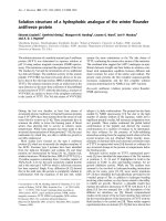
cells were transduced at ~90% efficiency with a SOX9-GFP
bicistronic retroviral vector (SOX9-SW1353), and controls
were transduced with a SOX9-free GFP-retrovirus (GFP-
SW1353) (Figure 1a). The SW1353 cells were of interest for
this study as their normal expression of SOX9 was much lower
than human chondrocytes in cartilage (relative to GAPDH),
whereas the level of SOX9 expression following transduction
was increased by 18-fold (Figure 1c) and approached the
level found in cartilage. There was no discernable change in
morphology following SOX9 transduction. Immunoblotting
confirmed that SOX9-SW1353 synthesised increased levels
of the SOX9 protein compared with controls (Figure 1b). The
cells also showed increased gene expression of SOX6 (up to
14-fold) and COL2A1 (up to 13-fold), but aggrecan expres-
sion was low and was unchanged by SOX9. The SW1353
cells expressed high levels of COL1A1 and this was reduced
6-fold by SOX9 transduction. The stimulation of both SOX6
and COL2A1 by SOX9 confirmed that SW1353 cells were
responsive to this factor, unlike other non-chondrocytic cells,
such as dermal fibroblasts, which failed to upregulate cartilage
matrix genes in response to SOX9 transduction [28].
Microarray analysis of SOX9-transduced SW1353 cells
Dual hybridisations were performed in quadruplicate (includ-
ing duplicated orientations of dye to sample) using probes
produced from single RNA samples from SOX9-SW1353 or
GFP-SW1353 cells. Extensive filtering of the normalised data
was carried out as described in the materials and methods
section. From the original 9,600 different genes on each array,
22 were found to be upregulated and 9 were downregulated
by SOX9. From these, eight of the most strongly regulated
genes were selected for further analysis (Table 2).
Real time PCR analysis of SOX9-regulated genes
Gene-expression analysis by quantitative real time (qRT) PCR
was used to confirm the gene changes identified by the micro-
array analysis. Statistically significant upregulation was
observed for SRPX (1.8-fold), S100A1 (7.9-fold), APOD (4.2-
fold) RGC32 (2.3-fold) and CRTL1 (2 fold) with analyses from
separately cultured SW1353 cells. Regulation of the expres-
sion of SPINT1, CRLF1 and MYBPH could not be confirmed.
Having previously shown that retroviral transduction with
SOX9 of passaged human OA chondrocytes re-activated their
potential to form cartilage matrix [15], we investigated the
expression of the novel genes identified in SW1353 cells in
human articular chondrocytes that had been expanded in mon-
olayer culture and transduced with SOX9-retrovirus. The
results showed significant upregulation (P < 0.05) of S100A1
(26.8-fold), CRTL1 (3.0-fold) and SRPX (1.7-fold) following
SOX9 transduction. Interestingly, SPINT1 was also signifi-
cantly upregulated in the chondrocytes (2.1-fold), which dif-
fered from the finding in the SW1353 cells. APOD was
Figure 1
Retroviral expression of SOX9 in SW1353 chondrosarcoma cellsRetroviral expression of SOX9 in SW1353 chondrosarcoma cells. (a)
Phase contrast micrograph demonstrating the morphology of SW1353
chondrosarcoma cells transduced with a retrovirus containing GFP or
bicistronic SOX9-GFP. Scale Bar = 50 μm. (b) Cell lysates from GFP-
or SOX9-SW1353 cells were analysed by western blotting using an
anti-SOX9 antibody. (c) Real time PCR analysis of cDNA derived from
green-fluorescent protein (GFP; black bars) or bicistronic SOX9-GFP
(grey bars) transduced SW1353 chondrosarcoma cells.
Available online />Page 5 of 10
(page number not for citation purposes)
expressed at very low level in transduced and control human
OA chondrocytes in monolayer culture, and although its
expression appeared to be increased slightly following SOX9
transduction, no statistical analysis was possible. MYBPH
expression was again unaffected by SOX9. Therefore, there
were examples of genes that displayed similar responses to
SOX9 transduction in both SW1353 cells and primary
chondrocytes, but also genes for which there were clear reg-
ulatory differences between the cell types.
Primary chondrocyte culture with decrease in SOX9
expression
To determine whether the expression of genes identified in this
study were altered by non-viral-mediated changes in SOX9
expression, we investigated in vitro cultures of freshly isolated
human articular chondrocytes (Figure 2). These cells were
from OA cartilage, and had a lower expression of SOX9 in cul-
ture than in tissue, but still higher (relative to GAPDH) than in
SW1353 cells. During monolayer culture of the OA chondro-
cytes there was a further 8–10-fold decrease in SOX9 expres-
sion, and we examined whether this was accompanied by any
change in expression of the newly identified genes (Figure 2).
A number of the genes including S100A1, RGC32, CRTL1
and SPINT1 were down regulated under these conditions,
and therefore correlated with the reduction in SOX9. SRPX, in
contrast, did not correlate with SOX9 in the monolayer cul-
tured HAC, and its expression increased with passage. The
expression of another gene, CRLF1, was unchanged during
the fall in SOX9. The expression of MYBPH and APOD (one
of the genes most strongly upregulated by SOX9 in SW1353
cells) were very low in these primary human articular chondro-
cytes, and significant regulation could not be identified.
Expression of SOX9-regulated genes in normal and
osteoarthritic cartilage
In a previous study [5] we showed that osteoarthritic cartilage
consistently showed reduced expression of SOX9 compared
with healthy age-matched control tissue. It was therefore of
great interest to understand whether the newly identified
genes were also altered in expression in OA cartilage. We
therefore probed globally amplified cDNA samples from nor-
mal and OA femoral knee cartilage [5] for their expression (Fig-
ure 3). Furthermore, the tissues samples analysed were paired
cartilage samples from high-load-bearing (MFC) and low-load-
bearing regions (LPC) of the same joints. SOX9 gene expres-
sion was reduced (P < 0.0001) in the osteoarthritic samples
compared with the age-matched controls, and there was no
difference between differently loaded sites. Of the genes
investigated, five were expressed at lower level in
osteoarthritic cartilage (CRTL1 (P < 0.01), SRPX (P <
0.0001), SPINT1 (P < 0.0001), RGC32 (P < 0.001) and
APOD (P < 0.0001)). One gene (CRLF1) was significantly
upregulated in OA tissue compared with normal tissue (P <
0.01), and S100A1 showed a wide range of expression and
no significant difference between OA and controls; however,
analysis of the results from all the cartilage samples showed
that its expression was correlated with SOX9. Most genes
investigated showed similar expression in both the more highly
loaded MFC and the lower loaded LPC sites. The exceptions
to this were APOD, which was further reduced (P < 0.01) in
expression in the more loaded and damaged cartilage, while
both S100A1 (p < 0.03) and CRLF1 (p < 0.02) were
expressed at higher levels in the more loaded tissue. The
analysis of cartilage oligomeric matrix protein (COMP) gene
expression showed that it was unaffected in OA (or location in
the joint), and demonstrated that there was no generalised
downregulation of all gene expression in OA chondrocytes.
Genomic analysis of potential SOX9 binding sites in
candidate genes
The candidate genes SPINT1, SRPX, APOD, RGC32,
CRTL1 and S100A1 were among those whose expression fol-
lowed that of SOX9 in most of the experimental systems that
we examined. Of these genes, CRTL1 and S100A1 have pre-
viously been shown to possess SOX9 binding sequences
within their promoter regions [9,29]. Potential SOX9 binding
Table 2
Candidate genes chosen following microarray analysis of SOX9 transduced SW1353 chondrosarcoma cells
Gene name GenBank accession number Fold upregulation Fold downregulation
Apolipoprotein D (APOD) NM_001647 22.89 NA
RGC32 NM_014059
10.82 NA
S100 calcium-binding protein A1 (S100A1) NM_006271
8.84 NA
Sushi-repeat-containing protein X chromosome (SRPX) NM_006307
3.47 NA
Cytokine receptor-like factor 1 (CRLF1) NM_004750
3.31 NA
Cartilage linking protein 1 (CRTL1) NM_001884
2.97 NA
Myosin-binding protein H (MYBPH) NM_004997
NA 4.23
Kunitz-type protease inhibitor (SPINT1) AF027205
NA 2.83
Genes demonstrated regulation consistently across quadruplicate microarray experiments. NA, not applicable.
Arthritis Research & Therapy Vol 9 No 5 Tew et al.
Page 6 of 10
(page number not for citation purposes)
sites in non-coding, conserved regions of the genome in and
around the other four gene loci were studied using rVista. This
tool identifies conserved transcription factor binding sites in
sequences based on homologies of such sites between differ-
ent species, and in these genes it identified binding site con-
servation in human, mouse and dog sequences (Table 3). The
analysis demonstrated conserved SOX9 binding regions in all
four candidate genes. As a control, analysis of the house-keep-
ing gene GAPDH revealed no potential SOX9 binding sites
common to all three genomic sequences.
Discussion
The transcription factor SOX9 has been shown to control the
transcription of a number of important cartilage matrix genes.
It is able to interact with a conserved cartilage-specific
enhancer element in the COL2A1 gene and can bind to
promoter and enhancer regions in a number of other cartilage
matrix genes [6-9]. This work has now identified a number of
genes whose expression was changed in SW1353 cells by
increasing SOX9 expression by retroviral transduction and
may similarly contain conserved SOX9 response elements
The human SW1353 cells have previously been used to eluci-
date cytokine regulation of ECM-degrading proteases as
model chondrocytes [27,30] and their SOX9 expression was
shown to be increased by fibroblast growth factors 1, 2 and 9
and decreased by IL1β and TNFα [31]. They have also been
used to identify cyclic AMP response element binding protein
Figure 2
Regulation of candidate genes during chondrocyte dedifferentiationRegulation of candidate genes during chondrocyte dedifferentiation. Real time PCR analysis of candidate gene expression in cDNA from human
articular chondrocytes at passage (P) 0, 1 or 2. Mean fold-change values (where P0 = 1) with standard errors are presented from chondrocytes cul-
tures obtained from 3 donors. * indicates significant difference in expression compared with passage 0 levels P < 0.05 by paired students t-test.
Available online />Page 7 of 10
(page number not for citation purposes)
and p300 as novel partners of SOX9 that bind at cartilage-
specific promoter sites [32]. Thus the SW1353 cells have
some features of chondrocytes, but as with other chondrocytic
cells in monolayer culture they expressed low levels of both
cartilage ECM genes and SOX9, 6 and 5 [33]. Their transduc-
tion of cytokine signals has also been reported to differ from
that seen in primary articular chondrocytes [33]. In this study
SOX9-transduction increased the expression of target genes
(such as COL2A1), although others appeared unaffected
(such as aggrecan). The SW1353 cells therefore appear to
lack some chondrocyte properties, but their response to
SOX9 transduction was clearly more chondrocyte-like than
Figure 3
Comparison of the expression levels of candidate genes in normal and osteoarthritic cartilageComparison of the expression levels of candidate genes in normal and osteoarthritic cartilage. Real time PCR analysis of candidate gene expression
in globally amplified cDNA representative of mRNA levels from normal (n = 8) or osteoarthritic (n = 15) human articular cartilage samples. Cartilage
for the analysis was derived from either the medial or lateral femoral condyles. NM = normal medial, NL = normal lateral, OM = osteoarthritic medial
and OL = osteoarthritic lateral. Symbols above bars indicate statistically significant regulation of that gene caused by:* disease (P < 0.05 mixed
effects regression model) or ᭜ joint location (P < 0.05 mixed effects regression model).
Arthritis Research & Therapy Vol 9 No 5 Tew et al.
Page 8 of 10
(page number not for citation purposes)
dermal fibroblasts, which showed no regulation cartilage
matrix genes in response to the overexpression of SOX9 [28].
From the initial microarray analysis we followed up gene-
expression changes by qRT-PCR analysis in SOX9-SW1353
cells to confirm their regulation. Investigation of changes in the
expression of this panel of genes in primary human chondro-
cytes following SOX9 transduction showed that some genes
showed evidence of similar control to SW1353 cells, although
some showed no comparable response. To extend these
observations we investigated the expression in articular
chondrocytes under conditions where the expression of
SOX9 was changed by both natural and pathological factors.
The expression was investigated in primary human chondro-
cytes cultured and passaged in monolayer, under which con-
ditions the expression of SOX9 progressively becomes
reduced. It was only after culture of the OA chondrocytes that
the expression of SOX9 became reduced to the level found in
the SW1353 cells before transduction. The change in expres-
sion during this fall in endogenous SOX9 expression showed
S100A1, RGC32, CRTL1 and SPINT1 to decrease, which
were therefore correlated with SOX9, as in SW1353 cells.
The identification of these SOX9-regulated genes led us to
probe a human normal and OA cartilage library of globally
amplified cDNA representing mRNA levels in chondrocytes in
cartilage taken from load bearing or non-load-bearing regions
from age-matched normal and OA human knees. SOX9 has
been shown to be downregulated in osteoarthritis, and this
may contribute to the pathological process by causing a
reduction in the expression of ECM genes [4,5]. We found
that many of the genes whose expression was altered by
SOX9 in SW1353 cells and/or isolated primary chondrocytes
displayed altered expression levels in OA cartilage (CRTL1 (P
< 0.01), SRPX (P < 0.0001), SPINT1 (P < 0.0001), RGC32
(P < 0.001) and APOD (P < 0.0001)) compared with age-
matched controls. It is worth noting that even a gene such as
COL2A1, which is known to have SOX9 regulatory elements,
has been demonstrated to poorly correlate with the expression
of SOX9 in control and osteoarthritic cartilage [4], suggesting
that in OA its expression is more dominantly controlled by
other factors. It was therefore more interesting to identify
genes such as CRTL1, RGC32, S100A1 and APOD, which
had a pattern of expression closely correlating with SOX9
expression levels in SW1353 cells, in primary chondrocytes,
and also in OA cartilage. SRPX generally correlated with
SOX9, except during chondrocyte dedifferentiation, which
may indicate that other factors predominantly influence it dur-
ing this process.
APOD, which was expressed at relatively low levels in
SW1353 cells, was expressed more strongly in cartilage, and
the expression was reduced in OA, which was consistent with
the decrease in SOX9. APOD encodes apolipoprotein D,
which is a protein component of low density lipoprotein in
human plasma [34], and is reported to be a transit protein in
the skin [35]. It may therefore have some function in cartilage
ECM. The finding that APOD is downregulated in OA agrees
with two previous microarray studies comparing normal and
OA tissue [36,37]. The present results showed further that
APOD expression was not only downregulated in OA, but was
also most strongly downregulated in the highly loaded, more
physically damaged cartilage. APOD expression thus corre-
lated with cartilage damage, whereas matrix genes, such as
CTRL1 and SOX9, were similarly changed in OA in both low-
loaded and high-loaded cartilage sites.
S100A1 encodes an intercellular calcium-binding protein,
which can control myocardial contractility [38] and has
recently been identified as an important SOX9 regulated gene
that controls the terminal differentiation of chondrocytes [29].
S100A1 has previously been reported to be downregulated in
osteoarthritis [36]. In the OA and control cartilage samples
investigated here, S100A1 had lower mean expression in OA,
but the difference was not statistically significant. However, its
expression was found to be significantly correlated with SOX9
when the results from all cartilage samples were analysed
(data not shown).
SRPX expression was increased by SOX9 transduction in
SOX9-SW1353 and in primary human articular chondrocytes,
and its expression was greatly reduced in OA cartilage. It
Table 3
Predicted SOX9 binding sites in candidate genes
a
Gene Conserved SOX9 binding site, relative to transcription start site Transcription start site (bp position based on homo sapiens
genome build 35.1)
APOD +2998 bp to +3011 bp +3110 bp to +3123 bp CHR3_RANDOM:544561
GAPDH None passed criteria CHR12:6513945
RGC32 +2534 bp to +2547 bp CHR13:40929712
SPINT1 -1183 bp to -1170 bp CHR15:38923534
SRPX +3939 bp to +3952 bp +5628 bp to +5641 bp +15773 bp to
+15786 bp
CHRX:37836348
a
Base pair (bp) positions are given relative to the transcription start site of each gene. In all instances, positive numbers describe sites within the
first intron of the gene.
Available online />Page 9 of 10
(page number not for citation purposes)
therefore correlated well with SOX9 expression, although dur-
ing chondrocyte dedifferentiation its expression increased
more than sevenfold by passage 2 and was clearly unrelated
to SOX9. This perhaps emphasises that any loss of chondro-
cyte phenotype in OA cartilage does not occur through a
mechanism closely related to the loss of phenotype that
occurs in these cells in monolayer culture. SRPX has a recog-
nised role in ocular biology and disease. The SRPX gene
encodes a putative membrane protein expressed abundantly
in the retina, and was discovered as a candidate gene respon-
sible for X-linked retinitis pigmentosa [39]. SOX9 has a poten-
tial regulatory role in the development of the retina, and may
regulate the synthesis of collagen type II in the vitreous of the
eye [40]. Furthermore, disrupted SOX9 expression in the 'odd
sex' transgenic mouse, which results in sex reversal, also
causes an eye phenotype with microphthalmia with cataracts
[41]. The expression of SRPX may therefore be regulated by
SOX9 during ocular development and may also have a role in
cartilage biology.
Despite being unable to confirm any regulation by SOX9 in
SW1353 by real time PCR, and with its expression unaffected
in primary chondrocytes by the transition to monolayer culture,
it was interesting that CRLF1 was significantly upregulated in
osteoarthritic cartilage. CRLF1 protein is a member of the
cytokine type I receptor family, and when expressed as a het-
erodimer with the cardiotrophin-like cytokine (CLC) can acti-
vate the membrane bound ciliary neurotrophic factor receptor-
α (CNTFRα), which causes an interaction between gp130
and leukaemia inhibitory factor receptor, leading to cell signal-
ling [42,43]. Further work characterising the expression of the
genes encoding CNTFRα and CLC in cartilage is required as
does the possibility that upregulation of CRLF1 expression
could have a use as a marker of OA.
This study identified genes whose expression in chondrocytes
was consistently correlated with changes in SOX9 expres-
sion. The results suggested that the expression of these genes
may be regulated by SOX9, and as SOX9 is essential for
chondrocyte phenotype, the novel genes with unknown func-
tion may help control the differentiated state of chondrocytes
within cartilage. The correlation of expression with SOX9
linked these genes to changes in cartilage in OA. As OA is
characterized by degenerative changes in cartilage it will be
important to establish how the changes in the expression of
SOX9-regulated genes contribute to the progressive loss of
chondrocyte function and the compromise in cartilage integrity
that occurs in OA.
Conclusion
We have identified genes in a human chondrosarcoma cell line
whose expression is altered by the overexpression of the chon-
drogenic transcription factor SOX9. Some of these genes
were similarly regulated in primary human chondrocytes in
response to changes in SOX9 induced by overexpression or
by dedifferentiation in culture. The expression of some of these
genes was also correlated with SOX9 expression in intact
human articular cartilage, and was therefore suppressed in OA
cartilage compared with age-matched control cartilage.
Competing interests
The authors declare that they have no competing interests.
Authors' contributions
SRT conceived, designed and performed the experimental
work associated with the microarray and was responsible for
the initial versions of this manuscript. CJB collected the normal
and OA cartilage and produced the cDNA libraries from fem-
oral cartilage. CMR undertook the laboratory work associated
with real time PCR analysis of the normal and OA cartilage
libraries. PDC performed the statistical analyses, designed
and validated the PCR primers, and supervised the project.
TEH supervised and oversaw the completion of the studies as
well as the writing of this manuscript. All authors read and
approved the final manuscript.
Acknowledgements
The authors wish to thank Andrew Hayes and Leo Zeef for technical and
analytical assistance with the microarray study and to the Human
Genome Mapping Project for kindly providing the arrays. This work was
funded by Biotechnology and Biological Sciences Research Council,
Medical Research Council and Engineering and Physical Sciences
Research Council, The Wellcome Trust (Research Leave Fellowship
GR067462MA to PDC) and the Arthritis Research Campaign (Clinical
Research Training Fellowship to CJB).
References
1. Zhao Q, Eberspaecher H, Lefebvre V, De Crombrugghe B: Paral-
lel expression of Sox9 and Col2a1 in cells undergoing
chondrogenesis. Dev Dyn 1997, 209:377-386.
2. Buckwalter JA, Saltzman C, Brown T: The impact of osteoarthri-
tis: implications for research. Clin Orthop Relat Res
2004:S6-15.
3. Aigner T, McKenna L: Molecular pathology and pathobiology of
osteoarthritic cartilage. Cell Mol Life Sci 2002, 59:5-18.
4. Aigner T, Gebhard PM, Schmid E, Bau B, Harley V, Poschl E:
SOX9 expression does not correlate with type II collagen
expression in adult articular chondrocytes. Matrix Biol 2003,
22:363-372.
5. Brew CJ, Andrew JG, Boot-Handford R, Hardingham TE: Late
osteoarthritic cartilage shows down regulation of SOX9 and
aggrecan expression but little evidence of chondrocyte
hypertrophy. Trans Orthop Res Soc 2004, 50:938.
6. Sekiya I, Tsuji K, Koopman P, Watanabe H, Yamada Y, Shinomiya
K, Nifuji A, Noda M: SOX9 enhances aggrecan gene promoter/
enhancer activity and is up-regulated by retinoic acid in a car-
tilage-derived cell line, TC6. J Biol Chem 2000,
275:10738-10744.
7. Zhang P, Jimenez SA, Stokes DG: Regulation of human COL9A1
gene expression. Activation of the proximal promoter region
by SOX9. J Biol Chem 2003, 278:117-123.
8. Bridgewater LC, Lefebvre V, de Crombrugghe B: Chondrocyte-
specific enhancer elements in the Col11a2 gene resemble the
Col2a1 tissue-specific enhancer. J Biol Chem 1998,
273:14998-15006.
9. Kou I, Ikegawa S: SOX9-dependent and -independent tran-
scriptional regulation of human cartilage link protein. J Biol
Chem 2004, 279:50942-50948.
10. Wagner T, Wirth J, Meyer J, Zabel B, Held M, Zimmer J, Pasantes
J, Bricarelli FD, Keutel J, Hustert E, et al.: Autosomal sex reversal
Arthritis Research & Therapy Vol 9 No 5 Tew et al.
Page 10 of 10
(page number not for citation purposes)
and campomelic dysplasia are caused by mutations in and
around the SRY-related gene SOX9. Cell 1994, 79:1111-1120.
11. Bi W, Deng JM, Zhang Z, Behringer RR, de Crombrugghe B: Sox9
is required for cartilage formation. Nat Genet 1999, 22:85-89.
12. Stokes DG, Liu G, Dharmavaram R, Hawkins D, Piera-Velazquez S,
Jimenez SA: Regulation of type-II collagen gene expression
during human chondrocyte de-differentiation and recovery of
chondrocyte-specific phenotype in culture involves Sry-type
high-mobility-group box (SOX) transcription factors. Biochem
J 2001, 360:461-470.
13. Hardingham T, Tew S, Murdoch A: Tissue engineering: chondro-
cytes and cartilage. Arthritis Res 2002, 4(Suppl 3):S63-68.
14. Li Y, Tew SR, Russell AM, Gonzalez K, Hardingham TE, Hawkins
RE: Transduction of human articular chondrocytes with aden-
oviral, retroviral and lentiviral vectors and the effects of
enhanced expression of SOX9. Tissue Eng 2004, 10:575-584.
15. Tew SR, Li Y, Pothacharoen P, Tweats LM, Hawkins RE, Hard-
ingham TE: Retroviral transduction with SOX9 enhances re-
expression of the chondrocyte phenotype in passaged oste-
oarthritic human articular chondrocytes. Osteoarthritis
Cartilage 2005, 13:80-89.
16. Lefebvre V, Huang W, Harley VR, Goodfellow PN, de Crombrug-
ghe B: SOX9 is a potent activator of the chondrocyte-specific
enhancer of the pro alpha1(II) collagen gene. Mol Cell Biol
1997, 17:2336-2346.
17. Kellgren JH, Lawrence JS: Radiological assessment of osteo-
arthrosis. Ann Rheum Dis 1957, 16:494-502.
18. MAXD [ />]
19. Yang YH, Dudoit S, Luu P, Lin DM, Peng V, Ngai J, Speed TP: Nor-
malization for cDNA microarray data: a robust composite
method addressing single and multiple slide systematic
variation. Nucleic Acids Res 2002, 30:e15.
20. MIAMExpress [ />]
21. Reno C, Marchuk L, Sciore P, Frank CB, Hart DA: Rapid isolation
of total RNA from small samples of hypocellular, dense con-
nective tissues. Biotechniques 1997, 22:1082-1086.
22. Flannery CR, Little CB, Caterson B, Hughes CE: Effects of cul-
ture conditions and exposure to catabolic stimulators (IL-1
and retinoic acid) on the expression of matrix metalloprotein-
ases (MMPs) and disintegrin metalloproteinases (ADAMs) by
articular cartilage chondrocytes. Matrix Biol 1999, 18:225-237.
23. Al-Taher A, Bashein A, Nolan T, Hollingsworth M, Brady G: Global
cDNA amplification combined with real-time RT-PCR: accurate
quantification of multiple human potassium channel genes at
the single cell level. Yeast 2000, 17:201-210.
24. Livak KJ, Schmittgen TD: Analysis of relative gene expression
data using real-time quantitative PCR and the 2(-Delta Delta
C(T)) Method. Methods 2001, 25:402-408.
25. Vista browser [ />]
26. Loots GG, Ovcharenko I, Pachter L, Dubchak I, Rubin EM: rVista
for comparative sequence-based discovery of functional tran-
scription factor binding sites. Genome Res 2002, 12:832-839.
27. Liacini A, Sylvester J, Li WQ, Zafarullah M: Mithramycin downreg-
ulates proinflammatory cytokine-induced matrix metallopro-
teinase gene expression in articular chondrocytes. Arthritis
Res Ther 2005, 7:R777-783.
28. Ikeda T, Kamekura S, Mabuchi A, Kou I, Seki S, Takato T, Naka-
mura K, Kawaguchi H, Ikegawa S, Chung UI: The combination of
SOX5, SOX6, and SOX9 (the SOX trio) provides signals suffi-
cient for induction of permanent cartilage. Arthritis Rheum
2004, 50:3561-3573.
29. Saito T, Ikeda T, Nakamura K, Chung UI, Kawaguchi H: S100A1
and S100B, transcriptional targets of SOX trio, inhibit terminal
differentiation of chondrocytes. EMBO Rep 2007.
30. Shi J, Schmitt-Talbot E, DiMattia DA, Dullea RG: The differential
effects of IL-1 and TNF-alpha on proinflammatory cytokine and
matrix metalloproteinase expression in human chondrosar-
coma cells. Inflamm Res 2004, 53:377-389.
31. Schaefer JF, Millham ML, de Crombrugghe B, Buckbinder L: FGF
signaling antagonizes cytokine-mediated repression of Sox9
in SW1353 chondrosarcoma cells. Osteoarthritis Cartilage
2003, 11:233-241.
32. Tsuda M, Takahashi S, Takahashi Y, Asahara H: Transcriptional
co-activators CREB-binding protein and p300 regulate
chondrocyte-specific gene expression via association with
Sox9. J Biol Chem 2003, 278:27224-27229.
33. Gebauer M, Saas J, Sohler F, Haag J, Soder S, Pieper M, Bartnik
E, Beninga J, Zimmer R, Aigner T: Comparison of the chondro-
sarcoma cell line SW1353 with primary human adult articular
chondrocytes with regard to their gene expression profile and
reactivity to IL-1beta. Osteoarthritis Cartilage 2005,
13:697-708.
34. Fielding PE, Fielding CJ: A cholesteryl ester transfer complex in
human plasma. Proc Natl Acad Sci USA 1980, 77:3327-3330.
35. Zeng C, Spielman AI, Vowels BR, Leyden JJ, Biemann K, Preti G:
A human axillary odorant is carried by apolipoprotein D. Proc
Natl Acad Sci USA 1996, 93:6626-6630.
36. Tardif G, Hum D, Pelletier JP, Boileau C, Ranger P, Martel-Pelletier
J:
Differential gene expression and regulation of the bone mor-
phogenetic protein antagonists follistatin and gremlin in nor-
mal and osteoarthritic human chondrocytes and synovial
fibroblasts. Arthritis Rheum 2004, 50:2521-2530.
37. Gebauer M, Saas J, Haag J, Dietz U, Takigawa M, Bartnik E, Aigner
T: Repression of anti-proliferative factor Tob1 in osteoarthritic
cartilage. Arthritis Res Ther 2005, 7:R274-284.
38. Most P, Bernotat J, Ehlermann P, Pleger ST, Reppel M, Borries M,
Niroomand F, Pieske B, Janssen PM, Eschenhagen T, et al.:
S100A1: a regulator of myocardial contractility. Proc Natl Acad
Sci USA 2001, 98:13889-13894.
39. Meindl A, Carvalho MR, Herrmann K, Lorenz B, Achatz H, Apfelst-
edt-Sylla E, Wittwer B, Ross M, Meitinger T: A gene (SRPX)
encoding a sushi-repeat-containing protein is deleted in
patients with X-linked retinitis pigmentosa. Hum Mol Genet
1995, 4:2339-2346.
40. Ihanamaki T, Saamanen AM, Suominen J, Pelliniemi LJ, Harley V,
Vuorio E, Salminen H: Expression of Sox9 and type IIA
procollagen during ocular development and aging in
transgenic Del1 mice with a mutation in the type II collagen
gene. Eur J Ophthalmol 2002, 12:450-458.
41. Qin Y, Kong LK, Poirier C, Truong C, Overbeek PA, Bishop CE:
Long-range activation of Sox9 in Odd Sex (Ods) mice. Hum
Mol Genet 2004, 13:1213-1218.
42. Elson GC, Lelievre E, Guillet C, Chevalier S, Plun-Favreau H,
Froger J, Suard I, de Coignac AB, Delneste Y, Bonnefoy JY, et al.:
CLF associates with CLC to form a functional heteromeric lig-
and for the CNTF receptor complex. Nat Neurosci 2000,
3:867-872.
43. Lelievre E, Plun-Favreau H, Chevalier S, Froger J, Guillet C, Elson
GC, Gauchat JF, Gascan H: Signaling pathways recruited by
the cardiotrophin-like cytokine/cytokine-like factor-1 compos-
ite cytokine: specific requirement of the membrane-bound
form of ciliary neurotrophic factor receptor alpha component.
J Biol Chem 2001, 276:22476-22484.