Báo cáo y học: "Human articular chondrocytes produce IL-7 and respond to IL-7 with increased production of matrix metalloproteinase-13" ppsx
Bạn đang xem bản rút gọn của tài liệu. Xem và tải ngay bản đầy đủ của tài liệu tại đây (1.46 MB, 10 trang )
Open Access
Available online />Page 1 of 10
(page number not for citation purposes)
Vol 10 No 1
Research article
Human articular chondrocytes produce IL-7 and respond to IL-7
with increased production of matrix metalloproteinase-13
David Long
1
, Simon Blake
2
, Xiao-Yu Song
2
, Michael Lark
2
and Richard F Loeser
1
1
Section of Molecular Medicine, Department of Internal Medicine, Wake Forest University School of Medicine, Medical Center Blvd, Winston-Salem,
North Carolina 27157, USA
2
Centocor Inc., Great Valley Parkway, Malvern, Pennsylvania 19355, USA
Corresponding author: Richard F Loeser,
Received: 29 Jun 2007 Revisions requested: 29 Aug 2007 Revisions received: 29 Jan 2008 Accepted: 20 Feb 2008 Published: 20 Feb 2008
Arthritis Research & Therapy 2008, 10:R23 (doi:10.1186/ar2376)
This article is online at: />© 2008 Long et al.; licensee BioMed Central Ltd.
This is an open access article distributed under the terms of the Creative Commons Attribution License ( />),
which permits unrestricted use, distribution, and reproduction in any medium, provided the original work is properly cited.
Abstract
Introduction Fibronectin fragments have been found in the
articular cartilage and synovial fluid of patients with osteoarthritis
and rheumatoid arthritis. These matrix fragments can stimulate
production of multiple mediators of matrix destruction, including
various cytokines and metalloproteinases. The purpose of this
study was to discover novel mediators of cartilage destruction
using fibronectin fragments as a stimulus.
Methods Human articular cartilage was obtained from tissue
donors and from osteoarthritic cartilage removed at the time of
knee replacement surgery. Enzymatically isolated chondrocytes
in serum-free cultures were stimulated overnight with the 110
kDa α5β1 integrin-binding fibronectin fragment or with IL-1, IL-
6, or IL-7. Cytokines and matrix metalloproteinases released into
the media were detected using antibody arrays and quantified
by ELISA. IL-7 receptor expression was evaluated by flow
cytometry, immunocytochemical staining, and PCR.
Results IL-7 was found to be produced by chondrocytes
treated with fibronectin fragments. Compared with cells isolated
from normal young adult human articular cartilage, increased IL-
7 production was noted in cells isolated from older adult tissue
donors and from osteoarthritic cartilage. Chondrocyte IL-7
production was also stimulated by combined treatment with the
catabolic cytokines IL-1 and IL-6. Chondrocytes were found to
express IL-7 receptors and to respond to IL-7 stimulation with
increased production of matrix metalloproteinase-13 and with
proteoglycan release from cartilage explants.
Conclusion These novel findings indicate that IL-7 may
contribute to cartilage destruction in joint diseases, including
osteoarthritis.
Introduction
The loss of cartilage matrix that occurs in osteoarthritis (OA) is
associated with a disturbance in the balance of anabolic (syn-
thetic) and catabolic (destructive) activities of the articular
chondrocytes [1]. There is increasing evidence that cytokines,
including IL-1, IL-6, and tumor necrosis factor (TNF)-α, play a
role in matrix destruction by enhancing chondrocyte catabolic
activity [2]. In addition to inducing matrix degrading enzymes
directly, these cytokines can also act by stimulating production
of additional proinflammatory cytokines. IL-6 is among the
cytokines produced by chondrocytes after IL-1 stimulation [3-
5]. These two cytokines have been shown to act synergisti-
cally to induce cartilage breakdown [6], suggesting that
chondrocytes have the ability to respond to co-stimulation with
multiple cytokine signals. A role for local production of
cytokines in the joint destruction that occurs in rheumatoid
arthritis (RA) is well established, and there is increasing evi-
dence for the role of cytokines in OA [7]. Determining which
cytokines are responsible for joint tissue destruction in arthritis
is the subject of continuing research.
IL-7 is a cytokine that produces a diverse array of biologic
effects. It was first described as a factor that promotes the
growth of B cells in mice [8]. Since then, much of the work on
DMEM = Dulbecco's modified Eagle's medium; ELISA = enzyme-linked immunosorbent assay; GAG = glycosaminoglycan; IL = interleukin; MMP =
matrix metalloproteinase; OA = osteoarthritis; PCR = polymerase chain reaction; PYK = proline-rich tyrosine kinase; RA = rheumatoid arthritis; RT =
reverse transcription; TIMP = tissue inhibitor of metalloproteinases; TNF = tumor necrosis factor.
Arthritis Research & Therapy Vol 10 No 1 Long et al.
Page 2 of 10
(page number not for citation purposes)
IL-7 has been focused on its importance within the context of
lymphocyte cell biology (for review [9,10]). IL-7 is required for
survival of peripheral T lymphocytes, possibly through negative
regulation of apoptosis in these cells. Other sites of IL-7
production include intestinal epithelial cells, keratinocytes,
endothelial cells, smooth muscle cells, and fibroblasts [9].
IL-7 has also been studied within the context of RA [10]. It has
been shown that IL-7 is produced at higher levels by fibro-
blast-like synoviocytes isolated from patients with RA and that
stimulation of these cells with the proinflammatory stimuli IL-1
and TNF-α upregulated production of IL-7 [11]. Other cells of
the synovial tissue, including synovial macrophages and syno-
vial T cells, have been shown to respond to IL-7 stimulation
with production of the inflammatory cytokines TNF-α and inter-
feron-γ [12]. It has also been demonstrated that levels of IL-7
in synovial fluid are increased in patients with RA [13]. In addi-
tion, IL-7 has been shown to induce bone loss by promoting
secretion of RANKL (receptor activator of nuclear factor-κB
ligand), a cytokine responsible for the formation of osteoclasts,
from T cells [14]. Collectively, these data point strongly to a
role for IL-7 in inflammatory joint disease, but a potential role
for IL-7 as a mediator of cartilage destruction has not been
reported.
Fibronectin fragments have been detected in cartilage and
synovial fluid samples from patients with RA or OA [15] and
are thought to play a role in cartilage destruction in arthritis by
stimulating chondrocytes to produce matrix metalloprotein-
ases (MMPs) as well as multiple cytokines and chemokines,
including IL-1, IL-6, IL-8, monocyte chemotactic protein-1, and
growth-related oncogene family members [5,16,17]. In the
present study, we screened for additional cytokines produced
by chondrocytes in response to fibronectin fragment stimula-
tion and identified IL-7. Levels of production were compared
using human articular chondrocytes isolated from nonarthritic
cartilage from young and old adults and from patients with OA.
The role of IL-1 and IL-6 in stimulating chondrocyte IL-7 pro-
duction was also determined, as was the ability of IL-7 to stim-
ulate chondrocytes directly. The results suggest a potential
role for IL-7 as a factor contributing to cartilage inflammation
and destruction in arthritis.
Materials and methods
Materials
Recombinant human proteins (IL-6, soluble IL-6 receptor, IL-
1β, and IL-7) were purchased from R&D Systems (Minneapo-
lis, MN, USA). Human MMP-13 ELISA, Human IL-7 Quantikine
High Sensitivity ELISA Kit, and Human IL-7 Biotinylated Fluor-
okine Kit were also from R&D Systems. Phospho-PYK-2 anti-
body was from BioSource (Camarillo, CA, USA). Total PYK2
antibody and 110 kDa fibronectin fragment were from Upstate
Biotechnology (Lake Placid, NY, USA). IL-7 receptor primers
and SybrGreen PCR Mastermix were from SuperArray Bio-
sciences (Frederick, MD, USA). RayBio Human Inflammation
Antibody Array III and Matrix Metalloproteinase Antibody Array
were from Raybiotech (Norcross, GA, USA). IL-6 neutralizing
antibody was produced by Centocor (Horsham, PA, USA). IL-
1 receptor antagonist (Anakinra) was a gift from Amgen (Thou-
sand Oaks, CA, USA). Nitrate/Nitrite Colorimetric Assay Kit
was from Cayman Chemical (Ann Arbor, MI, USA).
Tissue acquisition and chondrocyte cell culture
Human ankle and knee articular cartilage were obtained from
tissue donors within 48 hours of death through the Gift of
Hope Organ and Tissue Donor Network (Elmhurst, IL, USA) or
from the National Disease Research Interchange (Philadel-
phia, PA, USA), in accordance with institutional protocol. Each
donor specimen was graded for degenerative changes based
on the 5-point Collins scale (0 to 4), as modified by Muehle-
man and coworkers [18]. The OA cartilage was discarded tis-
sue obtained after knee replacement surgery. Cartilage was
dissected from the joints and digested in a sequential manner
with Pronase (Calbiochem, Gibbstown, NJ, USA) and then
overnight with collagenase, as previously described [19]. Via-
bility of isolated cells was determined using trypan blue, and
cells were counted using a hemocytometer. Monolayer cul-
tures were established by plating cells in six-well plates at 2 ×
10
6
cells/ml in Dulbecco's modified Eagle's medium (DMEM)/
Ham's F-12 medium supplemented with 10% fetal bovine
serum. Plates were maintained for about 5 to 7 days, with
feedings every 2 days until they reached 100% confluence
prior to experimental use.
Cartilage explant culture and stimulation
For explant cultures, full-thickness cartilage discs were
obtained using a 4 mm biopsy punch. Explants were cultured
for 72 hours in DMEM/Ham's F-12 (1/1) media supplemented
with 1% mini-ITS+ (5 nM insulin, 2 μg/ml transferrin, 2 ng/ml
selenous acid, 25 μg/ml ascorbic acid, and bovine serum albu-
min/linoleic acid at 420/2.1 μg/ml) for recovery. Wet weight of
tissue was then measured and explants were cultured at one
explant per well in a 12-well plate in 500 μl serum-free media
for 72 hours of stimulation. Cartilage matrix proteoglycan deg-
radation was estimated by measuring glycosaminoglycan
(GAG) release into the media using the dimethylmethylene
blue assay as previously described [19]. Nitric oxide release
was estimated by measuring nitrate levels in the medium using
a commercially available kit (Cayman Chemical). To test that
the assay was working properly, we stimulated one set of
explants with 10 ng/ml of IL-1β and detected 2.2 μmol/l nitrate
per milligram wet weight of tissue.
Chondrocyte stimulation
Medium was changed to serum-free DMEM/Ham's F-12
medium with antibiotics 18 hours (overnight) and again 2
hours before each experiment. Appropriate stimuli were then
added to cells. The following standard concentrations were
used for stimulation (unless otherwise indicated): 500 nmol/l
fibronectin fragment, 10 ng/ml IL-1β, 10 ng/ml IL-6 plus 20 ng/
Available online />Page 3 of 10
(page number not for citation purposes)
ml soluble IL-6 receptor, and 10 ng/ml IL-7. Inhibitor concen-
trations were 100 μg/ml IL-1 receptor antagonist and 500 ng/
ml IL-6 neutralizing antibody and, when used, these were
added 1 hour before stimulation. In experiments measuring
basal IL-7 production, medium was collected after 48 hours of
incubation in serum-free conditions. When storage was nec-
essary, 0.1% sodium azide was added to the medium before
storage at 4°C.
Antibody array
One milliliter of media was analyzed with the Human Inflamma-
tion Antibody Array III (Raybiotech), which can detect 40 dif-
ferent cytokines, or the Human Matrix Metalloproteinase
Antibody Array (Raybiotech), which can detect seven MMPs
and three tissue inhibitors of metalloproteinases (TIMPs). Both
membranes were spotted in duplicate with cytokine or MMP-
specific antibodies. Membranes were incubated with culture
media and analyzed in accordance with the manufacturer's
instructions.
ELISA
Medium was analyzed with either the Human MMP-13 or
Human IL-7 High Sensitivity ELISA (R&D Systems), in accord-
ance with the manufacturer's instructions. The minimum
detectable dose of IL-7 using this assay is reported as <0.1
pg/ml, with intra-assay and inter-assay precisions (coefficients
of variation) of 8.0 to 9.4 and 7.3 to 10.3 when using cell cul-
ture supernates. For the MMP-13 ELISA, medium was rou-
tinely diluted to obtain values that would fall within the range
of the standard curve.
Immunoblotting
Cells were washed with phosphate-buffered saline and lysed
with lysis buffer that contained 20 mmol/l Tris (pH 7.5), 150
mmol/l NaCl, 1 mmol/l EDTA, 1 mmol/l EGTA, 1% Triton X-
100, 2.5 mmol/l tetrapyrophosphate, 1 mmol/l glycerol phos-
phate, 1 mmol/l Na
3
VO
4
, 1 μl/ml leupeptin, and 1 mmol/l phe-
nylmethylsulfonyl fluoride. Lysates were centrifuged to remove
insoluble material, and the soluble protein concentration was
determined using BCA reagent (Pierce, Rockford, IL, USA).
Samples containing equal amounts of total protein were sep-
arated by SDS-PAGE, transferred to nitrocellulose, and
probed with anti-phospho-PYK2 antibody. Blots were then
stripped and probed with anti-total-PYK2 antibody to confirm
equal loading. Densitometry measurements were taken using
Kodak 1D image analysis software.
Real-time PCR analysis
Total RNA was isolated using the RNeasy Mini Kit (Qiagen,
Valencia, CA, USA). RNA from 10 different chondrocyte cul-
tures was pooled and genomic DNA contamination was
removed using Turbo DNA-free kit (Ambion, Austin, TX, USA),
in accordance with the manufacturer's instructions. Two
micrograms of DNA-free, pooled RNA was reverse transcribed
using an AMV reverse transcriptase and oligo dT primer at
42°C for 1 hour. Two microliters of RT reaction was then com-
bined in a reaction mixture with 1 μl specific primer pair, 12.5
μl 2× SybrGreen PCR Mastermix, and water to a final reaction
volume of 25 μl. Reactions were then run in triplicate with 40
cycles of amplification on an ABI Prism 7000 real-time PCR
machine (Applied Biosystems, Foster City, CA, USA). A nega-
tive control was included that contained primers, water and
Mastermix but no cDNA, and another negative control was
included that contained RNA that had not been reverse tran-
scribed in order to detect contaminating genomic DNA. An
amplification plot was generated using the ABI software. PCR
specificity was confirmed by dissociation curve analysis (data
not shown).
IL-7 binding assay
For flow cytometry analysis, chondrocytes were removed from
six-well dishes by trypsin digestion and for confocal micros-
copy analysis chondrocytes were examined directly in six-well
dishes. In both instances, cells were stained with fluorescently
labeled IL-7 using the Human IL-7 Biotinylated Fluorokine Kit
(R&D Systems), in accordance with the manufacturer's
instructions but with slight modifications. Briefly, cells were
washed twice with phosphate-buffered saline, followed by
incubation for 1 hour at 4°C with either 60 μl of biotinylated IL-
7 or 60 μl of biotinylated negative control reagent or 60 μl
biotinylated IL-7 complexed with a blocking antibody diluted in
wash buffer. Avidin-fluorescein 60 μl was then added to each
set of cells and incubation was continued for a further 30 min-
utes at 4°C. Cells were then washed three times with wash
buffer and examined by either flow cytometry or confocal
microscopy for green fluorescence using lasers with 488 nm
excitation and 530 nm emission wavelengths.
Statistical analysis
Unless indicated otherwise, results were analyzed using the
Student's t-test in StatView 5.0 (SAS Institute Inc., Cary, NC,
USA).
Results
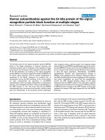
Chondrocytes produce IL-7 in response to fibronectin
fragment stimulation, aging, and OA
Using an antibody array method, one of the cytokines found to
be increased by fibronectin fragment stimulation was IL-7 (Fig-
ure 1a). This finding was confirmed by ELISA using additional
chondrocyte cultures (Figure 1b). In previously published
work, we showed that IL-1 production by chondrocytes
increases with increasing donor age [20]. Using the IL-7
ELISA, we also found a significant (r = 0.818, P = 0.014)
increase with age in the endogenous production of IL-7 by
chondrocytes cultured for 48 hours in serum-free medium
(Figure 2a). Although the younger donors all had Collin's
scores of 0, a correlation between Collin's score and IL-7 lev-
els was not evident in the older donors.
Arthritis Research & Therapy Vol 10 No 1 Long et al.
Page 4 of 10
(page number not for citation purposes)
We also considered the possibility that IL-7 production by
chondrocytes might be increased in cells isolated from OA
cartilage. A significant (P < 0.05) increase in the production of
endogenous IL-7 by isolated OA chondrocytes cultured in
serum-free medium was noted when compared with cells from
age-matched nonarthritic cartilage (Figure 2b).
Chondrocytes express the IL-7 receptor
Having shown that chondrocytes can produce IL-7, we next
wished to determine whether IL-7 could be acting in an auto-
crine or paracrine fashion in cartilage. Using fluorescently
labeled IL-7, examination by either flow cytometry (Figure 3a)
or confocal microscopy (Figure 3b) detected fluorescent IL-7
bound to chondrocytes. Similar results were noted using a
monoclonal antibody to the IL-7 receptor (data not shown). IL-
7 receptor expression by chondrocytes was also confirmed by
real-time PCR using RNA isolated from cartilage of 10 differ-
ent tissue donors (Figure 3c). Taken together, these lines of
evidence suggest that chondrocytes express the IL-7 receptor
and thus might be capable of responding to IL-7 in an auto-
crine or paracrine fashion.
Chondrocytes respond to IL-7 stimulation
Proline-rich tyrosine kinase (PYK)2 is a nonreceptor tyrosine
kinase that was previously shown to be activated in response
to IL-7 stimulation [21], and we previously showed that activa-
tion of PYK2 is required for chondrocyte fibronectin fragment
stimulated MMP-13 production [22]. Therefore, we wished to
determine whether PYK2 would be phosphorylated by
chondrocytes in response to IL-7 stimulation. In initial experi-
ments, chondrocytes were stimulated with 100 ng/ml recom-
binant IL-7 and cells were lysed at different time points over
the course of 2 hours. PYK2 phosphorylation was noted by 30
minutes and reached a maximum at 2 hours (Figure 4a). The
experiment was repeated using a 10 ng/ml concentration of IL-
7 with similar results (data not shown).
Figure 1
Chondrocytes produce IL-7 in response to stimulation with fibronectin fragmentsChondrocytes produce IL-7 in response to stimulation with fibronectin fragments. Human articular chondrocytes obtained from normal articular car-
tilage and cultured in serum-free media were treated overnight with 500 nmol/l of the 110 kDa fibronectin fragment (FN-f). Media was collected and
analyzed for cytokine production using (a) an inflammation antibody array or (b) an IL-7 ELISA. Results are representative of three experiments for
each result with different donor cells used in each experiment. The IL-7 spots on the array are shown in the red circles. (Other spots that were shown
to change after fibronectin fragment stimulation included IL-6, soluble IL-6 receptor [sIL-6R], interferon-inducible protein [IP]-10, and monocyte
chemotactic protein [MCP]-1.)
Available online />Page 5 of 10
(page number not for citation purposes)
We next determined whether IL-7-mediated PYK2 phosphor-
ylation was associated with production of matrix-degrading
enzymes, as we had previously shown using fibronectin frag-
ment stimulation. We chose a 10 ng/ml dose of IL-7 for further
experiments, based on previous dose-response studies
conducted in other cell types that found that 10 ng/ml was
required for stimulation of mononuclear and T-cell proliferation
[11,13] and TNF-α production [12]. Chondrocytes were
treated overnight with recombinant IL-7, and MMP secretion
into the media was analyzed with an MMP antibody array that
included MMP-1, -2, -3, -8, -9, -10 and -13, as well as TIMP-1,
-2 and -4. Interestingly, the only MMP on the array found to be
increased after IL-7 stimulation was MMP-13 (Figure 4b),
which suggests that IL-7 may be acting through a pathway dif-
ferent from those employed by other catabolic cytokines,
which upregulate multiple MMPs. None of the TIMPs were
increased after IL-7 stimulation. The IL-7 stimulation of MMP-
13 production was confirmed by ELISA using additional
chondrocyte cultures (Figure 4c). In cultures from three
donors, we also tested IL-7 at 0.1 ng/ml and found an almost
twofold increase in MMP-13 (data not shown). Although IL-7
has been shown to stimulate TNF-α production by monocytes
and CD4
+
T cells [12], we could not detect, by ELISA, TNF-α
in media from chondrocytes after overnight stimulation with IL-
7 (data not shown).
Several cytokines have been shown to act synergistically with
IL-1 to increase MMP-13 production. We therefore wished to
examine the ability of IL-7 to act synergistically with IL-1. As
shown in Figure 4c, IL-7 was not as potent as IL-1β but the
combination of IL-1 and IL-7 increased MMP-13 levels in the
media to a greater extent than did IL-1 treatment alone.
IL-7 causes proteoglycan release from cartilage explants
In order to further determine whether IL-7 might serve as a cat-
abolic mediator in articular cartilage, we stimulated cartilage
explants with 10 ng/ml IL-7 for 72 hours and measured GAG
release in the medium. Indeed, IL-7 caused a significant
increase in GAG release from cartilage explants relative to
controls (Figure 5a). Increased production of nitric oxide by
chondrocytes is also a characteristic of several catabolic
cytokines, including IL-1, but – unlike in explants treated with
IL-1β – we did not detect an increase in nitrate levels in media
from explants treated with IL-7 (Figure 5b).
The combination of IL-1 and IL-6 stimulates production
of IL-7 by chondrocytes
In previous studies we demonstrated that chondrocyte
fibronectin fragments stimulation increased production of sev-
eral cytokines and chemokines, including IL-1β and IL-6 [5],
which might be responsible for inducing IL-7 production in an
autocrine/paracrine manner. Therefore, chondrocytes were
pretreated for 1 hour with either 100 μg/ml IL-1 receptor
antagonist or 500 ng/ml IL-6 neutralizing antibody, or the com-
bination of both, before addition of fibronectin fragments. IL-6
neutralizing antibody alone reduced fibronectin fragment stim-
ulated IL-7 production, whereas the IL-1 receptor antagonist
showed no inhibition (Figure 6a). However, when both inhibi-
tors were added together, the combination completely
blocked IL-7 production (Figure 5a). This suggested that
chondrocyte IL-7 production was a result of the combined
effects of IL-1 and IL-6. To test this hypothesis, chondrocytes
were stimulated overnight with either recombinant IL-1β, IL-6
plus soluble IL-6 receptor (necessary to stimulate chondro-
cytes with IL-6), or the combination of the cytokines. Indeed,
the combination of the cytokines together was required to
induce IL-7 production (Figure 6b). These results suggest a
role for co-stimulation of chondrocyte IL-7 in response to IL-1
and IL-6.
Discussion
Although IL-7 has traditionally been thought of as a T-cell reg-
ulatory cytokine, in this report the ability of human articular
chondrocytes to produce IL-7, express an IL-7 receptor, and
respond to IL-7 stimulation was demonstrated. Chondrocyte
production of IL-7 was stimulated by catabolic and proinflam-
matory mediators, including the 110 kDa fibronectin fragment,
Figure 2
Effects of age and OA on chondrocyte production of IL-7Effects of age and OA on chondrocyte production of IL-7. Media was
collected 48 hours after changing to serum-free conditions in chondro-
cyte cultures established from (a) nonarthritic cartilage from 10 donors
of different ages or from (b) cartilage from age-matched nonarthritic (n
= 7) and osteoarthritic cartilage (n = 5). IL-7 was measured in the
media using ELISA. The relationship of age to IL-7 levels was analyzed
by Spearman correlation. The numbers in parentheses above the data
points in panel a are the Collin's scores for the donor samples. OA,
osteoarthritis.
Arthritis Research & Therapy Vol 10 No 1 Long et al.
Page 6 of 10
(page number not for citation purposes)
and by the combined actions of IL-1β and IL-6. The stimulation
of chondrocyte IL-7 production by fibronectin fragments
appeared to be part of an autocrine loop mediated by the frag-
ment stimulation of IL-1 and IL-6 production, because
inhibition of these cytokines blocked fragment stimulated IL-7
production. IL-7 stimulated chondrocytes to produce MMP-
13, a metalloproteinase that is responsible for degradation of
type II collagen in cartilage, and caused proteoglycan release
from cartilage explants. Additionally, increased production of
IL-7 was measured in cultures of osteoarthritic chondrocytes
relative to normal chondrocytes. These findings suggest a
potential involvement of IL-7 in the OA disease process.
To our knowledge, this is the first report of IL-7 protein produc-
tion and IL-7 receptor expression by articular chondrocytes. A
previous study used RT-PCR to detect IL-7 RNA in human
articular cartilage obtained from patients with RA but could not
detect IL-7 message in OA or normal cartilage [23]. A second
RT-PCR study confirmed IL-7 expression in RA cartilage but
also detected IL-7 message in two out of six cartilage samples
from OA patients, one out of five cartilage samples from
infants, and in all seven cartilage samples from mice aged 4–
8 days [24]. Mean levels of IL-7 in synovial fluid, measured
using ELISA, were reported to be 34 pg/ml in 44 RA patients
and 1.1 pg/ml in 10 patients with OA [13].
Based on the results from the inflammation antibody array (Fig-
ure 1a), we expected to find significantly higher levels of IL-7
than the low pg/ml range measured using the ELISA. The rea-
son for this discrepancy is not clear but could be due to the
Figure 3
Chondrocyte expression of IL-7 receptorsChondrocyte expression of IL-7 receptors. (a) Chondrocytes isolated from normal cartilage (n = 1) were incubated with a fluorescently labeled
recombinant IL-7 to demonstrate binding of IL-7 to the cell surface. Labeled cells were examined by flow cytometry. The peak that is shaded purple
with the black line shows cells stained with IL-7, the peak with the pink line shows blocking antibody negative control, and the peak with the green
line shows cells stained with the biotin negative control. (b) Chondrocytes isolated from normal cartilage were incubated with a fluorescently labeled
recombinant IL-7 as above. Labeled cells were examined by confocal microscopy. IL-7 staining is shown in green. Top left is the green channel, top
right is differential intermittent contrast, and bottom left is the merged image. Chondrocytes from eight different donors showed similar results. (c)
Pooled RNA isolated from 10 different sets of cultured chondrocytes was subjected to reverse transcription and real-time PCR with an IL-7 receptor
primer set. An amplification plot is shown to demonstrate positive signal. Amplified chondrocyte cDNA in triplicate is shown with the blue lines. Neg-
ative control with no reverse transcription of RNA before real-time PCR is shown with a red line. Negative control with no cDNA is shown with the
black line.
Available online />Page 7 of 10
(page number not for citation purposes)
different antibodies used to detect IL-7 in the two assays, or
perhaps the presence of binding molecules, such as soluble
IL-7 receptor or proteoglycans, that might have affected the
ELISA measurement differently from the membrane array.
However, the 1 to 2 pg/ml amount of IL-7 we detected in
chondrocytes stimulated with fibronectin fragments or IL-1
plus IL-6 is higher than the 0.33 pg/ml IL-7 reported to be pro-
duced by cultured RA synovial fibroblasts and is the same as
the amounts made by these cells after stimulation with IL-1β or
TNF-α [11].
The highest levels of IL-7 were noted in cultured cells estab-
lished from the cartilage of older tissue donors. In previous
work [20] we also noted an age-related increase in production
of IL-1β as well as increased production of MMP-13 in
response to IL-1 or fibronectin fragments. These findings sug-
gest an age-related increase in the proinflammatory
environment of cartilage that could contribute to cartilage
destruction and the development of arthritis in older adults.
In addition to the demonstration that chondrocytes express IL-
7 receptors and produce MMP-13 when cultured in the
presence of IL-7, the ability of chondrocytes to respond to IL-
7 (10 ng/ml) was demonstrated by examining phosphorylation
of a nonreceptor tyrosine kinase, namely PYK2. Activation of
PYK2 through IL-7 stimulation (50 ng/ml) was previously
reported in thymocytes [21]. Signaling mediated by PYK2 in
chondrocytes appears to be an important component of sev-
eral catabolic pathways. In addition to a role in fibronectin frag-
ments mediated MMP-13 production [22], PYK2 has been
shown to be involved in MMP-13 production by chondrocytes
stimulated with the inflammatory protein S100A4 through a
pathway involving intracellular calcium and reactive oxygen
species [25]. It has also been shown to be involved in
chondrocyte production of nitric oxide and MMP-3 induced by
monosodium urate monohydrate crystals [26].
Many cytokines have been identified as secretion products of
chondrocytes and their role in OA has become a subject of
Figure 4
Chondrocytes respond to IL-7 stimulation with increased PYK2 phosphorylation and production of MMP-13Chondrocytes respond to IL-7 stimulation with increased PYK2 phosphorylation and production of MMP-13. (a) Chondrocytes isolated from normal
adult cartilage were stimulated with 10 ng/mL recombinant IL-7 and lysates were made at indicated time points for immunoblotting with an antibody
to phosphorylated proline-rich tyrosine kinase (PYK)2 (Tyr402). The blot was then stripped and probed with total PYK2 antibody to confirm equal
loading. (b) Densitometric scanning of the blot shown in panel a. (c) Medium was collected from serum-free chondrocyte cultures after overnight
stimulation with 10 ng/ml recombinant IL-7 and examined for the presence of multiple matrix metalloproteinase (MMP) family members using an
MMP antibody array. MMP-13 spots are shown in circles. (d,e) Media was collected from serum-free chondrocyte cultures after overnight stimula-
tion with 10 ng/ml recombinant IL-7 or IL-1β, or the two together, and examined for the presence of MMP-13 using a commercially available ELISA.
Results are the mean of seven experiments.
Arthritis Research & Therapy Vol 10 No 1 Long et al.
Page 8 of 10
(page number not for citation purposes)
increasing interest [2,7]. Increased local cytokine activity may
also play an important role in the cartilage destruction that
occurs in RA. The principal cytokines receiving the most atten-
tion to date as mediators of cartilage destruction have been IL-
1β and TNF-α. However, chondrocytes have been shown to
produce a host of cytokines and inflammatory mediators, many
of which are also produced by monocytes/macrophages [27].
IL-7 can be added to this list of mediators. IL-7 is unlikely to be
a sole mediator of cartilage destruction in arthritis. However,
because IL-7 can stimulate cells to produce additional
cytokines, such as IL-6, IL-8 and TNF-α [10] and (as shown
here) can stimulate additional production of MMP-13 when
combined with IL-1β, it may be an important contributor to joint
tissue destruction in OA and RA.
Conclusion
IL-7 can be produced by articular chondrocytes, which also
express IL-7 receptors. Production of IL-7 is increased in
chondrocytes from older donors, from OA cartilage, and after
stimulation with fibronectin fragments, IL-1, and IL-6. Treat-
ment of chondrocytes with IL-7 stimulates PYK2 phosphoryla-
tion, increases the production of MMP-13, and results in GAG
release from cartilage explants. These findings suggest that IL-
7 may contribute to matrix destruction in arthritis.
Competing interests
Richard Loeser received a research grant from Centocor.
Simon Blake, Xiao-Yu, and Michael Lark are employees of
Centocor and own stock in the company.
Authors' contributions
DL designed and carried out experiments and helped to draft
the manuscript. SB, X-YS, and ML contributed to the design
of the study and interpretation of data. RL contributed to study
design, supervised the performance of experiments, inter-
preted data, and completed the writing of the manuscript. All
authors approved the content of the manuscript.
Acknowledgements
We wish to thank Drs Raghu Yammani, Michael Seeds and Hong Chen
for technical assistance and the Gift of Hope Organ and Tissue Donor
Network and the National Disease Research Interchange for providing
donor tissues. We thank Dr David Martin for assistance in obtaining OA
tissue. This work was supported by grants from the NIH (AR49003 and
AG16697) and Centocor.
References
1. Goldring MB: The role of the chondrocyte in osteoarthritis.
Arthritis Rheum 2000, 43:1916-1926.
2. Pelletier JP, Martel-Pelletier J, Abramson SB: Osteoarthritis, an
inflammatory disease: potential implication for the selection of
new therapeutic targets. Arthritis Rheum 2001, 44:1237-1247.
3. Nietfeld JJ, Wilbrink B, Helle M, van Roy JL, den Otter W, Swaak
AJ, Huber-Bruning O: Interleukin-1-induced interleukin-6 is
required for the inhibition of proteoglycan synthesis by inter-
leukin-1 in human articular cartilage. Arthritis Rheum 1990,
33:1695-1701.
4. Henrotin YE, De Groote DD, Labasse AH, Gaspar SE, Zheng SX,
Geenen VG, Reginster JY: Effects of exogenous IL-1 beta, TNF
alpha, IL-6, IL-8 and LIF on cytokine production by human artic-
ular chondrocytes. Osteoarthritis Cartilage 1996, 4:163-173.
5. Pulai JI, Chen H, Im HJ, Kumar S, Hanning C, Hegde PS, Loeser
RF: NF-κB mediates the stimulation of cytokine and chemok-
ine expression by human articular chondrocytes in response
to fibronectin fragments. J Immunol 2005, 174:5781-5788.
6. Rowan AD, Koshy PJ, Shingleton WD, Degnan BA, Heath JK, Ver-
nallis AB, Spaull JR, Life PF, Hudson K, Cawston TE: Synergistic
effects of glycoprotein 130 binding cytokines in combination
with interleukin-1 on cartilage collagen breakdown. Arthritis
Rheum 2001, 44:1620-1632.
7. Goldring MB: Osteoarthritis and cartilage: the role of cytokines.
Curr Rheumatol Rep 2000, 2:459-465.
8. Namen AE, Lupton S, Hjerrild K, Wignall J, Mochizuki DY,
Schmierer A, Mosley B, March CJ, Urdal D, Gillis S: Stimulation
of B-cell progenitors by cloned murine interleukin-7. Nature
1988, 333:571-573.
9. Jiang Q, Li WQ, Aiello FB, Mazzucchelli R, Asefa B, Khaled AR,
Durum SK: Cell biology of IL-7, a key lymphotrophin. Cytokine
Growth Factor Rev 2005, 16:513-533.
10. Hartgring SA, Bijlsma JW, Lafeber FP, van Roon JA: Interleukin-7
induced immunopathology in arthritis. Ann Rheum Dis 2006,
65(Suppl 3):iii69-iii74.
11. Harada S, Yamamura M, Okamoto H, Morita Y, Kawashima M, Aita
T, Makino H: Production of interleukin-7 and interleukin-15 by
Figure 5
IL-7 causes proteoglycan release, but not nitric oxide production, in cartilage explantsIL-7 causes proteoglycan release, but not nitric oxide production, in
cartilage explants. Cartilage explants were stimulated for 72 hours with
10 ng/ml recombinant human IL-7 before media collection. (a) Medium
was analyed for sulfated glycosaminoglycan (sGAG) using the dimeth-
ylmethylene blue assay and normalized for the wet weight of the tissue.
(b) Total nitrite was measured in the media as a marker for nitric oxide
production using commercially available colorimetric nitrate/nitrite
assay kit. Results represent four experiments.
Available online />Page 9 of 10
(page number not for citation purposes)
fibroblast-like synoviocytes from patients with rheumatoid
arthritis. Arthritis Rheum 1999, 42:1508-1516.
12. van Roon JA, Glaudemans KA, Bijlsma JW, Lafeber FP: Inter-
leukin 7 stimulates tumour necrosis factor alpha and Th1
cytokine production in joints of patients with rheumatoid
arthritis. Ann Rheum Dis 2003, 62:113-119.
13. van Roon JA, Verweij MC, Wijk MW, Jacobs KM, Bijlsma JW, Lafe-
ber FP: Increased intraarticular interleukin-7 in rheumatoid
arthritis patients stimulates cell contact-dependent activation
of CD4
+
T cells and macrophages. Arthritis Rheum 2005,
52:1700-1710.
14. Weitzmann MN, Cenci S, Rifas L, Brown C, Pacifici R: Interleukin-
7 stimulates osteoclast formation by up-regulating the T-cell
production of soluble osteoclastogenic cytokines. Blood
2000, 96:1873-1878.
15. Homandberg GA, Wen C, Hui F: Cartilage damaging activities
of fibronectin fragments derived from cartilage and synovial
fluid. Osteoarthritis Cartilage 1998, 6:231-244.
16. Homandberg GA, Hui F, Wen C, Purple C, Bewsey K, Koepp H,
Huch K, Harris A: Fibronectin-fragment-induced cartilage chon-
drolysis is associated with release of catabolic cytokines. Bio-
chem J 1997, 321:751-757.
17. Forsyth CB, Pulai J, Loeser RF: Fibronectin fragments and
blocking antibodies to alpha2beta1 and alpha5beta1 integrins
stimulate mitogen-activated protein kinase signaling and
increase collagenase 3 (matrix metalloproteinase 13) produc-
tion by human articular chondrocytes. Arthritis Rheum 2002,
46:2368-2376.
18. Muehleman C, Bareither D, Huch K, Cole AA, Kuettner KE: Prev-
alence of degenerative morphological changes in the joints of
the lower extremity. Osteoarthritis Cartilage 1997, 5:23-37.
19. Loeser RF, Todd MD, Seely BL: Prolonged treatment of human
osteoarthritic chondrocytes with insulin-like growth factor-I
stimulates proteoglycan synthesis but not proteoglycan matrix
accumulation in alginate cultures. J Rheumatol 2003,
30:1565-1570.
20. Forsyth CB, Cole A, Murphy G, Bienias JL, Im HJ, Loeser RF Jr:
Increased matrix metalloproteinase-13 production with aging
by human articular chondrocytes in response to catabolic
stimuli. J Gerontol A Biol Sci Med Sci 2005, 60:1118-1124.
21. Benbernou N, Muegge K, Durum SK: Interleukin (IL)-7 induces
rapid activation of Pyk2, which is bound to Janus kinase 1 and
IL-7Ralpha. J Biol Chem 2000, 275:7060-7065.
22. Loeser RF, Forsyth CB, Samarel AM, Im HJ: Fibronectin fragment
activation of proline-rich tyrosine kinase PYK2 mediates
integrin signals regulating collagenase-3 expression by
human chondrocytes through a protein kinase C-dependent
pathway. J Biol Chem 2003, 278:24577-24585.
23. Leistad L, Ostensen M, Faxvaag A: Detection of cytokine mRNA
in human, articular cartilage from patients with rheumatoid
arthritis and osteoarthritis by reverse transcriptase-polymer-
ase chain reaction. Scand J Rheumatol 1998, 27:61-67.
24. Tanabe BK, Abe LM, Kimura LH, Reinker KA, Yamaga KM:
Cytokine mRNA repertoire of articular chondrocytes from
arthritic patients, infants, and neonatal mice. Rheumatol Int
1996, 16:67-76.
25. Yammani RR, Carlson CS, Bresnick AR, Loeser RF: Increase in
production of matrix metalloproteinase 13 by human articular
chondrocytes due to stimulation with S100A4: role of the
receptor for advanced glycation end products. Arthritis Rheum
2006, 54:2901-2911.
26. Liu R, Liote F, Rose DM, Merz D, Terkeltaub R: Proline-rich tyro-
sine kinase 2 and Src kinase signaling transduce monoso-
dium urate crystal-induced nitric oxide production and matrix
metalloproteinase 3 expression in chondrocytes. Arthritis
Rheum 2004, 50:247-258.
27. Attur MG, Dave M, Akamatsu M, Katoh M, Amin AR: Osteoarthri-
tis or osteoarthrosis: the definition of inflammation becomes
Figure 6
Role for IL-1 and IL-6 in stimulation of IL-7 production by chondrocytesRole for IL-1 and IL-6 in stimulation of IL-7 production by chondrocytes. (a) Chondrocytes were pretreated with either an IL-6 neutralizing antibody or
the IL-1 receptor antagonist, or the combination of the two inhibitors, and then subsequently stimulated with fibronectin fragment. After overnight
stimulation media samples were collected and used for an inflammation antibody array. IL-7 spots are shown in red circles. (b) Chondrocytes were
stimulated with either IL-1β (10 ng/ml) or IL-6/soluble IL-6 receptor (10 ng/ml and 20 ng/ml) or the combination of cytokines. Medium was collected
and subsequently analyzed with an IL-7 ELISA.
Arthritis Research & Therapy Vol 10 No 1 Long et al.
Page 10 of 10
(page number not for citation purposes)
a semantic issue in the genomic era of molecular medicine.
Osteoarthritis Cartilage 2002, 10:1-4.