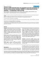
Báo cáo y học: "Systemic TNF blockade does not modulate synovial expression of the pro-inflammatory mediator HMGB1 in rheumatoid arthritis patients – a prospective clinical study" ppt
Bạn đang xem bản rút gọn của tài liệu. Xem và tải ngay bản đầy đủ của tài liệu tại đây (715.67 KB, 8 trang )
Open Access
Available online />Page 1 of 8
(page number not for citation purposes)
Vol 10 No 2
Research article
Systemic TNF blockade does not modulate synovial expression of
the pro-inflammatory mediator HMGB1 in rheumatoid arthritis
patients – a prospective clinical study
Erik Sundberg
1,2
, Cecilia Grundtman
2
, Erik af Klint
2
, Johan Lindberg
3
, Sofia Ernestam
2
, Ann-
Kristin Ulfgren
2
, Helena Erlandsson Harris
2
and Ulf Andersson
1
1
Department of Woman and Child Health, Pediatric Rheumatology Research Unit, Karolinska Institutet/Karolinska University Hospital, Stockholm,
Sweden
2
Department of Medicine, Rheumatology Unit, Karolinska Institutet/Karolinska University Hospital, Stockholm, Sweden
3
Department of Biotechnology, AlbaNova University Center, Royal Institute of Technology, Stockholm, Sweden
Corresponding author: Erik Sundberg,
Received: 21 Nov 2007 Revisions requested: 23 Jan 2008 Revisions received: 26 Feb 2008 Accepted: 17 Mar 2008 Published: 17 Mar 2008
Arthritis Research & Therapy 2008, 10:R33 (doi:10.1186/ar2387)
This article is online at: />© 2008 Sundberg et al.; licensee BioMed Central Ltd. This is an open access article distributed under the terms of the Creative Commons Attribution
License ( which permits unrestricted use, distribution, and reproduction in any medium, provided the
original work is properly cited.
Abstract
Introduction High-mobility group box chromosomal protein 1
(HMGB1) has recently been identified as an endogenous
mediator of arthritis. TNF and IL-1β, pivotal cytokines in arthritis
pathogenesis, both have the ability to induce the release of
HMGB1 from myeloid and dendritic cells. It was, therefore,
decided to investigate whether treatment based on TNF
blockade in rheumatoid arthritis (RA) affects the expression of
synovial HMGB1.
Methods Repeated arthroscopy-guided sampling of synovial
tissue was performed in nine patients with RA before and nine
weeks after initiation of anti-TNF mAb (infliximab) therapy.
Synovial biopsy specimens were analysed for HMGB1 protein
by immunohistochemical staining and for HMGB1 mRNA
expression by real-time reverse transcriptase PCR (RT-PCR).
Statistical evaluations were based on Wilcoxon's signed rank
tests or Spearman rank sum tests.
Results Aberrant, extranuclear HMGB1 and constitutive
nuclear HMGB1 expression, with histological signs of
inflammation, were evident in all biopsies obtained before
infliximab therapy. Signs of inflammation were still evident in the
second biopsies obtained nine weeks after initiation of infliximab
therapy. The cytoplasmic and extracellular expression of
HMGB1 decreased in five patients, remained unchanged in one
patient and increased in three patients, making the overall
change in HMGB1 protein expression not significant. No
correlation between the clinical response, as measured by
disease activity score calculated for 28 joints (DAS28) or the
American College of Rheumatology response criteria (ACR 20,
50, and 70), and the direction of change of HMGB1 expression
in individual patients could be discerned. In addition, infliximab
therapy did not alter HMGB1 mRNA synthesis.
Conclusion Pro-inflammatory HMGB1 expression during
rheumatoid synovitis was not consistently influenced by TNF-
blocking therapy with infliximab. This suggests that TNF is not
the main inducer of extranuclear HMGB1 during synovitis and
that HMGB1 may represent a TNF-independent molecule that
could be considered as a possible target for future therapeutic
intervention in RA.
Introduction
Rheumatoid arthritis (RA) is an autoimmune disease charac-
terised by chronic polyarticular inflammation leading to the
destruction of cartilage and subchondral bone. The pathogen-
esis of RA is complex, involving a wide range of endogenous
pro-inflammatory molecules including cytokines. Certain medi-
ators, with TNF as one causative molecule, can be success-
fully targeted in the treatment of chronic arthritis. TNF-blocking
therapy has been shown to dramatically reduce inflammation
and tissue destruction in many patients with RA [1-3].
ACR = American College of Rheumatology; DAS28 = Disease Activity Score calculated on 28 joints; HMGB1 = igh-mobility group box chromosomal
protein 1; IFN = Interferon; IL = Interleukinl; RA = Rheumatoid arthritis; RAGE = Receptor for advanced glycated end-products; mAb = Monoclonal
antibodies; RT-PCR = Reverse-transcriptase PCR; soluable RAGE = sRAGE; TLR = Toll-like receptor; TNF = Tumour necrosis factor.
Arthritis Research & Therapy Vol 10 No 2 Sundberg et al.
Page 2 of 8
(page number not for citation purposes)
However, it is also evident that anti-TNF therapy is not effective
in all patients and that many responders still present residual
signs of active disease. In order to improve the treatment of
chronic arthritis, a further search for additional potential target
molecules that act independently of TNF is highly warranted.
Recent findings have suggested that the high-mobility group
box chromosomal protein 1 (HMGB1) might be an important
molecule in the pathogenesis of arthritis [4-10]. Intranuclear
HMGB1 binds DNA and regulates transcription. In addition,
HMGB1 may be extracellularly translocated, thereby acting as
an inflammatory mediator of tissue invasion and tissue repair
[11-18]. HMGB1 may either be actively secreted from a wide
number of cell types following stimulation with inflammatory
mediators, including TNF, IL-1β, IFN-γ and multiple toll-like
receptor (TLR) ligands [15,19-23], or be passively released
from dying nucleated cells [12,13]. The extracellular effects of
HMGB1 are mediated via multiple receptors including the
receptor for advanced glycated end-products (RAGE), some
members of the TLR family and other as yet unidentified path-
ways [17,24-26]. Increased levels of HMGB1 are evident in
the synovial fluid of patients with RA and HMGB1 has been
shown to be abundantly expressed in an aberrant fashion in
rheumatoid synovial tissue [4,6]. Serum levels of HMGB1 are
also elevated in patients with RA and correlate with disease
activity [27]. In addition, intra-articular injections of HMGB1
trigger destructive arthritis in naive mice [5].
Different modes of HMGB1-blocking therapy, including neu-
tralising antibodies, antagonistic truncated HMGB1, soluable
RAGE (sRAGE), thrombomodulin or nuclear HMGB1 seques-
tration, have been successfully applied in studies of experi-
mental arthritides and sepsis [15,28-33]. It was recently
reported that gold salts interfere with the intracellular transport
mechanisms of HMGB1 and inhibit its release [34]. Oxaliplatin
is an antineoplastic platinum-based compound that generates
DNA adducts that strongly bind HMGB1. Therefore, gold salts
and oxaliplatin share the capacity to inhibit nuclear HMGB1
release via different mechanisms. Short-term oxaliplatin treat-
ment in collagen type-II-induced arthritis was recently studied
in mice and beneficial therapeutic effects coinciding with
nuclear HMGB1 retention were noted [35]. Once released,
HMGB1 might generate a positive feedback loop and induce
production of several pro-inflammatory cytokines such as IL-6,
IL-1β and TNF by macrophages and dendritic cells, thereby
sustaining prolonged inflammation [16,36].
In this pilot study the aim was to analyse to what extent extra-
nuclear HMGB1 expression depends on and relates to TNF
levels in RA, as previous studies have indicated that TNF can
induce HMGB1 release. Synovial biopsy specimens from
patients with RA were collected by arthroscopy before and
during therapy with TNF-specific mAb (infliximab) and the lev-
els of synovial expression of HMGB1 protein and mRNA were
evaluated.
The main findings were that synovial HMGB1 protein and
mRNA expression did not change in any consistent manner
after nine weeks of infliximab treatment.
Materials and methods
Patients, clinical assessment and therapy
Nine patients (seven females and two males) with RA diag-
nosed according to the revised American College of Rheuma-
tology (ACR) criteria [37] and active knee arthritis were
enrolled in a prospective clinical study. Informed consent was
obtained from all patients and the study was approved by the
local ethical committee at Karolinska University Hospital,
Stockholm, Sweden.
The median age of patients was 57 years (range 25 to 69
years) and the median disease duration was six years (range
0.6 to 18 years). The median duration of the current episode
of arthritis in the knee was 17.5 days (range 3 to 365 days; no
data for one patient). In all patients the disease activity score
calculated for 28 joints (DAS28) and the ACR response crite-
ria (ACR20, 50, and 70) were assessed at multiple time points
before and during therapy. The median DAS28 at inclusion
was 5.95 despite treatment with methotrexate (7.5 to 17.5
mg/week). Methotrexate doses were stable during the study
and for at least one month before the first arthroscopy. Four
patients received prednisone at stable doses (5 to 7.5 mg/
day) during the study period. One patient received
cyclosporine (150 mg/day) before and during the study
period.
Infliximab (Centocor BV, Leiden, The Netherlands) was given
as three intravenous infusions of 3 mg/kg in accordance with
the recommended standard-treatment protocol, with the first
infusion given 1 to 21 days after the first arthroscopy and the
subsequent infusions given two and six weeks later. The
results of synovial expression of IL-15 in response to infliximab
therapy using the same cohort of patients with RA have previ-
ously been published [38-40].
Synovial biopsies
Knee arthroscopy with multiple biopsies of synovial tissue of
the knee joint was performed in all patients 1 to 21 days before
the first infliximab infusion. A second arthroscopy was per-
formed at 8 to 10 weeks (median nine weeks) after the first
infusion, a time point when infliximab therapy is well estab-
lished and a clinical response can be evaluated [1].
All arthroscopies were performed by the same experienced
physician (EK). During the first arthroscopy, multiple synovial
tissue biopsies were taken from areas with signs of maximum
macroscopic inflammation, from the cartilage-pannus junction
and from synovial villi. Each biopsy site was documented pho-
tographically and mapped, allowing for follow-up biopsies to
be taken from the same areas. The biopsies were snap-frozen
within two minutes in liquid isopentane and stored at -70°C
Available online />Page 3 of 8
(page number not for citation purposes)
until sectioned. Serial cryostat sections (7 μm) were fixed for
20 minutes with 2% (v/v) formaldehyde and stored at -70°C.
Several biopsies were taken to secure sufficient material for
subsequent analyses. For each of the nine patients the best
biopsy pair with respect to morphology was selected for sub-
sequent immunohistochemical stainings. The staining was
always performed for samples taken before and after infliximab
treatment allowing for a pairwise comparison.
Immunohistochemistry
For indirect immunohistochemistry evaluation, tissue sections
were blocked for non-specific binding with H
2
O
2
and NaN
3
for
one hour, with normal goat serum (X0907, DAKO, Glostrup,
Denmark) for 15 minutes and with an Avidin/Biotin blocking kit
(SP-2001, Vector Laboratories, Burlingame, CA USA) accord-
ing to the manufacturer's instructions. Saponin (0.1% w/v in
HEPES pH 7.2) was added throughout the staining protocol.
An HMGB1-specific polyclonal peptide-affinity purified rabbit
antibody (PharMingen 556528, San Diego, CA, USA) was
used as the primary antibody at a final concentration of 0.5 μg/
ml. A non-specific rabbit antibody (X0902, DAKO, Glostrup,
Denmark) was used as the control. To identify blood vessels,
a human endothelium-specific mAb (anti-EN4, anti-human
CD31 from Sanbio, Bio-Zac, Uden, The Netherlands) was
used. As the negative control for CD31, an irrelevant mouse
IgG
1
mAb (DAKO, Glostrup, Denmark) was also used.
Sections were incubated with primary antibodies overnight.
The secondary antibody was a biotinylated goat anti-rabbit
IgG (BA-1000, Vector Laboratories, Burlingame, CA, USA)
diluted to 1:800 for HMGB1 detection, and a biotin goat anti-
mouse IgG
1
(DAKO, Glostrup, Denmark) was used for CD31
detection. Extra-avidin peroxidase (EAP) (Sigma, St. Louis,
MO, USA) with diaminobenzidine (DAB) (Vector Laboratories,
Burlingame, CA, USA) as substrate were used for visualisation
and sections were then counterstained with haematoxylin,
washed, dried and mounted with buffered glycerol.
Two evaluators (ES and CG), blinded to the order of the sec-
tions, performed a semi-quantitative analysis of the expression
of HMGB1. Scoring for each section was evaluated using a 0
to 4 scale with increments of 0.5. Index 0 corresponded to no
HMGB1 expression and 4 to the highest degree of HMGB1
protein expression. Separate analyses were performed for lin-
ing layers, vessels and cellular infiltrates in each of the sec-
tions. Nuclear, cytoplasmic and extracellular expression of
HMGB1 was also recorded from each tissue compartment.
Real-time RT-PCR
Due to a shortage of biopsy material, mRNA analysis was only
possible in six of the nine included patients. For first-strand
synthesis of each biopsy, 1 μg total RNA was mixed with 2 μl
20 TVN primer (4 μg/μl, Operon Biotechnology Inc. Huntsville,
AL USA). The primer sequences used were: for β-actin, for-
ward CCTTCGTGCCCCCCC and reverse GGAGAC-
CAAAAGCCTTCATACATC; and for HMGB1, forward
ATTGGTGATGTTGCGAAGAA and reverse GATCCACAG-
CAACTCCAGAA. The volume was adjusted to 15.5 μl using
RNase-free water, and the mixture was then incubated for 10
minutes at 70°C to denature the total RNA. The sample was
then incubated for two minutes on ice to allow the primers to
anneal and then spun briefly.
A 12.5 μl cDNA synthesis mixture, consisting of 6 μl 5× first-
strand buffer, 3 μl 0.1 M dithiothreitol (DTT), 2 μl Superscript
III (Invitrogen Corporation, Carlsbad, CA, USA), and 1.5 μl 10
mM deoxinucleoside triophosfate (dNTP) mix (Amersham Bio-
sciences, Piscataway, NJ, USA), was added to the sample.
The whole mixture was incubated at 46°C for first-strand syn-
thesis. After one hour the temperature was increased to 70°C
for 15 minutes to terminate the reaction. 2 U RNase H (Invitro-
gen Corporation, Carlsbad, CA, USA) was then added to
degrade the RNA. After RNase treatment the temperature was
increased to 70°C to inactivate RNases. All samples were
then diluted with RNase-free water to a final volume of 200 μl.
Real-time RT-PCR was performed using the iCycler system
from Bio-Rad Laboratories Inc. Hercules, CA, USA. Each reac-
tion was performed with a 3.0 μl template, 12.5 μl iQSYBR
Green Supermix (Bio-Rad Laboratories Inc. Hercules, CA,
USA), 300 nM primer and water to adjust the final volume to
25 μl. The PCR amplification steps were applied in the follow-
ing conditions: three minutes at 95°C, 40 cycles of 20 sec-
onds at 94°C, 30 seconds at 60°C and one minute at 72°C.
This was followed by melt curve analysis to ensure specific
amplification. The signal was calculated in all experiments
using 'PCR baseline subtracted relative flourescent unit
(RFU)'. All primers were designed using Primer3 and ampli-
cons were designed to span exon-exon junctions to minimise
contamination of genomic DNA [41]. Primer sequence infor-
mation is available in the supplemental material. Relative
expression between samples was calculated using the ΔΔCt
method and all samples were analysed in triplicate[42]. β-actin
was used as a reference housekeeping gene.
Statistical analysis
Wilcoxon's signed-rank test was used for the analysis of
matched pairs for protein data. as well as for the analysis of
mRNA data for the whole group. Spearman rank sum test was
utilised to statistically compare the degree of correlation
between the two persons evaluating HMGB1 protein expres-
sion by immunohistochemistry. p < 0.05 was considered to be
statistically significant.
Results
Clinical response and CD marker changes following
infliximab treatment
Patients were assessed for disease activity at baseline and
after three months using individual DAS28 and ACR scores
(Table 1). These data have been previously published [38].
Arthritis Research & Therapy Vol 10 No 2 Sundberg et al.
Page 4 of 8
(page number not for citation purposes)
Median DAS28 scores decreased from 5.95 to 4.41 (p <
0.01), the median tender joint count from 10 to 3 (p < 0.05),
the median swollen joint count from 14 to 2 (p < 0.01) and the
median serum C-reactive protein level decreased from 34 mg/
L to 19 mg/L (p = 0.08). Two patients fulfilled ACR70, one
patient fulfilled ACR50, three patients fulfilled ACR20 and
three patients were non-responders according to the ACR
criteria.
In repeated biopsies from each patient before and after inflixi-
mab treatment, expression of CD markers for T-cells (CD3),
macrophages (CD68) and activated macrophages (CD163)
were analysed. CD3 and CD163 were unaffected, whereas
the number of CD68-positive cells decreased as a conse-
quence of infliximab therapy (data not shown).
Synovial HMGB1 protein expression not influenced by
infliximab therapy
Aberrant synovial HMGB1 staining was evident in all nine
patients before and during infliximab treatment and was most
prominent in the lining layer, in areas with cellular infiltrates and
in certain blood vessel walls. Both an increased (Figures 1a,b)
and decreased (Figures 1c,d) presence of HMGB1 protein in
the synovia during therapy could be detected, but there was
no correlation with the clinical course of individual patients. In
the group of six patients who responded to therapy, three had
a decreased, two had an increased and one had an
unchanged level of protein expression of HMGB1.
There was no significant difference in the group in the overall
HMGB1 protein distribution before or after infliximab therapy
(Figure 2). Neither were any consistent changes (nuclear,
cytoplasmic or extracellular) recorded for HMGB1 expression
when different biopsy compartments, including lining layer,
cell infiltrates and endothelium, were analysed separately.
HMGB1 mRNA expression not influenced by infliximab
therapy
In the group of six patients studied by RT-PCR two individuals
had increased, two had decreased and two patients had
unchanged HMGB1 mRNA levels, with no correlation to clini-
cal outcomes. Taken together, the results indicated no
significant change of HMGB1 mRNA as a consequence of inf-
liximab therapy (Figure 3).
Table 1
Clinical assessment, response to infliximab treatment and HMGB1 expression.
Pat W Sex Age (years) Disease duration (years) CRP Medication DAS28 Clinical response HMGB1 protein HMGB1mRNA
MTX Pred DAS28 ACR ES CG
1 0 F 55 18 107 17.5 5 * 6.71 1.5 1
9 38 6.34 No 0 2 1.5
2 0 F 52 6 14 7.5 7.5 6.00 1.5 1.5
9 15 4.43 Mod 20 1 0.5 0
3 0 F 58 2 46 10 7.91 3 4
9 25 5.41 Mod 50 1 1.5 0
40F 57 4 7 10 4.94 2 2
9344.41No2022Up
5 0 M 56 7 44 15 5.95 2 2.5
972.90G703.53
6 0 F 69 1 69 15 5 7.39 2 3
9 7 1.79 G 70 1.5 2.5 Up
70F 66 14 34 10 5.64 2 2
9 16 4.73 Mod 0 1 0.5 Down
8 0 F 66 10 18 12.5 4.83 3.5 4
9 19 4.23 Mod 0 0.5 2 Down
9 0 M 25 0.6 26 17.5 7.5 5.62 .5 3
9253.91Mod2034
* Patient 1 also received 150 mg/day cyclosporine before and during the study period.Pat = Patient, W = week, CRP = C-reactive protein, MTX
= Methotrexate (mg/week), Pred = Prednisone (mg/day), DAS28 = disease activity score calculated on 28 joints, ACR = American College of
Rheumatology, HMGB1 = High-mobility group box chromosomal protein 1, ES = Erik Sundberg, CG = Cecilia Grundtman, F = Female, M =
Male, No = Non, Mod = Moderate, G = Good.
Available online />Page 5 of 8
(page number not for citation purposes)
Discussion
To the authors' knowledge, this is the first clinical study exam-
ining the effect of TNF blockade on synovial HMGB1 expres-
sion. The expression of synovial HMGB1 protein and mRNA in
RA synovitis remained unaffected by TNF blockade for nine
weeks and there was no correlation with the clinical course of
arthritis. This was somewhat surprising as the original discov-
ery of extranuclear and extracellular HMGB1 translocation
was derived from studies of macrophages stimulated by TNF,
IL-1β or endotoxin. Blocking intra-articular TNF by infliximab
might therefore theoretically inhibit synovial HMGB1 release.
However, this could not be verified from the results of the
present study. There are several additional molecules such as
IL-1β, IFN-γ, IFN-β, Nitric oxide and multiple TLR ligands that
are potent promotors of HMGB1 translocation and extracellu-
lar release, and expression of all these mediators may possibly
remain unchanged during infliximab therapy [43-45]. Support-
ing the clinical data for the TNF-independent release of
HMGB1 are results obtained using a sensitive HMGB1 Elis-
pot assay, which demonstrated that TNF has a distinctly infe-
rior capacity to stimulate HMGB1 release from cultured
macrophages compared with other molecules such as endo-
toxin, IFN-γ [46] or IL-1β (authors' unpublished data).
A similar arthroscopic-guided tissue sampling method and
immunohistochemical analysis have previously been used to
study the therapeutic effects of systemic infliximab therapy on
the synovial expression of other important pro-inflammatory
molecules [38,47]. Infliximab treatment in another small cohort
of patients with RA saw the number of TNF-synthesising cells
readily reduced and the production of IL-1α and IL-1β incon-
sistently down-regulated[47]. IL-15 was not inhibited by anti-
TNF therapy in the same cohort of patients with RA as evalu-
ated in the present study [38]. Therefore, beneficial clinical
results after infliximab therapy may occur despite the fact that
active cytokines such as IL-1, IL-15 and HMGB1 prevail dur-
ing synovitis. Using a similar methodology contrasting results
regarding HMGB1 expression in RA synovitis based on ther-
apy with intra-articular corticosteroid injections was recently
demonstrated [48]. Local intra-articular corticosteroid treat-
ment clearly down-regulated the aberrant extranuclear expres-
sion of HMGB1. Likewise, TNF and IL-1β, but not IL-1α, were
strongly suppressed by intra-articular corticosteroid therapy.
In the present study HMGB1 was analysed in a semi-quantita-
tive way using conventional manual microscopy. Computer-
ised image analysis was not an option because this
technology does not enable discrimination between the con-
stitutive nuclear HMGB1 expression and the aberrant pres-
ence of cytoplasmic and extracellular HMGB1 causing
pathology.
The question regarding a functional relationship between TNF
and HMGB1 in rheumatoid synovitis cannot be fully clarified in
the present study due to the relatively limited number of
patients with RA. However, it is proposed that the results can
be interpreted in at least two different ways: HMGB1 and TNF
could either act independently of each other; or HMGB1 could
act upstream of TNF in the pro-inflammatory cascade. The lat-
ter explanation is favoured, since it has previously been
reported that HMGB1 is a potent inducer of TNF production
in cultured macrophages and dendritic cells [16,49,50]. In
addition, it has recently been demonstrated that HMGB1 up-
regulates TNF transcription by direct binding to the TNF pro-
motor in osteoclasts [51]. Furthermore, successful HMGB1-
targeted therapies in experimental sepsis, arthritis and brain
ischaemia strongly down-regulate in vivo synthesis of TNF
[29,52,53]. Another possibility to explain the lack of correla-
tion between the clinical course and HMGB1 expression fol-
lowing infliximab treatment could be that HMGB1 may be
more dependent on the IL-1 pathway than the TNF pathway in
the pathogenesis of arthritis. This theory is also supported by
the fact that intra-articular HMGB1 injections cause arthritis in
wild-type mice but not in IL-1 type I-receptor deficient mice [5].
Considering that exaggerated and dysregulated HMGB1
release may lead to aggravated inflammation and tissue
destruction in chronic arthritis, the results of the present study
suggest that HMGB1 may serve as a possible TNF-independ-
ent target molecule for biological therapy. Future work is
required to elucidate the potential of this strategy.
Figure 1
HMGB1 synovial protein expression before and during infliximab ther-apy as detected by immunohistochemistryHMGB1 synovial protein expression before and during infliximab ther-
apy as detected by immunohistochemistry. Nuclear, cytoplasmic and
extracellular HMGB1 protein expression is evident in the RA synovia in
the lining layer as well as in cellular infiltrates and endothelium. (a)
Moderate aberrant synovial HMGB1 expression before the start of inf-
liximab therapy from patient number 5. (b) The second biopsy in patient
number 5 taken during infliximab treatment showing increased extranu-
clear HMGB1 staining. (c) A marked endothelial expression of HMGB1
is evident before infliximab therapy in synovitis obtained from patient
number 7. (d) Infliximab therapy for nine weeks resulted in reduced
endothelial HMGB1 expression in patient number 7. Original magnifica-
tion ×250 for (a) and (b), ×100 for (c) and (d).
Arthritis Research & Therapy Vol 10 No 2 Sundberg et al.
Page 6 of 8
(page number not for citation purposes)
Conclusion
A number of published observations have demonstrated that
the pro-inflammatory mediator HMGB1 has a functional
impact on the pathogenesis of arthritis. The results presented
here reveal an unaffected expression of HMGB1 at protein
and mRNA levels in synovia obtained from patients with RA
from repeated biopsies before and during well-established
TNF blockade with infliximab. These results are interpreted as
an indication of TNF-independent HMGB1 expression with
subsequent residual HMGB1 biological activity in RA synovitis
in spite of infliximab therapy. It is concluded that these findings
support attempts to generate novel HMGB1-targeted thera-
pies in chronic arthritis.
Competing interests
The authors declare that they have no competing interests.
Authors' contributions
UA, HEH, A-KU, ES and CG designed the study. EaK per-
formed the arthrosopies and synovial sampling. SE collected
clinical patient data. ES and CG performed the immunohisto-
chemical stainings and the immunohistochemical analysis. JL
performed the RT-PCR and mRNA analysis. ES, CG, UA and
HEH prepared the manuscript. All authors read and approved
the final manuscript.
Acknowledgements
The authors would like to thank Lars Ottosson, PhD, for valuable techni-
cal support and Associate Professor RA Harris for rewarding linguistic
Figure 2
Change of HMGB1 synovial protein expression before and during infliximab therapyChange of HMGB1 synovial protein expression before and during infliximab therapy. Immunohistochemical scoring of HMGB1 protein expression
was unchanged during the nine weeks of infliximab therapy. Values represent mean scores of highly concordant scoring recorded by the two inde-
pendent investigators (ES and CG). The correlation was statistically confirmed by Spearman rank sum tests with a correlation coefficient (r
s
)of 0.74
(significant at p < 0.002). Scoring for each section was evaluated using a 0 to 4 scale with increments of 0.5. Index 0 corresponds to no HMGB1
expression and 4 to highest degree of HMGB1 protein expression. Separate analyses were performed for lining layer, vessels and cellular infiltrates
in each of the sections. (a) Change of overall HMGB1 protein expression for each patient. (b) Change of HMGB1 expression in cellular infiltrates.
(c) Change of HMGB1 expression in lining layer. (d) Change of HMGB1 expression in endothelium.
Figure 3
Change of HMGB1 synovial mRNA expression before and after inflixi-mab therapyChange of HMGB1 synovial mRNA expression before and after inflixi-
mab therapy. HMGB1 mRNA expression as determined by reverse-
transcriptase PCR for six patients. Values are expressed as fold-change
of up- or down-regulation using a log
2
-scale. The last bar to the right
represents the average value for the whole group and the standard
deviation is indicated by whiskers.
Available online />Page 7 of 8
(page number not for citation purposes)
advice. Financial support was provided through the regional agreement
on medical training and clinical research (ALF) between Stockholm
county council and the Karolinska Institutet, King Gustaf V 80-year-foun-
dation, the Freemason Lodge Barnhuset in Stockholm, The Swedish
Research Council no K2005-74X-09082 and K2005-73X-14642, and
the Swedish Rheumatism Association.
References
1. Maini RN, Breedveld FC, Kalden JR, Smolen JS, Davis D, Macfar-
lane JD, Antoni C, Leeb B, Elliott MJ, Woody JN, Schaible TF, Feld-
mann M: Therapeutic efficacy of multiple intravenous infusions
of anti-tumor necrosis factor alpha monoclonal antibody com-
bined with low-dose weekly methotrexate in rheumatoid
arthritis. Arthritis Rheum 1998, 41:1552-1563.
2. Maini RN, Breedveld FC, Kalden JR, Smolen JS, Furst D, Weisman
MH, St Clair EW, Keenan GF, van der Heijde D, Marsters PA, Lip-
sky PE: Sustained improvement over two years in physical
function, structural damage, and signs and symptoms among
patients with rheumatoid arthritis treated with infliximab and
methotrexate. Arthritis Rheum 2004, 50:1051-1065.
3. Klareskog L, van der Heijde D, de Jager JP, Gough A, Kalden J,
Malaise M, Martin Mola E, Pavelka K, Sany J, Settas L, Wajdula J,
Pedersen R, Fatenejad S, Sanda M: Therapeutic effect of the
combination of etanercept and methotrexate compared with
each treatment alone in patients with rheumatoid arthritis:
double-blind randomised controlled trial. Lancet 2004,
363:675-681.
4. Kokkola R, Sundberg E, Ulfgren AK, Palmblad K, Li J, Wang H,
Ulloa L, Yang H, Yan XJ, Furie R, Chiorazzi N, Tracey KJ, Anders-
son U, Harris HE: High mobility group box chromosomal pro-
tein 1: a novel proinflammatory mediator in synovitis. Arthritis
Rheum 2002, 46:2598-2603.
5. Pullerits R, Jonsson IM, Verdrengh M, Bokarewa M, Andersson U,
Erlandsson-Harris H, Tarkowski A: High mobility group box chro-
mosomal protein 1, a DNA binding cytokine, induces arthritis.
Arthritis Rheum 2003, 48:1693-1700.
6. Taniguchi N, Kawahara K, Yone K, Hashiguchi T, Yamakuchi M,
Goto M, Inoue K, Yamada S, Ijiri K, Matsunaga S, Nakajima T,
Komiya S, Maruyama I: High mobility group box chromosomal
protein 1 plays a role in the pathogenesis of rheumatoid arthri-
tis as a novel cytokine. Arthritis Rheum 2003, 48:971-981.
7. Andersson U, Tracey KJ: HMGB1 as a mediator of necrosis-
induced inflammation and a therapeutic target in arthritis.
Rheum Dis Clin North Am 2004, 30:627-637.
8. Andersson U, Erlandsson-Harris H: HMGB1 is a potent trigger of
arthritis. J Intern Med 2004, 255:344-350.
9. Jiang W, Pisetsky DS: Mechanisms of Disease: the role of high-
mobility group protein 1 in the pathogenesis of inflammatory
arthritis. Nat Clin Pract Rheumatol 2007, 3:
52-58.
10. Palmblad K, Sundberg E, Diez M, Soderling R, Aveberger AC,
Andersson U, Harris HE: Morphological characterization of
intra-articular HMGB1 expression during the course of colla-
gen-induced arthritis. Arthritis Res Ther 2007, 9:R35.
11. Muller S, Scaffidi P, Degryse B, Bonaldi T, Ronfani L, Agresti A,
Beltrame M, Bianchi ME: New EMBO members' review: the dou-
ble life of HMGB1 chromatin protein: architectural factor and
extracellular signal. Embo J 2001, 20:4337-4340.
12. Scaffidi P, Misteli T, Bianchi ME: Release of chromatin protein
HMGB1 by necrotic cells triggers inflammation. Nature 2002,
418:191-195.
13. Rovere-Querini P, Capobianco A, Scaffidi P, Valentinis B, Catalan-
otti F, Giazzon M, Dumitriu IE, Muller S, Iannacone M, Traversari C,
Bianchi ME, Manfredi AA: HMGB1 is an endogenous immune
adjuvant released by necrotic cells. EMBO Rep 2004,
5:825-830.
14. Dumitriu IE, Baruah P, Valentinis B, Voll RE, Herrmann M, Nawroth
PP, Arnold B, Bianchi ME, Manfredi AA, Rovere-Querini P:
Release of high mobility group box 1 by dendritic cells con-
trols T cell activation via the receptor for advanced glycation
end products. J Immunol 2005, 174:7506-7515.
15. Wang H, Bloom O, Zhang M, Vishnubhakat JM, Ombrellino M, Che
J, Frazier A, Yang H, Ivanova S, Borovikova L, Manogue KR, Faist
E, Abraham E, Andersson J, Andersson U, Molina PE, Abumrad
NN, Sama A, Tracey KJ: HMG-1 as a late mediator of endotoxin
lethality in mice. Science 1999, 285:248-251.
16. Andersson U, Wang H, Palmblad K, Aveberger AC, Bloom O,
Erlandsson-Harris H, Janson A, Kokkola R, Zhang M, Yang H,
Tracey KJ: High mobility group 1 protein (HMG-1) stimulates
proinflammatory cytokine synthesis in human monocytes. J
Exp Med 2000, 192:565-570.
17. Rouhiainen A, Kuja-Panula J, Wilkman E, Pakkanen J, Stenfors J,
Tuominen RK, Lepantalo M, Carpen O, Parkkinen J, Rauvala H:
Regulation of monocyte migration by amphoterin (HMGB1).
Blood 2004, 104:1174-1182.
18. Limana F, Germani A, Zacheo A, Kajstura J, Di Carlo A, Borsellino
G, Leoni O, Palumbo R, Battistini L, Rastaldo R, Muller S, Pompilio
G, Anversa P, Bianchi ME, Capogrossi MC: Exogenous high-
mobility group box 1 protein induces myocardial regeneration
after infarction via enhanced cardiac C-kit+ cell proliferation
and differentiation. Circ Res 2005, 97:e73-83.
19. Semino C, Angelini G, Poggi A, Rubartelli A: NK/iDC interaction
results in IL-18 secretion by DCs at the synaptic cleft followed
by NK cell activation and release of the DC maturation factor
HMGB1. Blood 2005, 106:609-616.
20. Gardella S, Andrei C, Ferrera D, Lotti LV, Torrisi MR, Bianchi ME,
Rubartelli A: The nuclear protein HMGB1 is secreted by mono-
cytes via a non-classical, vesicle-mediated secretory pathway.
EMBO Rep 2002, 3:995-1001.
21. Bonaldi T, Talamo F, Scaffidi P, Ferrera D, Porto A, Bachi A, Rubar-
telli A, Agresti A, Bianchi ME: Monocytic cells hyperacetylate
chromatin protein HMGB1 to redirect it towards secretion.
Embo J 2003, 22:5551-5560.
22. Rendon-Mitchell B, Ochani M, Li J, Han J, Wang H, Yang H,
Susarla S, Czura C, Mitchell RA, Chen G, Sama AE, Tracey KJ,
Wang H: IFN-gamma induces high mobility group box 1 pro-
tein release partly through a TNF-dependent mechanism. J
Immunol 2003, 170:3890-3897.
23. Jiang W, Pisetsky DS: The role of IFN-alpha and nitric oxide in
the release of HMGB1 by RAW 264.7 cells stimulated with
polyinosinic-polycytidylic acid or lipopolysaccharide. J
Immunol 2006, 177:3337-3343.
24. Kokkola R, Andersson A, Mullins G, Ostberg T, Treutiger CJ,
Arnold B, Nawroth P, Andersson U, Harris RA, Harris HE: RAGE is
the major receptor for the proinflammatory activity of HMGB1
in rodent macrophages. Scand J Immunol 2005, 61:1-9.
25. Park JS, Svetkauskaite D, He Q, Kim JY, Strassheim D, Ishizaka A,
Abraham E: Involvement of toll-like receptors 2 and 4 in cellular
activation by high mobility group box 1 protein. J Biol Chem
2004, 279:7370-7377.
26. Yu M, Wang H, Ding A, Golenbock DT, Latz E, Czura CJ, Fenton
MJ, Tracey KJ, Yang H: HMGB1 signals through toll-like recep-
tor (TLR) 4 and TLR2. Shock 2006, 26:174-179.
27. Goldstein RS, Bruchfeld A, Yang L, Qureshi AR, Gallowitsch-
Puerta M, Patel NB, Huston BJ, Chavan S, Rosas-Ballina M,
Gregersen PK, Czura CJ, Sloan RP, Sama AE, Tracey KJ: Cholin-
ergic anti-inflammatory pathway activity and High Mobility
Group Box-1 (HMGB1) serum levels in patients with rheuma-
toid arthritis. Mol Med 2007, 13:210-215.
28. Wang H, Yang H, Czura CJ, Sama AE, Tracey KJ: HMGB1 as a
late mediator of lethal systemic inflammation. Am J Respir Crit
Care Med 2001, 164:1768-1773.
29. Kokkola R, Li J, Sundberg E, Aveberger AC, Palmblad K, Yang H,
Tracey KJ, Andersson U, Harris HE: Successful treatment of col-
lagen-induced arthritis in mice and rats by targeting extracel-
lular high mobility group box chromosomal protein 1 activity.
Arthritis Rheum 2003, 48:2052-2058.
30. Hofmann MA, Drury S, Hudson BI, Gleason MR, Qu W, Lu Y, Lalla
E, Chitnis S, Monteiro J, Stickland MH, Bucciarelli LG, Moser B,
Moxley G, Itescu S, Grant PJ, Gregersen PK, Stern DM, Schmidt
AM: RAGE and arthritis: the G82S polymorphism amplifies the
inflammatory response. Genes Immun 2002, 3:123-135.
31. Van de Wouwer M, Plaisance S, De Vriese A, Waelkens E, Collen
D, Persson J, Daha MR, Conway EM: The lectin-like domain of
thrombomodulin interferes with complement activation and
protects against arthritis. J Thromb Haemost 2006,
4:1813-1824.
32. Ulloa L, Ochani M, Yang H, Tanovic M, Halperin D, Yang R, Czura
CJ, Fink MP, Tracey KJ: Ethyl pyruvate prevents lethality in mice
with established lethal sepsis and systemic inflammation.
Proc Natl Acad Sci USA 2002, 99:12351-12356.
33. Chen G, Li J, Qiang X, Czura CJ, Ochani M, Ochani K, Ulloa L,
Yang H, Tracey KJ, Wang P, Sama AE, Wang H: Suppression of
Arthritis Research & Therapy Vol 10 No 2 Sundberg et al.
Page 8 of 8
(page number not for citation purposes)
HMGB1 release by stearoyl lysophosphatidylcholine:an addi-
tional mechanism for its therapeutic effects in experimental
sepsis. J Lipid Res 2005, 46:623-627.
34. Zetterstrom CK, Jiang W, Wahamaa H, Ostberg T, Aveberger AC,
Schierbeck H, Lotze MT, Andersson U, Pisetsky DS, Erlandsson
Harris H: Pivotal advance: inhibition of HMGB1 nuclear translo-
cation as a mechanism for the anti-rheumatic effects of gold
sodium thiomalate. J Leukoc Biol 2008, 83:31-38.
35. Ostberg T, Wahamaa H, Palmblad K, Ito N, Stridh P, Shoshan M,
Lotze MT, Harris HE, Andersson U: Oxaliplatin retains HMGB1
intranuclearly and ameliorates collagen type II-induced
arthritis. Arthritis Res Ther 2008, 10:R1.
36. Yang D, Chen Q, Yang H, Tracey KJ, Bustin M, Oppenheim JJ:
High mobility group box-1 protein induces the migration and
activation of human dendritic cells and acts as an alarmin. J
Leukoc Biol 2007, 81:59-66.
37. Arnett FC, Edworthy SM, Bloch DA, McShane DJ, Fries JF, Cooper
NS, Healey LA, Kaplan SR, Liang MH, Luthra HS, et al.: The Amer-
ican Rheumatism Association 1987 revised criteria for the
classification of rheumatoid arthritis. Arthritis Rheum 1988,
31:315-324.
38. Ernestam S, af Klint E, Catrina AI, Sundberg E, Engstrom M,
Klareskog L, Ulfgren AK: Synovial expression of IL-15 in rheu-
matoid arthritis is not influenced by blockade of tumour necro-
sis factor. Arthritis Res Ther 2006, 8:R18.
39. Catrina AI, af Klint E, Ernestam S, Catrina SB, Makrygiannakis D,
Botusan IR, Klareskog L, Ulfgren AK: Anti-tumor necrosis factor
therapy increases synovial osteoprotegerin expression in
rheumatoid arthritis. Arthritis Rheum 2006, 54:76-81.
40. Wittkowski H, Foell D, af Klint E, De Rycke L, De Keyser F, Frosch
M, Ulfgren AK, Roth J: Effects of intra-articular corticosteroids
and anti-TNF therapy on neutrophil activation in rheumatoid
arthritis. Ann Rheum Dis 2007, 66:1020-1025.
41. Rozen S, Skaletsky H: Primer3 on the WWW for general users
and for biologist programmers. Methods Mol Biol 2000,
132:365-386.
42. Schmittgen TD: Real-time quantitative PCR. Methods 2001,
25:
383-385.
43. Park JS, Gamboni-Robertson F, He Q, Svetkauskaite D, Kim JY,
Strassheim D, Sohn JW, Yamada S, Maruyama I, Banerjee A,
Ishizaka A, Abraham E: High mobility group box 1 protein inter-
acts with multiple Toll-like receptors. Am J Physiol Cell Physiol
2006, 290:C917-924.
44. Liu S, Stolz DB, Sappington PL, Macias CA, Killeen ME, Tenhunen
JJ, Delude RL, Fink MP: HMGB1 is secreted by immunostimu-
lated enterocytes and contributes to cytomix-induced hyper-
permeability of Caco-2 monolayers. Am J Physiol Cell Physiol
2006, 290:C990-999.
45. Harris HE, Raucci A: Alarmin(g) news about danger: workshop
on innate danger signals and HMGB1. EMBO Rep 2006,
7:774-778.
46. Wahamaa H, Vallerskog T, Qin S, Lunderius C, LaRosa G, Anders-
son U, Harris HE: HMGB1-secreting capacity of multiple cell lin-
eages revealed by a novel HMGB1 ELISPOT assay. J Leukoc
Biol 2007, 81:129-136.
47. Ulfgren AK, Andersson U, Engstrom M, Klareskog L, Maini RN,
Taylor PC: Systemic anti-tumor necrosis factor alpha therapy
in rheumatoid arthritis down-regulates synovial tumor necro-
sis factor alpha synthesis. Arthritis Rheum 2000,
43:2391-2396.
48. af Klint E, Grundtman C, Engstrom M, Catrina AI, Makrygiannakis
D, Klareskog L, Andersson U, Ulfgren AK: Intraarticular glucocor-
ticoid treatment reduces inflammation in synovial cell infiltra-
tions more efficiently than in synovial blood vessels. Arthritis
Rheum 2005, 52:3880-3889.
49. Messmer D, Yang H, Telusma G, Knoll F, Li J, Messmer B, Tracey
KJ, Chiorazzi N: High mobility group box protein 1: an endog-
enous signal for dendritic cell maturation and Th1 polarization.
J Immunol 2004, 173:307-313.
50. Semino C, Ceccarelli J, Lotti LV, Torrisi MR, Angelini G, Rubartelli
A: The maturation potential of NK cell clones toward autolo-
gous dendritic cells correlates with HMGB1 secretion. J Leu-
koc Biol 2007, 81:92-99.
51. Yamoah K, Brebene A, Baliram R, Inagaki K, Dolios G, Arabi A,
Majeed R, Amano H, Wang R, Yanagisawa R, Abe E: High Mobil-
ity Group Box Proteins (HMGBs) Modulate TNF{alpha}
Expression in Osteoclastogenesis via a Novel DNA Sequence.
Mol Endocrinol in press. 2008: Jan 24
52. Yang H, Ochani M, Li J, Qiang X, Tanovic M, Harris HE, Susarla
SM, Ulloa L, Wang H, DiRaimo R, Czura CJ, Wang H, Roth J, War-
ren HS, Fink MP, Fenton MJ, Andersson U, Tracey KJ:
Reversing
established sepsis with antagonists of endogenous high-
mobility group box 1. Proc Natl Acad Sci USA 2004,
101:296-301.
53. Liu K, Mori S, Takahashi HK, Tomono Y, Wake H, Kanke T, Sato Y,
Hiraga N, Adachi N, Yoshino T, Nishibori M: Anti-high mobility
group box 1 monoclonal antibody ameliorates brain infarction
induced by transient ischemia in rats. Faseb J 2007,
21:3904-3916.