Báo cáo y học: "Exogenous tumour necrosis factor α induces suppression of autoimmune arthritis" ppsx
Bạn đang xem bản rút gọn của tài liệu. Xem và tải ngay bản đầy đủ của tài liệu tại đây (650.54 KB, 10 trang )
Open Access
Available online />Page 1 of 10
(page number not for citation purposes)
Vol 10 No 1
Research article
Exogenous tumour necrosis factor α induces suppression of
autoimmune arthritis
Eugene Y Kim
1
, Howard H Chi
1
, Rajesh Rajaiah
1
and Kamal D Moudgil
1,2
1
Department of Microbiology and Immunology, University of Maryland School of Medicine, Baltimore, MD, USA.
2
Division of Rheumatology, Department of Medicine, University of Maryland School of Medicine, Baltimore, MD, USA.
Corresponding author: Kamal D Moudgil,
Received: 7 Jan 2008 Revisions requested: 14 Feb 2008 Revisions received: 12 Mar 2008 Accepted: 1 Apr 2008 Published: 1 Apr 2008
Arthritis Research & Therapy 2008, 10:R38 (doi:10.1186/ar2393)
This article is online at: />© 2008 Kim et al.; licensee BioMed Central Ltd.
This is an open access article distributed under the terms of the Creative Commons Attribution License ( />),
which permits unrestricted use, distribution, and reproduction in any medium, provided the original work is properly cited.
Abstract
Introduction Our previous studies showed that arthritic Lewis
(LEW) rats produced the highest levels of tumour necrosis
factor (TNF)α in the recovery phase of adjuvant arthritis (AA),
suggesting a correlation between high TNFα levels and reduced
severity of arthritis. To further explore this correlation, we
compared the TNFα secretion profile of the AA-resistant Wistar
Kyoto (WKY) rats with that of LEW rats, determined the effect
of exogenous TNFα on the course of AA in LEW rats, and
examined various mechanisms involved in TNFα-induced
disease modulation.
Methods A cohort each of LEW and WKY rats was immunised
subcutaneously with heat-killed Mycobacterium tuberculosis
H37Ra (Mtb). At different time points thereafter, subgroups of
rats were killed and their draining lymph node cells were tested
for cytokine production. Another group of LEW rats was injected
with TNFα intraperitoneally daily for a total of 10 injections, 3
before and 6 after Mtb challenge, and then observed for signs
of AA. In parallel, TNFα-treated rats were examined for changes
in other cytokines, in CD4+CD25+ T cell frequency, and in
indoleamine 2,3-dioxygenase (IDO) mRNA expression levels.
Results LEW rats displayed a TNFα secretion profile that was
opposite to that of the WKY rats. Furthermore, TNFα treatment
significantly downmodulated the severity of AA in LEW rats, and
decreased the interferon (IFN)-γ secretion in response to the
pathogenic determinant of the disease-related antigen. No
significant alterations were observed in other parameters tested.
Conclusion The role of endogenous TNFα in the induction and
propagation of arthritis is well established. However, exogenous
TNFα can downmodulate the course of AA, displaying an
immunoregulatory functional attribute of this cytokine.
Introduction
Rheumatoid arthritis (RA) is a chronic autoimmune disease
characterised by symmetrical joint involvement, synovial hyper-
plasia, neovascularisation, infiltration of the cartilage and
subchondral bone by the pannus tissue leading to erosions
and deformities [1-4]. Macrophages and T cells play a critical
role in initiating and propagating the disease process. The
cytokines tumour necrosis factor α (TNFα) and interleukin-1
(IL-1) mediate many of the inflammatory and tissue-damaging
activities within the joint [1-3,5]. The in vivo neutralisation of
these cytokines using the appropriate antibodies or decoy
receptors leads to significant amelioration of signs and symp-
toms of joint inflammation [2,6,7]. Specifically, therapeutic
strategies based on anti-TNFα antibodies or soluble TNFα
receptor (sTNFR) are currently being used in clinics for the
treatment of RA patients [7].
In the course of our preliminary studies in the rat adjuvant-
induced arthritis (AA) model of human RA [8-13], we observed
that the levels of TNFα produced by the arthritogenic epitope
of mycobacterial heat-shock protein 65 (Bhsp65) [10-12,14]
were highest in the recovery phase of the disease compared
to that at the onset or the peak phase of AA. This unexpected
correlation has formed the basis of subsequent experiments
described in the present work.
AA = adjuvant arthritis; Bhsp65 = mycobacterial heat shock protein 65; B177 = Bhsp65 peptide 177 to 191; B333 = Bhsp65 peptide 333 to 347;
HEL = hen egg white lysozyme; HEL65 = HEL peptide 65 to 78; Inc = incubation; LEW = Lewis; LNC = lymph node cells; Mtb = Mycobacterium
tuberculosis H37Ra; Ons = onset; Pk = peak; Rec = recovery; SI = stimulation index; sTNFR-I, soluble TNF receptor I; WKY = Wistar-Kyoto.
Arthritis Research & Therapy Vol 10 No 1 Kim et al.
Page 2 of 10
(page number not for citation purposes)
Our results show that the AA-susceptible Lewis (LEW) rats
given an arthritogenic stimulus (immunisation subcutaneously
with heat-killed Mycobacterium tuberculosis H37Ra, Mtb)
showed the highest levels of TNFα in the recovery phase of
AA, displaying a TNFα profile opposite to that of the AA-resist-
ant Wistar Kyoto (WKY) rats. Intriguingly, the pre-treatment of
LEW rats with TNFα injected intraperitoneally induced protec-
tion against AA. This protection was attributable in part to a
significant reduction of interferon (IFN)-γ production by the T
cells against the arthritogenic epitope 177 to 191 of Bhsp65
(B177). However, TNFα treatment did not have a significant
effect on IL-17 production [15,16], on the frequency of
CD4+CD25+Foxp3+ T cells (Treg) [4,17,18], or on the level
of expression of mRNA for indoleamine 2, 3-dioxygenase
(IDO), the enzyme involved in tryptophan-mediated tolero-
genic pathway [19,20]. Our results highlight a paradoxical
arthritis-regulatory function of exogenous TNFα.
Materials and methods
Animals
Lewis (LEW/Hsd) (RT.1
l
) and Wistar-Kyoto (WKY/NHsd)
(RT.1
l
) rats were purchased from Harlan Sprague-Dawley
(HSD) (Indianapolis, IN, USA and Madison, WI, USA, respec-
tively). Male, 4 to 6-week-old rats were used in this study.
These rats were housed in the vivarium of the University of
Maryland School of Medicine, Baltimore, MD, USA (UMB) and
were treated as per the guidelines of the institutional animal
care and use committee (IACUC) of UMB (protocol no.
0206011).
Antigens, mitogen and cytokine
Mycobacterial hsp65 (Bhsp65) peptides 177 to 191 (B177)
and 333 to 347 (B333), and HEL peptide 65 to 78 (HEL65)
were obtained from Macromolecular Resources and Global
Peptide Services (both at Fort Collins, CO, USA) [21,22]. The
recombinant Bhsp65 was expressed and purified, as well as
rendered free of endotoxin as described elsewhere [21,22].
Hen egg white lysozyme (HEL) and Concanavalin A (Con A)
were purchased from Sigma-Aldrich Co. (St Louis, MO, USA),
whereas purified protein derivative (PPD) was obtained from
Mycos Research (Fort Collins, CO, USA). Recombinant rat
TNFα was purchased from R&D Systems (Minneapolis, MN,
USA), and its endotoxin content was below 1 endotoxin unit
(EU)/μg. Units of TNFα were determined as ED
50
(1 U) = 15
pg.
Induction and evaluation of AA
LEW rats were immunised subcutaneously at the base of the
tail with heat-killed M. tuberculosis H37Ra (Mtb) (Difco,
Detroit, MI, USA) (1 mg/rat) suspended in oil (Sigma-Aldrich).
Beginning on day 7 after Mtb challenge, these rats were
observed and graded regularly for the severity of arthritis on
the basis of erythaema and swelling of the paws on a scale of
0 to 4 as described elsewhere [12,22]. The highest arthritic
score was 4 for each paw, with a maximum score of 16 per rat.
Different phases of AA were labelled as follows: incubation
(Inc), onset (Ons), peak (Pk), and recovery (Rec) phase.
Lymph node cell (LNC) proliferation assay
Arthritic LEW rats were killed at different phases of AA (Inc,
Ons, Pk, and Rec) and their draining lymph nodes (para-aortic,
inguinal, and popliteal) were harvested post-Mtb challenge.
For comparison, LNC of WKY rats immunised with Mtb were
harvested at the time points corresponding to different phases
of AA in LEW rats. Thereafter, a single-cell suspension of LNC
was prepared, and the cells were washed three times with
Hank's balanced salt solution (Invitrogen, Frederick, MD, USA)
[12,22]. These LNC were cultured (2.5 × 10
5
cells/well) for 4
days with or without antigen at 37°C in an atmosphere of 95%
air and 5% CO
2
in a flat-bottomed 96-well plate in HL-1
serum-free medium (Ventrex Laboratories, Portland, ME,
USA), which was supplemented with 2 mM L-glutamine, 100
U/ml penicillin G sodium, and 100 μg/ml streptomycin sulfate.
HEL, HEL65, or B333 served as negative control antigens,
whereas Con A or PPD was used as a positive control. The
antigens were used at a pre-titred final concentration of 25 ug/
ml that was determined to be optimal for comparison through
pilot experiments. After 4 days of culture, the cells were pulsed
with 1 μCi/well of [
3
H]-thymidine (International Chemical and
Nuclear, Irvine, CA, USA) and then harvested after 16 to 18 h.
The results were expressed either as counts per minute (cpm)
or as a stimulation index (SI = cpm of cells cultured with anti-
gen/cpm of cells in medium alone).
Collection of supernatant from LNC culture and testing
for cytokines by enzyme-linked immunosorbent assay
(ELISA)
The LNC harvested from Mtb-immunised LEW and WKY rats
were cultured in a 96-well plate as described above. These
LNC were then re-stimulated in vitro for 48 to 72 h with the
appropriate antigen, and the culture supernates were col-
lected thereafter [22]. These supernates were then tested by
ELISA using commercially available kits for the detection of
TNFα, IFN-γ and IL-10 (all from Biosource, Camarillo, CA,
USA), with lower detection limits (pg/ml) of 4, 13, and 10,
respectively. The results were expressed as pg/ml. For com-
parison of different groups, the background cytokine level was
deducted from the antigen-specific cytokine secretion (pg/ml
of cytokine from cells cultured with antigen – pg/ml of cytokine
from cells in medium alone; also referred to as Δ pg/ml)
[22,23].
Modulation of AA by in vivo TNFα treatment of LEW rats
TNFα was injected intraperitoneally daily into naive LEW rats
at 1 × 10
5
U/ml per injection beginning 3 days before immuni-
sation subcutaneously with Mtb on the fourth day. TNFα treat-
ment was continued through 6 days post Mtb injection for a
total of 10 injections. Control rats received equal number of
injections of phosphate-buffered saline (PBS) following the
same protocol as that used for TNFα injections, including Mtb
Available online />Page 3 of 10
(page number not for citation purposes)
injection after 3 days of starting PBS injection. Thereafter, all
rats were observed regularly for signs of arthritis, and the
severity of the disease was scored as described above.
Collection of sera and their testing for sTNFR-I and anti-
TNFα antibody by ELISA
Blood from LEW rats treated with TNFα in vivo as described
above along with that from control rats was collected either
from tail vein or via cardiac puncture. The serum was sepa-
rated from the clotted blood and tested in ELISA for the pres-
ence of sTNFR-I or anti-TNFα antibody. ELISA for sTNFR-I
(R&D Systems, Minneapolis, MN, USA) was performed follow-
ing the manufacturer's instructions, and the results were
expressed as pg/ml. ELISA for anti-TNFα antibody was set up
and optimised in-house. The ELISA plate (Greiner, Monroe,
NC, USA) was coated with 100 μl (0.1 μg/well) of TNFα (Bio-
source, Camarillo, CA, USA) overnight at 4°C. After washing
with PBS containing 0.05% Tween-20 (PBST), the wells were
blocked with 200 μl/well of 10% bovine serum albumin (BSA)
in PBST. Thereafter, the plate was washed, and 100 μl of
diluted rat sera (1:50, 1:100, 1:200, and 1:400) were added
per well and incubated at room temperature for 1 h. After
washings, 100 μl of horseradish peroxidase (HRP)-conju-
gated polyclonal anti-rat antibody (BD PharMingen, San
Diego, CA, USA) (1:2,500) was added per well. After 1 h at
room temperature, the plate was washed, and the colour was
developed by adding 30 μl/well of ABTS substrate (Bio-Rad,
Hercules, CA, USA) and incubating for 15 min. The colour
reaction was then stopped with 50 μl/well of 0.5 M H
2
SO
4
.
The OD
450
was measured using a Vmax microplate reader
(Molecular Devices, Sunnyvale, CA, USA).
Flow cytometric analysis of CD4
+
Foxp3
+
Treg and
peritoneal lavage cells
CD4+Foxp3+ T cells
TNFα-treated and Mtb-immunised LEW rats (test group) were
bled before and after the set of 10 injections of TNFα, and the
blood samples were collected under heparin. Thereafter, the
red blood cells (RBC) were lysed with ACK lysis buffer
(Sigma-Aldrich), and the remaining cells were surface-stained
first with anti-rat CD4-FITC (BD Biosciences, San Jose, CA,
USA), followed by permeabilisation and staining with anti-
mouse/rat Foxp3-PE (eBioscience, San Diego, CA, USA)
[17,18]. These stained cells were then analysed by fluores-
cence-activated cell sorting (FACS) using the FACS Caliber
and CellQuest software (both from BD Biosciences). A similar
procedure was followed when using LNC and spleen cells.
Peritoneal lavage cells
LEW rats were injected intraperitoneally daily for 4 days either
with TNFα or with PBS. The peritoneal cavity of these rats was
then flushed with PBS 3 h after the last injection, and 10 ml of
lavage fluid was collected. The lavage fluid was centrifuged to
collect the cells therein. These cells were then stained with
labelled antibodies against CD3 or CD11b/c followed by anal-
ysis by flow cytometry.
Determination of IL-17, IDO, and tryptophanyl-tRNA-
synthetase (TTS) mRNA levels in antigen-sensitised cells
by qRT-PCR
The draining LNC were harvested from TNFα- or PBS-treated
and Mtb immunised rats, and cultured for 48 h in the presence
or absence of the appropriate antigen. Total RNA was pre-
pared from 1 × 10
6
cells and reverse-transcribed using the
iScript cDNA synthesis kit (Bio-Rad Laboratories). The cDNA
thus obtained was amplified using an ABI Prism 7900HT
cycler (Applied Biosystems, Foster City, CA, USA) [24]. The
primers used in the assay for the detection of mRNAs for IL-
17, IDO, TTS, and hypoxanthine-guanine phosphoribosyl
transferase (HPRT) were designed using the Primer Express
2.0 program (Applied Biosystems) and were synthesised at
the UMB Biopolymer Core Facility. The mRNA levels of each
entity tested were normalised to the HPRT gene, and the rel-
ative gene expression levels were determined [24]. The results
were expressed as 'fold increase' over mRNA levels of cells
cultured in medium alone. We also confirmed the IDO mRNA
expression results in splenic adherent cells (macrophages and
dendritic cells).
Statistical analysis
The Student t test assuming equal or unequal variance (deter-
mined by the F test) was used as appropriate for the data to
test the statistical significance of the differences observed
among various test and control groups. A non-parametric Wil-
coxon rank sum test was employed to compare the arthritic
scores of any two groups of rats over the entire disease
course. The results were considered significant at p < 0.05.
Results
Arthritic LEW rats show highest levels of TNFα at the
recovery phase of AA, whereas AA-resistant WKY rats
exhibit an opposite profile
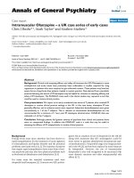
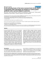
The results of ex vivo TNFα secretion (cytokine secretion with-
out any exogenously added antigen; Figure 1) showed that
there was a gradual increase in levels along with the progres-
sion (time post-Mtb injection) of AA in LEW rats with the high-
est level observed during Rec phase, while an opposite
pattern was observed in WKY rats. However, following re-
stimulation with Bhsp65, TNFα secretion was at a high level in
both LEW and WKY rats without significant changes during
the course of AA (Figure 1). Importantly, the level of TNFα
secreted in response to the pathogenic epitope B177 of
Bhsp65 was significantly increased at the Rec phase of AA in
the LEW rats, but at Inc phase in WKY rats. Overall, the high-
est level of TNFα secretion was observed during Rec phase in
LEW rats, but at Inc phase in WKY rats.
Arthritis Research & Therapy Vol 10 No 1 Kim et al.
Page 4 of 10
(page number not for citation purposes)
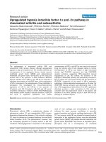
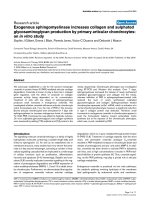
The severity of AA is downmodulated following in vivo
TNFα treatment of LEW rats
The above results showed that high TNFα levels correlate with
recovery from acute AA in LEW rats, and with resistance
against AA in WKY rats. To further examine this correlation, we
tested the effect of TNFα treatment on AA in the LEW rats.
Naïve LEW rats were given a total of 10 injections intraperito-
neally of TNFα (10
5
U/day) in PBS with three doses given
before Mtb-injection and then continued on the day (fourth
day) of Mtb injection and for 6 more days thereafter. After Mtb
injection, rats were observed regularly for signs of arthritis. The
control rats received 10 injections of PBS and were injected
with Mtb at the same time as the experimental rats. The results
(Figure 2) revealed that the TNFα-treated rats had a signifi-
cantly reduced severity of AA compared to that of the control
rats. This suppression of AA in the experimental group of rats
was evident before the peak of AA, and it continued for an
average of 7 days. Thus, treatment with TNFα, a pro-inflamma-
tory cytokine, significantly attenuated the severity of AA in the
LEW rats.
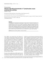
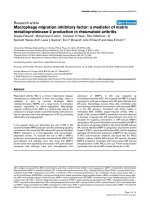
In vivo TNFα treatment of Mtb-immunised LEW rats
decreases IFN-γ secretion in response to the pathogenic
determinant B177 of Bhsp65
As TNFα treatment decreased the severity of AA, we tested
whether the suppression of AA involved any major changes in
the immune responsiveness to antigenic challenge. LEW rats
were treated with TNFα using the protocol described above,
including immunisation with Mtb or a control antigen (HEL/
IFA). After 9 days of antigenic challenge, the draining LNC of
these rats were harvested and tested for proliferative and
cytokine response using Bhsp65, HEL, using their peptides as
recall antigens. We obtained comparable (p > 0.05) numbers
of LNC from TNFα-treated and PBS-treated rats in both Mtb-
immunised and HEL-immunised groups (data not shown), sug-
gesting that, at the dose used, the injected TNFα did not lead
to a significant change in the number of cells (for example, via
apoptosis) in the draining lymph nodes. In the cohort of Mtb-
immunised rats, the LNC recall response to Bhsp65 and B177
in TNFα-treated rats was comparable to that of PBS-treated
control rats (Figure 3a). Similar results were obtained in the
Figure 1
Mycobacterium tuberculosis H37Ra (Mtb)-immunised Lewis (LEW) rats showed the highest level of tumour necrosis factor (TNF)α secretion during the Rec phase of adjuvant arthritis (AA), but Wistar-Kyoto (WKY) rats displayed an opposite profileMycobacterium tuberculosis H37Ra (Mtb)-immunised Lewis (LEW) rats showed the highest level of tumour necrosis factor (TNF)α secretion during
the Rec phase of adjuvant arthritis (AA), but Wistar-Kyoto (WKY) rats displayed an opposite profile. LEW (᭝) (n = 4 each) and WKY (▲) (n = 3
each) rats were killed at different time points after Mtb injection and their draining lymph node cells (LNC) were harvested. These LNC were cultured
for 48 h in a 96-well plate with or without the addition of any exogenous antigen. The supernates were then collected and analysed for TNFα by
enzyme-linked immunosorbent assay (ELISA). The LNC/culture supernates of individual rats were tested separately and then the results of each of
the two subgroups (LEW/WKY) were presented as pg/ml (mean ± SEM). For comparison, medium background was subtracted from antigen-
induced cytokine (Δ pg/ml). *p < 0.05 and **p ≤ 0.025, when levels of a particular cytokine at other phases of AA were compared with that at Inc
phase for the same rat strain (LEW/WKY); +, p ≤ 0.05, and ++, p ≤ 0.025, when cytokine levels were compared between LEW and WKY rats at the
corresponding phase of AA. Inc = incubation phase; Ons = onset phase; Pk = peak phase; and Rec = recovery phase. Testing of additional animals
following the above protocol yielded similar results.
Available online />Page 5 of 10
(page number not for citation purposes)
two groups of rats that were immunised with HEL (Figure 3b)
instead of Mtb. Furthermore, the results of cytokine testing
showed that IFN-γ secretion by LNC of TNF-treated, Mtb-
immunised LEW rats after B177 recall decreased significantly
compared to that of the PBS-treated control rats (Figure 3c).
This decrease in IFN-γ secretion in Mtb-immunised LEW rats
was specific to B177 as IFN-γ response to the control antigen
(HEL) in PBS-treated, HEL-immunised rats was comparable to
that of TNFα-treated, HEL-immunised rats (Figure 3d). As
there was no difference in the level of IL-10 secretion between
the two Mtb-immunised groups (Figure 3e,f), the decrease in
IFN-γ in response to B177 in TNFα-treated, Mtb-immunised
LEW rats steered the overall cytokine response towards a T
h
2
type. Thus, TNFα treatment neither resulted in a general non-
specific enhancement of antigen-specific proliferative T cell
response, nor induced a generalised immunosuppression.
Instead, the AA-protective effect of TNFα involved a decrease
of IFN-γ in response to the pathogenic epitope (B177) of
Bhsp65.
In another set of experiments, we examined whether TNFα
treatment had any effect on IL-17 production by Bhsp65- or
B177-reactive T cells. We tested IL-17 by qRT-PCR because
of rather limited reagents for the newer rat cytokines, including
IL-17. The level of IL-17 in TNFα-treated, Mtb-immunised rats
was comparable (p > 0.05) to that of PBS-treated, Mtb-immu-
nised rats (data not shown).
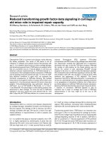
TNFα-treatment does not lead to any changes in serum
levels of sTNFR-I and anti-TNFα antibody
We examined two other parameters that might contribute to
TNFα-mediated protection against AA. First, the excessive
shedding of sTNFR-I [25,26], and second, the generation of
anti-TNFα antibody that might neutralise TNFα in vivo [27].
The levels of sTNFR-I (Figure 4A) as well as anti-TNFα anti-
bodies (Figure 4B) in sera of TNFα-treated, Mtb-immunised
LEW rats were comparable to that in sera of PBS-treated,
Mtb-immunised LEW rats.
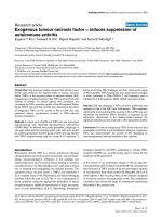
TNFα injections intraperitoneally do not induce any
preferential cell migration into the peritoneum
We also tested whether intraperitoneal injection of TNFα
might deviate the migration of T cells away from the joints into
the peritoneal cavity. Our results showed no difference in the
number/proportion of T cells (CD3) or macrophages/neu-
trophils (CD11b/c) infiltrating into the peritoneal cavity after
PBS treatment vs TNFα treatment intraperitoneally (Figure 5).
These results suggest the absence of a major shift in the
migration of T cells into the peritoneal cavity following TNFα
treatment.
The level of expression of mRNA for IDO as well as the
frequency of CD4+Foxp3+ T cells (Treg) is unaltered by
TNFα treatment
To gain further insights into the mechanisms by which TNFα
treatment might suppress AA, we compared the relative levels
of components of the two immunosuppressive pathways, the
IDO-TTS by qRT-PCR and the Treg by flow cytometry. IDO is
predominantly expressed in myeloid cells, and it catabolises
tryptophan [19,20,28]. By contrast, TTS binds to tryptophan
and makes it available for protein synthesis [19,20,28]. The
IDO-induced deprivation of tryptophan has been invoked in T
cell tolerance and suppression of T cell response. Similarly,
Treg can suppress the activity of pathogenic effector T cells
via cell-cell contact and immunomodulatory cytokines, TGF-β
and IL-10 [17,18]. Our results show that the levels of IDO
mRNA in Bhsp65-restimulated LNC of TNFα-treated rats
(1.51 fold compared to LNC in medium) were comparable (p
> 0.05) to that of PBS-treated rats (2.26 fold). Similarly, the
TTS mRNA levels in TNFα-treated versus PBS-treated rats
were 1.69 fold versus 2.6 fold, respectively, and this difference
was not significant (p > 0.05). The results for IDO mRNA test-
ing using splenic adherent cells (data not shown) were similar
to that obtained with LNC. In regard to Treg frequency, the
levels (mean ± SEM) were slightly lower in TNFα-treated rats
Figure 2
Downmodulation of adjuvant arthritis (AA) by in vivo tumour necrosis factor (TNF)α treatment of Lewis (LEW) ratsDownmodulation of adjuvant arthritis (AA) by in vivo tumour necrosis
factor (TNF)α treatment of Lewis (LEW) rats. LEW rats were injected
intraperitoneally daily either with 1 ml of 10
5
U/ml TNFα (n = 4; experi-
mental group; ■) or with 1 ml PBS (n = 8; controls; ᮀ) for 3 days
before the day of Mycobacterium tuberculosis H37Ra (Mtb) injection,
and then continued daily for 7 days, including the day of Mtb injection,
to a total of 10 injections. Thereafter, all rats were observed for signs of
AA, and the severity of arthritis was graded as described in Materials
and methods. The difference in the severity of arthritis during the
course of AA in the two groups of rats was statistically significant from
day 16 through day 25 (*p < 0.05; **p < 0.025). The difference
between the two rat groups was also significant (p < 0.05) when ana-
lysed by Wilcoxon rank sum test. Similar results were obtained in
repeat experiments. Also shown in the figure is a representative desig-
nation of different phases of the disease in the course of AA in the form
of days post-Mtb immunisation as follows: Inc = incubation, days 1 to
7; Ons = onset, days 8 to 10; Pk = peak, days 15 to 18; and Rec =
recovery, days 23 to 30.
Arthritis Research & Therapy Vol 10 No 1 Kim et al.
Page 6 of 10
(page number not for citation purposes)
(8.5% ± 0.4) than that of PBS-treated rats (10.2% ± 0.3), but
this difference was not statistically significant (p > 0.05).
Discussion
We observed that TNFα secretion in response to the arthri-
togenic epitope of Bhsp65 (B177) during the course of AA in
the LEW rat showed a paradoxically opposite profile in relation
to the disease severity. Considering the critical role of TNFα in
the initiation and propagation of arthritis, we had anticipated
that the level of TNFα might be high in the early phases of AA
(for example, Inc, Ons, and/or Pk), but relatively much lower in
the later phases (for example, Rec) of the disease. However,
the actual picture that was revealed was surprisingly reverse,
in that the arthritic LEW rats showed highest TNFα secretion
in the Rec phase of the disease compared to that at Ons or
Pk. This association of high TNFα levels with the decline of
inflammatory arthritis was also supported by the TNFα secre-
tion profile of the AA-resistant WKY rats. Unexpectedly, the
WKY rats secreted high levels of TNFα early after Mtb chal-
lenge, and the TNFα secretion then gradually declined with
time post-Mtb challenge, showing the reverse association of
disease activity/severity vs TNFα levels produced in response
to the pathogenic epitope of Bhsp65. However, this negative
correlation suggests but does not establish a causal relation-
ship between endogenous TNFα and protection against arthri-
tis. In this regard, our results of suppression of AA by
exogenous TNFα suggest that this cytokine also possesses an
immunoregulatory component. It is conceivable that the condi-
tions under which the same cytokine would manifest differen-
tial functional activities (pathogenic vs regulatory) might be
distinct, and these conditions have yet to be fully defined. We
propose that the concentration of TNFα is one of the critical
factors influencing the predominantly pathogenic vs protective
effect of the cytokine. Some of these factors are also revealed
in studies based on anti-TNF therapy in the AA model. Soluble
TNF-receptor (sTNF-RI) administered to LEW rats on days 9,
Figure 3
In vivo tumour necrosis factor (TNF)α treatment resulted in decreased interferon (IFN)-γ secretion by B177-restimulated LNC without affecting their proliferative responseIn vivo tumour necrosis factor (TNF)α treatment resulted in decreased interferon (IFN)-γ secretion by B177-restimulated LNC without affecting their
proliferative response. LEW rats were treated with PBS (ᮀ) or TNFα ( ) as described in the legend to Figure 2, with the exception that one sub-
group of rats was immunised with Mycobacterium tuberculosis H37Ra (Mtb) (a, c, e), whereas the other was injected with HEL/IFA (b, d, f). At day
9 after injection with Mtb or HEL, the draining LNC of these rats were harvested and tested in a proliferation assay ((a, b); n = 8 each). Peptide 333
to 347 of Bhsp65 (B333), peptide 65 to 78 of HEL (HEL65), and native HEL were used as control peptide/protein antigens. The results are pre-
sented as mean stimulation index (SI) ± SEM. In addition, the supernates collected after 72 h of culture of LNC of Mtb- or HEL-immunised rats were
tested by ELISA for IFN-γ (c, d) and interleukin (IL)-10 (e, f) (n = 5 each). The results of cytokine analysis are shown as Δ pg/ml (mean ± SEM). *, p
< 0.05 and **, p ≤ 0.025, when compared with the respective PBS control.
Available online />Page 7 of 10
(page number not for citation purposes)
11, and 13 of AA led to inhibition of AA, and the level of
suppression was dose-dependent [29]. Similarly, the treat-
ment of rats with sTNF-RI beginning on day 4 after disease
onset induced suppression of AA [30]. By contrast, in another
study, sTNF-RI treatment of DA rats on days 0, 2, and 4 post-
Mtb injection had no significant effect on early phase of AA
[31]. However, later in the course of AA, lower dose of sTNF-
RI exacerbated AA, while higher dose failed to alter the dis-
ease severity, supporting a concentration-dependent biologic
effect of this pro-inflammatory cytokine [31]. In a study con-
ducted in the CIA model, adenovirus-mediated gene delivery
of TNFR-IgG fusion protein initially suppressed arthritis but
subsequently exacerbated the disease [32]. Taken together,
these studies highlight both the disease-aggravating and the
disease-suppressing effects of TNFα.
We described above that systemic administration of TNFα
into LEW rats can downmodulate the course of clinical AA.
We ruled out the induction of any generalised immunosup-
pression due to chronic TNFα treatment by showing that
TNFα-treated, HEL-immunised LEW rats raised a robust pro-
liferative and cytokine response to the immunogen. Moreover,
we demonstrated that TNFα-treated LEW rats showed a sig-
nificant decrease in IFN-γ secretion in response to B177 with-
out much change in the proliferative response to the same
antigen. The ratio of IFN-γ to IL-10 showed a decrease, but this
skewing of the cytokine response was mainly because of a
decrease in IFN-γ levels rather than an increase in IL-10 secre-
tion. This decrease in IFN-γ levels could occur in part via
TNFα-mediated negative regulation of IL-12 production [33].
Although IFN-γ and TNFα are both pro-inflammatory cytokines,
but these cytokines might be regulated by different mecha-
nisms and also trigger differential effects [34] in a concentra-
tion-dependent mechanism. The downregulation of IFN-γ
production by the T cells following chronic TNFα exposure has
also been reported by other investigators [35]. However,
unlike for IFN-γ, we did not observe a significant change in IL-
17 response of Bhsp65- or B177-reactive T cells following
TNFα treatment. As Th1 and Th17 subsets of T cells are dis-
tinct lineages in regard to their differentiation and regulation by
different cytokines, a change in the production of one (IFN-γ)
but not the other (IL-17) cytokine after TNFα treatment of rats
is not an unexpected finding.
We also considered the earlier results of other investigators
showing that TNFα treatment can induce the shedding of sol-
uble TNF receptor I (sTNFR-I) from cell surface, which in turn
can bind circulating TNFα and suppress signals for continua-
tion of inflammation [25,26]. However, our analysis of sTNFR-
I in the sera of TNF-treated, Mtb-immunised LEW rats
excluded any significant change in sTNFR-I levels compared
to that of PBS-treated, Mtb-immunised LEW rats. Similarly, we
also ruled out the presence of circulating anti-TNFα antibodies
in the serum following TNFα injection, which in turn could neu-
tralise TNFα in vivo. We also excluded a major shift in the
migration of subsets of mononuclear cells into the peritoneal
cavity following TNFα injection intraperitoneally. Similarly, we
also ruled out any TNFα-induced enhancement of the level of
mRNA for IDO, the enzyme involved in IDO-tryptophan toler-
ance pathway and the level of CD4+CD25+ T cells (Treg). In
this study, we have tested only IDO mRNA expression but not
the IDO enzyme activity. Other investigators have demon-
strated that the induction of IDO activity is a two-step process,
with prostaglandin E2 causing an increase in IDO expression
Figure 4
Tumour necrosis factor (TNF)α treatment of Lewis (LEW) rats neither increased the release of soluble TNF receptor I (sTNFR-I) nor induced the generation of anti-TNFα antibodyTumour necrosis factor (TNF)α treatment of Lewis (LEW) rats neither
increased the release of soluble TNF receptor I (sTNFR-I) nor induced
the generation of anti-TNFα antibody. LEW rats were injectedintraperi-
toneally daily with 1 ml of either 10
5
U/ml TNFα or phosphate-buffered
saline (PBS) for 3 days before Mycobacterium tuberculosis H37Ra
(Mtb) immunisation, and then continued daily for a total of 10 injections.
At day 9 after Mtb immunisation, blood samples were collected from
these rats. The sera were then tested for sTNFR-I (A; n = 3+) and anti-
TNFα antibody (B; n = 3+) by enzyme-linked immunosorbent assay
(ELISA). Appropriate positive controls gave optimal results. The results
of sTNFR-I are presented as mean pg/ml ± SEM, and the results of
anti-TNFα antibody are presented as OD
450
(mean ± SEM). *p < 0.05
and †p ≤ 0.05, when naïve sera was compared with the PBS injected-
Mtb sera and TNF injected-Mtb sera, respectively.
Arthritis Research & Therapy Vol 10 No 1 Kim et al.
Page 8 of 10
(page number not for citation purposes)
and TNFα (or toll-like receptor ligands) leading to an increase
in IDO enzymatic activity [36]. Therefore, the precise contribu-
tion of IDO-tryptophan pathway to the TNFα-induced suppres-
sion of AA needs to be further explored. In regard to Treg,
there are limited reports on the effects of TNFα on Treg fre-
quency, and these revealed contrasting effects [37-39]. How-
ever, in RA patients, an increase in Treg numbers with anti-
TNFα treatment has been reported [38,40], which indirectly
supports our observed trend (but not significant) towards
decreased Treg numbers in TNFα-treated rats. TNFα may also
influence other important functions in vivo that have not been
addressed at this time in our study; for example, (a) apoptosis
within the target organ of pathogenic T cells that mediate
arthritis induction [41,42]; (b) alteration of the migration of
inflammatory cells into the joints by changing the expression of
adhesion molecules on endothelial cells [43]; (c) triggering of
the HPA axis by elevated systemic TNFα, leading to the
release of corticosteroids and suppression of TNFα in the tar-
get organ (the joints) [44,45]; (d) the induction of immunoreg-
ulatory cytokine IL-10, leading to the suppression of
pathogenic TNFα [46-48]; and (e) the modulation of dendritic
cells in vivo, which then present antigen favouring downregu-
lation of arthritis [49].
Our results highlight the immunoregulatory role of exogenous
TNFα in AA. Immune regulation by TNFα has been observed
in other models of autoimmune diseases as well. For example,
the downregulation of type 1 diabetes (T1D) in the non-obese
diabetic (NOD) mouse by CFA immunisation has been shown
to involve TNFα production and granzyme B/perforin-secret-
ing Treg [42,50]. In another study, TNFα expression within the
pancreas prevented diabetes in NOD mice [51], while sys-
temic treatment of TNFα in adult NOD mice decreased insuli-
tis as well as the incidence of diabetes [52]. However, the
modulation of diabetes by TNFα is influenced significantly by
the timing of administration or of the in vivo expression of
TNFα during the disease pathogenesis [53,54]. Similarly,
using the myelin oligodendrocyte glycoprotein (MOG)-
induced experimental autoimmune encephalomyelitis (EAE)
model, it has been shown that TNFα KO mice developed more
severe EAE, while TNFα treatment ameliorated the disease
[55]. Furthermore, studies in myocarditis have highlighted the
pathogenic as well as the protective roles of pro-inflammatory
cytokines [56,57]. In RA, there are several convincing pieces
of evidence to support the critical role of TNFα in mediating
the autoimmune inflammation [1,3,5], and accordingly, TNFα
antagonists are a significant addition to the therapeutic arse-
nal against RA [6,7]. However, our study has addressed the
understudied and under-appreciated protective or immu-
noregulatory role of exogenous TNFα against autoimmune
arthritis. These results have implications on our understanding
of the complex processes involved in the pathogenesis of
autoimmune arthritis as well as on the full range of effects on
immune responsiveness of individuals receiving anti-TNFα
agents for arthritis and other clinical conditions.
Conclusion
Pre-treatment of LEW rats with TNFα downmodulated the
severity of AA, and this TNFα induced protection against
arthritis involves suppression of IFN-γ production by the T cells
against the arthritogenic epitope of Bhsp65.
Figure 5
The composition of peritoneal lavage cells of tumour necrosis factor (TNF)α-treated Lewis (LEW) rats was comparable to that of phosphate-buffered saline (PBS)-treated ratsThe composition of peritoneal lavage cells of tumour necrosis factor (TNF)α-treated Lewis (LEW) rats was comparable to that of phosphate-buffered
saline (PBS)-treated rats. LEW rats (n = 4) were injected intraperitoneally daily with 1 ml of PBS (top panel) or TNFα (10
5
U) (bottom panel) for 4
days. After 3 h post the fourth injection, the peritoneal cavity was flushed with PBS and 10 ml of the peritoneal lavage fluid was collected. The cells
harvested from the lavage fluid were stained with appropriately labelled anti-CD3 or anti-CD11b/c antibody and analysed by fluorescence-activated
cell sorting (FACS). The results of one of the two independent experiments are shown in the figure. Both experiments yielded similar results.
Available online />Page 9 of 10
(page number not for citation purposes)
Competing interests
The authors declare that they have no competing interests.
Authors' contributions
EYK conducted most of the experimental work, designed
experiments, recorded and analysed the raw data, participated
in the interpretation of results as well as writing of the manu-
script. HHC contributed to the manuscript by designing and
conducting some of the experiments, and by recording, analys-
ing, and interpreting the results of those experiments. RR
designed and conducted some of the experiments, analysed
and interpreted their results, and participated in the writing of
the manuscript. KDM contributed by designing the experi-
ments, by analysing and interpreting the results, by writing of
the manuscript, and by arranging the grant support for this
study.
Acknowledgements
We thank Swamy Polumuri, Martin Flajnik, Peter Calabresi, John Sacci,
Dean Mann and Stefanie Vogel for their helpful critique and sugges-
tions. We gratefully acknowledge support from the National Institutes of
Health, Bethesda, MD (AI-047790 and AI-059623), and the Arthritis
Foundation, Atlanta, GA, USA.
References
1. Lipsky PE: Rheumatoid arthritis. In Harrison's Principles of Inter-
nal Medicine 16th edition. Edited by: Kasper D, Braunwald E,
Fauci A, Hauser S, Longo D, Jameson J. McGraw-Hill: New York;
2005:1968-1977.
2. Holmdahl R: Nature's choice of genes controlling chronic
inflammation. Ernst Schering Found Symp Proc 2006, 4:1-15.
3. Orozco C, Olsen NJ: Identification of patients with early rheu-
matoid arthritis: challenges and future directions. Clin Dev
Immunol 2006, 13:295-297.
4. Prakken BJ, Samodal R, Le TD, Giannoni F, Yung GP, Scavulli J,
Amox D, Roord S, de Kleer I, Bonnin D, Lanza P, Berry C, Massa
M, Billetta R, Albani S: Epitope-specific immunotherapy
induces immune deviation of proinflammatory T cells in rheu-
matoid arthritis. Proc Natl Acad Sci USA 2004, 101:4228-4233.
5. Berg WB van den, van Lent PL, Joosten LA, Abdollahi-Roodsaz S,
Koenders MI: Amplifying elements of arthritis and joint
destruction. Ann Rheum Dis 2007, 66(Suppl 3):iii45-48.
6. Feldmann M, Brennan FM, Foxwell BM, Maini RN: The role of TNF
alpha and IL-1 in rheumatoid arthritis. Curr Dir Autoimmun
2001, 3:188-199.
7. Feldmann M, Maini RN: Anti-TNF alpha therapy of rheumatoid
arthritis: what have we learned? Annu Rev Immunol 2001,
19:163-196.
8. Pearson CM: Development of arthritis, periarthritis and perios-
titis in rats given adjuvants. Proc Soc Exp Biol Med 1956,
91:95-101.
9. Taurog JD, Argentieri DC, McReynolds RA: Adjuvant arthritis.
Methods Enzymol 1988, 162:339-355.
10. van Eden W, Thole JE, Zee R van der, Noordzij A, van Embden JD,
Hensen EJ, Cohen IR: Cloning of the mycobacterial epitope rec-
ognized by T lymphocytes in adjuvant arthritis. Nature 1988,
331:171-173.
11. Quintana FJ, Carmi P, Mor F, Cohen IR: Inhibition of adjuvant
arthritis by a DNA vaccine encoding human heat shock protein
60. J Immunol 2002, 169:3422-3428.
12. Moudgil KD, Chang TT, Eradat H, Chen AM, Gupta RS, Brahn E,
Sercarz EE: Diversification of T cell responses to carboxy-ter-
minal determinants within the 65-kD heat-shock protein is
involved in regulation of autoimmune arthritis.
J Exp Med
1997, 185:1307-1316.
13. Ulmansky R, Cohen CJ, Szafer F, Moallem E, Fridlender ZG, Kashi
Y, Naparstek Y: Resistance to adjuvant arthritis is due to pro-
tective antibodies against heat shock protein surface epitopes
and the induction of IL-10 secretion. J Immunol 2002,
168:6463-6469.
14. Prakken BJ, Zee R van der, Anderton SM, van Kooten PJ, Kuis W,
van Eden W: Peptide-induced nasal tolerance for a mycobac-
terial heat shock protein 60 T cell epitope in rats suppresses
both adjuvant arthritis and nonmicrobially induced experimen-
tal arthritis. Proc Natl Acad Sci USA 1997, 94:3284-3289.
15. Ivanov II, McKenzie BS, Zhou L, Tadokoro CE, Lepelley A, Lafaille
JJ, Cua DJ, Littman DR: The orphan nuclear receptor RORγ t
directs the differentiation program of proinflammatory IL-17+
T helper cells. Cell 2006, 126:1121-1133.
16. Bettelli E, Oukka M, Kuchroo VK: T(H)-17 cells in the circle of
immunity and autoimmunity. Nat Immunol 2007, 8:345-350.
17. Sakaguchi S, Ono M, Setoguchi R, Yagi H, Hori S, Fehervari Z,
Shimizu J, Takahashi T, Nomura T: Foxp3+ CD25+ CD4+ natural
regulatory T cells in dominant self-tolerance and autoimmune
disease. Immunol Rev 2006, 212:8-27.
18. Shevach EM, DiPaolo RA, Andersson J, Zhao DM, Stephens GL,
Thornton AM: The lifestyle of naturally occurring CD4+ CD25+
Foxp3+ regulatory T cells. Immunol Rev 2006, 212:60-73.
19. Mellor AL, Munn DH: IDO expression by dendritic cells: toler-
ance and tryptophan catabolism. Nat Rev Immunol 2004,
4:762-774.
20. Grohmann U, Fallarino F, Bianchi R, Orabona C, Vacca C, Fioretti
MC, Puccetti P: A defect in tryptophan catabolism impairs tol-
erance in nonobese diabetic mice. J Exp Med 2003,
198:153-160.
21. Durai M, Kim HR, Moudgil KD: The regulatory C-terminal deter-
minants within mycobacterial heat shock protein 65 are cryptic
and cross-reactive with the dominant self homologs: implica-
tions for the pathogenesis of autoimmune arthritis. J Immunol
2004, 173:181-188.
22. Durai M, Gupta RS, Moudgil KD:
The T cells specific for the car-
boxyl-terminal determinants of self (rat) heat-shock protein 65
escape tolerance induction and are involved in regulation of
autoimmune arthritis. J Immunol 2004, 172:2795-2802.
23. Mia MY, Durai M, Kim HR, Moudgil KD: Heat shock protein 65-
reactive T cells are involved in the pathogenesis of non-anti-
genic dimethyl dioctadecyl ammonium bromide-induced
arthritis. J Immunol 2005, 175:219-227.
24. Toshchakov VU, Basu S, Fenton MJ, Vogel SN: Differential
involvement of BB loops of toll-IL-1 resistance (TIR) domain-
containing adapter proteins in TLR4-versus TLR2-mediated
signal transduction. J Immunol 2005, 175:494-500.
25. van Riemsdijk-van Overbeeke IC, Baan CC, Hesse CJ, Loonen EH,
Niesters HG, Zietse R, Weimar W: TNF-alpha: mRNA, plasma
protein levels and soluble receptors in patients on chronic
hemodialysis, on CAPD and with end-stage renal failure. Clin
Nephrol 2000, 53:115-123.
26. Xanthoulea S, Pasparakis M, Kousteni S, Brakebusch C, Wallach
D, Bauer J, Lassmann H, Kollias G: Tumor necrosis factor (TNF)
receptor shedding controls thresholds of innate immune acti-
vation that balance opposing TNF functions in infectious and
inflammatory diseases. J Exp Med 2004, 200:367-376.
27. Wildbaum G, Youssef S, Karin N: A targeted DNA vaccine aug-
ments the natural immune response to self TNF-alpha and
suppresses ongoing adjuvant arthritis. J Immunol 2000,
165:5860-5866.
28. Boasso A, Herbeuval JP, Hardy AW, Winkler C, Shearer GM: Reg-
ulation of indoleamine 2,3-dioxygenase and tryptophanyl-
tRNA-synthetase by CTLA-4-Fc in human CD4+ T cells. Blood
2005, 105:1574-1581.
29. Bendele AM, McComb J, Gould T, Frazier J, Chlipala E, Seely J,
Kieft G, Edwards CK 3rd: Effects of PEGylated soluble tumor
necrosis factor receptor type I (PEG sTNF-RI) alone and in
combination with methotrexate in adjuvant arthritic rats. Clin
Exp Rheumatol 1999, 17:553-560.
30. Schett G, Middleton S, Bolon B, Stolina M, Brown H, Zhu L, Pre-
torius J, Zack DJ, Kostenuik P, Feige U: Additive bone-protective
effects of anabolic treatment when used in conjunction with
RANKL and tumor necrosis factor inhibition in two rat arthritis
models. Arthritis Rheum 2005, 52:1604-1611.
31. Bush KA, Kirkham BW, Walker JS: The in vivo effects of tumour
necrosis factor blockade on the early cell mediated immune
events and syndrome expression in rat adjuvant arthritis.
Clin
Exp Immunol 2002, 127:423-429.
Arthritis Research & Therapy Vol 10 No 1 Kim et al.
Page 10 of 10
(page number not for citation purposes)
32. Quattrocchi E, Walmsley M, Browne K, Williams RO, Marinova-
Mutafchieva L, Buurman W, Butler DM, Feldmann M: Paradoxical
effects of adenovirus-mediated blockade of TNF activity in
murine collagen-induced arthritis. J Immunol 1999,
163:1000-1009.
33. Ma X, Trinchieri G: Regulation of interleukin-12 production in
antigen-presenting cells. Adv Immunol 2001, 79:55-92.
34. Ganster RW, Guo Z, Shao L, Geller DA: Differential effects of
TNF-alpha and IFN-gamma on gene transcription mediated by
NF-kappaB-Stat1 interactions. J Interferon Cytokine Res 2005,
25:707-719.
35. Aspalter RM, Wolf HM, Eibl MM: Chronic TNF-alpha exposure
impairs TCR-signaling via TNF-RII but not TNF-RI. Cell
Immunol 2005, 237:55-67.
36. Braun D, Longman RS, Albert ML: A two-step induction of
indoleamine 2,3 dioxygenase (IDO) activity during dendritic-
cell maturation. Blood 2005, 106:2375-2381.
37. Chen X, Baumel M, Mannel DN, Howard OM, Oppenheim JJ:
Interaction of TNF with TNF receptor type 2 promotes expan-
sion and function of mouse CD4+CD25+ T regulatory cells. J
Immunol 2007, 179:154-161.
38. Valencia X, Stephens G, Goldbach-Mansky R, Wilson M, Shevach
EM, Lipsky PE: TNF downmodulates the function of human
CD4+CD25hi T-regulatory cells. Blood 2006, 108:253-261.
39. Wu AJ, Hua H, Munson SH, McDevitt HO: Tumor necrosis fac-
tor-alpha regulation of CD4+CD25+ T cell levels in NOD mice.
Proc Natl Acad Sci USA 2002, 99:12287-12292.
40. Nadkarni S, Mauri C, Ehrenstein MR: Anti-TNF-alpha therapy
induces a distinct regulatory T cell population in patients with
rheumatoid arthritis via TGF-beta. J Exp Med 2007, 204:33-39.
41. Sheikh MS, Huang Y: Death receptor activation complexes: it
takes two to activate TNF receptor 1. Cell Cycle 2003,
2:550-552.
42. Qin HY, Chaturvedi P, Singh B: In vivo apoptosis of diabe-
togenic T cells in NOD mice by IFN-gamma/TNF-alpha. Int
Immunol 2004, 16:
1723-1732.
43. Ben-Horin S, Bank I: The role of very late antigen-1 in immune-
mediated inflammation. Clin Immunol 2004, 113:119-129.
44. Dunn AJ: Effects of cytokines and infections on brain
neurochemistry. Clin Neurosci Res 2006, 6:52-68.
45. Eskandari F, Webster JI, Sternberg EM: Neural immune path-
ways and their connection to inflammatory diseases. Arthritis
Res Ther 2003, 5:251-265.
46. Denys A, Udalova IA, Smith C, Williams LM, Ciesielski CJ, Camp-
bell J, Andrews C, Kwaitkowski D, Foxwell BM: Evidence for a
dual mechanism for IL-10 suppression of TNF-alpha produc-
tion that does not involve inhibition of p38 mitogen-activated
protein kinase or NF-kappa B in primary human macrophages.
J Immunol 2002, 168:4837-4845.
47. O'Shea JJ, Ma A, Lipsky P: Cytokines and autoimmunity. Nat
Rev Immunol 2002, 2:37-45.
48. Romagnani S: Regulation of the T cell response. Clin Exp
Allergy 2006, 36:1357-1366.
49. van Duivenvoorde LM, Louis-Plence P, Apparailly F, Voort EI van
der, Huizinga TW, Jorgensen C, Toes RE: Antigen-specific
immunomodulation of collagen-induced arthritis with tumor
necrosis factor-stimulated dendritic cells. Arthritis Rheum
2004, 50:3354-3364.
50. Qin HY, Mukherjee R, Lee-Chan E, Ewen C, Bleackley RC, Singh
B: A novel mechanism of regulatory T cell-mediated down-
regulation of autoimmunity. Int Immunol 2006, 18:1001-1015.
51. Grewal IS, Grewal KD, Wong FS, Picarella DE, Janeway CA Jr, Fla-
vell RA: Local expression of transgene encoded TNF alpha in
islets prevents autoimmune diabetes in nonobese diabetic
(NOD) mice by preventing the development of auto-reactive
islet-specific T cells. J Exp Med 1996, 184:1963-1974.
52. Jacob CO, Aiso S, Michie SA, McDevitt HO, Acha-Orbea H: Pre-
vention of diabetes in nonobese diabetic mice by tumor necro-
sis factor (TNF): similarities between TNF-alpha and
interleukin 1. Proc Natl Acad Sci USA 1990, 87:968-972.
53. Yang XD, Tisch R, Singer SM, Cao ZA, Liblau RS, Schreiber RD,
McDevitt HO: Effect of tumor necrosis factor alpha on insulin-
dependent diabetes mellitus in NOD mice. I. The early devel-
opment of autoimmunity and the diabetogenic process.
J Exp
Med 1994, 180:995-1004.
54. Christen U, Wolfe T, Mohrle U, Hughes AC, Rodrigo E, Green EA,
Flavell RA, von Herrath MG: A dual role for TNF-alpha in type 1
diabetes: islet-specific expression abrogates the ongoing
autoimmune process when induced late but not early during
pathogenesis. J Immunol 2001, 166:7023-7032.
55. Liu J, Marino MW, Wong G, Grail D, Dunn A, Bettadapura J, Slavin
AJ, Old L, Bernard CC: TNF is a potent anti-inflammatory
cytokine in autoimmune-mediated demyelination. Nat Med
1998, 4:78-83.
56. Lane JR, Neumann DA, Lafond-Walker A, Herskowitz A, Rose NR:
Role of IL-1 and tumor necrosis factor in coxsackie virus-
induced autoimmune myocarditis. J Immunol 1993,
151:1682-1690.
57. Fairweather D, Frisancho-Kiss S, Yusung SA, Barrett MA, Davis
SE, Steele RA, Gatewood SJ, Rose NR: IL-12 protects against
coxsackievirus B3-induced myocarditis by increasing IFN-
gamma and macrophage and neutrophil populations in the
heart. J Immunol 2005, 174:261-269.