Báo cáo y học: "Mesenchymal stem cells in arthritic disease" pot
Bạn đang xem bản rút gọn của tài liệu. Xem và tải ngay bản đầy đủ của tài liệu tại đây (1.01 MB, 12 trang )
Page 1 of 12
(page number not for citation purposes)
Available online />Abstract
Mesenchymal stem cells (MSCs), the nonhematopoietic progenitor
cells found in various adult tissues, are characterized by their ease
of isolation and their rapid growth in vitro while maintaining their
differentiation potential, allowing for extensive culture expansion to
obtain large quantities suitable for therapeutic use. These
properties make MSCs an ideal candidate cell type as building
blocks for tissue engineering efforts to regenerate replacement
tissues and repair damaged structures as encountered in various
arthritic conditions. Osteoarthritis (OA) is the most common
arthritic condition and, like rheumatoid arthritis (RA), presents an
inflammatory environment with immunological involvement and this
has been an enduring obstacle that can potentially limit the use of
cartilage tissue engineering. Recent advances in our under-
standing of the functions of MSCs have shown that MSCs also
possess potent immunosuppression and anti-inflammation effects.
In addition, through secretion of various soluble factors, MSCs can
influence the local tissue environment and exert protective effects
with an end result of effectively stimulating regeneration in situ.
This function of MSCs can be exploited for their therapeutic
application in degenerative joint diseases such as RA and OA. This
review surveys the advances made in the past decade which have
led to our current understanding of stem cell biology as relevant to
diseases of the joint. The potential involvement of MSCs in the
pathophysiology of degenerative joint diseases will also be
discussed. Specifically, we will explore the potential of MSC-based
cell therapy of OA and RA by means of functional replacement of
damaged cartilage via tissue engineering as well as their anti-
inflammatory and immunosuppressive activities.
Introduction
Mesenchymal stem cells (MSCs), also known in the literature
as bone marrow stem cells, skeletal stem cells, and
multipotent mesenchymal stromal cells, are nonhematopoietic
progenitor cells isolated from adult tissues, and are charac-
terized in vitro by their extensive proliferative ability in an
uncommitted state while retaining the potential to
differentiate along various lineages of mesenchymal origin,
including chondrocyte, osteoblast, and adipocyte lineages, in
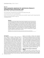
response to appropriate stimuli (Figure 1). Since the first study
by Friedenstein and colleagues [1] more than 40 years ago,
the field of MSC investigation has gained increasing attention
and popularity, particularly in the past decade. Using
‘mesenchymal stem cell’ as a key word in a PubMed search,
we retrieved 271 papers from 1998, 1,714 in 2007, and
1,185 in 2008 as of 19 July 2008. Initial studies focused on
MSC characterization, tissue origin, and the basic biology of
MSC growth and differentiation regulation. These studies led
to the realization that MSCs can be easily isolated from
various tissue sources, readily expanded in culture, and
appropriately differentiated under suitable stimulation. These
characteristics make MSCs an ideal candidate cell type for
tissue engineering efforts aiming to regenerate replacement
tissues for diseased structures. Further studies discovered
that the regenerative effects of MSCs do not merely rely on
their ability to structurally contribute to tissue repair. MSCs
possess potent immunomodulatory and anti-inflammatory
effects, and through either direct cell-cell interaction or
secretion of various factors, MSCs can exert a tremendous
effect on local tissue repair through modulating local
environment and activation of endogenous progenitor cells.
These features make MSC-based cell therapy a hotly pursued
subject of investigation in regenerative medicine.
1. Biology of mesenchymal stem cells
Characteristics and tissue distribution
Originally derived from bone marrow [1,2], MSCs and MSC-
like cells have been identified to exist in and can be isolated
from a large number of adult tissues, where they are
postulated to carry out the function of replacing and regener-
ating local cells that are lost to normal tissue turnover, injury,
or aging. These tissues include adipose, periosteum, synovial
membrane, synovial fluid (SF), muscle, dermis, deciduous
Review
Mesenchymal stem cells in arthritic diseases
Faye H Chen and Rocky S Tuan
Cartilage Biology and Orthopaedics Branch, National Institute of Arthritis, and Musculoskeletal and Skin Diseases, National Institutes of Health,
Department of Health and Human Services, Building 50, 50 South Dr., Bethesda, MD 20892, USA
Corresponding author: Rocky S Tuan,
Published: 10 October 2008 Arthritis Research & Therapy 2008, 10:223 (doi:10.1186/ar2514)
This article is online at />© 2008 BioMed Central Ltd
3-D = three-dimensional; BMP = bone morphogenetic protein; CIA = collagen-induced arthritis; ECM = extracellular matrix; FLS = fibroblast-like
synoviocyte; GVHD = graft-versus-host disease; IFN-γ = interferon-gamma; IL = interleukin; MHC = major histocompatibility complex; MMP = matrix
metalloproteinase; MSC = mesenchymal stem cell; NF-κB = nuclear factor-kappa-B; NK = natural killer; OA = osteoarthritis; PBMC = peripheral
blood mononuclear cell; PTHrP = parathyroid hormone-related protein; RA = rheumatoid arthritis; SF = synovial fluid; TGF-β = transforming growth
factor-beta; TNF-α = tumor necrosis factor-alpha; Treg = regulatory T cell.
Page 2 of 12
(page number not for citation purposes)
Arthritis Research & Therapy Vol 10 No 5 Chen and Tuan
teeth, pericytes, trabecular bone, infrapatellar fat pad, and
articular cartilage (reviewed in [3-5]). Despite the intense
research on MSCs, however, there is no uniformly accepted
clear and specific definitive phenotype or surface markers for
the prospective isolation of MSCs. Instead, MSCs are
defined retrospectively by a constellation of characteristics in
vitro, including a combination of phenotypic markers and
multipotential differentiation functional properties. The
minimal requirement for a population of cells to qualify as
MSCs, as suggested by the International Society for Cyto-
therapy, is threefold: (a) they must be plastic adherent under
standard culture conditions, (b) they should express CD105,
CD73, and CD90 and lack the expression of CD45, CD34,
CD14 or CD11b, CD79α or CD19, and HLA-DR surface
molecules, and (c) they should possess tripotential meso-
dermal differentiation capability into osteoblasts, chondro-
cytes, and adipocytes [6]. While this minimal set of standard
criteria was meant to foster a more uniform characterization
of MSCs and facilitate the exchange of data among
investigators, it will probably require modification as evolving
research gives rise to new knowledge. Although plastic
adherence serves as the most commonly used and simple
isolation procedure, various positive and negative surface
markers (for example, Stro-1, CD146/melanoma cell adhesion
molecule, CD271/low-affinity nerve growth factor, and stage-
specific embryonic antigen-4 [7]) have also been used to
enrich MSC yield and homogeneity. Recently, Buhring and
colleagues [8] described a panel of surface markers, including
CD140b (platelet-derived growth factor receptor-D), CD340
(HER-2/erbB2), and CD349 (frizzled-9) in conjunction with
CD217, that can be used for MSC enrichment. However, the
enriched cell fractions are still heterogeneous, and the majority
of isolated cells are not clonogenic.
Although MSCs isolated from different tissues show similar
phenotypic characteristics, it is not clear whether these are
the same MSCs, and they clearly show different propensities
in proliferation and differentiation potentials in response to
stimulation with various growth factors. A study that
compared human MSCs derived from bone marrow,
periosteum, synovium, skeletal muscle, and adipose tissue
revealed that synovium-derived MSCs exhibited the highest
capacity for chondrogenesis, followed by bone marrow-
derived and periosteum-derived MSCs [9]. Isolation methods,
culture surface, medium, and seeding density as well as
treatment with various growth factors influence the expansion
and differentiation and immunogenic properties of MSCs
[10]. Donor age and disease stage can also influence MSC
yield, proliferation rate, and differentiation potential. Of
particular relevance to rheumatic diseases, some studies
have shown that age, rheumatoid arthritis (RA), and advanced
osteoarthritis (OA) disease stage adversely affect MSCs
derived from the bone marrow of patients, with significantly
reduced proliferative capacity and chondrogenic activity
compared with those from young healthy donors, although
these findings are debated [11-13]. In one study, bone
marrow-derived MSCs from RA and OA patients showed
chondrogenic potential similar to that of MSCs isolated from
healthy donors [14]. In another study, compared with MSCs
from healthy donors, MSCs from individuals with RA showed
similar frequency, differentiation potential, survival, and
immunophenotypic characteristics, but RA patient MSCs
showed impaired clonogenic and proliferative potential with
premature telomere length loss [13]. However, irrespective of
age or OA disease etiology, it has been found that a sufficient
number of MSCs with adequate chondrogenic differentiation
potential can be isolated. Therefore, a therapeutic application
of MSCs for cartilage regeneration of RA and OA lesions
seems feasible.
Mesenchymal stem cell differentiation potential and
control
MSCs are characterized by their intrinsic self-renewal capacity
which is reflected in its clonogenic property and multilineage
differentiation potential. Under defined conditions, MSCs can
differentiate into chondrocytes, osteoblasts, and adipocytes,
and they also serve as hematopoiesis-supporting stromal cells
[2,15] (Figure 1). MSCs have also been reported, albeit
controversially, to differentiate into myocytes and cardio-
myocytes and even into cells of nonmesodermal origin,
including hepatocytes and neurons [16].
MSC chondrogenesis is a complex process and an active
area of research. Much of our understanding of the relevant
Figure 1
Multilineage differentiation potential of mesenchymal stem cells
(MSCs). Under appropriate conditions, MSCs are able to differentiate
into cell types of different lineages, including bone, cartilage, adipose,
muscle, tendon, and stroma. The arrows are presented as bidirectional,
indicating that differentiated MSCs are capable of dedifferentiation and
transdifferentiation. Adapted from [89].
Page 3 of 12
(page number not for citation purposes)
molecules and processes stems from our knowledge of
healthy cartilage homeostasis as well as cartilage formation in
the developing limb [17]. The standard experimental model
consists of a three-dimensional (3-D) culture of MSCs, as
high-density cell pellet or micromass culture or in a 3-D
scaffold, under the stimulation of suitable chondrogenic
factors. Elements including activations of various intracellular
signaling pathways (mitogen-activated protein kinases and
Smads) and transcription factors (sox9, L-sox5, and L-sox6),
production and interaction with extracellular matrix (ECM)
proteins (collagen type II, aggrecan, and cartilage oligomeric
matrix protein), activities of soluble bioactive factors such as
growth factors, cytokines, chemokines, and hormones, and
effects of environmental factors such as mechanical loading
and oxygen tension all affect chondrogenic differentiation of
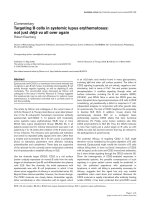
MSCs (Figure 2). One of the most important molecules
intrinsic to the assumption of the cartilaginous phenotype is
the transcription factor sox9. In bone marrow-derived MSCs,
expression of exogenous sox9 led to increased proteoglycan
deposition [18].
Growth factors that have regulatory effects on MSCs include
members of the transforming growth factor-beta (TGF-β)
superfamily, the insulin-like growth factors, the fibroblast
growth factors, the platelet-derived growth factor, and Wnts.
Among these growth factors, TGF-βs, including TGF-β1,
TGF-β2, and TGF-β3, as well as bone morphogenetic
proteins (BMPs) are the most potent inducers to promote
chondrogenesis of MSCs. For human MSCs, TGF-β2 and
TGF-β3 were shown to be more active than TGF-β1 in
promoting chondrogenesis in that, although cellular content
is similar after culture, significantly more proteoglycans and
collagen type II can be produced [19]. BMPs, known for their
involvement in cartilage formation, act alone or in concert with
other growth factors to induce or enhance MSC
chondrogenic differentiation. For example, BMP-2, BMP-4, or
BMP-6, combined with TGF-β3, induced chondrogenic
phenotype in cultured human bone marrow-derived MSC
pellets, with BMP-2 seemingly the most effective [20]. For
adipose tissue-derived MSCs, due to their lack of expression
of TGF-β type I receptor and reduced expression of BMP-2,
BMP-4, and BMP-6 when compared with bone marrow
MSCs, supplementation with BMP-6 and TGF-β seems to be
optimal for their chondrogenic differentiation, with BMP-6
stimulating stronger chondrogenic differentiation compared
with TGF-β [21]. Wnt signaling pathway protein poly-
morphism and altered gene expression have recently been
associated with RA and OA [22,23]. Canonical Wnt
signaling in coordination with TGF-β and BMP signaling has
been shown to enhance MSC differentiation [24,25]. In
addition, canonical and noncanonical Wnts have been shown
to cross-talk with each other in regulating stem cell
proliferation and osteogenic differentiation [26].
While MSCs can be induced to undergo chondrogenic
differentiation, with current systems and knowledge, the end
result is often less than desirable, with inferior cartilage-
related properties coupled with problematic terminal
differentiation. In one study, bovine MSCs were compared
directly with articular chondrocytes from the same animals for
their cartilage-forming capacity [27]. Both cell types were
cast into an agarose hydrogel system and cultured under the
same chondrogenic conditions with the stimulation of TGF-β.
While MSCs underwent chondrogenic differentiation as
indicated by cartilage ECM expression, the amount and
mechanical properties of the ECM were inferior to those
produced by the chondrocytes. These results suggest that
further optimization is needed for the successful use of MSCs
for cartilage tissue engineering. The other challenge in
controlling MSC chondrogenesis is the premature hyper-
tropic terminal differentiation of MSCs undergoing chondro-
genic differentiation. Hypertropic maturation of MSCs is
characterized by the premature expression of collagen type X,
matrix metalloproteinase-13 (MMP-13), and alkaline phospha-
tase activity that is normally found in growth plate cartilage
but not in stable healthy articular cartilage. The expression of
collagen type X can be detected early during MSC chondro-
genesis, and it is debatable whether its expression does
signal true hypertrophic differentiation [28]; however, it has
been correlated with the unstable transient nature of trans-
planted tissue in vivo, which leads to vascular invasion and
calcification [29]. Various factors are involved in the regula-
tion of hypertropic differentiation. The TGF-β family of growth
factors and their intracellular signaling molecules are involved
in chondrogenesis, including terminal differentiation [30].
TGF-β can inhibit chick sternal chondrocyte terminal differen-
tiation, as shown by suppression of expression of collagen
type X and alkaline phosphatase [31]. On the other hand,
BMP-2 can induce terminal differentiation [32,33], and in
chick sternal chondrocytes, this process can be inhibited by
the BMP antagonist chordin [33]. It has been shown that the
combination of isolation and culture condition as well as the
use of different BMPs can influence the outcome and extent
of MSC chondrogenesis progression as well as their terminal
hypertrophy [34]. Furthermore, similar to growth plate
development in which hypertrophic maturation is under the
regulation of a feedback loop involving Indian hedgehog and
parathyroid hormone-related protein (PTHrP) [35], PTHrP
also plays a regulatory role in MSC terminal differentiation.
When human bone marrow MSCs from OA patients were
cultured in a 3-D polyglycolic acid scaffold in the presence of
TGF-β3, upregulated expression of collagen type X was
significantly suppressed by the presence of PTHrP whereas
expression of other cartilage-specific matrix proteins was not
affected [36].
Taken together, these findings suggest a complex interplay of
extracellular growth factor molecules, signal transduction
pathways, and transcription factor networks for the control of
MSC chondrogenesis. Optimization of chondrogenesis to
generate stable cartilage suitable for clinical use is likely cell
source-dependent and will likely be a function of cellular
Available online />context, microenvironment as well as properties, dose, and
timing of the molecules administered to the cells [4,37].
Immunoregulatory properties of
mesenchymal stem cells
A very important property of MSCs, especially for their use in
rheumatic diseases, is their potent immunosuppressive and
anti-inflammatory functions that have been demonstrated
both in vitro and in vivo. Due to the scarcity of MSCs,
especially the apparent decrease in quantity and quality with
age and diseases, as well as the fact that patient-derived
MSCs have the same genetic defects as the patient, it is
sometimes desirable to consider using allogeneic MSCs for
therapy. Traditionally, allogeneic cell treatment has required
accompanying immunosuppression therapy. However, in the
case of MSCs, this may not always be necessary as it has
Arthritis Research & Therapy Vol 10 No 5 Chen and Tuan
Page 4 of 12
(page number not for citation purposes)
Figure 2
Use of mesenchymal stem cells (MSCs) as cell therapy for cartilage tissue repair and regeneration. The two potential approaches of MSC-based
cartilage repair and regeneration are illustrated. The first is ex vivo cartilage tissue engineering, in which a replacement tissue is constructed in vitro
using MSCs combined with scaffold under appropriate environmental stimuli. The second is in vivo cartilage regeneration via MSC cell therapy
using its anti-inflammatory and immunosuppressive effects. As shown in this figure, MSCs are expanded and injected locally into the affected joint.
MSCs can be applied systematically as well. MSCs, due to their potential regenerative functions as indicated, will help to influence the
microenvironment to aid in the regeneration of the cartilage.
been shown that MSCs can be used to modulate host
immune systems and confer immune suppression function.
However, caution should be exercised as this field of
research is still maturing and conflicting results have been
obtained in different systems from different labs.
First, MSCs are hypoimmunogenic and can evade the host
immune elimination. MSCs express low (fetal) to intermediate
(adult) major histocompatibility complex (MHC) class I
molecules and do not express MHC class II molecules on
their cell surface, although an intracellular pool of MHC class
II molecules can be stimulated to be expressed on the cell
surface by interferon-gamma (IFN-γ) [38]. However, since
MSCs do not express any costimulatory molecules, including
B7-1 (CD80), B7-2 (CD86), or CD40, they do not activate
alloreative T cells [39]. After differentiation into adipocytes,
osteoblasts, and chondrocytes, MSCs continue to express
MHC class I but not class II molecules on their cell surface,
even under stimulation, and continue to be nonimmunogenic
[38]. These properties suggest that MSCs should be able to
be transplanted to an allogeneic host without immune
rejection and that in vivo MSC cell therapy and tissue-
engineered cartilage construct using allogeneic MSCs
transplanted in vivo in hypoimmunogenic biomaterial
scaffolds should not elicit a host immune response. However,
the immune privilege of MSCs seems to be limited. A few
studies in mouse systems have reported that, in vivo,
allogeneic mismatched MSCs were rejected by the host and
could not form ectopic bone, while syngeneic recipient
allowed ectopic bone formation, despite the fact that, in vitro,
the MSCs showed immunosuppressive activity [40,41].
MSCs not only evade detection and elimination by the
immune system but can further modulate and suppress allo-
reactivity through modulating most major immune cell
activities [38,39,42-53]. In vitro, MSCs inhibit T-cell prolifera-
tion and activation in response to mitogenic or antigenic
stimulation in a dose-dependent manner. Numerous studies
[38,39,42-48] have shown that MSCs, as well as their
differentiated progenies of adipocytes, osteoblasts, or
chondrocytes, inhibit proliferation of allogeneic lymphocytes.
Both naïve and memory T cells as well as CD4
+
and CD8
+
T cells in mixed lymphocyte cultures were suppressed.
Furthermore, MSCs suppress CD8
+
T cell-mediated lysis.
T cells were found to be anergic and arrest in the G
0
-G
1
phase of the cell cycle.
In addition to T cells, MSCs exert proliferation inhibitory
effects on B cells [49], natural killer (NK) cells [50,51], and
dendritic cells [44,45,52,53]. In addition to the effect on
proliferation, MSCs can further interfere and affect cellular
differentiation and maturation and function of the immune
cells [44,45,52,53]. MSCs inhibit the maturation and
decrease the expression of presentation molecules and
costimulatory molecules of antigen-presenting cells [53].
MSCs can also inhibit B-cell antibody production [49]. In the
case of NK cells, MSCs can suppress their proliferation,
cytokine secretion, and cytotoxicity [45,50,51]. Furthermore,
MSCs not only have a direct inhibitory effect on T cells but
also affect the first critical step of immune response in that
they can inhibit the differentiation and maturation of the
antigen-presenting cells and cause the dendritic cells to
switch cytokine secretion profile to decrease their secretion
of proinflammatory cytokines such as tumor necrosis factor-
alpha (TNF-α), IFN-γ, and interleukin-12 (IL-12) and, impor-
tantly, increase production of IL-10 which is suppressive and
tolerogenic and a potent inducer of regulatory T cells (Tregs)
[44,45,53]. In addition, it has been reported [45] that human
MSCs caused an increase in the proportion of Tregs present.
Overall, the effect of MSCs on the immune cells is to skew
the immune response toward a tolerant and anti-inflammatory
phenotype. These immunomodulative effects seem not to be
limited to MSCs but are shared by other mesenchymal cells.
Progenies of MSC differentiation as well as various stromal
cells from different tissues, including chondrocytes and fibro-
blasts, have also been shown to have immunosuppressive
effects under certain conditions [38,46].
The mechanism of the immunomodulatory effects of MSCs is
not completely understood, although both direct and indirect
effects have been suggested through either cell-cell
interaction or soluble factors that create a local immuno-
suppressive environment. MSCs alter the cytokine secretion
profile of dendritic cells, naïve and effector T cells, and NK
cells to induce a more anti-inflammatory or tolerant pheno-
type. Secretion of the proinflammatory cytokines, TNF-α and
IFN-γ, is decreased whereas that of the more suppressive IL-
4 and IL-10 is stimulated [45]. Other factors involved have
been shown to include hepatocyte growth factor, TGF-β1, IL-
10, IL-6, prostaglandin E
2
, nitric oxide, and possibly
indoleamine 2,3-dioxygnease. Although the precise mecha-
nism has yet to be clarified (reviewed in [42,43]), the body of
evidence suggests that MSCs are immunosuppressive and
anti-inflammatory and can be transplanted between MHC-
incompatible individuals.
The immunosuppressive effects of MSCs have also been
demonstrated in vivo. The first of such studies was carried
out in baboons in which systematic administration of
allogeneic MSCs was used to prolong skin graft [47]. In an
animal model of experimental autoimmune encephalomyelitis
that mimics human multiple sclerosis, MSC administration
strikingly ameliorated disease. MSCs were effective when
administered at disease onset and at the peak of disease but
not after disease stabilization. This effect was believed to be
mediated through inducing T-cell anergy [48]. The
immunosuppressive function of MSCs has also been shown
to be effective in humans. In one report, MSCs were used to
treat severe steroid-refractory graft-versus-host disease
(GVHD), resulting in the disappearance of GVHD in six out of
eight patients, with their survival rate being significantly better
than that of patients not treated with MSCs [54]. In animal
Available online />Page 5 of 12
(page number not for citation purposes)
models, MSC implantations improved outcomes of renal,
lung, and cardiac injuries, at least partially by shifting the
microenvironment at the injury sites from proinflammatory to
anti-inflammatory [55-57]. In a murine pulmonary fibrosis
model, MSCs inhibited bleomycin-induced inflammation and
fibrosis within the lungs. This was shown to be due primarily
to the secretion of IL-1 receptor antagonist by MSCs [56].
MSC-conditioned medium was shown to block proliferation
of an IL-1α-dependent T-cell line and inhibit production of
TNF-α by activated macrophages in vitro. Furthermore, MSC
administration was more effective than recombinant IL-1
receptor antagonist delivered via either adenoviral infection or
osmotic pumps in inhibiting bleomycin-induced increases in
TNF-α, IL-1α, and trafficking of lymphocytes and neutrophils
into the lung [56]. These successful animal studies have led
to additional human studies, which include phase I/II clinical
trials on GVHD, acute myocardial infarction, end-stage
ischemic heart diseases, osteogenesis imperfecta, multiple
sclerosis, and open bone fracture (see [58] for review and
[59] for a list of ongoing clinical trials).
The studies on the effect of MSCs on immunomodulation,
along with other studies, also attest to another critical aspect
regarding the function of MSCs, that is, the trophic effects of
MSCs. In most in vivo studies except for those using in vitro
engineered constructs, significant engraftment of MSCs was
not observed whereas the potent beneficial effects of MSCs
were obvious. It thus appears that MSCs can secrete soluble
factors that can be anti-inflammatory, immunomodulatory, and
supportive for tissue repair through activating the
regenerative potential of the endogenous progenitor cells. In
line with this notion, MSCs have been used in vivo for
enhancing the engraftment of other tissues (for example,
hematopoietic stem cells). MSCs can support hematopoiesis
through the secretion of cytokines and have the capacity to
maintain and expand lineage-specific colony-forming units
from CD34
+
marrow cells in long-term bone marrow culture
[60,61], and when cotransplanted, can enhance hemato-
poietic stem cell engraftment and increase the success of
hematopoietic stem cell transplantation in clinical outcomes
[62-64]. It is reasonable to anticipate that MSC therapy in
conjunction with hematopoietic stem cell transplantation can
be used for treating autoimmune diseases, such as RA, to
possibly bypass the immunoablasive conditioning step and
tissue toxicity as a result of the immunomodulation function of
MSCs. This is expected to be an intensely pursued area of
research in the next few years.
The immune-suppressive function of MSCs brings caution to
its use under certain conditions. One of the concerns relates
to the potential interplay between MSCs and tumors. It has
been shown that MSCs, especially mouse MSCs, will
accumulate cytogenetic aberrations and become neoplastic
after a few passages in culture [65,66]. Human MSCs seem
to be more stable in culture during the standard in vitro
culture time of 6 to 8 weeks; however, they can also undergo
spontaneous transformation following long-term in vitro
culture (4 to 5 months) involving the mesenchymal-epithelial
transition process [67]. Therefore, care should be taken when
MSCs are expanded for clinical use. This is especially true for
the potential allogeneic ‘off-the-shelf’ approach, whereas
autologous MSC treatment should not require such a long
expansion time when enough original material is used. There
has also been some debate on the effect of in vitro expanded
MSCs on tumor growth. MSCs have the capacity to engraft
into multiple tissues in vivo, especially to sites of injury and
inflammation, including primary tumor and tissue sites of
metastasis. The effect of MSCs on tumor growth has been
somewhat controversial. There are reports that MSCs
promote tumor growth and metastasis as well as studies to
the contrary (reviewed in [68]). The contradictory results
probably relate to the different tumors and models used and
to the differences stemming from the heterogeneity and
different culture methods of MSCs. Nevertheless, the ability
of MSCs to target tumors has given rise to a potential
therapeutic way of cancer therapy to specifically deliver
antitumor drugs in situ. MSCs genetically modified to express
antitumor factors, including IL-12 and antagonist for hepatic
growth factor, have been used. The therapeutic application
for MSCs on tumor growth requires further investigation to
rule out the potential side effects of MSCs.
2. Mesenchymal stem cells in rheumatic
diseases
The ease of isolation and expansion and the multipotential
differentiation capacity, especially the chondrogenic differen-
tiation property of MSCs, make MSCs the cell type of choice
for articular cartilage tissue engineering that aims to replace
and regenerate the diseased structure in joint diseases. In
addition, their immunomodulatory and anti-inflammatory
functions make MSCs the ideal candidate for cell therapy to
treat diseases with inflammatory features such as those
encountered in OA and RA, although research in this area is
just starting to gain momentum. Therefore, MSCs are actively
being considered as candidate cells for the treatment of
arthritic joint diseases both as a structural substitute and as a
stand-alone cell therapy or as a combination thereof
(Figure 2). The involvement of MSCs in OA and RA and their
potential use for their treatment are discussed below.
Mesenchymal stem cell and osteoarthritis
OA is the most common type of arthritis. It is estimated that
26.9 million Americans 25 years old or older have clinical OA
of some joints, with a higher percentage of affliction in the
older population [69]. Its clinical manifestations include joint
pain and impairment to movement, and surrounding tissues
are often affected with local inflammation. The etiology of OA
is not completely understood; however, injury, age, and
genetics have been considered among the risk factors. OA is
a progressively debilitating disease that affects mostly
cartilage, with associated changes in bone. Cartilage has
limited intrinsic healing and regenerative capacities. Current
Arthritis Research & Therapy Vol 10 No 5 Chen and Tuan
Page 6 of 12
(page number not for citation purposes)
pharmacologic treatment for early OA has seen limited
success, and various surgical procedures, including
debridement, drilling, osteochondral transplantation, autolo-
gous perichondral and periosteal grafts, and autologous
chondrocyte implantation, are able to relieve pain temporarily
but eventually fail [70]. Due to the increasing incidence of OA
and the aging population coupled with inefficient therapeutic
choices, novel cartilage repair strategies are in need.
The availability of large quantities of MSCs and their potential
for ready chondrogenic differentiation after prolonged in vitro
expansion have made MSCs the most hopeful candidate
progenitor cell source for cartilage tissue engineering. MSCs
loaded on a 3-D scaffold under appropriate differentiation
cues can undergo chondrogenic differentiation, and the
resulting construct can be used as a replacement tissue for
cartilage repair (Figure 2). In vitro cartilage tissue engineering
has attracted a lot of research effort and attention from
biologists, engineers, and clinicians in the past 10 years. The
regulation and control of this process have been extensively
reviewed above and elsewhere and readers are referred to
these publications for additional information [4,71,72]. In
addition to being used for structural replacement as the aim
of cartilage tissue engineering in cartilage repair, MSCs have
been used directly in cell therapy for OA cartilage repair in
situ. OA is associated with progressive and often severe
inflammation. For tissue engineering or cell therapy to be
successful, measures must be taken to control such an
inflammatory environment. Because MSCs have been shown
to possess anti-inflammatory function, they are also a suitable
cell type for this purpose. Several characteristics of MSCs
make them attractive in this respect. First, MSCs have been
shown to be able to migrate and engraft onto multiple
musculoskeletal tissues, especially sites of injury, and undergo
site-specific differentiation. More importantly, while there,
MSCs can exert significant effects on local environment and
resident endogenous tissue progenitor cells through direct or
indirect interactions and soluble factors. In addition, MSCs
have shown potent anti-inflammatory and immunosuppressive
activities. Taken together, these properties make MSCs a
promising candidate for cell therapy for diseases that often
involve the immune system, such as OA and RA (Figure 2).
A study by Murphy and colleagues [73] employing MSCs in a
goat OA model highlighted the regenerative effect of MSC
cell therapy in OA. Trauma-induced OA was simulated in this
model by unilateral excision of the medial meniscus and
resection of the anterior cruciate ligament, followed by
exercise. Autologous MSCs in hyaluronan solution were
injected intra-articularly to test their effect. In the control
animals without MSCs, OA development was observed as
expected, with substantial fibrillation and erosion of large
areas of articular cartilage, accompanied by osteophyte
formation and changes to the subchondral bone. In the MSC-
treated joints, there was marked regeneration of the medial
meniscus and decreased cartilage destruction and bone
changes. Injected labeled MSCs were not observed to be
engrafted on articular cartilage. Labeled MSCs were seen
engrafted in the neomeniscus, though not in a large enough
quantity to account for the majority of the newly formed
tissue. These findings suggested that the beneficial effect of
MSCs on cartilage protection and on OA progression was
not due to the direct structural contribution of MSCs. Based
on knowledge gained from other systems, it is possible that
the injected MSCs in this case acted to induce endogenous
progenitor cells through various direct or indirect interactions
to regenerate meniscus, which in turn retarded cartilage
degeneration associated with OA. Based on the goat study, a
procedure using direct injection of adult stem cells into the
patient’s knee to repair meniscus and prevent OA
progression is currently in a phase I/II clinical trial.
The above study highlights another challenge in using MSCs
systematically or locally for arthritis prevention and treatment,
that is, the inefficient engraftment of MSCs to the articular
cartilage. In one experiment, the engraftment, survival, and
long-term fate of human MSCs were assessed after in utero
transplantation in sheep, and transplanted cells were shown
to persist and undergo site-specific differentiation into
chondrocytes, adipocytes, myocytes and cardiomyocytes,
bone marrow stromal cells, and thymic stroma. However,
although most of the animals had human cell engraftment in
various tissues, cartilage-specific engraftment was not
efficient [74]. In another study, plastic adherence-enriched
bone marrow mesenchymal precursor cells were syste-
matically transplanted via tail vein injection into irradiated
mice [75]. After 1 to 5 months, the donor cells were found in
bone, cartilage, and lung in addition to marrow and spleen.
When chondrocytes were isolated from xiphoid and articular
cartilage by microscopic dissection, progeny of the donor
cells accounted for 2.5% of the isolated chondrocytes.
Although donor cells were found to engraft onto articular
cartilage of irradiated mice, albeit at low efficiency, assays of
control nonirradiated mice revealed very low levels of the
donor cells at the same time points [75]. In studies with
different models of induced arthritis, including a trauma-
induced goat OA model [73] and a mouse model of collagen-
induced arthritis (CIA) [76], transplanted cells were not
detected in joint cartilage. Investigation into the mechanisms
of MSC trafficking and homing, possibly through the
regulation of various chemokines and receptors, as well as
adhesion molecules and their receptors (reviewed in [77]), is
currently an actively pursued area of research and will likely
provide insights into means of increasing engraftment of
MSCs onto articular cartilage for more efficient treatment of
arthritis. Despite the low engraftment efficiency, MSC-based
procedures have been found to exert a therapeutic effect in
various disease models, including arthritis, possibly through
their trophic effect and their anti-inflammatory and immuno-
suppressive activities, which can significantly affect the local
environment and resident endogenous tissue progenitor cells
in carrying out the regenerative function.
Available online />Page 7 of 12
(page number not for citation purposes)
Mesenchymal stem cell and rheumatoid
arthritis
RA is a complex multisystem autoimmune disease charac-
terized by cartilage and bone destruction associated with
local production of inflammatory mediators, such as TNF-α
and IL-1β. The etiology of RA is not completely understood,
and multiple cells are thought to contribute to the pathogenic
progression, with T cells [78] and fibroblast-like synoviocytes
(FLSs) [79] playing central roles in orchestrating the disease
progression of inflammation and tissue damage. Although it is
still debatable, RA is believed to be a T cell-driven inflam-
matory synovitis disease in which T cells and synoviocytes
participate in a complex network of cell- and mediator-driven
events leading to joint destruction. Both antigen-activated
CD4
+
T helper 1 (Th1) and CD8
+
T cells are reported to be
involved in the pathogenesis of RA. After being triggered and
activated, T cells stimulate monocytes, macrophages, and
FLSs to produce inflammatory mediators, including IL-1,
TNF-α, IFN-γ, and IL-6, and secrete MMPs, leading to the
systemic inflammation that eventually results in joint
destruction [78,80]. Pharmacological interventions aiming at
reducing inflammation, including methotrexate and anti-TNF-α
drugs (infliximab, adalimumab, and etanercept), have been
used to treat RA symptoms [81]. Recently, for patients who
do not respond to conventional treatment, autologous
hematopoietic stem cell transplantation after immune ablation
treatment has become an option. However, this comes with a
high risk of side effects, including mortality. Joint destruction
in RA and the anti-inflammatory and immune-suppressive
properties of MSCs suggest that RA may be a candidate
disease for cartilage and bone repair using MSC therapy.
MSCs have been identified in synovium and SF that share
characteristics of bone marrow derived-MSCs, with clono-
genic and multipotential differentiation potentials. The origin
of SF-MSCs is not clear. From gene array profiling, it has
been observed that SF-MSCs are more similar to synovial
MSCs than bone marrow MSCs [82]. This finding can
suggest that SF-MSCs are derived from synovium instead of
bone marrow or are the result of phenotypic changes due to
their local environment. Furthermore, the relationship between
FLS and MSC is not fully elucidated. It has been reported
that a fraction of the RA FLS population shows properties
that are associated with MSCs in that they can differentiate
into chondrocytes, osteoblasts, adipocytes, and muscle cells
despite the pathological condition [83-85]. By means of a
mouse model of bone marrow transplantation in which bone
marrow cells from green fluorescence protein-transgenic
donor mice were transplanted into lethally irradiated recipient
mice, it was shown that normal FLSs contain a minor fraction
(1.2%) of bone marrow-derived mesenchymal cells. At the
onset of CIA in a mouse model of RA, before inflammation,
primitive bone marrow stromal cells migrated from the bone
marrow into the affected joint cavity and appeared to
contribute to synovial proliferation, and this process is
dependent on the proinflammatory cytokine TNF-α [83].
Upon CIA development, the arthritic FLSs contain a
substantial portion (33.7%) of bone marrow-derived cells
[84]. These cells can differentiate in vitro into various mesen-
chymal cell types, but inflammatory cytokines such as IL-1β
prevent the multilineage differentiation. The transcription
factor nuclear factor-kappa-B (NF-κB), which can be
activated by proinflammatory cytokines, plays a key role in the
repression of osteogenic and adipogenic differentiation of
arthritic FLS. Furthermore, specific activation of NF-κB
profoundly enhances FLS proliferation, motility, and secretion
of matrix-degrading MMP-13. Therefore, it is proposed that
arthritic FLSs are, in fact, MSCs which are arrested at early
stages of differentiation by inflammation activation of NF-κB
[84]. In another study, MSCs from RA and healthy donors were
compared. RA MSCs showed frequency, differentiation
potential, survival, and immunophenotypic characteristics
similar to those of normal MSCs, but impaired clonogenic and
proliferative potential with premature telomere length loss [13].
Currently, the biological roles MSCs play in RA patho-
physiology are unknown. However, MSCs isolated from RA
patients and patients with other autoimmune diseases seem
to be similar to normal MSCs in that they are clonogenic and
possess multipotential differentiation capacity. More impor-
tantly, they can also inhibit the proliferation of autologous and
allogeneic peripheral blood mononuclear cells (PBMCs) in a
dose-dependent manner. The inhibition was observed with
MSCs and PBMCs either from healthy donors or from
patients suffering from autoimmune diseases [86]. This
indicates that MSCs from RA patients can potentially be used
for immunomodulatory cell therapy. Recently, in a more
specific study, allogeneic MSCs were tested against T cells
from RA patients which react to collagen type II [87]. MSCs
or MSC-differentiated chondrocytes were able to inhibit
collagen type II-stimulated T-cell proliferation and activation in
a dose-dependent manner. In addition, MSCs and their
chondrocyte progeny alike inhibited the secretion of
proinflammatory cytokines IFN-γ and TNF-α by CD4
+
and
CD8
+
cells while increasing the secretion of IL-10 and
restoring the secretion of IL-4. It was also shown that TGF-β
played a significant role in the inhibitory effects of MSCs in
this case.
So far, the in vivo use of MSCs for treating RA has generated
mixed results. CIA is an experimental autoimmune disease
that shares several clinical and histological features with RA.
CIA can be elicited in susceptible strains of rodents and
nonhuman primates by immunization with collagen type II, the
major matrix constituent protein of articular cartilage. In a CIA
mouse model, a single injection of MSCs prevented the
occurrence of severe irreversible damage to bone and
cartilage [76]. Using cell tracking, donor cells were not
detected in joints of treated mice, suggesting that the
injected MSCs did not restore tissue integrity by mechanisms
of direct tissue repair. At the end of the experiment, cells
were not evident in peritoneal or secondary lymphoid organs,
Arthritis Research & Therapy Vol 10 No 5 Chen and Tuan
Page 8 of 12
(page number not for citation purposes)
although cells were detected in the intermediate time point. In
terms of mechanism, MSC treatment induced hyporespon-
siveness of T lymphocytes from MSC-treated mice in that
they showed basal in vitro proliferation and mitogen-induced
and collagen type II-recalled proliferation compared with T cells
from non-MSC-treated animals. MSC treatment modulated
the expression of proinflammatory cytokines. In particular,
serum concentration of TNF-α was significantly decreased. It
was suggested that MSCs exerted their immunomodulatory
function by educating antigen-specific Tregs. In MSC-treated
immunized mice, CD4
+
CD25
+
CD27
+
Tregs were increased
significantly compared with non-MSC-treated mice, and
Tregs from these mice inhibited proliferation of T lymphocytes
when proliferation was recalled using collagen type II. These
results suggest an effective therapeutic approach to target
the pathogenic mechanism of autoimmune arthritis using
allogeneic MSCs.
In another CIA study, mouse stem cell line C3H10T1/2 did
not confer any benefit. In vitro experiments showed that the
addition of TNF-α was sufficient to reverse the immuno-
suppressive effect of MSCs on T-cell proliferation [88]. These
data suggest that environmental parameters, in particular
those related to inflammation, may influence the immuno-
suppressive properties of MSCs.
Conclusion
The potential use of MSCs as building blocks for joint tissue
replacement via tissue engineering and their newly uncovered
potential for direct cell therapy by virtue of their trophic and
anti-inflammatory and immunosuppressive properties (Figure 2)
have generated a lot of enthusiasm in orthopaedics and
rheumatology communities. A large body of research has
produced exciting data, leading to the hope of their potential
application. However, controversy still exists, and a great deal
of work needs to be done before MSCs can be accepted for
clinical therapeutic applications.
Research on MSCs and their use in various rheumatic
diseases has been clearly gaining attention and momentum.
The need for successful therapy in treating these diseases
warrants more investment in research and development, both
at the fundamental level of basic biology and in more
translational studies. Fundamental knowledge of MSC
identification, isolation, culture, and differentiation still
requires extensive and intensive studies. The lack of an
unambiguous definition and isolation of MSCs and the
heterogeneity of MSCs alone, resulting in inadequately
defined cell populations isolated by different groups, most
likely contributed to some of the different and often
contradictory results reported so far. For cartilage tissue
engineering, the principal challenge is to find the optimal and
most effective cues for cartilage formation in vitro, be it
growth factors tailored for the specific MSCs, bioactive
scaffolds, or the enhancing environmental factors, with the
goal of generating a stable replacement articular cartilage
tissue that has appropriate mechanical properties and can
integrate with the host tissues with proper stable long-term
functions. Research on the in vivo MSC niche and the
regulation of this microenvironment will prove to be of pivotal
importance to determine how best to use MSCs to modulate
the local environment and endogenous progenitor cells for
repair and regeneration purposes. It is clear that the evolving
and rapidly developing research on the immunomodulatory
and anti-inflammatory effects of MSCs will improve our
knowledge of the mechanism and regulation of this
phenomenon. While caution should be exercised in the
clinical application of MSC therapy on arthritic patients,
contingent upon the confirmation of additional conclusive
animal studies, we believe that MSCs offer great hope in
relieving the disease burden of degenerative joint diseases
through their application in the form of replacement tissue as
well as local or systemic cell therapy.
Competing interests
The authors declare that they have no competing interests.
Acknowledgements
This work was supported by the Intramural Research Program of the
National Institute of Arthritis and Musculoskeletal and Skin Diseases,
National Institutes of Health (Bethesda, MD, USA) (Z01 AR41131).
References
1. Friedenstein AJ, Piatetzky-Shapiro I, Petrakova KV: Osteogenesis
in transplants of bone marrow cells. J Embryol Exp Morphol
1966, 16:381-390.
2. Pittenger MF, Mackay AM, Beck SC, Jaiswal RK, Douglas R,
Mosca JD, Moorman MA, Simonetti DW, Craig S, Marshak DR:
Multilineage potential of adult human mesenchymal stem
cells. Science 1999, 284:143-147.
3. Mimeault M, Batra SK: Recent progress on tissue-resident
adult stem cell biology and their therapeutic implications.
Stem Cell Rev 2008, 4:27-49.
4. Chen FH, Rousche KT, Tuan RS: Technology insight: adult
stem cells in cartilage regeneration and tissue engineering.
Nat Clin Pract Rheumatol 2006, 2:373-382.
5. Bianco P, Robey PG, Simmons PJ: Mesenchymal stem cells:
revisiting history, concepts, and assays. Cell Stem Cell 2008,
2:313-319.
6. Dominici M, Le Blanc K, Mueller I, Slaper-Cortenbach I, Marini F,
Krause D, Deans R, Keating A, Prockop D, Horwitz E: Minimal
criteria for defining multipotent mesenchymal stromal cells.
Available online />Page 9 of 12
(page number not for citation purposes)
This article is part of a special collection of reviews, The
Scientific Basis of Rheumatology: A Decade of
Progress, published to mark Arthritis Research &
Therapy’s 10th anniversary.
Other articles in this series can be found at:
/>The Scientific Basis
of Rheumatology:
A Decade of Progress
The International Society for Cellular Therapy position state-
ment. Cytotherapy 2006, 8:315-317.
7. Jones E, McGonagle D: Human bone marrow mesenchymal
stem cells in vivo. Rheumatology (Oxford) 2008, 47:126-131.
8. Buhring HJ, Battula VL, Treml S, Schewe B, Kanz L, Vogel W:
Novel markers for the prospective isolation of human MSC.
Ann N Y Acad Sci 2007, 1106:262-271.
9. Sakaguchi Y, Sekiya I, Yagishita K, Muneta T: Comparison of
human stem cells derived from various mesenchymal tissues:
superiority of synovium as a cell source. Arthritis Rheum 2005,
52:2521-2529.
10. Sotiropoulou PA, Perez SA, Salagianni M, Baxevanis CN,
Papamichail M: Characterization of the optimal culture condi-
tions for clinical scale production of human mesenchymal
stem cells. Stem Cells 2006, 24:462-471.
11. Murphy JM, Dixon K, Beck S, Fabian D, Feldman A, Barry F:
Reduced chondrogenic and adipogenic activity of mesenchy-
mal stem cells from patients with advanced osteoarthritis.
Arthritis Rheum 2002, 46:704-713.
12. Sethe S, Scutt A, Stolzing A: Aging of mesenchymal stem cells.
Ageing Res Rev 2006, 5:91-116.
13. Kastrinaki MC, Sidiropoulos P, Roche S, Ringe J, Lehmann S, Kri-
tikos H, Vlahava VM, Delorme B, Eliopoulos GD, Jorgensen C,
Charbord P, Häupl T, Boumpas DT, Papadaki HA: Functional,
molecular and proteomic characterisation of bone marrow
mesenchymal stem cells in rheumatoid arthritis. Ann Rheum
Dis 2008, 67:741-749.
14. Dudics V, Kunstar A, Kovacs J, Lakatos T, Geher P, Gomor B,
Monostori E, Uher F: Chondrogenic potential of mesenchymal
stem cells from patients with rheumatoid arthritis and
osteoarthritis: measurements in a microculture system. Cells
Tissues Organs 2008, PMID:18562787.
15. Prockop DJ: Marrow stromal cells as stem cells for non-
hematopoietic tissues. Science 1997, 276:71-74.
16. Jiang Y, Jahagirdar BN, Reinhardt RL, Schwartz RE, Keene CD,
Ortiz-Gonzalez XR, Reyes M, Lenvik T, Lund T, Blackstad M, Du J,
Aldrich S, Lisberg A, Low WC, Largaespada DA, Verfaillie CM:
Pluripotency of mesenchymal stem cells derived from adult
marrow. Nature 2002, 418:41-49.
17. Tuan RS: Biology of developmental and regenerative skeleto-
genesis. Clin Orthop Relat Res 2004, 427:S105-117.
18. Tsuchiya H, Kitoh H, Sugiura F, Ishiguro N: Chondrogenesis
enhanced by overexpression of sox9 gene in mouse bone
marrow-derived mesenchymal stem cells. Biochem Biophys
Res Commun 2003, 301:338-343.
19. Barry F, Boynton RE, Liu B, Murphy JM: Chondrogenic differenti-
ation of mesenchymal stem cells from bone marrow: differen-
tiation-dependent gene expression of matrix components.
Exp Cell Res 2001, 268:189-200.
20. Sekiya I, Larson BL, Vuoristo JT, Reger RL, Prockop DJ: Compari-
son of effect of BMP-2, -4, and -6 on in vitro cartilage forma-
tion of human adult stem cells from bone marrow stroma. Cell
Tissue Res 2005, 320:269-276.
21. Hennig T, Lorenz H, Thiel A, Goetzke K, Dickhut A, Geiger F,
Richter W: Reduced chondrogenic potential of adipose tissue
derived stromal cells correlates with an altered TGFbeta
receptor and BMP profile and is overcome by BMP-6. J Cell
Physiol 2007, 211:682-691.
22. Rousche KT, Basksh D, Tuan RS: Wnt signaling for targeted
therapies in rheumatology. In Further Targeted Therapies in
Rheumatology. Edited by Smolen JS, Lipsky PE. London, UK:
Taylor & Francis; 2007.
23. Sen M: Wnt signalling in rheumatoid arthritis. Rheumatology
(Oxford) 2005, 44:708-713.
24. Zhou S, Eid K, Glowacki J: Cooperation between TFG-beta and
Wnt pathways during chondrocyte and adipocyte differentia-
tion of human marrow stromal cells. J Bone Miner Res 2004,
19:463-470.
25. Tuli R, Tuli S, Nandi S, Huang X, Manner PA, Hozack WJ, Daniel-
son KG, Hall DJ, Tuan RS: Transforming growth factor-b-medi-
ated chondrogenesis of human mesenchymal progenitor
cells involves N-cadherin and mitogen-activated protein
kinase and Wnt signaling cross-talk. J Biol Chem 2003, 278:
41227-41236.
26. Baksh D, Tuan RS: Canonical and non-canonical Wnts differen-
tially affect the development potential of primary isolate of
human bone marrow mesenchymal stem cells. J Cell Physiol
2007, 212:817-826.
27. Mauck RL, Yuan X, Tuan RS: Chondrogenic differentiation and
functional maturation of bovine mesenchymal stem cells in
long-term agarose culture. Osteoarthritis Cartilage 2006, 14:
179-189.
28. Mwale F, Stachura D, Roughley P, Antoniou J: Limitations of
using aggrecan and type X collagen as markers of chondro-
genesis in mesenchymal stem cell differentiation. J Orthop
Res 2006, 24:1791-1798.
29. Pelttari K, Winter A, Steck E, Goetzke K, Hennig T, Ochs BG,
Aigner T, Richter W: Premature induction of hypertrophy
during in vitro chondrogenesis of human mesenchymal stem
cells correlates with calcification and vascular invasion after
ectopic transplantation in SCID mice. Arthritis Rheum 2006,
54:3254-3266.
30. Mello MA, Tuan RS: Effects of TGF-beta1 and triiodothyronine
on cartilage maturation: in vitro analysis using long-term high-
density micromass cultures of chick embryonic limb mes-
enchymal cells. J Orthop Res 2006, 24:2095-2105.
31. Ferguson CM, Schwarz EM, Reynolds PR, Puzas JE, Rosier RN,
O’Keefe RJ: Smad2 and 3 mediate transforming growth factor-
beta1-induced inhibition of chondrocyte maturation.
Endocrinology 2000, 141:4728-4735.
32. Valcourt U, Gouttenoire J, Moustakas A, Herbage D, Mallein-Gerin
F: Functions of transforming growth factor-beta family type I
receptors and Smad proteins in the hypertrophic maturation
and osteoblastic differentiation of chondrocytes. J Biol Chem
2002, 277:33545-33558.
33. Zhang D, Ferguson CM, O’Keefe RJ, Puzas JE, Rosier RN,
Reynolds PR: A role for the BMP antagonist chordin in endo-
chondral ossification. J Bone Miner Res 2002, 17:293-300.
34. Taipaleenmaki H, Suomi S, Hentunen T, Laitala-Leinonen T, Saa-
manen AM: Impact of stromal cell composition on BMP-
induced chondrogenic differentiation of mouse bone marrow
derived mesenchymal cells. Exp Cell Res 2008, 314:2400-
2410.
35. Vortkamp A, Lee K, Lanske B, Segre GV, Kronenberg HM, Tabin
CJ: Regulation of rate of cartilage differentiation by Indian
hedgehog and PTH-related protein. Science 1996, 273:613-
622.
36. Kafienah W, Mistry S, Dickinson SC, Sims TJ, Learmonth I, Hollan-
der AP: Three-dimensional cartilage tissue engineering using
adult stem cells from osteoarthritis patients. Arthritis Rheum
2007, 56:177-187.
37. Lozito T, Kolf C, Tuan RS: Microenvironmental regulation of
adult mesenchymal stem cells. In Regulatory Networks in Stem
Cells. Edited by Rajasekhar VK and Vemuri MC. New York, NY:
Humana Press/Springer; 2008.
38. Le Blanc K, Tammik C, Rosendahl K, Zetterberg E, Ringden O:
HLA expression and immunologic properties of differentiated
and undifferentiated mesenchymal stem cells. Exp Hematol
2003, 31:890-896.
39. Tse WT, Pendleton JD, Beyer WM, Egalka MC, Guinan EC: Sup-
pression of allogeneic T-cell proliferation by human marrow
stromal cells: implications in transplantation. Transplantation
2003, 75:389-397.
40. Prigozhina TB, Khitrin S, Elkin G, Eizik O, Morecki S, Slavin S:
Mesenchymal stromal cells lose their immunosuppressive
potential after allotransplantation. Exp Hematol 2008,
PMID:18619727.
41. Eliopoulos N, Stagg J, Lejeune L, Pommey S, Galipeau J: Allo-
geneic marrow stromal cells are immune rejected by MHC
class I- and class II-mismatched recipient mice. Blood 2005,
106:4057-4065.
42. Le Blanc K, Ringden O: Immunomodulation by mesenchymal
stem cells and clinical experience. J Intern Med 2007, 262:
509-525.
43. Uccelli A, Pistoia V, Moretta L: Mesenchymal stem cells: a new
strategy for immunosuppression? Trends Immunol 2007, 28:
219-226.
44. Beyth S, Borovsky Z, Mevorach D, Liebergall M, Gazit Z, Aslan H,
Galun E, Rachmilewitz J: Human mesenchymal stem cells alter
antigen-presenting cell maturation and induce T-cell unre-
sponsiveness. Blood 2005, 105:2214-2219.
45. Aggarwal S, Pittenger MF: Human mesenchymal stem cells
modulate allogeneic immune cell responses. Blood 2005,
105:1815-1822.
Arthritis Research & Therapy Vol 10 No 5 Chen and Tuan
Page 10 of 12
(page number not for citation purposes)
46. Jones S, Horwood N, Cope A, Dazzi F: The antiproliferative
effect of mesenchymal stem cells is a fundamental property
shared by all stromal cells. J Immunol 2007, 179:2824-2831.
47. Bartholomew A, Sturgeon C, Siatskas M, Ferrer K, McIntosh K,
Patil S, Hardy W, Devine S, Ucker D, Deans R, Moseley A,
Hoffman R: Mesenchymal stem cells suppress lymphocyte
proliferation in vitro and prolong skin graft survival in vivo. Exp
Hematol 2002, 30:42-48.
48. Zappia E, Casazza S, Pedemonte E, Benvenuto F, Bonanni I,
Gerdoni E, Giunti D, Ceravolo A, Cazzanti F, Frassoni F, Mancardi
G, Uccelli A: Mesenchymal stem cells ameliorate experimental
autoimmune encephalomyelitis inducing T-cell anergy. Blood
2005, 106:1755-1761.
49. Corcione A, Benvenuto F, Ferretti E, Giunti D, Cappiello V, Caz-
zanti F, Risso M, Gualandi F, Mancardi GL, Pistoia V, Uccelli A:
Human mesenchymal stem cells modulate B-cell functions.
Blood 2006, 107:367-372.
50. Spaggiari GM, Capobianco A, Abdelrazik H, Becchetti F, Mingari
MC, Moretta L: Mesenchymal stem cells inhibit natural killer-
cell proliferation, cytotoxicity, and cytokine production: role of
indoleamine 2,3-dioxygenase and prostaglandin E
2
. Blood
2008, 111:1327-1333.
51. Sotiropoulou PA, Perez SA, Gritzapis AD, Baxevanis CN,
Papamichail M: Interactions between human mesenchymal
stem cells and natural killer cells. Stem Cells 2006, 24:74-85.
52. Ramasamy R, Fazekasova H, Lam EW, Soeiro I, Lombardi G,
Dazzi F: Mesenchymal stem cells inhibit dendritic cell differen-
tiation and function by preventing entry into the cell cycle.
Transplantation 2007, 83:71-76.
53. Jiang XX, Zhang Y, Liu B, Zhang SX, Wu Y, Yu XD, Mao N:
Human mesenchymal stem cells inhibit differentiation and
function of monocyte-derived dendritic cells. Blood 2005, 105:
4120-4126.
54. Ringdén O, Uzunel M, Rasmusson I, Remberger M, Sundberg B,
Lönnies H, Marschall HU, Dlugosz A, Szakos A, Hassan Z,
Omazic B, Aschan J, Barkholt L, Le Blanc K: Mesenchymal stem
cells for treatment of therapy-resistant graft-versus-host
disease. Transplantation 2006, 81:1390-1397.
55. Togel F, Hu Z, Weiss K, Isaac J, Lange C, Westenfelder C:
Administered mesenchymal stem cells protect against
ischemic acute renal failure through differentiation-indepen-
dent mechanisms. Am J Physiol Renal Physiol 2005, 289:F31-
42.
56. Ortiz LA, Dutreil M, Fattman C, Pandey AC, Torres G, Go K,
Phinney DG: Interleukin 1 receptor antagonist mediates the
antiinflammatory and antifibrotic effect of mesenchymal stem
cells during lung injury. Proc Natl Acad Sci U S A 2007, 104:
11002-11007.
57. Guo J, Lin GS, Bao CY, Hu ZM, Hu MY: Anti-inflammation role
for mesenchymal stem cells transplantation in myocardial
infarction. Inflammation 2007, 30:97-104.
58. Giordano A, Galderisi U, Marino IR: From the laboratory bench
to the patient’s bedside: an update on clinical trials with mes-
enchymal stem cells. J Cell Physiol 2007, 211:27-35.
59. ClinicalTrials.gov homepage [].
60. Majumdar MK, Thiede MA, Haynesworth SE, Bruder SP, Gerson
SL: Human marrow-derived mesenchymal stem cells (MSCs)
express hematopoietic cytokines and support long-term
hematopoiesis when differentiated toward stromal and
osteogenic lineages. J Hematother Stem Cell Res 2000, 9:841-
848.
61. Cheng L, Qasba P, Vanguri P, Thiede MA: Human mesenchymal
stem cells support megakaryocyte and pro-platelet formation
from CD34(+) hematopoietic progenitor cells. J Cell Physiol
2000, 184:58-69.
62. Koc ON, Gerson SL, Cooper BW, Dyhouse SM, Haynesworth
SE, Caplan AI, Lazarus HM: Rapid hematopoietic recovery after
coinfusion of autologous-blood stem cells and culture-
expanded marrow mesenchymal stem cells in advanced
breast cancer patients receiving high-dose chemotherapy. J
Clin Oncol 2000, 18:307-316.
63. Almeida-Porada G, Porada CD, Tran N, Zanjani ED: Cotransplan-
tation of human stromal cell progenitors into preimmune fetal
sheep results in early appearance of human donor cells in cir-
culation and boosts cell levels in bone marrow at later time
points after transplantation. Blood 2000, 95:3620-3627.
64. Ringden O, Uzunel M, Sundberg B, Lonnies L, Nava S, Gustafs-
son J, Henningsohn L, Le Blanc K: Tissue repair using allo-
geneic mesenchymal stem cells for hemorrhagic cystitis,
pneumomediastinum and perforated colon. Leukemia 2007,
21:2271-2276.
65. Tolar J, Nauta AJ, Osborn MJ, Panoskaltsis Mortari A, McElmurry
RT, Bell S, Xia L, Zhou N, Riddle M, Schroeder TM, Westendorf
JJ, McIvor RS, Hogendoorn PC, Szuhai K, Oseth L, Hirsch B, Yant
SR, Kay MA, Peister A, Prockop DJ, Fibbe WE, Blazar BR:
Sarcoma derived from cultured mesenchymal stem cells.
Stem Cells 2007, 25:371-379.
66. Miura M, Miura Y, Padilla-Nash HM, Molinolo AA, Fu B, Patel V,
Seo BM, Sonoyama W, Zheng JJ, Baker CC, Chen W, Ried T, Shi
S: Accumulated chromosomal instability in murine bone
marrow mesenchymal stem cells leads to malignant transfor-
mation. Stem Cells 2006, 24:1095-1103.
67. Rubio D, Garcia S, De la Cueva T, Paz MF, Lloyd AC, Bernad A,
Garcia-Castro J: Human mesenchymal stem cell transforma-
tion is associated with a mesenchymal-epithelial transition.
Exp Cell Res 2008, 314:691-698.
68. Lazennec G, Jorgensen C: Concise review: adult multipotent
stromal cells and cancer: risk or benefit? Stem Cells 2008, 26:
1387-1394.
69. Lawrence RC, Felson DT, Helmick CG, Arnold LM, Choi H, Deyo
RA, Gabriel S, Hirsch R, Hochberg MC, Hunder GG, Jordan JM,
Katz JN, Kremers HM, Wolfe F; National Arthritis Data Work-
group: Estimates of the prevalence of arthritis and other
rheumatic conditions in the United States. Part II. Arthritis
Rheum 2008, 58:26-35.
70. Hunziker EB: Articular cartilage repair: basic science and clini-
cal progress. A review of the current status and prospects.
Osteoarthritis Cartilage 2002, 10:432-463.
71. Chen FH, Tuan RS: Adult stem cells for cartilage tissue engi-
neering and regeneration. Curr Rhematol Rev 2008, 4:149-
154.
72. Noth U, Steinert AF, Tuan RS: Technology insight: adult mes-
enchymal stem cells for osteoarthritis therapy. Nat Clin Pract
Rheumatol 2008, 4:371-380.
73. Murphy JM, Fink DJ, Hunziker EB, Barry FP: Stem cell therapy in
a caprine model of osteoarthritis. Arthritis Rheum 2003, 48:
3464-3474.
74. Liechty KW, MacKenzie TC, Shaaban AF, Radu A, Moseley AM,
Deans R, Marshak DR, Flake AW: Human mesenchymal stem
cells engraft and demonstrate site-specific differentiation
after in utero transplantation in sheep. Nat Med 2000, 6:1282-
1286.
75. Pereira RF, Halford KW, O’Hara MD, Leeper DB, Sokolov BP,
Pollard MD, Bagasra O, Prockop DJ: Cultured adherent cells
from marrow can serve as long-lasting precursor cells for
bone, cartilage, and lung in irradiated mice. Proc Natl Acad Sci
U S A 1995, 92:4857-4861.
76. Augello A, Tasso R, Negrini SM, Cancedda R, Pennesi G: Cell
therapy using allogeneic bone marrow mesenchymal stem
cells prevents tissue damage in collagen-induced arthritis.
Arthritis Rheum 2007, 56:1175-1186.
77. Fox JM, Chamberlain G, Ashton BA, Middleton J: Recent
advances into the understanding of mesenchymal stem cell
trafficking. Br J Haematol 2007, 137:491-502.
78. Fournier C: Where do T cells stand in rheumatoid arthritis?
Joint Bone Spine 2005, 72:527-532.
79. Mor A, Abramson SB, Pillinger MH: The fibroblast-like synovial
cell in rheumatoid arthritis: a key player in inflammation and
joint destruction. Clin Immunol 2005, 115:118-128.
80. Fox DA: The role of T cells in the immunopathogenesis of
rheumatoid arthritis: new perspectives. Arthritis Rheum 1997,
40:598-609.
81. Toussirot E, Wendling D: The use of TNF-alpha blocking
agents in rheumatoid arthritis: an update. Expert Opin Pharma-
cother 2007, 8:2089-2107.
82. Morito T, Muneta T, Hara K, Ju YJ, Mochizuki T, Makino H,
Umezawa A, Sekiya I: Synovial fluid-derived mesenchymal
stem cells increase after intra-articular ligament injury in
humans. Rheumatology (Oxford) 2008, 47:1137-1143.
83. Marinova-Mutafchieva L, Williams RO, Funa K, Maini RN, Zvaifler
NJ: Inflammation is preceded by tumor necrosis factor-depen-
dent infiltration of mesenchymal cells in experimental arthri-
tis. Arthritis Rheum 2002, 46:507-513.
84. Li X, Makarov SS: An essential role of NF-kappaB in the
Available online />Page 11 of 12
(page number not for citation purposes)
‘tumor-like‘ phenotype of arthritic synoviocytes. Proc Natl
Acad Sci U S A 2006, 103:17432-17437.
85. Jones EA, English A, Henshaw K, Kinsey SE, Markham AF, Emery
P, McGonagle D: Enumeration and phenotypic characteriza-
tion of synovial fluid multipotential mesenchymal progenitor
cells in inflammatory and degenerative arthritis. Arthritis
Rheum 2004, 50:817-827.
86. Bocelli-Tyndall C, Bracci L, Spagnoli G, Braccini A, Bouchenaki
M, Ceredig R, Pistoia V, Martin I, Tyndall A: Bone marrow mes-
enchymal stromal cells (BM-MSCs) from healthy donors and
auto-immune disease patients reduce the proliferation of
autologous- and allogeneic-stimulated lymphocytes in vitro.
Rheumatology (Oxford) 2007, 46:403-408.
87. Zheng ZH, Li XY, Ding J, Jia JF, Zhu P: Allogeneic mesenchymal
stem cell and mesenchymal stem cell-differentiated chondro-
cyte suppress the responses of type II collagen-reactive T
cells in rheumatoid arthritis. Rheumatology (Oxford) 2008, 47:
22-30.
88. Djouad F, Fritz V, Apparailly F, Louis-Plence P, Bony C, Sany J,
Jorgensen C, Noel D: Reversal of the immunosuppressive
properties of mesenchymal stem cells by tumor necrosis
factor alpha in collagen-induced arthritis. Arthritis Rheum
2005, 52:1595-1603.
89. Tuan RS, Boland G, Tuli R: Adult mesenchymal stem cells and
cell based tissue engineering. Arthritis Res Ther 2003, 5:32-45.
Arthritis Research & Therapy Vol 10 No 5 Chen and Tuan
Page 12 of 12
(page number not for citation purposes)