Báo cáo y học: "Therapeutic efficacy of intra-articular adrenomedullin injection in antigen-induced arthritis in rabbits" ppt
Bạn đang xem bản rút gọn của tài liệu. Xem và tải ngay bản đầy đủ của tài liệu tại đây (4.14 MB, 10 trang )
Open Access
Available online />Page 1 of 10
(page number not for citation purposes)
Vol 10 No 6
Research article
Therapeutic efficacy of intra-articular adrenomedullin injection in
antigen-induced arthritis in rabbits
Toshiyuki Okura
1
, Kousuke Marutsuka
2
, Hiroaki Hamada
1
, Tomohisa Sekimoto
1
,
Tsuyoshi Fukushima
3
, Yujiro Asada
2
, Kazuo Kitamura
4
and Etsuo Chosa
1
1
Division of Orthopedic Surgery, Department of Medicine of Sensory and Motor Organs, Faculty of Medicine, University of Miyazaki, 5200 Kihara,
Kiyotake, Miyazaki 889-1692, Japan
2
Section of Pathophysiology, Department of Pathology, Faculty of Medicine, University of Miyazaki, 5200 Kihara, Kiyotake, Miyazaki 889-1692, Japan
3
Section of Oncopathology and Regenerative Biology, Department of Pathology, Faculty of Medicine, University of Miyazaki, 5200 Kihara, Kiyotake,
Miyazaki 889-1692, Japan
4
Division of Circulatory and Body Fluid Regulation, Department of Internal Medicine, Faculty of Medicine, University of Miyazaki, 5200 Kihara, Kiyotake,
Miyazaki 889-1692, Japan
Corresponding author: Etsuo Chosa,
Received: 12 Jun 2008 Revisions requested: 17 Jul 2008 Revisions received: 15 Oct 2008 Accepted: 13 Nov 2008 Published: 13 Nov 2008
Arthritis Research & Therapy 2008, 10:R133 (doi:10.1186/ar2550)
This article is online at: />© 2008 Okura et al.; licensee BioMed Central Ltd.
This is an open access article distributed under the terms of the Creative Commons Attribution License ( />),
which permits unrestricted use, distribution, and reproduction in any medium, provided the original work is properly cited.
Abstract
Introduction Adrenomedullin is a potent vasodilatory and
hypotensive peptide as well as an endogenous
immunomodulatory factor with predominantly anti-inflammatory
effects. The purpose of the present study was to evaluate the
therapeutic effects of adrenomedullin in rabbits with antigen-
induced arthritis, an experimental model of rheumatoid arthritis.
Methods Following the induction of arthritis in both knee joints
by ovalbumin injection into the joint spaces of pre-immunized
rabbits, increasing daily doses of adrenomedullin were injected
into the knee joint spaces or saline was injected into the
contralateral knee joint spaces as the control. For time-course
experiments, adrenomedullin and saline were injected into the
knee joint spaces daily for 7 days and 20 days. The degree of
joint swelling and the histological change in the knee joints
injected with adrenomedullin were compared with the control
knee joints. Histological evaluation of the infrapatellar fat pads
and synovial tissue was performed. TNFα, IL-6, vascular
endothelial growth factor and transforming growth factor-beta
mRNA levels in the synovial tissue were measured using real-
time quantitative PCR.
Results Daily injections of adrenomedullin into the knee joint
spaces of rabbits with antigen-induced arthritis decreased joint
swelling. Histological examination revealed that adrenomedullin
reduced edematous changes and the infiltration of inflammatory
cells in the synovial tissues. Analysis of mRNA levels showed
that adrenomedullin significantly reduced TNFα mRNA
expression by 21% to 49% in a dose-dependent manner, and
dose-dependently increased IL-6 mRNA expression by 45% to
121%.
Conclusions These results suggest that daily injections of
adrenomedullin into the knee joint spaces of rabbits with
antigen-induced arthritis ameliorated the inflammatory response
in arthritic joints. Adrenomedullin may thus be useful as a
treatment for rheumatoid arthritis; however, the effect of
adrenomedullin on IL-6 production in the synovial tissue may be
an undesirable adverse effect in rheumatoid arthritis therapy.
Introduction
Rheumatoid arthritis (RA) is a chronic and systemic inflamma-
tory disorder affecting multiple joints. The causes of RA are not
fully understood, and the treatment has not been completely
established. The cytokine network, consisting of many inflam-
matory cytokines, mediates the chronic inflammatory process,
including that in RA. The balance between proinflammatory
cytokines and anti-inflammatory cytokines is important in
determining the grade and extent of inflammation. Considera-
ble progress has been reported in the use of biological agents
that mediate the pathogenesis of RA, especially antibodies to
TNFα and soluble TNFα receptors [1,2].
AIA: antigen-induced arthritis; AM: adrenomedullin; H & E: hematoxylin and eosin; IL: interleukin; PCR: polymerase chain reaction; RA: rheumatoid
arthritis; RT: reverse transcriptase; TGFβ: transforming growth factor beta; TNF: tumor necrosis factor; VEGF: vascular endothelial growth factor.
Arthritis Research & Therapy Vol 10 No 6 Okura et al.
Page 2 of 10
(page number not for citation purposes)
Adrenomedullin (AM) is a 52-amino-acid peptide, which was
originally isolated from extracts of human pheochromocytoma
using elevated platelet cAMP activity as an indicator [3].
Besides its potent vasodilatory and hypotensive effects, AM is
also known to have other multiple regulatory functions. Several
studies have suggested that AM acts as an endogenous
immunomodulatory factor, with predominantly anti-inflamma-
tory effects. It has been reported that AM reduces the secre-
tion of TNFα from activated macrophages [4-6]. In addition,
AM has been shown to ameliorate colitis in murine models
[7,8]. Moreover, AM was reported to abrogate arthritis in a
murine model via an inhibitory effect on the T helper type 1-
driven autoimmune and inflammatory responses [9].
We and other investigators have reported that elevated AM
levels are found in plasma, joint fluid, and the synovium in RA
[10,11]. From the observations of the anti-inflammatory effects
of AM, it is speculated that the body responds to an inflamma-
tory condition and attempts to ameliorate arthritis by increas-
ing the secretion of AM.
The aim of the present study was to investigate the therapeutic
effects of AM in an animal model of RA in vivo. We used rab-
bits with antigen-induced arthritis (AIA), an experimental
model of RA [12,13]. We showed that daily injections of AM
into the knee joint spaces of rabbits with AIA decreased joint
swelling. Histological examination revealed that AM reduced
edematous changes and the infiltration of inflammatory cells in
the synovial tissues. Analysis of mRNA levels in the synovial
tissue demonstrated that AM significantly reduced the TNFα
mRNA level, but increased the IL-6 mRNA level. These results
suggest that, although AM ameliorated joint pathology in the
rabbit AIA model, the effect of AM on IL-6 production might be
an adverse effect in RA therapy.
Materials and methods
Animals
Female Japanese white rabbits (Kyudo Co., Ltd, Saga, Japan)
weighing 3.1 to 3.5 kg were used in the study. The rabbits
were housed in a temperature-controlled and humidity-control-
led room and were maintained on standard pellet chow and
tap water. All experiments were performed under the regula-
tions of the Animal Research Committee of Miyazaki
University.
Induction of antigen-induced arthritis
The AIA rabbit model was developed as described by
Consden and colleagues [13]. Briefly, rabbits were anesthe-
tized by an intravenous injection of pentobarbital sodium and
were immunized by 1.2 ml intradermal injections of 6 mg/ml
ovalbumin (Sigma-Aldrich, St Louis, MO, USA) in saline emul-
sified with an equal volume of TiterMax Gold (TiterMax, Nor-
cross, GA, USA). The rabbits were re-immunized in the same
manner 30 days later. Seven days after the second immuniza-
tion, the rabbits underwent skin testing following a 0.1 ml intra-
dermal injection of a solution of 200 μg/ml ovalbumin in saline.
Animals exhibiting a welt of 13 mm or greater after 24 hours
were confirmed as 'immunized'. Twelve days after the second
immunization, the 'immunized' rabbits were anesthetized and
arthritis was induced by 0.5 ml bilateral knee intra-articular
injections of a solution of 20 mg/ml ovalbumin in saline.
Treatment protocol
Twenty-four hours after arthritis induction, the rabbits were
anesthetized and different doses of AM (1 ng to 3 μg; Peptide
Institute Inc., Osaka, Japan) dissolved in 0.3 ml saline were
injected into the knee joint spaces or 0.3 ml saline was
injected into the contralateral knee joint spaces as controls.
For time-course experiments, AM and saline were injected into
the knee joint spaces daily for 7 days and 20 days. The rabbits
were sacrificed on day 8 (n = 5 in each group) and day 21 (n
= 3 in each group).
Measurement of adrenomedullin in plasma
To evaluate the effect of intra-articular injection of AM on the
blood concentration, whole-blood samples (total 1 ml) were
taken from a peripheral artery in the rabbit ear using a 22-
gauge needle before and 15, 30, 60 and 120 minutes after
intra-articular injection of 3 μg AM. Blood samples were trans-
ferred into tubes containing 1 mg/ml disodium ethylenedi-
amine tetraacetic acid and 500 kallikrein inhibitory units/ml
aprotinin, and were centrifuged for 15 minutes at 1670 g. The
plasma was stored at -30°C until assayed. Plasma AM con-
centration was measured using an immunoenzymometric
assay kit [14].
Joint swelling
To evaluate the grade of arthritis/inflammation, joint swelling
was assessed by measuring the maximum diameter of the
swollen joint using calipers. The swelling was compared with
that at the same level on the contralateral knee, treated with
saline.
Histological evaluation
For histological evaluation, rabbits were given an overdose of
pentobarbital 8 days and 21 days after arthritis induction. The
infrapatellar fat pads were harvested from dissected knees
and were cut longitudinally, perpendicular to the patella liga-
ment in the middle of the infrapatellar fat pad. The tissues were
fixed in 10% buffered formaldehyde and embedded in paraffin
wax, and sections 3 μm thick were obtained. The specimens
were stained with H & E and Mallory–Azan stains. The area of
the infrapatellar fat pad was measured using AxioVision soft-
ware (release 4.3; ZEISS, Oberkochen, Germany). Inflamma-
tory cells, including lymphocytes and plasma cells, were
counted in the superficial and deep portions of the infrapatellar
fat pads (three fields under ×200 magnification in each por-
tion) in H & E-stained specimens. The inflammatory cell count
was performed by two independent observers.
Available online />Page 3 of 10
(page number not for citation purposes)
To measure the collagen volume, the images of sections with
Mallory–Azan stain were projected onto a color imaging anal-
ysis system (Mac SCOPE version 2.3.2; Mitani, Fukui, Japan).
In each section, 10 separate sites were analyzed at ×40 mag-
nification. The collagen volume fraction was obtained by calcu-
lating the mean ratio of connective tissue to the total tissue
area.
Measurement of cytokine mRNA
Total RNA was extracted from the infrapatellar fat pad with
TRIzol reagent (Invitrogen, Carlsbad, CA, USA) according to
the manufacturer's protocol and was then reverse-transcribed
into cDNA with the SuperScript First-Strand Synthesis Sys-
tem for RT-PCR kit (Invitrogen). To measure rabbit TNFα, IL-6,
vascular endothelial growth factor (VEGF), transforming
growth factor beta (TGFβ), and β-actin mRNA levels, we used
the quantitative RT-PCR method of real-time quantitative PCR.
Table 1 presents the sequences of the primers for TNFα [Gen-
Bank:M12845
], IL-6 [GenBank: AF169176], VEGF [Gen-
Bank:AY196796
], TGFβ [GenBank:AB020217], and β-actin
[GenBank:AF309819
] [15]. PCR was performed in a LightCy-
cler (Roche, Basel, Switzerland) using the SYBR Premix Ex
Taq kit (Takara Bio, Shiga, Japan) according to the manufac-
turer's instructions. We obtained data from three independent
experiments. The mRNA levels were compared after they had
been normalized relative to those of β-actin.
Measurement of TNFα and IL-6
Protein extracts were isolated by homogenization of infrapatel-
lar fat pads (50 mg tissue/ml) in 50 mmol/l Tris–HCl, pH 7.4,
with 0.5 mmol/l dithiothreitol, and 10 μl/ml protease inhibitor
cocktail (Sigma-Aldrich). The samples were centrifuged at
30,000 × g for 20 minutes and stored at -30°C until assayed.
TNFα and IL-6 levels in the protein extracts were measured
using ELISA kits for human TNFα and IL-6 (R&D Systems,
Minneapolis, MN, USA) according to Zagariya and colleagues
[16]. The TNFα level in the protein extracts was also measured
by SDS-PAGE and western blotting using Armenian hamster
anti-mouse TNFα monoclonal antibody (Santa Cruz Biotech-
nology, Santa Cruz, CA, USA). We could not, however, obtain
worthwhile data by these methods (data not shown). It was
considered that these ELISA kits and the anti-mouse TNFα
monoclonal antibody may not cross-react with rabbit IL-6 and
TNFα, or that TNFα and IL-6 levels in the protein extracts were
lower than the detection limits of these assays.
Statistical analysis
In all experiments, we compared values for AM-treated knees
with control knees from the same animal. All data are
expressed as the mean ± standard error. The differences were
analyzed using the Mann–Whitney U test. P < 0.05 was con-
sidered statistically significant.
Results
Adrenomedullin concentration in plasma
We measured the plasma AM concentration before and 15,
30, 60 and 120 minutes after intra-articular injection of 3 μg
AM (n = 6). No significant change, however, was observed in
the plasma concentration of AM (Figure 1). The intra-articular
injection of AM did not therefore increase the level of AM in
plasma.
Joint swelling
To evaluate the anti-inflammatory effect of AM on arthritis, we
used calipers to measure joint swelling in AM-treated knees
and compared the swelling with that at the same level on the
contralateral knees, treated with saline. In rabbits with AIA
treated with daily injections of AM or saline into the knee joint
spaces for 7 days, 3 μg AM significantly reduced joint swelling
Table 1
Primers for real-time PCR
Gene GenBank accession number Product (base pairs) Oligonucleotide sequences (forward and reverse
primers)
TNFα
a
[GenBank:M12845] 252 AGCCCACGTAGTAGCAAACCC
TTGATGGCAGAGAGGAGGTTGA
IL-6 [GenBank:AF169176
] 93 CCGGCGGTGAATAATGAGAC
CCTGAACTTGGCCTGAAGGTG
Vascular endothelial growth factor [GenBank:AY196796
] 91 AATGATGAAAGCCTGGAGTGTGTG
CTATGTGCTGGCCCTGGTGA
Transforming growth factor beta [GenBank:AB020217
] 136 AAGGACCTGGGCTGGAAGTG
CCGGGTTGTGCTGGTTGTA
β-Actin [GenBank:AF309819
] 183 CCATGTACGTGGCCATCCAG
TCTTCATGAGGTAGTCGGTCAGGTC
a
Primer source was Reno and colleagues [15].
Arthritis Research & Therapy Vol 10 No 6 Okura et al.
Page 4 of 10
(page number not for citation purposes)
compared with contralateral knees after day 5. No significant
decrease in joint swelling was observed, however, in knees
treated with <0.1 μg AM (Figures 2a and 3). In rabbits with
AIA treated for 20 days with daily injections of AM or saline
into the knee joint spaces, 0.1 μg and 3 μg AM showed a ten-
dency to reduce joint swelling throughout the experiment –
and significantly decreased joint swelling on days 12 and 16
and on days 8, 12 and 16, respectively, compared with con-
tralateral knees (Figure 2b). Daily intra-articular injections of 1
ng and 0.01 μg AM, however, did not ameliorate joint swelling
(data not shown).
Histological findings
To evaluate the effect of AM on synovial tissue and intra-artic-
ular tissue in the inflamed joints, we examined the infrapatellar
fat pads by histology. The infrapatellar fat pads harvested from
control knees on day 8 showed a dense inflammatory reaction,
including edematous changes in the synovial interstitium, intra-
cellular edema in the infrapatellar fat pads, hyperplasia of syn-
ovial surface cells and widespread infiltration of inflammatory
cells in the infrapatellar fat pads (Figure 4d,e,f). In contrast,
these inflammatory reactions were suppressed in the knees
treated with AM for 7 days. In particular, edematous changes
in the synovial interstitium, intracellular edema in the infrapatel-
lar fat pads and infiltration of inflammatory cells in the deep
portion of the infrapatellar fat pads were significantly reduced
(Figure 4a,b,c). The infrapatellar fat pads harvested from con-
trol knees on day 21 also showed a severe inflammatory reac-
tion. Edematous changes in the synovial interstitium,
hyperplasia of synovial surface cells and widespread infiltra-
Figure 1
Sequential concentrations of plasma adrenomedullin following intra-articular adrenomedullin injection in rabbits with antigen-induced arthritisSequential concentrations of plasma adrenomedullin following
intra-articular adrenomedullin injection in rabbits with antigen-
induced arthritis. Whole-blood samples (total 1 ml) were taken from a
peripheral artery in the rabbit ear using a 22-gauge needle before and
15, 30, 60 and 120 minutes after intra-articular injection of 3 μg
adrenomedullin (AM). The plasma AM concentration was measured
using an immunoenzymometric assay kit (n = 6). White circles, plasma
AM levels in rabbits; black circles, average plasma AM levels at each
time point after intra-articular injection of 3 μg AM. Data expressed as
the mean ± standard error of the mean.
Figure 2
Adrenomedullin reduced joint swelling in rabbits with antigen-induced arthritisAdrenomedullin reduced joint swelling in rabbits with antigen-
induced arthritis. Rabbits with antigen-induced arthritis (AIA) were
treated with daily injections of adrenomedullin (AM) or saline (control)
into the knee joint spaces beginning 24 hours after arthritis onset. Joint
swelling was defined as the increase in knee diameter from normal and
was compared with that at the same level on the contralateral knee,
treated with saline. (a) Joint swelling progress in rabbits with AIA
treated with daily intra-articular injections of AM or saline for 7 days (n
= 5 in each group). Daily intra-articular injections of 3 μg AM signifi-
cantly decreased joint swelling compared with contralateral knees after
day 5. No significant decrease in joint swelling was observed in knees
treated with <0.1 μg AM. (b) Joint swelling progress in rabbits with AIA
treated with daily intra-articular injections of AM or saline for 20 days (n
= 3 in each group). Daily intra-articular injections of 0.1 μg and 3 μg
AM showed a tendency to reduce joint swelling throughout the experi-
ment, and significantly decreased joint swelling on days 12 and 16 and
on days 8, 12 and 16, respectively, compared with contralateral control
knees. Daily intra-articular injections of 1 ng and 0.01 μg AM did not
ameliorate joint swelling throughout the experiments (data not shown).
Data expressed as the mean ± standard error of the mean. *P < 0.05
and **P < 0.01, compared with contralateral knees.
Available online />Page 5 of 10
(page number not for citation purposes)
tion of inflammatory cells throughout the infrapatellar fat pads
were observed (Figure 5d,e,f). In the knees treated with AM for
20 days, these inflammatory reactions were ameliorated. AM
treatment significantly suppressed infiltration of inflammatory
cells in the deep portion of the infrapatellar fat pads (Figure
5a,b,c).
The total number of inflammatory cells that infiltrated the infra-
patellar fat pad was significantly reduced by 26% at 0.1 μg
AM on day 21 (Figure 6a). Daily intra-articular injections of 3
μg AM significantly suppressed the total number of inflamma-
tory cells infiltrating the infrapatellar fat pad by 38% and 23%
at day 8 and day 21, respectively, and suppressed the infiltra-
tion of inflammatory cells in the deep portion of the infrapatellar
fat pad by 49% and 54% at day 8 and day 21, respectively,
compared with the controls (Figure 6).
To examine the effect of AM on tissue edema, we measured
the total area of the infrapatellar fat pad using software. Daily
intra-articular injections of 3 μg AM significantly decreased the
total area of the infrapatellar fat pad by 15% and 20% at day
8 and day 21, respectively, compared with the controls (Figure
7).
To observe the effect of AM on fibrosis of the infrapatellar fat
pads harvested on day 21, we examined the collagen volume
ratio of the infrapatellar fat pad histologically using Mal-
lory–Azan staining. The collagen volume ratio was significantly
increased in AM-treated knees by 39% and 31% at 0.1 μg and
3 μg AM, respectively, compared with control knees (Figures
8 and 9). The effects of AM on these pathological tissue
changes, however, were not observed in knees treated with
low-dose AM.
Cytokines
To elucidate the mechanism of the anti-inflammatory effects of
AM in inflamed joints, we investigated the effect of AM on
cytokine mRNA expression linked to AIA. Treatment with AM
Figure 3
Macroscopic pathology of joint swelling in rabbits with antigen-induced arthritisMacroscopic pathology of joint swelling in rabbits with antigen-
induced arthritis. (a) Photograph taken before arthritis onset. (b) Pho-
tograph taken 24 hours after arthritis onset. (c) The left knee of the rab-
bit with antigen-induced arthritis (AIA) was treated with daily intra-
articular injections of 1 ng adrenomedullin (AM) for 7 days and the right
knee was treated with daily intra-articular injections of saline for 7 days.
Photograph taken 8 days after arthritis onset. (d) The left knee of the
rabbit with AIA was treated with daily intra-articular injections of 3 μg
AM for 7 days and the right knee was treated with daily intra-articular
injections of saline for 7 days. Photograph taken 8 days after arthritis
onset.
Figure 4
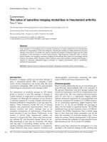
Histological analysis of infrapatellar fat pad harvested from rabbit knees 8 days after arthritis onsetHistological analysis of infrapatellar fat pad harvested from rabbit
knees 8 days after arthritis onset. Rabbits with antigen-induced
arthritis (AIA) were treated with daily injections of adrenomedullin (AM)
or saline (control) into the knee joint spaces for 7 days. The infrapatellar
fat pads were harvested from rabbit knees 8 days after arthritis onset.
Tissues were sectioned longitudinally perpendicular to the patella liga-
ment in the middle of the tissue, and were stained with H & E. (a), (b),
(c) AIA rabbit knee was treated with daily intra-articular injections of 3
μg AM for 7 days. (a) Low-magnification image (×100). (b), (c) High-
magnification images (×400) of the superficial portion and the deep
portion of (a), respectively. (d), (e), (f) The contralateral knee of (a), (b)
and (c) was treated with daily intra-articular injections of saline for 7
days. (d) Low-magnification image (×100). (e), (f) High-magnification
images (×400) of the superficial portion and the deep portion of (d),
respectively. Arrows indicate inflammatory cells. Bar = 50 μm.
Arthritis Research & Therapy Vol 10 No 6 Okura et al.
Page 6 of 10
(page number not for citation purposes)
reduced TNFα mRNA expression in a dose-dependent man-
ner. Daily intra-articular injections of 3 μg AM significantly sup-
pressed the TNFα mRNA level by 21% and 49% at day 8 and
day 21, respectively, compared with controls (Figure 10a). In
contrast, AM dose-dependently increased IL-6 mRNA expres-
sion. Daily intra-articular injections of 3 μg AM significantly
increased the IL-6 mRNA level by 45% and 121% at day 8 and
day 21, respectively, compared with controls (Figure 10b).
Although the VEGF mRNA level was suppressed by 10% at 3
μg AM on day 8, we did not observe a dose-dependent effect
of AM on VEGF mRNA expression (Figure 10d). AM treatment
did not significantly alter the TGFβ mRNA level (Figure 10c).
Figure 5
Histological analysis of infrapatellar fat pad harvested from rabbit knees 21 days after arthritis onsetHistological analysis of infrapatellar fat pad harvested from rabbit
knees 21 days after arthritis onset. Rabbits with antigen-induced
arthritis (AIA) were treated with daily injections of adrenomedullin (AM)
or saline (control) into the knee joint spaces for 20 days. The infrapatel-
lar fat pads were harvested from rabbit knees 21 days after arthritis
onset. The tissues were sectioned longitudinally perpendicular to the
patella ligament in the middle of the tissue, and were stained with H &
E. (a), (b), (c) AIA rabbit knee was treated with daily intra-articular
injections of 3 μg AM for 20 days. (a) Low-magnification image (×100).
(b), (c) High-magnification images (×400) of the superficial portion and
the deep portion of (a), respectively. (d), (e), (f) The contralateral knee
of (a), (b) and (c) was treated with daily intra-articular injections of
saline for 20 days. (d) Low-magnification image (×100). (e), (f) High-
magnification images (×400) of the superficial portion and the deep
portion of (d), respectively. Arrows indicate inflammatory cells. Bar =
50 μm.
Figure 6
Effect of adrenomedullin on the infiltration of inflammatory cells in the infrapatellar fat padEffect of adrenomedullin on the infiltration of inflammatory cells in
the infrapatellar fat pad. (a) Total number of inflammatory cells that
infiltrated the infrapatellar fat pad (three sites in the superficial portion,
three sites in the deep portion). The total number of inflammatory cells
that infiltrated the infrapatellar fat pad was significantly reduced by
26% with daily intra-articular injections of 0.1 μg adrenomedullin (AM)
on day 21. Daily intra-articular injections of 3 μg AM significantly sup-
pressed the total number of inflammatory cells by 38% and 23% at day
8 and day 21, respectively. (b) Number of inflammatory cells that infil-
trated the deep portion of the infrapatellar fat pad (three sites). Daily
intra-articular injections of 3 μg AM significantly suppressed the infiltra-
tion of inflammatory cells in the deep portion of the infrapatellar fat pad
by 49% and 54% at day 8 and day 21, respectively. Open and closed
columns represent the data at day 8 (n = 5 in each group) and at day
21 (n = 3 in each group), respectively. Data expressed as the mean ±
standard error of the mean. *P < 0.05 and **P < 0.01, compared with
contralateral knees.
Available online />Page 7 of 10
(page number not for citation purposes)
Discussion
In the present study we have shown that daily injections of AM
into the knee joint spaces of rabbits with AIA ameliorated the
inflammatory response associated with the disease. Treatment
with AM reduced joint swelling, and reduced the expression of
TNFα mRNA, edematous changes and the number of infiltrat-
ing inflammatory cells in the synovial tissue. To the best of our
knowledge, this is the first report to show the effects of daily
intra-articular injections of AM in rabbits with AIA.
We observed that AM suppressed joint swelling (Figures 2
and 3). Histologically, AM treatment reduced edematous
changes and increased the ratio of connective tissue in the
infrapatellar fat pad (Figures 7, 8 and 9). A previous study
showed that TNFα induced cytoskeletal reorganization of
endothelial cells and increased endothelial permeability by
stimulating TNF receptors 1 and 2 [17]. In addition, TNFα
facilitates the ability of VEGF to promote excessive vascular
permeability [18]. TNFα also suppresses the expression of
matrix genes and the induction of connective tissue growth
factor by TGFβ during the wound healing response [19].
TNFα therefore aggravates edematous changes and sup-
presses the fibrotic response of the tissue. Moreover, AM was
shown to reduce endothelial hyperpermeability induced by
hydrogen peroxide, thrombin, and Escherichia coli hemolysin
[20].
Two research groups reported recently that AM signaling defi-
ciency in mice resulted in midgestation death and massive
edema. The cause of this edema was shown to be a result of
fragility and hyperpermeability of blood vessels in one group
and to be a failure of lymphatic vessel growth in the other
[21,22]. The evidence from these studies suggests that AM
plays an important role in preventing edema. From these
observations, we speculate that AM not only suppresses the
production of TNFα, but also directly and indirectly inhibits
edematous changes in the inflamed joint.
Although RA is a chronic and systemic inflammatory disorder
of unknown etiology, TNFα has been shown to play a central
role in the pathogenesis of RA [1,2,23]. TNFα stimulates the
proliferation of synovial cells and the production of matrix met-
alloproteinases by chondrocytes and synovial cells, and
induces the release of other proinflammatory cytokines, lead-
ing to joint destruction [23,24]. We have shown that daily
injections of AM into the knee joint spaces of rabbits with AIA
suppressed the expression of TNFα mRNA in the synovial tis-
sue in a dose-dependent manner (Figure 10a). It has been
Figure 7
Effect of adrenomedullin on the total area of the infrapatellar fat padEffect of adrenomedullin on the total area of the infrapatellar fat
pad. The infrapatellar fat pads were sectioned longitudinally perpendic-
ular to the patella ligament in the middle of the tissue, and the total tis-
sue area was determined using software. Daily intra-articular injections
of 3 μg adrenomedullin (AM) significantly reduced the total tissue area
by 15% and 20% at day 8 and day 21, respectively. Open and closed
columns represent the data at day 8 (n = 5 in each group) and day 21
(n = 3 in each group), respectively. Data expressed as the mean ±
standard error of the mean. *P < 0.05 and **P < 0.01, compared with
contralateral knees.
Figure 8
Histological analysis of infrapatellar fat-pad sections stained with Mallory – Azan from rabbits with antigen-induced arthritisHistological analysis of infrapatellar fat-pad sections stained with Mallory – Azan from rabbits with antigen-induced arthritis. Rabbits with
antigen-induced arthritis (AIA) were treated with daily injections of adrenomedullin (AM) or saline (control) into the knee joint spaces for 20 days. The
infrapatellar fat pads were harvested from rabbit knees 21 days after arthritis induction. The tissues were sectioned longitudinally perpendicular to
the patella ligament in the middle of the tissue, and were stained with Mallory – Azan. (a) AIA rabbit knee was treated with daily intra-articular injec-
tions of 3 μg AM for 20 days. (b) AIA rabbit knee was treated with daily intra-articular injections of 1 ng AM for 20 days. (c) The contralateral knee of
(a) was treated with daily intra-articular injections of saline for 20 days. Photographs taken at ×40 magnification. Bar = 500 μm.
Arthritis Research & Therapy Vol 10 No 6 Okura et al.
Page 8 of 10
(page number not for citation purposes)
reported that AM suppressed the secretion of TNFα from
lipopolysaccharide-stimulated RAW 264.7 macrophages and
NR8383 macrophages [4-6]. Because the major source of
TNFα in inflamed synovial tissue of RA is due to macrophages
[25], it is plausible that AM suppresses the production of
TNFα from activated macrophages in inflamed synovial tissue.
On the contrary, we found that AM increased IL-6 mRNA
expression in the synovial tissue (Figure 10b). Our results
agree with previous findings on the effects of AM on IL-6 pro-
duction. AM is reported to augment the production of IL-6 from
NR8383 cells and Swiss 3T3 fibroblast cells stimulated with
lipopolysaccharide or cytokines [4,26]. Several observations
support the concept that IL-6 is an anti-inflammatory cytokine
[27]. IL-6 has been shown to have a suppressive effect on
TNFα and IL-1β production in peripheral blood mononuclear
cells and exerts its anti-inflammatory effects in hepatitis by
reducing the production of TNF [28,29]. Our results therefore
lead us to speculate that the mechanism involved in the anti-
inflammatory effects of AM is related to suppression of TNFα
in inflamed synovial tissue directly or through IL-6 production.
Overproduction of IL-6 has been observed and is known to
cause unfavorable clinical symptoms in immune-inflammatory
diseases such as RA. Overproduction of IL-6 induces the pro-
duction of rheumatoid factors and increases antibody levels,
the platelet count, C-reactive protein levels, and serum amy-
loid A protein levels in RA [30]. Treatment with a humanized
anti-IL-6 receptor antibody has also been shown to reduce RA
disease activity [30,31]. The effect of AM on IL-6 production
might therefore be an undesirable adverse effect in RA ther-
apy. Plasma AM levels have been reported to increase with RA
disease activity and in the acute or flare phase of myocardial
infarction and sepsis [10,11,32,33]. Recent studies have
shown that AM administration in the acute phase reaction of
several disease models produced significant protective
effects in organs against inflammation and oxidative stress
[34-36]. Miyashita and colleagues reported that AM adminis-
tration to prevent ischemic brain damage in mice less than 72
hours after the ischemic event showed significant therapeutic
effects, whereas AM administration more than 72 hours after
stroke onset produced no significant therapeutic effects [37].
From these observations and our study findings, we speculate
that the effects of AM may be dependent on the tissue envi-
ronment and the disease state; that is, the role and effects of
AM in inflammation may change during the inflammatory proc-
ess. AM acts as a strong anti-inflammatory agent in the acute
or flare phase of inflammation, but in the chronic phase of
inflammation AM may act not only as an anti-inflammatory
agent but also as a proinflammatory agent. It is therefore
important to consider the time of administration, the route of
administration and the dosage schedule of AM in the treat-
ment of RA.
Conclusion
In the present study, the effects of daily intra-articular injec-
tions of AM into the knees of rabbits with AIA were examined.
The results suggest that AM suppresses the inflammatory
response in inflamed joints by inhibiting the expression of
TNFα mRNA and increasing IL-6 mRNA level.
Although AM may have anti-inflammatory properties, the effect
of AM on IL-6 production in inflamed synovial tissue might be
an undesirable adverse effect in RA therapy. Further research
is necessary to investigate the drug effects, the time of admin-
istration and the dosage schedules of intra-articular injection
of AM in the treatment of RA.
Competing interests
The authors declare that they have no competing interests.
Authors' contributions
TO and KM had full access to all of the study data and take full
responsibility for the integrity of the data and the accuracy of
the data analysis. EC and HH conceived the study, and partic-
ipated in the study design. TS helped to develop the animal
model and draft the manuscript. TF helped to carry out real-
time PCR and perform statistical analyses. YA performed the
histological evaluation. KK measured the level of AM in plasma
and participated in the study design.
Figure 9
Quantitative evaluation of collagen volume in the infrapatellar fat padQuantitative evaluation of collagen volume in the infrapatellar fat
pad. To measure the collagen volume, the sections with Mallory – Azan
stain were projected onto a color imaging analysis system. In each sec-
tion, 10 separate sites were analyzed and the collagen volume fraction
was obtained by calculating the mean ratio of connective tissue to the
total tissue area. The collagen volume ratio was increased in adrenom-
edullin (AM)-treated knees by 39% and 31% at 0.1 μg and 3 μg AM,
respectively. Data expressed as the mean ± standard error of the mean.
*P < 0.05, compared with contralateral knees.
Available online />Page 9 of 10
(page number not for citation purposes)
Acknowledgements
The authors would like to thank Dr Atsushi Yamashita for his helpful dis-
cussion, Ms Kyoko Ohashi for technical support in the western blot anal-
ysis, and Ms Mariko Tokashiki for technical support in measurement of
the plasma AM concentration. The present study was supported by
Grants-in Aid for Scientific Research from the Ministry of Education in
Japan (No. 16591498).
References
1. Moreland LW, Baumgartner SW, Schiff MH, Tindall EA, Fleis-
chmann RM, Weaver AL, Ettlinger RE, Cohen S, Koopman WJ,
Mohler K, Widmer MB, Blosch CM: Treatment of rheumatoid
arthritis with a recombinant human tumor necrosis factor
receptor (p75)-Fc fusion protein. N Engl J Med 1997,
337:141-147.
2. Elliott MJ, Maini RN, Feldmann M, Long-Fox A, Charles P, Katsikis
P, Brennan FM, Walker J, Bijl H, Ghrayeb J, Woody JN: Treatment
of rheumatoid arthritis with chimeric monoclonal antibodies to
tumor necrosis factor alpha. Arthritis Rheum 1993,
36:1681-1690.
3. Kitamura K, Kangawa K, Kawamoto M, Ichiki Y, Nakamura S, Mat-
suo H, Eto T: Adrenomedullin: a novel hypotensive peptide iso-
lated from human pheochromocytoma. Biochem Biophys Res
Commun 1993, 192:553-560.
4. Wong LY, Cheung BM, Li YY, Tang F: Adrenomedullin is both
proinflammatory and antiinflammatory: its effects on gene
expression and secretion of cytokines and macrophage
migration inhibitory factor in NR8383 macrophage cell line.
Endocrinology 2005, 146:1321-1327.
5. Kubo A, Minamino N, Isumi Y, Katafuchi T, Kangawa K, Dohi K,
Matsuo H: Production of adrenomedullin in macrophage cell
line and peritoneal macrophage. J Biol Chem 1998,
273:16730-16738.
6. Wu R, Zhou M, Wang P: Adrenomedullin and adrenomedullin
binding protein-1 downregulate TNF-α in macrophage cell line
and rat Kupffer cells. Regul Pept 2003, 112:19-26.
7. Gonzalez-Rey E, Fernandez-Martin A, Chorny A, Delgado M: Ther-
apeutic effect of urocortin and adrenomedullin in a murine
model of Crohn's disease. Gut 2006, 55:824-832.
8. Ashizuka S, Ishikawa N, Kato J, Yamaga J, Inatsu H, Eto T, Kitamura
K: Effect of adrenomedullin administration on acetic acid-
induced colitis in rats. Peptides 2005, 26:2610-2615.
9. Gonzalez-Rey E, Chorny A, O'Valle F, Delgado M: Adrenomedul-
lin protects from experimental arthritis by down-regulating
inflammation and Th1 response and inducing regulatory T
cells. Am J Pathol 2007, 170:263-271.
10. Chosa E, Hamada H, Kitamura K, Eto T, Tajima N:
Increased
plasma and joint tissue adrenomedullin concentrations in
patients with rheumatoid arthritis compared to those with
osteoarthritis. J Rheumatol 2003, 30:2553-2556.
11. Yudoh K, Matsuno H, Kimura T: Plasma adrenomedullin in rheu-
matoid arthritis compared with other rheumatic diseases.
Arthritis Rheum 1999, 42:1297-1298.
12. Dumonde DC, Glynn LE: The production of arthritis in rabbits by
an immunological reaction to fibrin. Br J Exp Pathol 1962,
43:373-383.
Figure 10
Effect of adrenomedullin on cytokine mRNA expression linked to antigen-induced arthritisEffect of adrenomedullin on cytokine mRNA expression linked to antigen-induced arthritis. Expression levels of TNFα, IL-6, transforming
growth factor beta (TGFβ), and vascular endothelial growth factor (VEGF) mRNA in the infrapatellar fat pads were determined by real-time quantita-
tive PCR. (a) Adrenomedullin (AM) treatment reduced TNFα mRNA expression in a dose-dependent manner. Daily intra-articular injections of 3 μg
AM significantly suppressed the TNFα mRNA level by 21% and 49% at day 8 and day 21, respectively. (b) AM increased IL-6 mRNA expression in
a dose-dependent manner. Daily intra-articular injections of 3 μg AM significantly increased the IL-6 mRNA level by 45% and 121% at day 8 and day
21, respectively. (c) AM treatment did not alter the TGFβ mRNA level. (d) Although the VEGF mRNA level was suppressed by 10% at 3 μg AM on
day 8, a dose-dependent effect of AM on VEGF mRNA expression was not observed. Open and closed columns represent the data at day 8 (n = 5
in each group) and day 21 (n = 3 in each group), respectively. Data expressed as the mean ± standard error of the mean. *P < 0.05 and **P < 0.01,
compared with contralateral knees
Arthritis Research & Therapy Vol 10 No 6 Okura et al.
Page 10 of 10
(page number not for citation purposes)
13. Consden R, Doble A, Glynn LE, Nind AP: Production of a chronic
arthritis with ovalbumin. Its retention in the rabbit knee joint.
Ann Rheum Dis 1971, 30:307-315.
14. Kita T, Kitamura K, Hashida S, Morishita K, Eto T: Plasma
adrenomedullin is closely correlated with pulse wave velocity
in middle-aged and elderly patients. Hypertens Res 2003,
26:887-893.
15. Reno C, Boykiw R, Martinez ML, Hart DA: Temporal alterations
in mRNA levels for proteinases and inhibitors and their poten-
tial regulators in the healing medial collateral ligament. Bio-
chem Biophys Res Commun 1998, 252:757-763.
16. Zagariya A, Bhat R, Navale S, Chari G, Vidyasagar D: Inhibition of
meconium-induced cytokine expression and cell apoptosis by
pretreatment with captopril. Pediatrics 2006, 117:1722-1727.
17. Ferrero E, Zocchi MR, Magni E, Panzeri MC, Curnis F, Rugarli C,
Ferrero ME, Corti A: Roles of tumor necrosis factor p55 and p75
receptors in TNF-α-induced vascular permeability. Am J Phys-
iol Cell Physiol 2001, 281:c1173-c1179.
18. Clauss M, Sunderkötter C, Sveinbjörnsson B, Hippenstiel S, Wil-
luweit A, Marino M, Haas E, Seljelid R, Scheurich P, Suttorp N,
Grell M, Risau W: A permissive role for tumor necrosis factor in
vascular endothelial growth factor-induced vascular
permeability. Blood 2001, 97:1321-1329.
19. Leask A, Abraham DJ: TGF-β signaling and the fibrotic
response. FASEB J 2004, 18:816-827.
20. Hippenstiel S, Witzenrath M, Schmeck B, Hocke A, Krisp M, Krüll
M, Seybold J, Seeger W, Rascher W, Schütte H, Suttorp N:
Adrenomedullin reduces endothelial hyperpermeability. Circ
Res 2002, 91:618-625.
21. Ichikawa-Shindo Y, Sakurai T, Kamiyoshi A, Kawate H, Iinuma N,
Yoshizawa T, Koyama T, Fukuchi J, Iimuro S, Moriyama N,
Kawakami H, Murata T, Kangawa K, Nagai R, Shindo T: The GPCR
modulator protein RAMP2 is essential for angiogenesis and
vascular integrity. J Clin Invest 2008, 118:29-39.
22. Fritz-Six KL, Dunworth WP, Li M, Caron KM:
Adrenomedullin sig-
naling is necessary for murine lymphatic vascular
development. J Clin Invest 2008, 118:40-50.
23. Arend WP, Dayer JM: Inhibition of the production and effects of
interleukin-1 and tumor necrosis factor alpha in rheumatoid
arthritis. Arthritis Rheum 1995, 38:151-160.
24. Nishimoto N, Ito A, Ono M, Tagoh H, Matsumoto T, Tomita T, Ochi
T, Yoshizaki K: IL-6 inhibits the proliferation of fibroblastic syn-
ovial cells from rheumatoid arthritis patients in the presence
of soluble IL-6 receptor. Int Immunol 2000, 12:187-193.
25. Chu CQ, Field M, Feldmann M, Maini RN: Localization of tumor
necrosis factor alpha in synovial tissues and at the carti-
lage–pannus junction in patients with rheumatoid arthritis.
Arthritis Rheum 1991, 34:1125-1132.
26. Isumi Y, Minamino N, Kubo A, Nishimoto N, Yoshizaki K, Yoshioka
M, Kangawa K, Matsuo H: Adrenomedullin stimulates inter-
leukin-6 production in Swiss 3T3 cells. Biochem Biophys Res
Commun 1998, 244:325-331.
27. Tilg H, Dinarello CA, Mier JW: IL-6 and APPs: anti-inflammatory
and immunosuppressive mediators. Immunol Today 1997,
18:428-432.
28. Schindler R, Mancilla J, Endres S, Ghorbani R, Clark SC, Dinarello
CA: Correlations and interactions in the production of inter-
leukin-6 (IL-6), IL-1, and tumor necrosis factor (TNF) in human
blood mononuclear cells: IL-6 suppresses IL-1 and TNF. Blood
1990, 75:40-47.
29. Mizuhara H, O'Neill E, Seki N, Ogawa T, Kusunoki C, Otsuka K,
Satoh S, Niwa M, Senoh H, Fujiwara H: T cell activation-associ-
ated hepatic injury: mediation by tumor necrosis factors and
protection by interleukin 6. J Exp Med 1994, 179:1529-1537.
30. Nishimoto N, Kishimoto T, Yoshizaki K: Anti-interleukin 6 recep-
tor antibody treatment in rheumatic disease. Ann Rheum Dis
2000, 59(Suppl 1):i21-i27.
31. Nishimoto N, Yoshizaki K, Miyasaka N, Yamamoto K, Kawai S,
Takeuchi T, Hashimoto J, Azuma J, Kishimoto T: Treatment of
rheumatoid arthritis with humanized anti-interleukin-6 recep-
tor antibody: a multicenter, double-blind, placebo-controlled
trial. Arthritis Rheum 2004, 50:
1761-1769.
32. Kobayashi K, Kitamura K, Hirayama N, Date H, Kashiwagi T,
Ikushima I, Hanada Y, Nagatomo Y, Takenaga M, Ishikawa T, Ima-
mura T, Koiwaya Y, Eto T: Increased plasma adrenomedullin in
acute myocardial infarction. Am Heart J 1996, 131:676-680.
33. Hirata Y, Mitaka C, Sato K, Nagura T, Tsunoda Y, Amaha K,
Marumo F: Increased circulating adrenomedullin, a novel
vasodilatory peptide, in sepsis. J Clin Endocrinol Metab 1996,
81:1449-1453.
34. Kawai J, Ando K, Tojo A, Shimosawa T, Takahashi K, Onozato ML,
Yamasaki M, Ogita T, Nakaoka T, Fujita T: Endogenous adrenom-
edullin protects against vascular response to injury in mice.
Circulation 2004, 109:1147-1153.
35. Nakamura R, Kato J, Kitamura K, Onitsuka H, Imamura T, Cao Y,
Marutsuka K, Asada Y, Kangawa K, Eto T: Adrenomedullin
administration immediately after myocardial infarction amelio-
rates progression of heart failure in rats. Circulation 2004,
110:426-431.
36. Yang S, Zhou M, Fowler DE, Wang P: Mechanisms of the bene-
ficial effect of adrenomedullin and adrenomedullin-binding
protein-1 in sepsis: down-regulation of proinflammatory
cytokines. Crit Care Med 2002, 30:2729-2735.
37. Miyashita K, Itoh H, Arai H, Suganami T, Sawada N, Fukunaga Y,
Sone M, Yamahara K, Yurugi-Kobayashi T, Park K, Oyamada N,
Sawada N, Taura D, Tsujimoto H, Chao TH, Tamura N, Mukoyama
M, Nakao K: The neuroprotective and vasculo-neuro-regenera-
tive roles of adrenomedullin in ischemic brain and its thera-
peutic potential. Endocrinology 2006, 147:1642-1653.