Báo cáo y học: " Insights into endocrine-immunological disturbances in autoimmunity and their impact on treatment" doc
Bạn đang xem bản rút gọn của tài liệu. Xem và tải ngay bản đầy đủ của tài liệu tại đây (887.8 KB, 7 trang )
Available online />Page 1 of 7
(page number not for citation purposes)
Abstract
The neuroendocrine immune (NEI) system is regarded as a
fundamental network for the maintenance of health status
(homeostasis), and it plays an important role in several systemic
diseases, including autoimmune disorders. Among the major
players of NEI pathways are steroid hormones of the adrenal
(cortisol) and gonadal glands (sex hormones), neurohormones
such as melatonin, and more recently the vitamin D endocrine
system. Estrogens, melatonin and chronic stress (inducing
decreased adrenal glucocorticoid release over a long time) strongly
modulate the NEI system and stimulate the immune response. The
vitamin D endocrine system is regarded as a potential immuno-
suppressive factor. Consequently, estrogens (especially in patients
affected by B-cell-driven immunity) and melatonin should be
avoided, and glucocorticoids (as replacement therapy) and vitamin
D are allowed in the treatment of autoimmunity.
Introduction
The neuroendocrine immune (NEI) system is generally con-
sidered a fundamental network and its integrity is essential for
the maintenance of health status in humans [1]. As a con-
sequence, several systemic diseases, including autoimmune
disorders, originate from the altered balance/activation of the
NEI system.
Modulators of the immune system include different hormones,
and major players of NEI pathways are steroid hormones of
the adrenal and gonadal glands, as well as neurohormones
such as melatonin (MLT) [1]. Steroid hormones are not
stored in endocrine glands in the form of the final bioactive
hormones, but their precursor cholesterol is metabolized by
different enzyme steps leading to cortisol, the bioactive highly
antiinflammatory endogenous glucocorticoid. The enzyme
steps are regulated by microenvironmental factors such as
cytokines. In the tissue, cortisol is degraded to cortisone,
which can be reactivated by the 11β-hydroxysteroid de-
hydrogenase type 1 [2]. In addition, the adrenal glands
produce the major androgen precursors dehydroepiandro-
sterone and androstenedione, which can be converted into
active sex hormones such as testosterone and estrogens in
peripheral tissues.
Gonadal glands mainly synthesize sex hormones such as
testosterone (in the testicles) and estrogens (in the ovaries),
again from the precursor cholesterol. Testosterone can also
be viewed as a precursor of estrogens in tissue with high
aromatase activity (that is, rheumatoid synovial tissue).
Interestingly, another hormone arising from cholesterol,
namely vitamin D and its endocrine system, is involved in
various biological processes that modulate immune res-
ponses (mainly immunosuppressive), and has an important
role in autoimmune diseases [3].
Role of glucocorticoids in autoimmunity and
inflammation
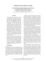
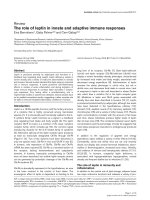
Several factors are involved in the pathogenesis of auto-
immune rheumatic diseases, including genetic aspects,
chronic infections, sex hormones (estrogens) and stress
(Figure 1).
In particular, stress (that is, interpersonal, severe surgery,
chronic infections) is now recognized as an important risk
factor [4]. Patients with insufficient stress response axes
demonstrate paradoxically decreased stress responses and,
consequently, proinflammatory side effects [5]. The loss of
adequate stress responses is reflected by low serum levels of
cortisol and also low concentrations of norepinephrine in the
tissue (nerve fiber loss) [1,5]. Minor stress and probably also
Review
Insights into endocrine-immunological disturbances in
autoimmunity and their impact on treatment
Maurizio Cutolo
1
and Rainer H Straub
2
1
Research Laboratories and Clinical Academic Unit of Rheumatology, University of Genova Italy, Viale Benedetto XV 6, 16132 Genova, Italy
2
Laboratory of NeuroEndocrinoImmunology, Department of Internal Medicine I, University Hospital, F.J Strauss-Allee 11, D-93042 Regensburg,
Germany
Corresponding author: Maurizio Cutolo,
Published: 6 April 2009 Arthritis Research & Therapy 2009, 11:218 (doi:10.1186/ar2630)
This article is online at />© 2009 BioMed Central Ltd
IFN = interferon; IL = interleukin; MLT = melatonin; NEI = neuroendocrine immune; 1,25(OH)
2
D
3
= 1,25-dihydroxyvitamin D
3
; RA = rheumatoid
arthritis; SLE = systemic lupus erythematosus; TNF = tumor necrosis factor; VDR = vitamin D receptor.
Arthritis Research & Therapy Vol 11 No 2 Cutolo and Straub
Page 2 of 7
(page number not for citation purposes)
major stress are therefore not accompanied by an adequate
stress response, especially over a long time, which leads to
inadequately low concentrations of stress axes mediators
(that is, endogenous glucocorticoids) in relation to the
immune/inflammatory situation [5].
Therapeutically, a reduction in stress episodes or a change of
stress management must be implemented.
Based on their decreased concentrations in chronic diseases,
glucocorticoids are now considered important disease-
modifying antirheumatic drugs at least in rheumatoid arthritis
(RA) [1,6]. In addition, mild exercise and training and a
decrease in the proinflammatory load – as obtained also by
the use of anticytokine therapy – can normalize stress axes,
leading to favorable responses and decreased appeal of
exogenous glucocorticoids [7].
Suppression of the hypothalamic–pituitary–adrenal axis,
however – especially in polymyalgia rheumatica, an inflam-
matory syndrome affecting older people – plays an important
pathogenetic role [8]. In fact, older people per se represent a
condition of endocrine senescence including adrenal
hypofunction, and the presence of chronic stress represents
a harmful stimulus to seriously compromised endogenous
glucocorticoid production. Interestingly, serum cytokine
(mainly IL-6) and steroidal hormone patterns suggest that
patients with polymyalgia rheumatica have an intensive
inflammatory reaction (much higher then in RA), and
glucocorticoid administration represents the most effective
replacement treatment [9].
Insights into therapeutic optimization of
neuroendocrine immune system modulation
in autoimmunity
It has been known for many decades that disease symptoms
in immune-mediated diseases such as RA follow obvious
circadian rhythms, with an increase of activity in the early
morning hours, abatement during the day, and a smaller new
increase in the early evening [10].
A number of articles have reported a temporal relationship
between elevated levels of proinflammatory cytokines and
symptoms of RA, such as morning stiffness [11,12]. Several
of these cytokines are highly elevated in patients with active
RA in the early hours of the day, but after noon their levels are
almost zero. Their release pattern and serum concentrations,
respectively, are modulated by hormones and neuronal
pathways coordinated by a subordinate neuroendocrine
centre in the hypothalamus, which follows a strict 24-hour
daily cycle.
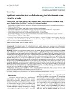
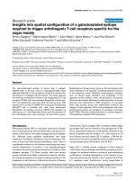
The closest similarity with the daily pattern of RA symptoms,
such as morning stiffness, joint pain and functional disability,
seems to exist for serum concentrations of IL-6 (Figure 2).
Proinflammatory hormones (that is, MLT) start to rise during the
night before the onset of RA symptoms and before an
endogenous cortisol rise in these patients [11] (Figure 2). The
role of IL-6 in the regulation of inflammatory and immune
responses, particularly in RA, is well established, but an
increased production of other proinflammatory cytokines such
as TNF, IL-1, IL-8, IL-12 and IL-17 has also been reported [13].
RA has also been considered characterized by an inadequate
antiinflammatory response that may contribute to morning
stiffness. This lack of anti-inflammatory response is not yet
totally understood. Cortisol secretion and glucocorticoid
receptor density, however, have been reported to be altered
in patients with RA. Furthermore, changes of peripheral
metabolism of endogenous glucocorticoids may also
contribute to the early morning manifestation of the disease
symptoms in RA [13].
Based on these considerations, the usual administration of
glucocorticoids between 06:00 and 08:00 hours has been
suggested as not optimal [14]. This could simply be too late
since the night-time pathophysiological processes have
already led to inflammation, pain and subjective symptoms.
Consequently, it has been hypothesized that it could be
easier to prevent the circadian increase of proinflammatory
cytokine levels, and therefore the consequently observed
clinical signs and symptoms of the disease, than to treat
these signs and symptoms once they are established in the
morning [10].
Figure 1
Factors involved in the pathogenesis of autoimmune rheumatic
diseases. The major risk factors in autoimmunity include specific
genetic background, chronic infections as triggers of the immune
response, estrogens as general enhancers of the immune response (at
least B-cell-driven), and chronic stress over a long time inducing a
decreased adrenal glucocorticoid release.
A new modified-release delivery system has been developed
that adapts the release of the administered glucocorticoids to
the circadian rhythms of endogenous cortisol and disease
symptoms to improve glucocorticoid therapy [15]. The efficacy
and safety of a new modified-release prednisone tablet (intake
at 22:00 hours, release at 02:00 hours) compared with
immediate-release prednisone (intake at 07:00 hours) in RA
patients was recently tested. The mean relative change in
duration of morning stiffness of the joints from baseline to
end of treatment (12 weeks) was significantly higher with
modified-release prednisone than with immediate-release
prednisone (–22.7% vs. –0.4%; difference = 22.4% (95%
confidence interval = 0.49 to 44.30); P = 0.045). Patients in
the modified-release prednisone group achieved a mean
reduction of morning stiffness of 44.0 minutes (standard
deviation = 136.6 minutes) compared with baseline.
The absolute difference between the treatment groups was
29.2 minutes (95% confidence interval = –2.59 to 61.9) in
favor of modified-release prednisone (P = 0.072). The safety
profile did not differ between treatments [15].
Optimization of the timing (night) and dosage (low) of
glucocorticoids has been recently recommended to improve
the tolerance and efficacy in autoimmune rheumatic diseases
[16].
Insights into neurohormonal effects of
melatonin in autoimmunity
MLT is an important neurohormone mainly synthesized by the
pineal gland with a circadian rhythm that peaks between
01:00 and 03:00 hours.
At physiological concentrations in human peripheral blood
mononuclear cells, MLT has been reported to stimulate the
production of IFNγ, IL-1, IL-2, IL-6 and IL-12, but not production
of IL-4 [17]. In addition, MLT was found to enhance production
of inflammatory cytokines from cultured human monocytes/
macrophages, including IL-12, and to turn the MLT/IL-2
connection towards the enhancement of T-cell immunity [18].
MLT serum levels at 20:00 and 08:00 hours have been found
to be significantly higher in patients with RA than in healthy
control individuals (P <0.05). MLT was found to be detec-
table at high concentration in synovial fluids from patients
who had RA, and binding sites for MLT were present in
synovial macrophages [18]. Interestingly, IFNγ, IL-1, IL-6, IL-2,
IL-12 and TNF production reach their peak during the night
and early morning, shortly after MLT serum levels are highest
and plasma cortisol is lowest [19] (Figure 2). This is in line
with the hypothesis that MLT upregulates cytokine production
and immune system activity [18].
Since until recently there was still a need to obtain clinically-
based evidence about the possible role of MLT as a disease-
promoting or a disease-protecting hormone in RA, a double-
blind placebo-controlled study investigating the effects of
MLT administration in patients with RA was initiated [20]. The
results obtained were somewhat disappointing and surpri-
sing, as the authors stated in the discussion by considering
MLT an in vitro potent antioxidant [20]. MLT was therefore
expected to decrease oxidative processes such as lipid
peroxidation that decreased in RA patients, but the erythro-
cyte sedimentation rate and neopterin levels increased
compared with patients treated with placebo. This observa-
tion is consistent with an antioxidant effect, but also suggests
some proinflammatory activity [20]. In addition, no reduction
of RA disease activity was observed, and the disease
appeared to be worse in some MLT-treated RA patients.
Serum TNF was recently found to be higher in Northern
European patients with RA than in their controls and was
found to be significantly correlated with the early increased
serum MLT concentrations, at least during the winter [21].
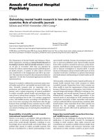
The increased serum concentrations and circadian rhythm of
MLT and a relative adrenal insufficiency in chronic RA (low
cortisol) therefore allow at least T-helper type 1 cytokines to
be produced in higher amounts during the late night under
the enhancing effect of the pineal hormone (Figure 3).
In conclusion, the translation from basic research to clinical
medicine clearly showed that MLT treatment does not
improve RA and must be avoided [22].
Insights into estrogen involvement in
neuroendocrine immune disturbances and
autoimmunity
Generally, based on epidemiological and immunological
evaluations, estrogens enhance the humoral response
Available online />Page 3 of 7
(page number not for citation purposes)
Figure 2
Daily pattern of rheumatoid arthritis symptoms for serum
concentrations of IL-6. Proinflammatory hormones (that is, melatonin)
start to rise during the early night before the onset of rheumatoid
arthritis symptoms and before the endogenous cortisol rise in these
patients. Grey shading, trigger time for the IL-6 rise.
(antibody production, T-helper type 2 immune response,
B-cell immunity); but, at the same time, estrogens might
inhibit T cells and macrophages at normal to high estrogen
concentrations [23]. The translation of the study results on
modulatory effects of estrogens obtained from animal and in
vitro investigations to the human condition, however, is
always difficult and complex [24]. In addition, in most animal
studies only 17β-estradiol was used as the investigated
estrogen and in most human studies a crude mixture of
conjugated estrogens was used, which can have proinflam-
matory effects, as recently reviewed [25].
Different concentrations used in in vitro or in vivo testing
might also render estrogen friend or foe in immune/
inflammatory conditions. Moreover, different cells involved in
the immune/inflammatory response react in an opposite
manner to different estrogen concentrations. In addition, the
expression of estrogen receptors (estrogen receptor alpha or
estrogen receptor beta) might be quite different under
inflammatory conditions depending on the microenvironment
and the type of disease. Generally, estrogens enhance cell
proliferation and reduce cell apoptosis [26]. Finally, the role
of local estrogen concentrations and the type of peripheral
estrogen metabolites at the level of inflammatory foci is of
great importance in order to explain the sometime opposite
modulatory effects exerted by these hormones on the
immune/inflammatory reaction [27].
Immunological evidence suggests that female gonadal
hormones exert an important role in the etiology and course
of chronic autoimmune diseases since the menstrual cycle,
pregnancy and menopausal status are recognized as signifi-
cant influencing factors [28].
Generally, the immune supportive role exerted by estrogens is
evident in trauma/sepsis and some chronic autoimmune
disorders such as systemic lupus erythematosus (SLE) or
Sjögren syndrome [28]. Interestingly, studies in women using
oral contraceptives versus those not using oral contracep-
tives demonstrated no significant increased risk of developing
SLE, but hormone replacement therapy in postmenopausal
women seems to increase the risk of developing SLE [29].
Oral contraceptive use was not associated with changes in
the disease course in premenopausal women with SLE, at
least in the nonactive phase, but hormone replacement
therapy increased the risk of mild flares in postmenopausal
patients [23]. This information indicates that the positive
effect of estrogens on B cells does not play a role in
premenopausal women with normal menstrual cycles and low
disease activity, but estrogens mildly stimulate SLE in women
with postmenopausal levels of estrogens [23].
Nevertheless, mainly for strictly B-cell-dependent diseases,
the female to male preponderance can be explained by the
propagating effects of estrogens [30]. On the other hand,
because men never experience high estrogen (or proges-
terone) levels like women during pregnancy, the apparent
gender dimorphism of chronic inflammatory diseases during
the reproductive period of women can be explained. In
addition, higher androgen levels in men most often exert
inhibitory effects on many immune phenomena (the opposite
for low androgen levels; that is, in Klinefelter syndrome),
which is an other important argument why women with low
androgen levels are protected from infectious diseases but
are more prone to B-cell-dependent autoimmunity [31,32].
In conclusion, in the presence of active immune-mediated
diseases such as SLE (or antiphospholipid syndrome and
others), the administration of estrogens should be avoided.
Vitamin D endocrine system in autoimmunity
The discovery of the vitamin D receptor (VDR), a member of
the nuclear hormone receptor superfamily, in the cells of the
immune system suggested that vitamin D could have
immunoregulatory properties [3].
The vitamin D endocrine system is involved in various bio-
logical processes that modulate immune responses, and
plays an important role in autoimmune diseases [33]
(Figure 3). In addition to exerting direct modulatory effects on
T-cell and B-cell function, VDR agonists influence the
phenotype and function of dendritic cells, thereby promoting
tolerogenic properties that favor the induction of regulatory,
rather than effector, T cells [34]. VDR agonistic effects have
been demonstrated in several experimental models and could
be utilized to treat several autoimmune diseases and other
Arthritis Research & Therapy Vol 11 No 2 Cutolo and Straub
Page 4 of 7
(page number not for citation purposes)
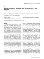
Figure 3
Altered balance between nocturnal hormones in rheumatoid arthritis.
The altered balance between nocturnal hormone production in chronic
diseases such as rheumatoid arthritis (RA) is characterized by
increased levels and steady-state duration of melatonin (enhancer of
the immune/inflammatory reaction) and by decreased adrenal cortisol
availability (downregulator of the immune/inflammatory reaction).
immune-mediated pathologies that are characterized by
chronic inflammatory responses [3].
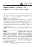
1,25-Dihydroxyvitamin D
3
(1,25(OH)
2
D
3
) – which is pro-
duced by macrophages, dendritic cells, T cells and B cells –
seems to contribute physiologically, via the VDR expressed in
these cell types, to the autocrine and paracrine regulation of
both innate and adaptive immune responses [35-37]
(Figure 4). This tight control of bioactive hormone production
by cells of the immune system itself further supports the
relevance of the vitamin D endocrine system in the
modulation of immune responses in health and disease. A
physiological role for vitamin D in the immune system is also
supported by the presence of the VDR in primary lymphoid
organs. The primary lymphoid organs (bone marrow and
thymus) are the centers where the immune system develops
and differentiates [38].
T cells have been shown to play fundamental roles in auto-
immune diseases. Quiescent CD4
+
T cells express VDRs at
low numbers, which increase fivefold after activation [39].
The effects of 1,25(OH)
2
D
3
on the acquired, antigen-specific
immune response are characterized by inhibition of T-lympho-
cyte activation, particularly of the T-helper type 1 arm. Treat-
ment of CD4 T cells with 1,25(OH)
2
D
3
inhibits T-helper
type 1 cell proliferation and cytokine production [40]. Other
observations demonstrated inhibition of both T-helper type 1
and T-helper type 2 cell cytokine production, including
inhibition of IL-4 [41,42].
Interestingly, 1,25(OH)
2
D
3
has been found to regulate the
proliferation of activated B cells and their subsequent
differentiation, as well as enhancing IL-10 expression, further
suggesting its role in the maintenance of B-cell homeostasis;
this regulation also shows that the correction of vitamin D
deficiency may be useful in the treatment of B-cell-mediated
autoimmune disorders as well as allergic immune responses
[37,43]. Addition of 1,25(OH)
2
D
3
was also shown to inhibit
the expression of IL-6, an important factor that stimulates T-
helper type 17 cells, which seem considered a critical
component of the autoimmune reaction [44].
Interestingly, vitamin D has been shown to inhibit antibody
secretion and autoantibody production in B cells [45]. In
vitro, 1,25(OH)
2
D
3
stimulates phagocytosis and killing of
bacteria by macrophages but suppresses the antigen-
presenting capacity of these cells and of dendritic cells [46].
Vitamin D has been found to promote the induction of mono-
cytic differentiation to macrophages and to modulate macro-
phage responses, preventing them from releasing proinflam-
matory cytokines and chemokines [47]. In synthesis, the most
evident effects of the vitamin D hormone on the immune
system seem to be in the control of T-helper type-1-driven
autoimmunity and partially the T-helper type-2-driven immune
response.
Decreased vitamin D plasma levels have been linked to
autoimmune diseases in humans [41]. A large population-
based study (Nurses Health Study I and II) showed recently
that women in the highest quintile of vitamin D intake had a
40% reduced rate of developing multiple sclerosis [48].
Experimentally it has been shown that vitamin D deficiency
exacerbates both inflammatory bowel disease and multiple
sclerosis in animals, and vitamin D hormone has been shown
to suppress experimental encephalitis and colitis in mice [41].
Low serum levels of vitamin D might be related, among other
factors, to prolonged daily darkness (reduced activation of
the provitamin D by ultraviolet B sunlight), to different genetic
background (that is, vitamin D receptor polymorphism) and to
nutritional factors. This might also explain the latitude-related
prevalence of autoimmune diseases such SLE and RA by
considering the potential immunosuppressive roles of vitamin D
[49].
The 25(OH)D
3
plasma levels have recently been found to be
inversely correlated with RA disease activity, showing a
circannual rhythm (more severe in winter) [50]. In addition,
greater intake of vitamin D was associated with a lower risk of
RA, as well as a significant clinical improvement being
strongly correlated with the immunomodulating potential in
vitamin-D-treated RA patients [50].
Patients with SLE have multiple risk factors for vitamin D
deficiency, and the disease complexity seems to correlate
Available online />Page 5 of 7
(page number not for citation purposes)
Figure 4
Downregulatory and upregulatory influences exerted by vitamin D on
the immune response. Synthetic presentation of the major
downregulatory (yellow) and upregulatory (with) influences exerted by
vitamin D on the immune response. Effects on T lymphocytes and B
lymphocytes and related cytokines, as well as on antigen-presenting
cells (APC) and dendritic cells (DC). NKT, natural killer T cells; TGFβ1,
transforming growth factor beta 1; Th, T-helper type cells; Treg,
regulatory T cells.
with lower 25-OH vitamin D plasma levels [51]. Considera-
tion of the possibility of vitamin D deficiency and its treatment
should therefore be mandatory in SLE patients, but also in
patients with undifferentiated connective tissue disease [51-
54].
Conclusions
Presently, by considering some of the major players of the NEI
system, it is evident that estrogens, MLT and chronic stress
(chronic stress inducing a decreased adrenal glucocorticoid
release over a long time) can support the immune response. In
contrast, the integrity of the vitamin D endocrine system is
regarded as a potential immunosuppressive hormonal system.
Estrogens (especially in patients affected by B-cell-driven
immunity) and MLT should consequently be avoided, and
glucocorticoids (as replacement therapy) and vitamin D are
allowed in the treatment of autoimmunity.
Competing interests
The authors declare that they have no competing interests.
References
1. Cutolo M, Straub RH, Bijlsma JW: Neuroendocrine-immune
interactions in synovitis. Nat Clin Pract Rheumatol 2007, 3:627-
634.
2. Buttgereit F, Zhou H, Seibel MJ: Arthritis and endogenous glu-
cocorticoids: the emerging role of the 11
ββ
-HSD enzymes. Ann
Rheum Dis 2008, 67:1201-1203.
3. Adorini L, Penna G: Control of autoimmune diseases by the
vitamin D endocrine system. Nat Clin Pract Rheumatol 2008, 4:
404-412.
4. Elenkov IJ, Chrousos GP: Stress system – organization, physi-
ology and immunoregulation. Neuroimmunomodulation 2006,
13:257-267.
5. Cutolo M, Straub RH: Stress as a risk factor in the pathogene-
sis of rheumatoid arthritis. Neuroimmunomodulation 2006, 13:
277-282.
6. Bijlsma JW, Hoes JN, Van Everdingen AA, Verstappen SM,
Jacobs JW: Are glucocorticoids DMARDs? Ann NY Acad Sci
2006, 1069:268-274.
7. Straub RH, Pongratz G, Cutolo M, Wijbrandts CA, Baeten D,
Fleck M, Atzeni F, Grunke M, Kalden JR, Schölmerich J, Lorenz
HM, Tak PP, Sarzi-Puttini P: Increased cortisol relative to
adrenocorticotropic hormone predicts improvement during
anti-tumor necrosis factor therapy in rheumatoid arthritis.
Arthritis Rheum 2008, 58:976-978.
8. Cutolo M, Cimmino MA, Sulli A: Polymyalgia rheumatica vs late-
onset rheumatoid arthritis. Rheumatology (Oxford) 2009,
48:93-95.
9. Straub RH, Cutolo M: Further evidence for insufficient hypo-
thalamic–pituitary–glandular axes in polymyalgia rheumatica.
J Rheumatol 2006, 33:1219-1223.
10. Straub RH, Cutolo M: Circadian rhythms in rheumatoid arthri-
tis: implications for pathophysiology and therapeutic manage-
ment. Arthritis Rheum 2007, 56:399-408.
11. Cutolo M, Seriolo B, Craviotto C, Pizzorni C, Sulli A: Circadian
rhythms in RA. Ann Rheum Dis 2003, 62:593-596.
12. Arvidson NG, Gudbjornsson B, Elfman L, Ryden AC, Totterman
TH, Hallgren R: Circadian rhythm of serum interleukin-6 in
rheumatoid arthritis. Ann Rheum Dis 1994, 53:521-524.
13. Cutolo M, Straub RH, Buttgereit F: Circadian rhythms of noctur-
nal hormones in rheumatoid arthritis: translation from bench
to bedside. Ann Rheum Dis 2008, 67:905-908.
14. Bijlsma JW, Jacobs J: Innovative use of glucocorticoids in
patients with rheumatoid arthritis. Lancet 2008,
371:183-184.
15. Buttgereit F, Doering G, Schaeffler A, Witte S, Sierakowski S,
Gromnica-Ihle E, Jeka S, Krueger K, Szechinski J, Alten R: Effi-
cacy of modified-release versus standard prednisone to
reduce duration of morning stiffness of the joints in rheuma-
toid arthritis (CAPRA-1): a double-blind, randomised con-
trolled trial. Lancet 2008, 371:183-184.
16. Hoes JN, Jacobs JW, Boers M, Boumpas D, Buttgereit F, Caeyers
N, Choy EH, Cutolo M, Da Silva JA, Esselens G, Guillevin L, Haf-
strom I, Kirwan JR, Rovensky J, Russell A, Saag KG, Svensson B,
Westhovens R, Zeidler H, Bijlsma JW: EULAR evidence-based
recommendations on the management of systemic glucocor-
ticoid therapy in rheumatic diseases. Ann Rheum Dis 2007,
66:1560-1567.
17. Garcia-Maurino S, Gonzalez-Haba MG, Calvo JR, Rafii-El-Idrissi
M, Sanchez-Margalet V, Goberna R, Guerrero JM: Melatonin
enhances IL-2, IL-6, and IFN-
γγ
production by human circulat-
ing CD4
+
cells. J Immunol 1997, 159:574-581.
18. Cutolo M, Villaggio B, Candido F, Valenti S, Giusti M, Felli L, Sulli
A, Accardo S: Melatonin influences interleukin-12 and nitric
oxide production by primary cultures of rheumatoid synovial
macrophages and THP-1 cells. Ann NY Acad Sci 1999, 876:
246-254.
19. Petrovsky N, McNair P, Harrison LC: Diurnal rhythms of pro-
inflammatory cytokines: regulation by plasma cortisol and
therapeutic implications. Cytokine 1998, 10:307-312.
20. Forrest CM, Mackay GM, Stoy N, Stone TW, Darlington LG:
Inflammatory status and kynurenine metabolism in rheuma-
toid arthritis treated with melatonin. Br J Clin Pharmacol 2007,
64:517-526.
21. Cutolo M, Maestroni GJ, Otsa K, Aakre O, Villaggio B, Capellino
S, Montagna P, Fazzuoli L, Veldi T, Peets T, Hertens E, Sulli A:
Circadian melatonin and cortisol levels in rheumatoid arthritis
patients in winter time: a north and south Europe comparison.
Ann Rheum Dis 2005, 64:212-216.
22. Maestroni G, Otsa K, Cutolo M: Melatonin treatment does not
improve rheumatoid arthritis. Br J Clin Pharmacol 2008, 65:
797-798.
23. Straub RH: The complex role of estrogens in inflammation.
Endocr Rev 2007, 28:521-574.
24. Cutolo M: Sex and rheumatoid arthritis: mouse model versus
human disease. Arthritis Rheum 2007, 56:1-3.
25. Cutolo M, Capellino S, Straub RH: Oestrogens in rheumatic
diseases: friend or foe? Rheumatology (Oxford) 2008,
47(Suppl 3):iii2-iii5.
26. Castagnetta LA, Carruba G, Granata OM, Stefano R, Miele M,
Schmidt M, Cutolo M, Straub RH: Increased estrogen formation
and estrogen to androgen ratio in the synovial fluid of
patients with rheumatoid arthritis. J Rheumatol 2003, 30:2597-
2605.
27. Cutolo M, Sulli A, Capellino S, Villaggio B, Montagna P, Seriolo B,
Straub RH: Sex hormones influence on the immune system:
basic and clinical aspects in autoimmunity. Lupus 2004, 13:
635-638.
28. Hughes GC, Clark EA: Regulation of dendritic cells by female
sex steroids: relevance to immunity and autoimmunity.
Autoimmunity 2007, 40:470-481.
29. Cohen-Solal JF, Jeganathan V, Grimaldi CM, Peeva E, Diamond B:
Sex hormones and SLE: influencing the fate of autoreactive B
cells. Curr Top Microbiol Immunol 2006, 305:67-88.
30. Cutolo M, Capellino S, Sulli A, Serioli B, Secchi ME, Villaggio B,
Straub RH: Estrogens and autoimmune diseases. Ann NY
Acad Sci 2006, 1089:538-547.
31. Cutolo M, Sulli A, Capellino S, Villaggio B, Montagna P, Seriolo B,
Straub RH: Sex hormones influence on the immune system:
basic and clinical aspects in autoimmunity. Lupus 2004,
13:635-638.
32. Ganesan K, Selvam R, Abhirami R, Raju KV, Manohar BM,
Puvanakrishnan R: Gender differences and protective effects of
testosterone in collagen induced arthritis in rats. Rheumatol
Int 2008, 28:345-353.
33. Adorini L: Intervention in autoimmunity: the potential of
vitamin D receptor agonists. Cell Immunol 2007, 233:115-124.
34. van Etten E, Mathieu C: Immunoregulation by 1,25-dihydroxyvi-
tamin D3: basic concepts. J Steroid Biochem Mol Biol 2005,
97:93-101.
35. Hewison M, Freeman L, Hughes SV, Evans KN, Bland R, Eliopou-
los AG, Kilby MD, Moss PA, Chakraverty R: Differential regula-
tion of vitamin D receptor and its ligand in human
monocyte-derived dendritic cells. J Immunol 2003, 170:5382-
Arthritis Research & Therapy Vol 11 No 2 Cutolo and Straub
Page 6 of 7
(page number not for citation purposes)
5390.
36. Cadranel J, Garabedian M, Milleron B, Guillozo H, Akoun G,
Hance AJ: 1,25(OH)
2
D
3
production by T lymphocytes and alve-
olar macrophages recovered by lavage from normocalcemic
patients with tuberculosis. J Clin Invest 1990, 85:1588-1593.
37. Chen S, Sims GP, Chen XX, Gu YY, Chen S, Lipsky PE: Modula-
tory effects of 1,25-dihydroxyvitamin D
3
on human B cell dif-
ferentiation. J Immunol 2007, 179:1634-1647.
38. Deluca HF, Cantorna MT. Vitamin D: its role and uses in
immunology. FASEB J 2001, 15:2579-2585.
39. Mahon BD, Wittke A, Weaver V, Cantorna MT: The targets of
vitamin D depend on the differentiation and activation status
of CD4 positive T cells. J Cell Biochem 2003, 89:922-932.
40. Boonstra A, Barrat FJ, Crain C, Heath VL, Savelkoul HF, O’Garra
A: 1
αα
,25-Dihydroxyvitamin d3 has a direct effect on naive
CD4(+) T cells to enhance the development of Th2 cells.
J Immunol 2001, 167:4974-4980.
41. Cantorna MT, Mahon BD: Mounting evidence for vitamin D as
an environmental factor affecting autoimmune disease preva-
lence. Exp Biol Med (Maywood) 2004, 229:1136-1142.
42. Staeva-Vieira TP, Freedman LP: 1,25-dihydroxyvitamin D3
inhibits IFN
γγ
and IL-4 levels during in vitro polarization of
primary murine CD4
+
T cells. J Immunol 2002, 168:1181-1189.
43. Heine G, Niesner U, Chang HD, Steinmeyer A, Zügel U, Zuberbier
T, Radbruch A, Worm M: 1,25-dihydroxyvitamin D(3) promotes
IL-10 production in human B cells. Eur J Immunol 2008, 38:
2210-2218
44. Stockinger B: Th17 cells: an orphan with influence 4. Immunol
Cell Biol 2007, 85:83-84.
45. Linker-Israeli M, Elstner E, Klinenberg JR, Wallace DJ, Koeffler HP:
Vitamin D(3) and its synthetic analogs inhibit the sponta-
neous in vitro immunoglobulin production by SLE-derived
PBMC. Clin Immunol 2001, 99:82-93.
46. Griffin MD, Lutz WH, Phan VA, Bachman LA, McKean DJ, Kumar
R: Potent inhibition of dendritic cell differentiation and matu-
ration by vitamin D analogs. Biochem Biophys Res Commun
2000, 270:701-708.
47. Helming L, Bose J, Ehrchen J, Chiebe S, Frahm T, Geffers R,
Probst-Kepper M, Balling R, Lengeling A: 1
αα
,25-Dihydroxyvita-
min D3 is a potent suppressor of interferon gamma-mediated
macrophage activation. Blood 2005, 106:4351-4358.
48. Munger KL, Zhang SM, O’Reilly E, Hernán MA, Olek MJ, Willett
WC, Ascherio A: Vitamin D intake and incidence of multiple
sclerosis. Neurology 2004, 62:60-65.
49. Cutolo M, Otsa K, Uprus M, Paolino S, Seriolo B: Vitamin D in
rheumatoid arthritis. Autoimmun Rev 2007, 7:59-64.
50. Cutolo M, Otsa K, Laas K, Yprus M, Lehtme R, Secchi ME, Sulli A,
Paolino S, Seriolo B: Circannual vitamin D serum levels and
disease activity in rheumatoid arthritis: Northern versus
Southern Europe. Clin Exp Rheumatol 2006, 24:702-704.
51. Ruiz-Irastorza G, Egurbide MV, Olivares N, Martinez-Berriotxoa A,
Aguirre C: Vitamin D deficiency in systemic lupus erythemato-
sus: prevalence, predictors and clinical consequences.
Rheumatology (Oxford) 2008, 47:920-923.
52. Zold E, Szodoray P, Gaal J, Kappelmayer J, Csathy L, Gyimesi E,
Zeher M, Szegedi G, Bodolay E: Vitamin D deficiency in undif-
ferentiated connective tissue disease. Arthritis Res Ther 2008,
10(5):R123.
53. Cutolo M: Vitamin D or hormone D deficiency in autoimmune
rheumatic diseases, including undifferentiated connective
tissue disease. Arthritis Res Ther 2008, 6:123.
54. Cutolo M, Otsa K, Paolino S, Yprus M, Veldi T, Seriolo B: Vitamin
D involvement in rheumatoid arthritis and systemic lupus ery-
thaematosus. Ann Rheum Dis 2009, 68:446-447.
Available online />Page 7 of 7
(page number not for citation purposes)