Báo cáo y học: "Asporin, a susceptibility gene in osteoarthritis, is expressed at higher levels in the more degenerate human intervertebral disc" ppsx
Bạn đang xem bản rút gọn của tài liệu. Xem và tải ngay bản đầy đủ của tài liệu tại đây (1.78 MB, 7 trang )
Open Access
Available online />Page 1 of 7
(page number not for citation purposes)
Vol 11 No 2
Research article
Asporin, a susceptibility gene in osteoarthritis, is expressed at
higher levels in the more degenerate human intervertebral disc
Helen E Gruber, Jane A Ingram, Gretchen L Hoelscher, Natalia Zinchenko, Edward N Hanley Jr
and Yubo Sun
Department of Orthopedic Surgery, Carolinas Medical Center, PO Box 32861, Charlotte, NC 28232, USA
Corresponding author: Helen E Gruber,
Received: 23 Dec 2008 Revisions requested: 9 Feb 2009 Revisions received: 11 Feb 2009 Accepted: 27 Mar 2009 Published: 27 Mar 2009
Arthritis Research & Therapy 2009, 11:R47 (doi:10.1186/ar2660)
This article is online at: />© 2009 Gruber et al.; licensee BioMed Central Ltd.
This is an open access article distributed under the terms of the Creative Commons Attribution License ( />),
which permits unrestricted use, distribution, and reproduction in any medium, provided the original work is properly cited.
Abstract
Introduction Asporin, also known as periodontal ligament-
associated protein 1 (PLAP1), is a member of the family of small
leucine-rich proteoglycan (SLRP) family. It is present within the
cartilage extracellular matrix (ECM), and is reported to have a
genetic association with osteoarthritis. Its D14 allele has
recently been found to be associated with lumbar disc
degeneration in Asian subjects. There have been no studies,
however, of this gene's normal immunohistochemical
localization within the human intervertebral disc, or of expression
levels in Caucasian individuals with disc degeneration.
Methods Studies were approved by our human subjects
Institutional Review Board. Methods included
immunohistochemical localization of asporin in the disc of
humans and the sand rat (a small rodent with spontaneous age-
related disc degeneration), and Affymetrix microarray analysis of
asporin gene expression in vivo and in vitro.
Results Immunohistochemical studies of human discs revealed
that some, but not all, cells of the outer annulus expressed
asporin. Fewer cells in the inner annulus contained asporin, and
it was rarely present in cells in the nucleus pulposus. Similar
patterns were found for the presence of asporin in lumbar discs
of sand rats. Substantial relative gene expression levels were
seen for asporin in both disc tissue and in annulus cells grown
in three-dimensional culture. More degenerate human discs
(Thompson grade 4) showed higher expression levels of asporin
than did less degenerate (grade 1, 2 and 3) discs, P = 0.004.
Conclusions In the discs of Caucasian subjects studied here,
and in the sand rat, greater immunolocalization levels were
found in the outer compared to inner annulus. Localization was
rare in the nucleus. Gene expression studies showed greatest
expression of asporin in the more degenerate human discs in
vivo.
Introduction
Asporin, also known as periodontal ligament-associated pro-
tein 1 (PLAP1), is an interesting, recently discovered leucine-
rich protein that is a member of the family of small leucine-rich
proteoglycan (SLRP) family and is associated with the extra-
cellular matrix (ECM) in cartilage, meniscus and several other
tissues [1,2]. The normal asporin allele contains 13 aspartic
acid repeats in a 382 amino acid protein, and is designated
D13. There are now three polymorphisms thought to be
strongly associated with osteoarthritis (OA) susceptibility;
these occur in the asporin gene (ASPN), the secreted frizzled-
related protein 3 gene (FRZB), and the calmodulin 1 gene
(CALM1) [3].
Recent studies have identified populations of individuals with
osteoarthritis of the knee and asporin alleles with 14 aspartic
acid repeats in the N-terminal region of the protein (desig-
nated D14). Work by Kizawa et al. first identified an associa-
tion of the ASPN single nucleotide polymorphisms (SNPs)
with knee and hip OA in Japanese patients [4]. As shown by
Iida et al., there are at least 19 SNPs in asporin in Japanese
patients with OA [5]. Associations of the ASPN SNPs with
OA have been studied by Shi et al. [6] and by Nakamura et al.
[7].
In non-Asian populations, however, recent studies appear to
show a lack of association of the ASPN SNPs with OA sus-
ceptibility, as reflected in the work by Rodriguez-Lopez et al. in
ASPN: asporin gene; CALM1: calmodulin 1 gene; CHTN: Cooperative Human Tissue Network; ECM: extracellular matrix; FRZB: secreted frizzled-
related protein 3 gene; GEO: Gene Expression Omnibus; SLRP: small leucine-rich proteoglycan; SNP: single nucleotide polymorphism.
Arthritis Research & Therapy Vol 11 No 2 Gruber et al.
Page 2 of 7
(page number not for citation purposes)
Spanish Caucasians [8] and work by Mustafa et al. in British
Caucasians [9].
Our laboratory has become interested in asporin in the human
intervertebral disc following the finding of Song et al. of an
association of the ASPN D14 allele with disc degeneration in
Asians [10]. Our literature search was not able to identify any
studies showing immunolocalization of asporin in the human
disc. The objectives of the present study, therefore, were to
determine the localization patterns of asporin within the human
and sand rat intervertebral disc, and to assess asporin expres-
sion in the human disc in vivo and in vitro.
Materials and methods
Clinical study population
Experimental study of human disc specimens was approved
prospectively by the authors' Human Subjects Institutional
Review Board at Carolinas Medical Center. The need for
informed consent was waived since disc tissue was removed
as part of routine surgical practice. Scoring of disc degenera-
tion utilized the Thompson scoring system; this system scores
disc degeneration over the spectrum from a healthy disc
(Thompson grade I) to discs with advanced degeneration
(grade V, the most advanced stage of degeneration) [11].
Patient specimens were derived from surgical disc proce-
dures performed on individuals with herniated discs and
degenerative disc disease. Surgical specimens were trans-
ported to the laboratory in sterile tissue culture medium. Care
was taken to remove all granulation tissue and to sample only
disc tissue. Non-surgical control donor disc specimens were
obtained via the National Cancer Institute Cooperative Human
Tissue Network (CHTN); they were shipped overnight to the
laboratory in sterile tissue culture medium and processed as
described below. Specimen procurement from the CHTN was
included in our approved protocol by our human subjects Insti-
tutional Review board.
Sand rat intervertebral disc tissue
Animal studies were carried out following approval by our Insti-
tutional Animal Care and Use Committee. Psammomys
obesus, the sand rat, is studied in our laboratory as a model of
spontaneous, age-related disc degeneration. Colony housing
and animal diet have been previously described [12,13].
Spines from seven animals were analyzed in the present study
of immunolocalization of asporin. Lumbar spines were
removed immediately after rats were killed, fixed in 10% neu-
tral buffered formalin, decalcified, and embedded in paraffin.
Discs were individually embedded as mid-sagittal sections
and also as en face sections individual discs. Sections were
processed as described below for immunohistochemical stud-
ies.
Expression of asporin in vivo and in vitro
Analysis of human disc tissue was carried out as previously
described using laser capture microdissection methods [14].
Cells cultured in three dimensions and in monolayers were
assayed for gene expression using the Affymetrix microarray
system (Affymetrix, Santa Clara, CA, USA). Total RNA was
extracted from cells using the TRIzol reagent (Gibco,
Carlsbad, CA, USA), reverse transcribed to double-stranded
cDNA, subjected to two rounds of transcription, and hybrid-
ized to the DNA microarray in the Affymetrix Fluidics Station
400. Affymetrix human U133 X3P arrays were used. The
GCOS Affymetrix GeneChip Operating System (version 1.2,
Affymetrix) was used for determining gene expression levels of
asporin [GenBank:NM_017680.1
]. Gene array data have
been uploaded to the Gene Expression Omnibus database
[GEO:GSE15227].
Immunolocalization of asporin
Slides were deparaffinized in xylene and hydrated through
graded alcohols to distilled water. The remainder of the proce-
dure was performed using the Dako AutostainerPlus (Dako,
Carpenteria, CA, USA). Endogenous peroxidase was blocked
using 3%H
2
O
2
(Sigma, St Louis, MO, USA). Slides were incu-
bated for 1 h with polyclonal anti-asporin (GenWay Biotech,
San Diego, CA, USA) at a 1:400 dilution. Secondary antibody
was LSAB2 Link Antibody (Dako) applied for 10 minutes fol-
lowed by peroxidase-conjugated streptavidin (Dako) for 10
minutes, and DAB (Dako) for 5 minutes. Some sand rat spine
specimens and three-dimensional cultured human disc cells
were prepared using the Vector VIP (Vector Laboratories, Bur-
lingame, CA, USA) red chromagen. Universal Negative Con-
trol Rabbit (Dako) was used as a negative control. A positive
control from a young rat articular cartilage joint was also
included with each run. Slides were removed from the stainer,
rinsed in water, counterstained with light green, dehydrated,
cleared and mounted with resinous mounting media.
Three-dimensional culture of human annulus cells
Human annulus cells were cultured in monolayer or three-
dimensional culture in a collagen sponge as previously
described [15,16] for 2 weeks, and cultures terminated for
harvest of mRNA and immunohistochemistry studies as
described above.
Statistical analyses
Standard statistical analyses were performed utilizing InStat
(GraphPad Software, San Diego, CA, USA). Correlation anal-
yses and means ± standard deviations (SD) were calculated.
P = 0.05 was considered to be the level of significance.
Results
In all, 19 discs from 15 subjects were examined with immuno-
histochemical procedures to localize asporin. Table 1 summa-
rizes the lumbar site and subject age and gender. The majority
of the surgical disc specimens we receive come from grade III
and IV discs. However, the present study does include four of
the healthier grade I to II discs (mean subject age 35.3 years),
and two of the most degenerate grade V (most degenerate)
Available online />Page 3 of 7
(page number not for citation purposes)
discs (mean subject age 38.5 years). Mean age for the six
grade III discs was 45.5 years, and 53 years for the five grade
IV discs.
Asporin immunolocalization
The first portion of our study of asporin and the intervertebral
disc tested for the presence of asporin in human and sand rat
discs using immunohistochemistry. Localization was seen
within disc cells, but not in the extracellular disc matrix.
As shown in Figure 1 for the human outer annulus, many cells
show positive cytoplasmic immunolocalization of asporin.
Some adjacent cells, however, did not show the presence of
asporin (Figure 1a, arrow). Similar findings were seen in the
more sparse cell population of the inner annulus as illustrated
in Figure 1b. Cells present in clusters in the inner annulus were
also studied; as noted in other disc areas, some, but not all,
cells were positive for asporin immunolocalization (Figure 2a).
In the nucleus pulposus, rare positive cells were present (Fig-
ure 2b). In the inner annulus, both positive and negative local-
ization was present in cells that had concentric layers of
extracellular matrix surrounding them (Figure 3).
The sand rat disc was also examined for asporin localization in
sections of disc cut en face. Patterns of immunolocalization for
the presence of asporin were similar to those seen in the
human disc: the greatest localization was present in the outer
annulus, with fewer cells positive in the deepest region of the
inner annulus and nucleus pulposus (Figure 4).
The presence of asporin was also detected in vitro in monol-
ayer cultured human annulus cells (Figure 5a), and in annulus
cells cultured in a three-dimensional collagen sponge (Figure
5c), which more closely mimics the in vivo microenvironment.
Cells in monolayer and three-dimensional culture all showed
positive asporin localization. (Figure 5b,d shows monolayer
and three-dimensional culture negative controls, respectively).
Asporin gene expression in human discs in vivo and in
vitro
In the second part of our study of asporin and the intervertebral
disc, asporin gene expression was evaluated in human disc tis-
sue and in human annulus cells cultured in three-dimensional
culture and in monolayer.
Table 1
Demographic features for specimens studied with immunocytochemistry
Subject Age (years)/gender Site Thompson grade Other information
1 45/F L4 to 5 1.5 CHTN: unknown
1 45/F L3 to 4 II CHTN: unknown
2 21/M L5 to S1 II Surgical specimen
3 40/M L4 to 5 2.5 CHTN: MI
4 33/F L1 to 2 III CHTN: PE
5 46/F L4 to 5 III Surgical specimen
6 53/M L3 to 4 III Surgical specimen
7 29/F L5 to S1 III Surgical specimen
8 54/M L4 to 5 III Surgical specimen
9 58/M L2 to 3 III Surgical specimen
4 33/F L3 to 4 IV CHTN: PE
10 59/F L3 to 4 IV Surgical specimen
10 59/F L1 to 2 IV Surgical specimen
10 59/F L2 to 3 IV Surgical specimen
11 78/M L3 to 4 IV Surgical specimen
12 56/F L2 to 3 IV Surgical specimen
13 39/F L4 to 5 IV Surgical specimen
14 44/F L5 to S1 V Surgical specimen
15 39/M L5 to S1 V Surgical specimen
CHTN, Cooperative Human Tissue Network; F, female; L, lumbar; M, male; MI, myocardial infarction; PE, pulmonary embolism; S, sacral.
Arthritis Research & Therapy Vol 11 No 2 Gruber et al.
Page 4 of 7
(page number not for citation purposes)
mRNA from 15 subjects, mean age 44.9 years, was harvested
using laser microdissection as previously described [14]. An
average asporin relative expression value of 7,123 was
present in these specimens (ranging from 105 to 43,268). No
correlation was present with asporin expression levels and
age. However, the relationship between asporin expression
Thompson grades approached significance (P = 0.0516). Fur-
ther analysis showed that expression levels in the most degen-
erate discs studied here (grade 4) were significantly greater
than that seen in healthier grade 1, 2 and 3 discs (Figure 6) (P
= 0.004).
Gene expression was also analyzed in annulus cells cultured
in three dimensions. These cells were derived from 13 human
surgical specimens (mean subject age 45.6, range 23 to 72
years); subject demographic data are presented in Table 2. An
average asporin relative expression value of 2,158 was
present in these specimens (ranging from 135 to 6,251). No
correlation was present between asporin expression levels
and Thompson grade.
Discussion
To date, although the D14 allele of asporin has been reported
to be associated with lumbar disc degeneration in Asian sub-
jects [10], there have been no studies on the location of
asporin in the intervertebral disc, or on the levels of gene
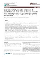
Figure 1
Presence of asporinPresence of asporin. (a) Localization of asporin in the outer annulus of
the human disc. Note that there are some cells that do not show
asporin localization (arrow). (b) The presence of asporin is shown in the
inner annulus of the human disc; note a nearby cell without asporin
content (arrow). (c) Negative control. (d) Positive control showing
localization of asporin in articular chondrocytes of the rat humerus (a, b,
c × 260; d × 300).
Figure 2
Cells with and cells without the presence of asporin are shown here in clusters in the inner annulus ((a) × 470)Cells with and cells without the presence of asporin are shown here in
clusters in the inner annulus ((a) × 470). Rare cells show immunolocal-
ization of asporin in the nucleus pulposus ((b) × 230).
Available online />Page 5 of 7
(page number not for citation purposes)
expression in disc tissue, in Caucasian subjects. Novel work
reported here shows the presence of asporin associated with
cells in the outer annulus and, less frequently, with cells in the
inner annulus and nucleus pulposus. We also found that the
asporin gene was expressed in healthy and degenerated
discs, and by annulus cells in monolayer and three-dimen-
sional cell culture.
Our studies showed significantly higher asporin expression
levels in more degenerate intervertebral discs; these findings
are similar to those presented from recent work by Kizawa et
al. in cartilage from osteoarthritic subjects [4]. They found high
Figure 3
Cells with concentric rings of extracellular matrix in the inner annulus illustrated here show the presence or absence (arrow) of asporin (× 820)Cells with concentric rings of extracellular matrix in the inner annulus
illustrated here show the presence or absence (arrow) of asporin (×
820).
Figure 4
Sectioned en face, this sand rat lumbar vertebral specimen shows simi-lar asporin localization to that seen in the human discSectioned en face, this sand rat lumbar vertebral specimen shows simi-
lar asporin localization to that seen in the human disc. The greatest
asporin presence is in the outer annulus, with fewer cells showing
asporin content in the inner annulus (arrows mark cells negative for
asporin immunolocalization) (× 360).
Figure 5
Immunolocalization of asporin in vitroImmunolocalization of asporin in vitro. First panel: (a) annulus cells in
monolayer show uniform localization; (b) monolayer negative control;
(c) three-dimensional cultured annulus cells also show expression of
asporin in all cells; (d) three-dimensional culture negative control. (a
and b, × 66; c and d, × 275).
Figure 6
Asporin expression levels were significantly greater in more degenerate Thompson grade 4 discs vs expression levels in healthier grade 1, 2, and 3 discs (P = 0.004)Asporin expression levels were significantly greater in more degenerate
Thompson grade 4 discs vs expression levels in healthier grade 1, 2,
and 3 discs (P = 0.004).
Arthritis Research & Therapy Vol 11 No 2 Gruber et al.
Page 6 of 7
(page number not for citation purposes)
levels in osteoarthritic subjects, but much lower levels in con-
trol subjects. They also found that a chondrogenic cell line with
overexpression of asporin showed suppression of chondro-
cyte marker genes aggrecan and type II collagen during
chondrocyte differentiation. These cells also showed suppres-
sion of TGFβ-mediated expression of aggrecan and type II col-
lagen. Overexpression of asporin also was associated with
proteoglycan content of the ECM. These findings led Kizawa
et al. to suggest that asporin may play a major role in modulat-
ing chondrocyte matrix homeostasis.
Our studies presented here have several shortcomings that
should be mentioned. Since the majority of the disc specimens
which we received are Thompson grade III and IV, it is unfortu-
nate that we did not have any of the most degenerate grade V
specimens which could be used for gene expression studies.
However, in spite of this problem, the data did show signifi-
cantly greater asporin expression in the more degenerate
grade IV specimens studied here as compared to expression
in grades I, II and III (Figure 6, P = 0.004).
Secondly, we have not yet been able to examine our study
population for the presence of the asporin polymorphisms now
known to be present in Asian patients with disc degeneration
[10]. Such studies are now underway. It is also important that
further research be carried out at the cellular level to elucidate
the function and role of asporin in the progression of disc
degeneration. Final points for future work include studies of
expression in nucleus pulposus cells. Another important future
project would be to carry out an analysis in disc as was
reported by Kizawa et al. [4] in cartilage. Another shortcoming
is that we were not able to carry out a complete analysis of the
localization of asporin in the aging/degenerating lumbar spine
of the sand rat; we look forward to this future analysis.
Conclusions
In the discs of Caucasian subjects studied here, and in the
sand rat (an small rodent with spontaneous age-related disc
degeneration), greater immunolocalization was found in the
outer, compared to inner, annulus. Localization was rare in the
nucleus. Similar patterns were found for the presence of
asporin in lumbar discs of sand rats. Gene expression studies
showed greatest expression of asporin in the more degenerate
human discs in vivo. Asporin was also expressed in human
annulus cells cultured in monolayer and three-dimensional cul-
ture.
Competing interests
The authors declare that they have no competing interests.
Authors' contributions
HEG and EN conceived the study and participated in design
and coordination. HEG wrote the manuscript. YS and GH
assisted with gene expression studies. YS assisted with a
review of asporin in osteoarthritis. NZ and JAI performed his-
tology and immunocytochemistry. All authors read and
approved the final manuscript.
Acknowledgements
We wish to thank the Carolinas Back Pain Research Endowment for
support. This research was performed at Carolinas Medical Center,
Charlotte, NC, USA.
Table 2
Demographic features of subjects whose annulus cells were studied in three-dimensional gene expression analyses
Subject Age (years)/gender Site Thompson grade Other information
1 23/M L5 to S1 I Surgical specimen
2 45/F T5 to T6 II Surgical specimen
3 52/F L2 to L3 III CHTN: cardiac dysfunction
4 36/F L5 to S1 III Surgical specimen
5 31/M L5 to S1 III Surgical specimen
6 46/F L2 to L3 III Surgical specimen
7 28/F L5 to S1 III Surgical specimen
8 65/F T12 to L1 IV Surgical specimen
9 32/F L5 to S1 IV Surgical specimen
10 60/M L4 to L5 IV Surgical specimen
11 59/F L3 to L4 IV Surgical specimen
12 72/F L4 to 5 IV Surgical specimen
13 45/M L5 to S1 V Surgical specimen
CHTN, Cooperative Human Tissue Network; F, female; L, lumbar; M, male; S, sacral.
Available online />Page 7 of 7
(page number not for citation purposes)
References
1. Lorenzo P, Aspberg A, Önnerfjord P, Bayliss MT, Neame PJ,
Heinegård D: Identification and characterization of asporin. A
novel member of the leucine-rich repeat protein family closely
related to decorin and biglycan. J Biol Chem 2001,
276:12201-12211.
2. Henry SP, Takanosu M, Boyd TC, Mayne PM, Eberspaecher H,
Zhou W, DeCrombrugghe B, Hook M, Mayne R: Expression pat-
tern and gene characterization of asporin. A newly discovered
member of the leucine-rich repeat protein family. J Biol Chem
2001, 276:12212-12221.
3. Loughlin J: Polymorphism in signal transduction is a major
route through which osteoarthritis susceptibility is acting.
Curr Opin Rheumatol 2005, 17:629-633.
4. Kizawa H, Kou I, Iida A, Sudo A, Miyamoto Y, Fukuda A, Mabuchi
A, Kotani A, Kawakami A, Yamamoto S, Uchida A, Nakamura K,
Notoya K, Nakamura Y, Ikegawa S: An aspartic acid repeat poly-
morphism in asporin inhibits chondrogenesis and increases
susceptibility to osteoarthritis. Nat Genet 2005, 37:138-144.
5. Iida A, Kizawa H, Nakamura Y, Ikegawa S: High-resolution SNP
map of ASPN, a susceptibility gene for osteoarthritis. J Hum
Genet 2006, 51:151-154.
6. Shi D, Nakamura T, Dai J, Yi L, Qin J, Chen D, Xu Z, Wang Y, Ike-
gawa S, Jiang Q: Association of the aspartic acid-repeat poly-
morphism in the asporin gene with age at onset of knee
osteoarthritis in Han Chinese population. J Hum Genet 2007,
52:664-667.
7. Nakamura T, Shi D, Tzetis M, Rodriguez-Lopez J, Miyamoto Y, Tse-
zou A, Gonzalez A, Jiang Q, Kamatani N, Loughlin J, Ikegawa S:
Meta-analysis of association between the ASPN D-repeat and
osteoarthritis. Hum Mol Genet 2007, 16:1676-1681.
8. Rodriguez-Lopez J, Pombo-Suarez M, Liz M, Gomez-Reino JJ,
Gonzalea A: Lack of association of a variable number of aspar-
tic acid residues in the asporin gene with osteoarthritis sus-
ceptibility: case-control studies in Spanish Caucasians.
Arthritis Res Ther 2006, 8:R55.
9. Mustafa Z, Dowling B, Chapman K, Sinsheimer JS, Carr A, Lough-
lin J: Investigating the aspartic acid (D) repeat of asporin as a
risk factor for osteoarthritis in a UK Caucasian population.
Arthritis Rheum 2005, 52:3502-3506.
10. Song YQ, Cheung KM, Ho DW, Poon SCS, Chiba K, Kawaguchi
Y, Hirose Y, Alini M, Grad S, Yee AE, Leong JC, Luk KD, Yip S-P,
Karppinen J, Cheah KS, Sham P, Ikegawa S, Chan D: Association
of the asporin D14 allele with lumbar-disc degeneration in
Asians. Am J Hum Genet 2008, 82:744-747.
11. Thompson JP, Pearce RH, Schechter MT, Adams ME, Tsang IKY,
Bishop PB: Preliminary evaluation of a scheme for grading the
gross morphology of the human intervertebral disc. Spine
1990, 15:411-415.
12. Gruber HE, Johnson T, Norton HJ, Hanley EN Jr: The sand rat
model for disc degeneration: radiologic characterization of
age-related changes. Cross-sectional and prospective analy-
ses. Spine 2002, 27:230-234.
13. Gruber HE, Ashraf N, Kilburn J, Williams C, Norton HJ, Gordon BE,
Hanley EN: Vertebral endplate architecture and vasculariza-
tion: application of micro-computerized tomography, a vascu-
lar tracer, and immunocytochemistry in analyses of disc
degeneration in the aging sand rat. Spine 2005,
30:2593-2600.
14. Gruber HE, Mougeot JL, Hoelscher GL, Ingram JA, Hanley EN Jr:
Microarray analysis of laser capture microdissected annulus
cells from the human intervertebral disc. Spine 2007,
32:1181-1187.
15. Hagiwara H, Aoki T, Yoshimi T: Immunoelectron microscopic
analysis of chondroitin sulfates during calcification in the rat
growth plate cartilage. Histochemistry 1995, 103:213-220.
16. Gruber HE, Leslie K, Ingram J, Norton HJ, Hanley EN Jr: Cell-
based tissue engineering for the intervertebral disc: in vitro
studies of human disc cell gene expression and matrix pro-
duction within selected cell carriers. Spine J 2004, 4:44-55.