Báo cáo y học: "Candida albicans induces cyclo-oxygenase 2 expression and prostaglandin E2 production in synovial fibroblasts through an extracellular-regulated kinase 1/2 dependent pathway" pps
Bạn đang xem bản rút gọn của tài liệu. Xem và tải ngay bản đầy đủ của tài liệu tại đây (1.6 MB, 9 trang )
Open Access
Available online />Page 1 of 9
(page number not for citation purposes)
Vol 11 No 2
Research article
Candida albicans induces cyclo-oxygenase 2 expression and
prostaglandin E2 production in synovial fibroblasts through an
extracellular-regulated kinase 1/2 dependent pathway
Herng-Sheng Lee
1
, Chung-Shinn Lee
2
, Chi-Jung Yang
2
, Sui-Long Su
3
and Donald M Salter
4
1
Department of Pathology, Tri-Service General Hospital and National Defense Medical Center, No. 325, Sec. 2, Chenggong Rd, Neihu District, Taipei
City 114, Taiwan
2
Graduate Institute of Pathology and Parasitology, National Defense Medical Center, No. 161, Minchun E. Rd, Neihu District, Taipei City 114, Taiwan
3
School of Public Health, National Defense Medical Center, No. 161, Minchun E. Rd, Neihu District, Taipei City 114, Taiwan
4
Osteoarticular Research Group, Centre for Inflammation Research, Queens Medical Research Institute, 47 Little France Crescent, Edinburgh, EH16
4TJ, UK
Corresponding author: Herng-Sheng Lee,
Received: 19 Sep 2008 Revisions requested: 6 Nov 2008 Revisions received: 17 Mar 2009 Accepted: 29 Mar 2009 Published: 29 Mar 2009
Arthritis Research & Therapy 2009, 11:R48 (doi:10.1186/ar2661)
This article is online at: />© 2009 Lee et al.; licensee BioMed Central Ltd.
This is an open access article distributed under the terms of the Creative Commons Attribution License ( />),
which permits unrestricted use, distribution, and reproduction in any medium, provided the original work is properly cited.
Abstract
Introduction Synovial cells are potential sources of
inflammatory mediators in bacterial-induced arthritis but their
involvement in the inflammatory response to Candida albicans-
induced septic arthritis is largely unknown.
Methods Primary cultures of rat synovial fibroblasts were
infected with C. albicans (ATCC90028). Immunocytochemistry,
western blotting, and RT-PCR were performed to assess cyclo-
oxygenase 2 induction. Phosphorylation of extracellular-
regulated kinase (ERK1/2) following infection in the absence or
presence of U0126 was assessed by western blotting whilst
prostaglandin E2 production was measured by ELISA. Nuclear
factor κB (NFκB) translocation was evaluated by an
electrophoretic mobility shift assay.
Results Infection of synovial fibroblasts with C. albicans
resulted in cyclo-oxygenase 2 expression and prostaglandin E2
production. Cyclo-oxygenase 2 expression and prostaglandin
E2 production was dependent upon extracellular-regulated
kinase 1/2 phosphorylation, associated with activation of NFκB
and significantly elevated in the presence of laminarin, an
inhibitor of dectin-1 activity. Synovial fibroblasts adjacent to C.
albicans hyphae aggregates appeared to be the major
contributors to the increased levels of cyclo-oxygenase 2 and
phosphorylated extracellular-regulated kinase 1/2.
Conclusions C. albicans infection of synovial fibroblasts in vitro
results in upregulation of cyclo-oxygenase 2 and prostaglandin
E2 by mechanisms that may involve activation of extracellular-
regulated kinase 1/2 and are associated with NFκB activation.
Introduction
Infectious arthritis is a potentially serious disease that may
cause rapid destruction of the joint and produce permanent
deformities. Articular structures can be affected by mycotic
infections through direct inoculation, contiguous spread, or
hematogenous dissemination [1-4]. Of the various Candida
species, Candida albicans is most commonly associated with
fungal arthritis, especially in immunocompromized individuals
[4-7]. Typically infection predominates in large weight-bearing
joints, most often the knee [8]. Experimental arthritis in
Sprague-Dawley rats with intravenous administration of C.
albicans demonstrates that Candida arthritis involves not only
joint tissues but also adjacent bones [9]. In mice, direct inoc-
ulation of joints with C. albicans results in a rapidly progressive
septic arthritis that also exacerbates collagen-induced arthritis
[10]. Fungal infection may also induce and exacerbate autoim-
mune diseases such as rheumatoid arthritis potentially through
effects of β-glucans, polysaccharides in the cell wall of fungi,
on inflammatory and immune responses [11].
AEC: 3-amino-9-ethylcarbazole; BSA: bovine serum albumin; COX-2: cyclo-oxygenase 2; EDTA: ethylenediaminetetraacetic acid; ELISA: enzyme-
linked immunosorbent assay; EMSA: electrophoretic-mobility shift assay; ERK: extracellular-regulated kinase; GAPDH: glyceraldehyde-3-phosphate
dehydrogenase; HRP: horseradish peroxidase; IL: interleukin; JNK: c-Jun N-terminal kinase; MEK: mitogen-activated protein kinase; NFκB: nuclear
factor κB; PBS: phosphate-buffered saline; PGE
2
: prostaglandin E
2
; PKC: protein kinase C; PVDF: polyvinylidene difluoride; RT-PCR: reverse tran-
scription polymerase chain reaction; SD: Sprague-Dawley; TBST: Tris-buffered saline/Tween; TLR: Toll-like receptor; TNF: tumor necrosis factor.
Arthritis Research & Therapy Vol 11 No 2 Lee et al.
Page 2 of 9
(page number not for citation purposes)
Cyclo-oxygenase 2 (COX-2) is a key enzyme involved in joint
inflammation through production of prostaglandins. COX-2 is
induced in human joint tissues, including chondrocytes and
synoviocytes, by inflammatory stimuli such as interleukin 1β
(IL1β), IL17, and tumor necrosis factor (TNF) [12-20] and has
roles in cartilage degradation and synovial angiogenesis
[16,21]. Micro-oganisms of all types, mostly bacterial infec-
tions, can produce an infectious arthritis associated with
COX-2 induction and prostaglandin E
2
(PGE
2
) production
[22-24]. In response to C. albicans infection HeLa cells [25],
vascular endothelial cells [26], and macrophages in vitro [27]
have been shown to express COX-2. The signal transduction
pathways resulting in COX-2 expression may involve Toll-like
receptor (TLR) 2 and 4 [25,28], which activate a variety of sig-
naling molecules including p38 [29], c-Jun N-terminal kinase
(JNK) [29,30], extracellular-regulated kinase (ERK) [31,32],
protein kinase C (PKC), and activated nuclear factor κB
(NFκB) [25,30]. More recently dectin-1 the receptor for β-glu-
can a fungal wall component has been shown to be involved
in the induction of cytokines and chemokines possibly by col-
laborating with TLRs [33].
Although it is well documented that C. albicans may induce
joint inflammation and destruction, the detailed inflammatory
responses and associated mechanisms are largely unknown.
The present study was undertaken to establish a model to
examine COX-2 induction in synovial fibroblasts following C.
albicans infection in vitro.
Materials and methods
Synovial fibroblast isolation and culture
Male Sprague-Dawley (SD) rats (8 weeks old, 280 to 300 g)
were obtained from BioLASCO Taiwan (Taipei, Taiwan). All
experiments were approved by the local Institutional Review
Board and performed in adherence to the National Institutes of
Health Guidelines for the treatment of laboratory animals. The
synovium of knee joints was aseptically removed from normal
SD rats, cut into small fragments and incubated with antimi-
crobial solution (500 IU/ml penicillin/streptomycin; Gibco Inv-
itrogen, Burlington, Ontario, Canada) for 1 h, washed with
sterile phosphate-buffered saline (PBS) before digestion with
3 mg/ml collagenase type H (Sigma, St Louis, MO, USA) at
37°C for 12 h. The resultant cell suspension was centrifuged
at 2,500 rpm for 10 minutes following which the supernatant
was discarded and the pellet resuspended in PBS. After fur-
ther centrifugation at 1,000 rpm for 10 minutes, cells were
resuspended and seeded in 20 ml of Ham's F12 medium
(Sigma) containing 10% fetal bovine serum (Gibco Invitrogen)
and 100 IU/ml penicillin/streptomycin (Gibco Invitrogen). The
synovial cells were then cultured in a humidified 5% CO
2
atmosphere at 37°C until confluent, detached with 0.05%
trypsin/ethylenediaminetetraacetic acid (EDTA) (Gibco Invitro-
gen) and seeded at a density of 2 × 10
5
cells/dish in 60 mm
tissue culture dishes (Orange scientific, Braine-l'Alleud, Bel-
gium) for further experimental procedures.
C. albicans preparation
C. albicans (ATCC 90028) was grown on Sabouraud dex-
trose agar (BD Microbiology System, Sparks, MD, USA) at
25°C. After a 16-h culture, colonies were suspended in PBS
(Gibco Invitrogen) and prepared to the desired density of 1 ×
10
3
to 1 × 10
7
yeasts/ml.
Experimental protocol for C. albicans incubation with
synovial fibroblasts
Dishes of synovial fibroblasts were placed in serum-free media
(3 ml) overnight and then treated with either 200 μL PBS or
200 μL suspension of C. albicans (2 × 10
2
to 2 × 10
6
yeasts/
dish) in 5% CO
2
atmosphere at 37°C for 6 or 12 h. In some
experiments synovial fibroblasts were pre-incubated with
U0126 (Cell Signaling Technology, Beverly, MA, USA), a
mitogen-activated protein kinase (MEK)1/2 inhibitor, at a con-
centration of 20 μM for 2 h; laminarin (Sigma) a β-glucan
receptor blocking agent and specific inhibitor of dectin-1 activ-
ity at a concentration of 10 mg/ml for 1 h. MG-132 (Calbio-
chem, San Diego, CA, USA) as a NFκB inhibitor was co-
incubated with synovial fibroblasts at a concentration of 35
μM. For the trans-well experiments (Transwell, Corning Incor-
porated, Corning, NY, USA), synovial fibroblasts were seeded
in the upper chamber and C. albicans were plated in the lower
chamber overnight, and then interacted for 12 h. In controls C.
albicans were omitted from the lower chamber.
Immunocytochemistry
After a 12-h co-culture of synovial fibroblasts and C. albicans,
cells and fungi on dish were washed with ice-cold PBS twice
and then fixed using 2 ml of a 1:1 methanol/acetone mixture
per dish for 5 minutes at -20°C. Cells were then stained by
immunocytochemistry. Immunodetection for COX-2 was per-
formed with a standard avidin-biotin-peroxidase complex
detection kit (DakoCytomation, Glostrup, Denmark). Dishes
were washed twice with PBS and blocked by incubation with
200 μL 1% non-immune horse serum (Vector Laboratories,
Burlingame, CA, USA) in 1% bovine serum albumin (BSA) in
antibody diluent (DakoCytomation) for 30 minutes at room
temperature. The solution was poured off and the cells incu-
bated sequentially with anti-COX-2 epitope specific antibody
(1:200) (Lab Vision Corporation, Cheshire, UK) or anti-phos-
pho-ERK1/2 (1:100) (Cell Signaling) for 60 minutes, bioti-
nylated secondary antibody (1:200) for 45 minutes, and
horseradish peroxidase (HRP)-conjugated streptavidin for 20
minutes. Between each incubation cells were washed with
Tris-buffered saline/Tween (TBST) (12.5 mM Tris/HCl, pH
7.6, 137 mM NaCl, 0.1% Tween 20) three times. The chro-
mogen 3-amino-9-ethylcarbazole (AEC) was then added for
15 minutes and finally counterstained with Mayer's hematoxy-
lin. The cells were mounted with a coverslip and visualized
under light microscopy.
Available online />Page 3 of 9
(page number not for citation purposes)
Reverse transcription PCR
Total RNA was isolated from cells after a 12-h co-culture of
synovial fibroblasts and C. albicans using easy-BLUE Total
RNA Extraction Kit (iNtRON Biotechnology, Gyeonggi-do,
Korea). For first strand cDNA synthesis, 3 μg of total RNA was
used in a single-round RT reaction (total volume 20 μl), con-
taining 0.75 μg oligo(dT)
14
primer, 1 mM deoxynucleosides
(dNTPs), 1 × first strand buffer, 0.4 mM dithiothreitol (DTT), 40
units RNaseOut recombinant ribonuclease inhibitor, and 200
units of superscript II reverse transcriptase (Gibco Invitrogen).
The reverse transcription reaction was performed at 42°C for
2 h, followed by 95°C for 5 minutes. PCR was run using 0.9
μl of the reverse transcription reaction mixture as template, 0.4
mM of gene specific primers, 1 × PCR buffer, 0.25 mM
dNTPs, and 1.5 units of Taq DNA polymerase (BioMan, Taipei,
Taiwan). The amplification was carried out at 94°C for 1
minute, then for 30 cycles at 94°C for 1 minute, 56°C for 1
minute, and 72°C for 1 minute followed by a final extension at
72°C for 10 minutes. All PCR products were size-fractionated
by a 1.5% agarose gel electrophoresis, and DNA bands were
visualized by staining the gel with 0.1 μg/ml ethidium bromide.
The bands were analyzed using gel documentation system
(Bio-Profil, Bio-1D version 99; Viogene, Sunnyvale, CA, USA).
The values were expressed as ratio of the band intensity of the
target gene to glyceraldehyde-3-phosphate dehydrogenase
(GAPDH) and the ratio of the band intensity of COX-2/
GAPDH in the control condition was normalized to 1. Variance
and P values were analyzed by Alphaimager 1220 V5.5 (Alpha
Innotech Corporation, San Leandro, CA, USA). A Student t
test was used for statistical comparison between groups. A P
value of less than 0.05 was considered statistically significant.
The primers used were as follows: COX-2 5'-GTCTCT-
CATCTGCAATAATGTG-3' (sense) and 5'-ATCTGTGT-
GGGTACAAATTTG-3' (antisense) ([GenBank:S67722
];
PCR product 801 base pairs (bp)); GAPDH 5'-CCCATCAC-
CATCTTCCAGGAG-3' (sense) and 5'-GTTGTCATGGAT-
GACCTTGGCC-3' (antisense) ([GenBank:X02231
]; PCR
product 284 bp).
Analysis of COX-2, ERK1/2 and phospho-ERK1/2
expression
Following C. albicans infection cells for 12 h were immediately
washed with ice-cold PBS containing 100 μM Na
3
VO
4
(Sigma) and lysed in situ with ice-cold lysis buffer at 4°C for
15 minutes. Lysis buffer contained 1% Igepal (Sigma), 100
μM Na
3
VO
4
, and a protease inhibitor cocktail tablet (Roche
Diagnostics, Mannheim, Germany). Whole cell lysates were
collected after centrifugation at 14,500 rpm for 15 minutes.
Protein concentration was determined by the Lowry method.
Equal amounts of protein (20 μg) were loaded onto 10% SDS
polyacrylamide gels and were transferred to polyvinylidene dif-
luoride (PVDF) membranes (Millipore Immobilon-P, Sigma).
Membranes were blocked overnight at 4°C with 2% BSA in
TBST. After washing three times with TBST, blots were incu-
bated for 1 h at room temperature with primary antibody (anti-
COX-2, 1/1,000 dilution; anti-total ERK1/2, 1/2,000 dilution;
anti-phospho-ERK1/2, 1/2,000 dilution) diluted with 2% BSA
in TBST. After washing six times with TBST, the blots were
then incubated with HRP-labeled secondary antibody (1/
1,000 dilution) for 1 h at room temperature. Membranes were
rewashed extensively and binding was detected using
Enhanced Chemiluminescense western blotting detection
system (Amersham Pharmacia Biotech, Piscataway, NJ, USA),
according to the manufacturer's instructions. Anti-ERK1/2 and
phospho-ERK1/2 antibodies were from Cell Signaling Tech-
nology. Mouse monoclonal antibody tubulin Ab-4 (primary anti-
body, 1/5,000 dilution; secondary antibody, 1/20,000
dilution) (Lab Vision) served as internal control. The band was
semiquantified by densitometry using systems as described
above.
Activation of NFκB by electrophoretic-mobility shift
assay (EMSA)
Cells were infected with 2 × 10
5
C. albicans at 37°C for 6 h.
Nuclear and cytoplasmic extracts of synovial fibroblasts were
prepared using NE-PER nuclear and cytoplasmic extraction
reagents according to the manufacturer's protocols (Pierce,
Rockford, IL, USA). A non-radioactive EMSA was performed
using an EMSA kit according to the manufacturer's instruc-
tions (Panomics, Redwood City, CA, USA). Nuclear protein (8
μg) was used to bind biotinylated oligonucleotides containing
the NFκB binding site for 30 minutes at room temperature. The
blank control was nuclear extracts being replaced with water.
A competition/cold control was set up by adding non-biotin-
labeled cold probes to the reaction. Samples were separated
in a non-denaturing polyacrylamide gel (6%, with 2.5% glyc-
erol) and blotted on a Biodyne B Pre-cut Modified Nylon mem-
brane (Pierce). The biotin was labeled with alkaline
phosphatase-conjugated streptavidin and alkaline phos-
phatase was detected with Enhanced Chemiluminescense
western blotting detection system (Amersham). The band was
semiquantified by densitometry using systems as described
above.
Measurement of PGE
2
, IL1β, and TNFα production in
culture medium
Cells were infected with 2 × 10
5
C. albicans in the presence
or absence of U0126 (20 μM, pre-incubation for 2 h) at 37°C
for 12 h. The culture supernatant was harvested, and PGE
2
,
IL1β, and TNFα concentrations were measured by ELISA
(R&D Systems, Minneapolis, MN, USA) according to the man-
ufacturer's instructions.
Results
COX-2 induction by C. albicans infection
The effect of C. albicans on COX-2 expression by synovial
fibroblasts was assessed at the molecular and protein level.
Extraction of total RNA from synovial fibroblasts was per-
formed after 12-h co-culture of synovial fibroblasts with differ-
Arthritis Research & Therapy Vol 11 No 2 Lee et al.
Page 4 of 9
(page number not for citation purposes)
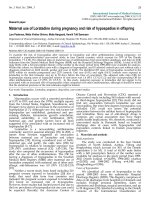
ent seeding densities of C. albicans and COX-2 induction
examined by RT-PCR. Addition of C. albicans to synovial
fibroblasts increased COX-2 expression in a dose dependent
manner. A significant increase in COX-2 expression over basal
conditions was seen at a dose of 2 × 10
4
yeasts/dish (2.03 ±
0.74-fold increase, P = 0.0185) with no further increase when
higher numbers of yeast were added (Figure 1a). The expres-
sion of COX-2 protein showed a similar pattern to that of
mRNA expression (Figure 1b).
To ascertain whether COX-2 induction was mediated by pro-
duction of a soluble mediator in the system culture medium
was collected from co-cultures of synovial fibroblasts and C.
albicans and added directly to non-infected synovial fibrob-
lasts. No change in COX-2 expression was seen. The levels of
IL1β and TNFα production were also undetectable (data not
shown).
ERK1/2 activation is necessary for C. albicans induction
of COX-2 expression
COX-2 expression by proinflammatory cytokines is associated
with ERK1/2 and NFκB activation. To establish if similar
events were occurring with C. albicans infection of synovial
fibroblasts a series of experiments were undertaken to identify
whether either ERK1/2 or NFκB were activated under the
experimental conditions that result in increased COX-2
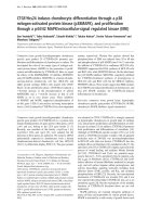
expression. The results are shown in Figure 2. Co-incubation
of synovial fibroblasts resulted in ERK1/2 activation in a dose
dependent manner. Significant levels of ERK1/2 phosphoryla-
tion were identified with the addition of C. albicans at doses
of 2 × 10
4
yeasts/dish and above (Figure 2a). Following co-
culture of synovial fibroblasts with C. albicans at 2 × 10
5
yeasts/dish for 6 h, NFκB electrophoretic-mobility shift
showed activation of NFκB (Figure 2b).
Figure 1
Cyclo-oxygenase 2 (COX-2) expression following co-culture of Cand-ida albicans with synovial fibroblastsCyclo-oxygenase 2 (COX-2) expression following co-culture of Cand-
ida albicans with synovial fibroblasts. COX-2 expression by synovial
fibroblasts was assessed after 12-h co-culture of synovial fibroblasts
with different seeding densities of C. albicans. (a) Gene expression. A
representative agarose gel demonstrating COX-2 mRNA expression as
assessed by reverse transcription polymerase chain reaction (RT-PCR).
Glyceraldehyde 3-phosphate dehydrogenase (GAPDH) served as
internal control. The graph shows the results of densitometric analysis
of DNA bands expressed as the mean ± standard deviation (SD) of the
relative fold change in COX-2/GAPDH ratio with the ratio of the control
condition normalized to 1 (N = 6, * P < 0.05). (b) Protein expression. A
representative western blot of COX-2 protein expression following
infection by C. albicans with tubulin as an internal protein loading con-
trol. The graph shows the results of densitometric analysis of bands
expressed as the mean ± SD of the relative change in COX-2/tubulin
ratio with the ratio of the control condition normalized to 1 (N = 5, * P <
0.05).
Figure 2
Activation of extracellular-regulated kinase (ERK1/2) and nuclear factor κB (NFκB) following infection of synovial fibroblasts with Candida albi-cansActivation of extracellular-regulated kinase (ERK1/2) and nuclear factor
κB (NFκB) following infection of synovial fibroblasts with Candida albi-
cans. (a) Synovial fibroblasts were infected for 12 h with C. albicans
and levels of total and phosphorylated ERK1/2 (P-ERK1/2) assessed.
Tubulin served as protein loading control. Shown is a representative
western blot from one of five experiments. (b) Synovial fibroblasts were
infected for 6 h with C. albicans and NFκB activation assessed by
electrophoretic-mobility shift assay (EMSA). Upper panel: NFκB EMSA.
The first lane of the NFκB series is the blank control. The lower series is
the cold control. Lower panel: semiquantitative analysis of EMSA band
density (N = 3, *P < 0.05).
Available online />Page 5 of 9
(page number not for citation purposes)
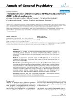
We next examined whether COX-2 expression was regulated
by ERK1/2 activation. Synovial fibroblasts were pretreated
with U0126, a MEK1/2 inhibitor, at a concentration of 20 μM
for 2 h before addition of C. albicans (2 × 10
5
yeasts/dish for
12 h) (Figure 3a). C. albicans increased ERK1/2 phosphoryla-
tion and COX-2 expression in the absence but not the pres-
ence of U0126. U0126 by itself had no effect on COX-2
expression or ERK1/2 phosphorylation. MG132 as an NFκB
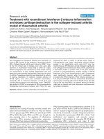
inhibitor suppressed the COX-2 expression (Figure 3b). Immu-
nohistochemistry (Figure 4) demonstrates increased phospho-
ERK1/2 and COX-2 expression in synovial fibroblasts to which
C. albicans are adherent. However, the cells without C. albi-
cans attachment demonstrated only very weak positivity. In the
presence of U0126 no expression of phospho-ERK1/2 or
COX-2 is demonstrable in the infected synovial fibroblasts.
PGE
2
production
To assess whether increased expression of COX-2 was asso-
ciated with changes in prostaglandin production levels of
PGE
2
released into the media was measured (Figure 5). In the
presence of C. albicans infection PGE
2
release into the media
was significantly increased over basal levels. This effect of C.
albicans was suppressed by the addition of U0126.
Laminarin effect and trans-well experiment
To assess whether COX-2 induction was dependent on inter-
actions with the dectin-1 receptor synovial fibroblasts were
infected with C. albicans in the presence of laminarin (Figure
6). Laminarin had no effect on levels of synovial fibroblast
COX-2 mRNA in the absence of C. albicans. Infection of syn-
ovial fibroblasts with C. albicans resulted in a 2 ± 0.3-fold
increase in COX-2 gene expression (P < 0.05). In the pres-
ence of laminarin there was a lower, 1.6 + 0.3-fold, but signif-
Figure 3
The effect of extracellular-regulated kinase (ERK) and nuclear factor κB (NFκB) inhibition on cyclo-oxygenase 2 (COX-2) production following infec-tion of synovial fibroblasts with Candida albicansThe effect of extracellular-regulated kinase (ERK) and nuclear factor κB (NFκB) inhibition on cyclo-oxygenase 2 (COX-2) production following infec-
tion of synovial fibroblasts with Candida albicans. Following infection of synovial fibroblasts for 12 h with 2 × 10
5
yeasts/dish in the absence or pres-
ence of (a) the mitogen-activated protein kinase (MEK)1/2 inhibitor U0126 or (b) the NFκB inhibitor MG-132 COX-2 protein levels were assessed
by western blotting. Shown is a representative blots from N = 3 experiments. The graphs shows the results of densitometric analysis of bands
expressed as the mean ± standard deviation (SD) of the relative fold change in band density with the ratio of the control condition normalized to 1 (N
= 3, *,
#
P < 0.05).
Arthritis Research & Therapy Vol 11 No 2 Lee et al.
Page 6 of 9
(page number not for citation purposes)
icant (P < 0.05) increase in COX-2 gene expression when
synovial fibroblasts were infected with C. albicans. This 20%
decrease in COX-2 gene expression by laminarin was statisti-
cally significantly (P < 0.05, synovial fibroblasts + C. albicans
vs synovial fibroblasts + C. albicans + laminarin) (Figure 6a).
COX-2 gene expression was significantly upregulated (1.71 ±
0.3-fold increase, P < 0.05) (Figure 6b) when synovial fibrob-
lasts and C. albicans were co-cultured in different trans-well
chambers. This indicates that direct contact may have only a
minor contribution to the elevation of COX-2 gene expression
seen when synovial fibroblasts are infected with C. albicans.
Discussion
The present study has demonstrated that synovial fibroblast
expression of COX-2, under the control of ERK1/2, is induced
following C. albicans infection. Upregulation of COX-2 is
associated with NFκB activation and appears to be more
prominent in synovial fibroblasts adjacent to fungal colonies.
The finding that ERK1/2 phosphorylation occurs on exposure
of synovial fibroblasts to C. albicans is consistent with obser-
vations of interactions of C. albicans with inflammatory and
epithelial cells. Phagocytosis of C. albicans by macrophages
results in ERK phosphorylation [29] and TNFα production by
monocytes exposed to C. albicans is ERK dependent [32].
Figure 4
Immunocytochemical detection of cyclo-oxygenase 2 (COX-2) and phospho-extracellular-regulated kinase (ERK)1/2 expression in synovial fibrob-lasts infected with Candida albicans for 12 hImmunocytochemical detection of cyclo-oxygenase 2 (COX-2) and phospho-extracellular-regulated kinase (ERK)1/2 expression in synovial fibrob-
lasts infected with Candida albicans for 12 h. Synovial fibroblasts infected with C. albicans in the absence (c) or presence (d) of the mitogen-acti-
vated protein kinase (MEK)1/2 inhibitor U0126 were immunostained for COX-2 and phosphorylated ERK1/2. (a) Negative control with omitted
primary antibody. (b) Control synovial fibroblasts. (c) Synovial fibroblasts infected with C. albicans. (d) Synovial fibroblasts infected with C. albicans
in the presence of U0126 (insert × 1,000; others × 400).
Available online />Page 7 of 9
(page number not for citation purposes)
TLR2 appears to be the major receptor mediating PGE
2
pro-
duction by mouse macrophages in response to C. albicans
[28]. C. albicans increases COX-2 expression in HeLa cells
with roles for both TLR2 and TLR4 being identified [25]. Sim-
ilar mechanisms are likely to be involved in the induction of
COX-2 and PGE
2
production in the current study.
Toll-like receptors have been shown to be involved in synovial
inflammation in a wide range of inflammatory joint diseases
including rheumatoid arthritis [34,35], Lyme arthritis [36], and
streptococcal cell wall-induced arthritis [37]. TLR signaling is
also likely to be involved in mediating proinflammatory
responses and subsequent tissue destruction in fungal arthri-
tis. The basic cell wall structure of C. albicans consists of a lin-
ear β-glucan backbone from which there are covalently
attached branches of additional β-glucan and mannoproteins.
Mannoproteins, highly antigenic proteins with large numbers
of mannose groups attached have been shown to induce pro-
inflammatory cytokine production in murine macrophages and
human mononuclear cells [38]. Mannose-containing molecular
patterns are also strong inducers of COX-2 expression and
Figure 5
The effect of Candida albicans infection of prostaglandin E2 (PGE
2
) production by synovial fibroblastsThe effect of Candida albicans infection of prostaglandin E2 (PGE
2
)
production by synovial fibroblasts. Synovial fibroblasts were infected
with C. albicans 2 × 10
5
yeasts/dish for 12 h in the absence or pres-
ence of mitogen-activated protein kinase (MEK1)/2 inhibitor U0126
and the supernatants were collected for assessment of PGE
2
produc-
tion by ELISA. N = 5, C. albicans vs phosphate-buffered saline (PBS),
*P < 0.05; C. albicans + U0126 vs C. albicans alone, #P < 0.05.
Figure 6
Requirement for dectin-1 and cell-cell interactions in upregulation of cyclo-oxygenase 2 (COX-2) expression (measured by RT-PCR) in synovial fibroblasts infected with Candida albicansRequirement for dectin-1 and cell-cell interactions in upregulation of cyclo-oxygenase 2 (COX-2) expression (measured by RT-PCR) in synovial
fibroblasts infected with Candida albicans. (a) Synovial fibroblasts were infected with C. albicans in the absence or presence of 10 mg/ml laminarin
and COX-2 gene expression assessed. In the presence of C. albicans COX-2 gene expression by synovial fibroblasts is increased (*P < 0.05 C.
albicans vs phosphate-buffered saline (PBS)). This increase in COX-2 gene expression is decreased by around 20% by laminarin (#P < 0.05 lami-
narin + C. albicans vs C. albicans alone). (b) Synovial fibroblasts and C. albicans were seeded in different chambers of trans-well plates overnight
followed by 12 h of chamber interaction and subsequent assessment of COX-2 gene expression in synovial fibroblasts (lanes 1 and 2) and C. albi-
cans (lane 3). Lane 1: synovial fibroblasts in the upper chamber and no C. albicans in the lower chamber. Lane 2: synovial fibroblasts in the upper
chamber and C. albicans in the lower chamber. Lane 3: empty upper chamber and C. albicans in the lower chamber. RT-PCR using the RNA from
C. albicans showed no band of COX-2 and glyceraldehyde-3-phosphate dehydrogenase (GAPDH) (N = 5, * P < 0.05).
Arthritis Research & Therapy Vol 11 No 2 Lee et al.
Page 8 of 9
(page number not for citation purposes)
PGE
2
production in human macrophages [39]. TLR have
important roles in the induction of cytokines by fungi with TLR4
recognition of O-linked mannosyl residues present in the C.
albicans cell wall are thought to be particularly important.
Phospholipomannan, present in the cell surface of C. albicans,
has been shown to be recognized by TLR2 [40]. Cytokine
induction by C. albicans may also be through recognition of β-
glucan by the dectin-1 (dentritic cell-associated C-type lectin-
1)/TLR2 receptor complex [38].
Dectin-1, a major β-glucan receptor, has a number of antimi-
crobial functions in phagocytes including induction of
cytokines and chemokines, possibly by collaborating with
TLRs, involvement in endocytosis and phagocytosis and pro-
duction of the respiratory burst [33]. Rat dectin-1 is involved in
immune responses against fungi [41]. Laminarin, a soluble
form of glucan blocks signaling through dectin-1 [42]. Lami-
narin decreases TNFα production by macrophages in
response to zymosan and C. albicans infection [43]. In the cur-
rent study laminarin partially blocked the increase in COX-2
mRNA that is seen when synovial fibroblasts are infected with
C. albicans. This indicates that signaling through dectin-1 has
a partial role in the upregulation of COX-2 gene expression.
This may be through direct contact between C. albicans and
synovial fibroblasts as the elevation of COX-2 gene expression
was similar in trans-well experiments and experiments with
laminarin where contact between C. albicans and synovial
fibroblasts was possible.
Conclusions
We show for the first time that COX-2 induction and PGE
2
production occurs following infection of C. albicans to syno-
vial fibroblasts and that this requires ERK1/2 activation and is
associated with NFκB activation. These interactions may sig-
nificantly contribute to the detrimental inflammatory/catabolic
activities of synovial fibroblasts in septic arthritis induced by C.
albicans and other fungi.
Competing interests
The authors declare that they have no competing interests.
Authors' contributions
HSL conceived of the study, participated in its design and
coordination, participated in the interpretation of results, and
predominantly drafted the manuscript. CSL supervised the
experiments by CJY. CJY carried out the RT-PCR, western
blotting, EMSA, immunocytochemistry, and ELISA. SLS per-
formed the statistical analysis. DMS helped to discuss the
results and draft the manuscript. All authors read and
approved the final manuscript.
Acknowledgements
This study was supported by a grant from the National Science Council
and National Defense Medical Center, Tri-Service General Hospital, Tai-
wan (NSC95-2320-B-016-018-MY2, NSC97-2320-B-016-009-MY3,
DOD96-13-03 and DOD97-08-06).
References
1. Adachi Y, Okazaki M, Ohno N, Yadomae T: Enhancement of
cytokine production by macrophages stimulated with (1→3)-
β-D-glucan, grifolan (GRN), isolated from Grifola frondosa.
Biol Pharm Bull 1994, 17:1554-1560.
2. Gathe JC Jr, Harris RL, Garland B, Bradshaw MW, Williams TW
Jr: Candida osteomyelitis. Report of five cases and review of
the literature. Am J Med 1987, 82:927-937.
3. Lazzarini L, Manfrin V, De Lalla F: Candidal prosthetic hip infec-
tion in a patient with previous candidal septic arthritis. J
Arthroplasty 2004, 19:248-252.
4. Kohli R, Hadley S: Fungal arthritis and osteomyelitis. Infect Dis
Clin North Am 2005, 19:831-851.
5. Bayer AS, Guze LB: Fungal arthritis. I. Candida arthritis: diag-
nostic and prognostic implications and therapeutic considera-
tions. Semin Arthritis Rheum 1978, 8:142-150.
6. Katzenstein D: Isolated Candida arthritis: report of a case and
definition of a distinct clinical syndrome. Arthritis Rheum 1985,
28:1421-1424.
7. Vicari P, Feitosa Pinheiro R, Chauffaille Mde L, Yamamoto M,
Figueiredo MS: Septic arthritis as the first sign of Candida trop-
icalis fungaemia in an acute lymphoid leukemia patient. Braz
J Infect Dis 2003, 7:426-428.
8. Yousefzadeh DK, Jackson JH: Neonatal and infantile candidal
arthritis with or without osteomyelitis: a clinical and radio-
graphical review of 21 cases. Skeletal Radiol 1980, 5:77-90.
9. Nakamura Y, Masuhara T, Ito-Kuwa S, Aoki S: Induction of exper-
imental Candida arthritis in rats. J Med Vet Mycol 1991,
29:179-192.
10. Yordanov M, Danova S, Ivanovska N: Inflammation induced by
inoculation of the joint with Candida albicans.
Inflammation
2004, 28:127-132.
11. Hida S, Miura NN, Adachi Y, Ohno N: Cell wall β-glucan derived
from Candida albicans acts as a trigger for autoimmune arthri-
tis in SKG mice. Biol Pharm Bull 2007, 30:1589-1592.
12. Farahat MN, Yanni G, Poston R, Panayi GS: Cytokine expression
in synovial membranes of patients with rheumatoid arthritis
and osteoarthritis. Ann Rheum Dis 1993, 52:870-875.
13. Fernandes JC, Pelletier JM, Pelletier JP: The role of cytokines in
osteoarthritis pathophysiology. Biorheology 2002,
39:237-246.
14. Faour WH, He Y, He QW, de Ladurantaye M, Quintero M, Mancini
A, Di Battista JA: Prostaglandin E(2) regulates the level and sta-
bility of cyclooxygenase-2 mRNA through activation of p38
mitogen-activated protein kinase in interleukin-1 β-treated
human synovial fibroblasts. J Biol Chem 2001,
276:31720-31731.
15. Dinarello CA: The IL-1 family and inflammatory diseases. Clin
Exp Rheumatol 2002, 20:S1-13.
16. Woods JM, Mogollon A, Amin MA, Martinez RJ, Koch AE: The role
of COX-2 in angiogenesis and rheumatoid arthritis. Exp Mol
Pathol 2003, 74:282-290.
17. LeGrand A, Fermor B, Fink C, Pisetsky DS, Weinberg JB, Vail TP,
Guilak F: Interleukin-1, tumor necrosis factor alpha, and inter-
leukin-17 synergistically up-regulate nitric oxide and prostag-
landin E2 production in explants of human osteoarthritic knee
menisci. Arthritis Rheum 2001, 44:2078-2083.
18. Kojima F, Naraba H, Miyamoto S, Beppu M, Aoki H, Kawai S:
Membrane-associated prostaglandin E synthase-1 is upregu-
lated by proinflammatory cytokines in chondrocytes from
patients with osteoarthritis. Arthritis Res Ther 2004,
6:R355-365.
19. van Doornum S, Ryan PF: Clinical manifestations of gout and
their management. Med J Aust 2000, 172:493-497.
20. Nalbant S, Chen LX, Sieck MS, Clayburne G, Schumacher HR:
Prophylactic effect of highly selective COX-2 inhibition in
acute monosodium urate crystal induced inflammation in the
rat subcutaneous air pouch.
J Rheumatol 2005, 32:1762-1764.
21. Martel-Pelletier J, Pelletier JP, Fahmi H: Cyclooxygenase-2 and
prostaglandins in articular tissues. Semin Arthritis Rheum
2003, 33:155-167.
22. Eren A, Ugutmen E, Ozkan K, Turhan Y, Eceviz E, Cilli F: Chondro-
protective effect of salicylate and chloroquine in pyogenic sep-
tic arthritis. Adv Ther 2008, 25:133-142.
23. Funk JL, Frye JB, Oyarzo JN, Kuscuoglu N, Wilson J, McCaffrey G,
Stafford G, Chen G, Lantz RC, Jolad SD, Sólyom AM, Kiela PR,
Available online />Page 9 of 9
(page number not for citation purposes)
Timmermann BN: Efficacy and mechanism of action of turmeric
supplements in the treatment of experimental arthritis. Arthri-
tis Rheum 2006, 54:3452-3464.
24. Sano H, Hla T, Maier JA, Crofford LJ, Case JP, Maciag T, Wilder
RL: In vivo cyclooxygenase expression in synovial tissues of
patients with rheumatoid arthritis and osteoarthritis and rats
with adjuvant and streptococcal cell wall arthritis. J Clin Invest
1992, 89:97-108.
25. Deva R, Shankaranarayanan P, Ciccoli R, Nigam S: Candida albi-
cans induces selectively transcriptional activation of cyclooxy-
genase-2 in HeLa cells: pivotal roles of Toll-like receptors, p38
mitogen-activated protein kinase, and NF-kappa B. J Immunol
2003, 171:3047-3055.
26. Filler SG, Pfunder AS, Spellberg BJ, Spellberg JP, Edwards JE Jr:
Candida albicans stimulates cytokine production and leuko-
cyte adhesion molecule expression by endothelial cells. Infect
Immun 1996, 64:2609-2617.
27. Suram S, Brown GD, Ghosh M, Gordon S, Loper R, Taylor PR,
Akira S, Uematsu S, Williams DL, Leslie CC: Regulation of
cytosolic phospholipase A2 activation and cyclooxygenase 2
expression in macrophages by the β-glucan receptor. J Biol
Chem 2006, 281:5506-5514.
28. Villamón E, Roig P, Gil ML, Gozalbo D: Toll-like receptor 2 medi-
ates prostaglandin E(2) production in murine peritoneal mac-
rophages and splenocytes in response to Candida albicans.
Res Microbiol 2005, 156:115-118.
29. Ibata-Ombetta S, Jouault T, Trinel PA, Poulain D: Role of extracel-
lular signal-regulated protein kinase cascade in macrophage
killing of Candida albicans. J Leukoc Biol 2001, 70:149-154.
30. Choi JH, Choi EK, Park SJ, Ko HM, Kim KJ, Han SJ, Choi IW, Im
SY: Impairment of p38 MAPK-mediated cytosolic phospholi-
pase A2 activation in the kidneys is associated with patho-
genicity of Candida albicans. Immunology 2007, 120:173-181.
31. Tang N, Liu L, Kang K, Mukherjee PK, Takahara M, Chen G,
McCormick TS, Cooper KD, Ghannoum M: Inhibition of mono-
cytic interleukin-12 production by Candida albicans via selec-
tive activation of ERK mitogen-activated protein kinase. Infect
Immun 2004, 72:2513-2520.
32. Wellington M, Dolan K, Haidaris CG: Monocyte responses to
Candida albicans are enhanced by antibody in cooperation
with antibody-independent pathogen recognition. FEMS
Immunol Med Microbiol 2007, 51:70-83.
33. Tsoni SV, Brown GD: β-Glucans and dectin-1. Ann NY Acad Sci
2008, 1143:45-60.
34. Huang Q, Ma Y, Adebayo A, Pope RM: Increased macrophage
activation mediated through Toll-like receptors in rheumatoid
arthritis. Arthritis Rheum 2007, 56:2192-2201.
35. Sacre SM, Andreakos E, Kiriakidis S, Amjadi P, Lundberg A, Gid-
dins G, Feldmann M, Brennan F, Foxwell BM: The Toll-like recep-
tor adaptor proteins MyD88 and Mal/TIRAP contribute to the
inflammatory and destructive processes in a human model of
rheumatoid arthritis. Am J Pathol 2007, 170:518-525.
36. Sobek V, Birkner N, Falk I, Würch A, Kirschning CJ, Wagner H,
Wallich R, Lamers MC, Simon MM: Direct Toll-like receptor 2
mediated co-stimulation of T cells in the mouse system as a
basis for chronic inflammatory joint disease. Arthritis Res Ther
2004, 6:R433-446.
37. Joosten LA, Koenders MI, Smeets RL, Heuvelmans-Jacobs M,
Helsen MM, Takeda K, Akira S, Lubberts E, Loo FA van de, Berg
WB van den: Toll-like receptor 2 pathway drives streptococcal
cell wall-induced joint inflammation: critical role of myeloid dif-
ferentiation factor 88. J Immunol 2003, 171:6145-6153.
38. Netea MG, Gow NA, Munro CA, Bates S, Collins C, Ferwerda G,
Hobson RP, Bertram G, Hughes HB, Jansen T, Jacobs L, Buurman
ET, Gijzen K, Williams DL, Torensma R, McKinnon A, MacCallum
DM, Odds FC, Meer JW Van der, Brown AJ, Kullberg BJ: Immune
sensing of Candida albicans requires cooperative recognition
of mannans and glucans by lectin and Toll-like receptors. J
Clin Invest 2006, 116:1642-1650.
39. Fernández N, Alonso S, Valera I, Vigo AG, Renedo M, Barbolla L,
Crespo MS: Mannose-containing molecular patterns are
strong inducers of cyclooxygenase-2 expression and prostag-
landin E2 production in human macrophages. J Immunol 2005,
174:8154-8162.
40. Roeder A, Kirschning CJ, Rupec RA, Schaller M, Weindl G, Kort-
ing HC: Toll-like receptors as key mediators in innate antifun-
gal immunity. Med Mycol 2004, 42:485-498.
41. Kato Y, Adachi Y, Ohno N:
Characterization of rat β-glucan
receptor dectin-1. Microbiol Immunol 2008, 52:418-428.
42. Gantner BN, Simmons RM, Canavera SJ, Akira S, Underhill DM:
Collaborative induction of inflammatory responses by dectin-
1 and Toll-like receptor 2. J Exp Med 2003, 197:1107-1117.
43. Plaine A, Yáñez A, Murciano C, Gaillardin C, Gil ML, Richard ML,
Gozalbo D: Enhanced proinflammatory response to the Cand-
ida albicans gpi7 null mutant by murine cells. Microbes Infect
2008, 10:382-389.