Báo cáo y học: "Type I interferon receptor controls B-cell expression of nucleic acid-sensing Toll-like receptors and autoantibody production in a murine model of lupus" doc
Bạn đang xem bản rút gọn của tài liệu. Xem và tải ngay bản đầy đủ của tài liệu tại đây (746.08 KB, 10 trang )
Open Access
Available online />Page 1 of 10
(page number not for citation purposes)
Vol 11 No 4
Research article
Type I interferon receptor controls B-cell expression of nucleic
acid-sensing Toll-like receptors and autoantibody production in a
murine model of lupus
Donna L Thibault
1,2
, Kareem L Graham
1
, Lowen Y Lee
1
, Imelda Balboni
1,3
, Paul J Hertzog
4
and
Paul J Utz
1
1
Department of Medicine, Division of Immunology and Rheumatology, Stanford University School of Medicine, 269 Campus Drive, CCSR 2250,
Stanford, CA, 94305, USA
2
Current address: Genentech, Inc., 1 DNA Way, South San Francisco, CA 94080, USA
3
Department of Pediatrics, Division of Pediatric Rheumatology, Stanford University School of Medicine, 300 Pasteur Drive, Boswell Building A085,
Stanford, CA, 94305, USA
4
Centre for Functional Genomics and Human Disease, Monash Institute of Medical Research, 27-31 Wright Street, Clayton, Victoria 3168, Australia
Corresponding author: Donna L Thibault,
Received: 25 Feb 2009 Revisions requested: 3 Apr 2009 Revisions received: 22 May 2009 Accepted: 22 Jul 2009 Published: 22 Jul 2009
Arthritis Research & Therapy 2009, 11:R112 (doi:10.1186/ar2771)
This article is online at: />© 2009 Thibault et al.; licensee BioMed Central Ltd.
This is an open access article distributed under the terms of the Creative Commons Attribution License ( />),
which permits unrestricted use, distribution, and reproduction in any medium, provided the original work is properly cited.
Abstract
Introduction Systemic lupus erythematosus (SLE) is a chronic
autoimmune disease characterized by the production of high-
titer IgG autoantibodies directed against nuclear autoantigens.
Type I interferon (IFN-I) has been shown to play a pathogenic
role in this disease. In the current study, we characterized the
role of the IFNAR2 chain of the type I IFN (IFN-I) receptor in the
targeting of nucleic acid-associated autoantigens and in B-cell
expression of the nucleic acid-sensing Toll-like receptors
(TLRs), TLR7 and TLR9, in the pristane model of lupus.
Methods Wild-type (WT) and IFNAR2
-/-
mice were treated with
pristane and monitored for proteinuria on a monthly basis.
Autoantibody production was determined by autoantigen
microarrays and confirmed using enzyme-linked immunosorbent
assay (ELISA) and immunoprecipitation. Serum immunoglobulin
isotype levels, as well as B-cell cytokine production in vitro, were
quantified by ELISA. B-cell proliferation was measured by
thymidine incorporation assay.
Results Autoantigen microarray profiling revealed that pristane-
treated IFNAR2
-/-
mice lacked autoantibodies directed against
components of the RNA-associated autoantigen complexes
Smith antigen/ribonucleoprotein (Sm/RNP) and ribosomal
phosphoprotein P0 (RiboP). The level of IgG anti-single-
stranded DNA and anti-histone autoantibodies in pristane-
treated IFNAR2
-/-
mice was decreased compared to pristane-
treated WT mice. TLR7 expression and activation by a TLR7
agonist were dramatically reduced in B cells from IFNAR2
-/-
mice. IFNAR2
-/-
B cells failed to upregulate TLR7 as well as
TLR9 expression in response to IFN-I, and effector responses to
TLR7 and TLR9 agonists were significantly decreased as
compared to B cells from WT mice following treatment with IFN-
α.
Conclusions Our studies provide a critical link between the IFN-
I pathway and the regulation of TLR-specific B-cell responses in
a murine model of SLE.
Introduction
Autoantibodies directed against nucleic acid-associated
autoantigens are characteristic of the autoimmune disease
systemic lupus erythematosus (SLE). The role of the type I
interferon (IFN-I) system in the pathogenesis of both human
and murine SLE has been studied extensively (reviewed in [1]).
Many SLE autoantigens contain nucleic acids and act as
endogenous ligands for nucleic acid-sensing Toll-like recep-
ANA: anti-nuclear autoantibody; ELISA: enzyme-linked immunosorbent assay; FBS: fetal bovine serum; GAM-Ig: goat-anti-mouse-immunoglobulin;
GAPDH: glyceraldehyde-3-phosphate dehydrogenase; HRP: horseradish peroxidase; IFN-I: type I interferon; IFNAR: interferon-I receptor; IL-6: inter-
leukin-6; IRF9: interferon regulatory factor 9; ODN: oligodeoxynucleotide; OVA: ovalbumin; PBS: phosphate-buffered saline; PDC: plasmacytoid den-
dritic cell; RiboP: ribosomal phosphoprotein P0; RNP: ribonucleoprotein; SAM: significance analysis of microarrays; SLE: systemic lupus
erythematosus; Sm: Smith antigen; snRNP: small nuclear ribonucleoprotein; SOCS1: suppressor of cytokine signaling 1; ssDNA: single-stranded
DNA; TAM: Tyro-3, Axl, and Mer; TLR: Toll-like receptor; WT: wild-type.
Arthritis Research & Therapy Vol 11 No 4 Thibault et al.
Page 2 of 10
(page number not for citation purposes)
tors (TLRs) [2]. Ligation of TLR9 by DNA-associated autoanti-
gens or TLR7 by RNA-associated autoantigens induces
secretion of IFN-I by plasmacytoid dendritic cells (PDCs) and
activates autoreactive B cells [3-12]. Production of anti-DNA
autoantibodies requires TLR9, and the production of anti-ribo-
nucleoprotein (anti-RNP) autoantibodies requires TLR7
[13,14]. A duplication of the TLR7 gene in Yaa mice is suffi-
cient for the induction of autoantibodies against RNA-associ-
ated targets [15,16], although some studies suggest that
other genes in this locus contribute to autoimmunity in this
model [17,18]. TLRs control isotype switching to pathogenic
IgG isotypes in SLE as MyD88
-/-
and TLR9
-/-
SLE mice lack
autoantibodies of the IgG2a and IgG2b subclasses [19].
Mice treated with a single intraperitoneal injection of the min-
eral oil pristane develop a lupus-like disease characterized by
the production of autoantibodies directed against many lupus
autoantigens, including DNA/histones and components of the
U1 small nuclear RNP (snRNP)/Smith antigen (Sm) complex
[20]. Autoantibodies directed against this complex are associ-
ated with both human and murine lupus [21], and the RNA
component can serve as an endogenous ligand for TLR7
[3,5,6,8-10]. Importantly, pristane-treated TLR7
-/-
mice fail to
develop isotype-switched anti-snRNP/Sm autoantibodies
[14]. Pristane treatment results in the formation of lipogranulo-
mas and the overexpression of IFN-inducible genes [22],
which closely resembles the IFN-I-induced gene expression
signature seen in blood cells derived from human patients with
SLE [23,24] and is dependent on TLR7 [25]. In addition, treat-
ment with pristane induces apoptosis in vivo, providing a
potential source of autoantigens [26], including RNPs and
nucleosomes.
All subtypes of IFN-I bind to the IFN-I receptor (IFNAR), which
is composed of two chains: IFNAR1 and IFNAR2. The IFNAR2
chain exists in both transmembrane and soluble isoforms and
is critical for ligand binding and signal transduction through
the receptor [27,28]. Negative regulators of IFN and other
proinflammatory cytokine signaling, including suppressor of
cytokine signaling 1 (SOCS1) and the Tyro-3, Axl, and Mer
(TAM) receptors, have been shown to associate with, and reg-
ulate signaling through, the IFNAR1 chain [29,30]. Signaling
through the IFNAR results in activation of the IFN-stimulated
gene factor 3 (ISGF3) heterotrimeric complex, composed of
STAT1, STAT2, and IFN regulatory factor 9 (IRF9) [31]. We
have previously shown that the IFN-I signaling molecules IRF9
and STAT1 are required for the production of IgG autoanti-
bodies in the pristane model and mediate the IFN-I-inducible
expression of TLR7 and TLR9 in B cells [32]. We also noted
a requirement for these molecules for isotype switching to the
pathogenic IgG2a isotype in this model. Nacionales and col-
leagues [33] demonstrated that mice deficient in the IFNAR1
chain of the receptor fail to develop anti-Sm/RNP and anti-
chromatin autoantibodies in the pristane model, although TLR
responses were not characterized in these mice. Also, isotype
analysis of antigen-specific autoantibodies was not performed.
Interestingly, pristane-treated IFNAR1
-/-
mice produced normal
serum levels of IgG2a, and a high percentage developed anti-
nuclear autoantibodies (ANAs).
In the present study, we characterized the role of the IFNAR2
chain of the IFNAR in the pristane model. Pristane-treated
IFNAR2
-/-
mice developed high titers of total serum IgM
accompanied by significantly lower levels of the pathogenic
IgG2a isotype. Pristane-treated IFNAR2
-/-
mice failed to
develop IgG autoantibodies directed against both RNA- and
DNA-associated autoantigens. TLR7 expression and activa-
tion by TLR7 agonists were completely abolished in IFNAR2
-/
-
B cells, demonstrating that B-cell activation through TLR7
requires IFNAR2. In addition, B cells from IFNAR2
-/-
mice
failed to upregulate TLR9 expression and activation following
incubation with IFN-I. Our results demonstrate a novel role for
the IFNAR2 chain of the IFNAR in TLR7- and TLR9-specific B-
cell responses and in the production of autoantibodies
directed against nucleic acid-associated targets.
Materials and methods
Mice and treatment
BALB/cJ mice were purchased from The Jackson Laboratory
(Bar Harbor, ME, USA). IFNAR2
-/-
mice on the BALB/c back-
ground were provided by Paul J Hertzog (Monash University,
Clayton, Australia) [30]. Mice were maintained under standard
conditions at the Stanford University Research Animal Facility.
Female mice 8 to 10 weeks of age were given a single 0.5 mL
intraperitoneal injection of pristane (Sigma-Aldrich, St. Louis,
MO, USA) or phosphate-buffered saline (PBS). Sera were col-
lected before injection and at 4-week intervals. Proteinuria was
monitored by dipstick analysis using Albustix (Bayer Corp.,
Elkhart, IN, USA) on a monthly basis. All animal experiments
were approved by, and performed in compliance with, the
guidelines of the Institutional Animal Care and Use Committee.
Autoantigen microarrays
Antigens were printed in ordered arrays on FAST slides
(Whatman, now part of GE Healthcare, Piscataway, NJ, USA).
Arrays were blocked with PBS containing 3% fetal bovine
serum (FBS) and 0.05% Tween-20 (Sigma-Aldrich) overnight
at 4°C. Arrays were probed with 1:300 dilutions of mouse
serum for 1 hour at 4°C followed by washing and incubation
with a 1:2,000 dilution of cyanine 3-conjugated goat anti-
mouse (GAM)-IgG/IgM (Jackson ImmunoResearch Laborato-
ries, Inc., West Grove, PA, USA). Arrays were scanned using
a GenePix 4000B scanner (Molecular Devices Corporation,
Sunnyvale, CA, USA). The median pixel intensities of individual
features were determined using GenePix Pro version 6.0, and
background values were subtracted. The data were expressed
as normalized median net digital fluorescence units, represent-
ing median values from eight replicate features on each array
normalized to the median intensity of eight GAM-Ig features.
Significance analysis of microarrays (SAM) [34] was applied
Available online />Page 3 of 10
(page number not for citation purposes)
to the dataset. A hierarchical clustering algorithm [35] using
the uncentered correlation similarity metric and complete link-
age method was applied, and results were depicted as a heat-
map and dendogram generated using Java Treeview software
[36]. A full list of antigens included on the array and detailed
protocols are provided [see Additional data file 1] [37].
Enzyme-linked immunosorbent assays
For anti-single-stranded DNA (anti-ssDNA) enzyme-linked
immunosorbent assays (ELISAs), Nunc MaxiSorp plates (Nal-
gene, a brand of Thermo Scientific Nunc, Rochester, NY,
USA) were coated with 10 μg/mL calf thymus DNA (Sigma-
Aldrich). For anti-Sm/RNP and anti-ribosomal phosphoprotein
P0 (anti-RiboP) ELISAs, plates were coated with 1 μg/mL Sm/
RNP or RiboP (Diarect AG, Freiburg, Germany). Wells were
incubated with sera diluted 1:250 in PBS containing 3% FBS
and 0.05% Tween-20 followed by incubation with horseradish
peroxidase (HRP)-conjugated GAM-IgM or GAM-IgG (South-
ernBiotech, Birmingham, AL, USA). Tetramethylbenzidine
(Pierce, Rockford, IL, USA) was added, and optical density val-
ues were determined at 450 nm.
To determine levels of total serum Ig isotypes, plates were
coated with 5 μg/mL GAM-Ig (H+L) (SouthernBiotech) over-
night at 4°C. Wells were incubated with 1:5,000,000 dilution
for IgG, or 1:500,000 dilution for all other isotypes, of sera in
PBS containing 3% FBS and 0.05% Tween-20 followed by
isotype-specific HRP-conjugated GAM-Ig (SouthernBiotech).
Standard curves were constructed using mouse Ig isotype
standards (SouthernBiotech), and total levels were deter-
mined.
Real-time quantitative polymerase chain reaction
Splenocytes were harvested from age- and gender-matched
wild-type (WT) and IFNAR2
-/-
mice. B cells were negatively
selected using magnetic beads (Miltenyi Biotec, Bergisch
Gladbach, Germany). Cells were more than 95% pure, as
assessed by flow cytometry (B220
+
biotin
-
; data not shown). B
cells were cultured in RPMI supplemented with
L-glutamine (2
mM), sodium pyruvate (1 mM), nonessential amino acids (0.1
mM), penicillin (100 U/mL), streptomycin (0.1 mg/mL), 2-ME
(5 × 10
-5
M), and FBS (10%) in the presence or absence of
1,000 IU/mL recombinant IFN-α (Calbiochem, now part of
EMD Biosciences, Inc., San Diego, CA, USA) for 4 hours.
RNA was extracted using RNeasy Mini kit (Qiagen Inc., Valen-
cia, CA, USA). RNA (10 ng) was amplified using one-step
QuantiTect SYBR Green reverse transcription-polymerase
chain reaction (Qiagen Inc.) and 0.5 μM forward and reverse
primers using an Opticon2 continuous fluorescence detector
(MJ Research, now part of Bio-Rad Laboratories, Inc., Her-
cules, CA, USA). The fold change in expression of each tran-
script normalized to glyceraldehyde-3-phosphate
dehydrogenase (GAPDH) was determined using the 2
-ΔΔCt
method. QuantiTect Primer Assay sets for murine TLR7, TLR9,
and GAPDH were purchased from Qiagen Inc.
Proliferation assay
Splenocytes were harvested at the conclusion of the study 12
months following pristane injection, and B cells were purified
as above. Cells were stimulated with 1 μM ODN1826 or 1 mM
Loxoribine (InvivoGen, San Diego, CA, USA). Sixteen hours
following stimulation, wells were pulsed with 1 μCi [
3
H]TdR
(Amersham, now part of GE Healthcare) and harvested 24
hours following stimulation. Incorporated radioactivity was
measured using a betaplate scintillation counter.
Interleukin-6 production
B cells were purified, cultured, and stimulated as above. After
24 hours in culture, supernatants were assayed for production
of interleukin-6 (IL-6) by sandwich ELISA using a commercially
available ELISA kit (BD Pharmingen, San Diego, CA, USA).
For IFN-α pretreatment studies, B cells were incubated in the
presence or absence of 1,000 IU/mL IFN-α for 24 hours. TLR
ligands were then added as above, and IL-6 concentration in
the supernatant was determined 24 hours following stimula-
tion.
Results
Proteinuria
To address the role of IFN-I in the development of autoimmu-
nity in the pristane model of SLE, WT and IFNAR2
-/-
mice were
treated with either pristane or PBS as a negative control. WT
BALB/c mice treated with pristane develop an immune com-
plex-mediated glomerulonephritis [38]. The development of
proteinuria in the mice, a measure of kidney disease, was
therefore assessed. Over the course of 12 months, 5 of 10
(50%) pristane-treated WT mice developed proteinuria,
whereas none of 10 (0%) pristane-treated IFNAR2
-/-
mice
developed proteinuria (Table 1). These data suggest that IFN-
I signaling through IFNAR2 is critical for the development of
kidney damage in the pristane model of SLE. Because the
development of kidney disease in the pristane model is not as
severe as in other spontaneous models of SLE, such as the
(NZB × NZW)F1 or the MRL/lpr models, we focused our stud-
ies instead on the mechanisms of autoantigen selection and
on the role of IFN-I and TLRs in this process.
Table 1
Development of proteinuria
Genotype Treatment Number Proteinuria
a
(percentage)
WT PBS 5 0 (0)
WT Pristane 10 5 (50)
IFNAR2
-/-
PBS 4 0 (0)
IFNAR2
-/-
Pristane 10 0 (0)
b
a
Proteinuria is at least 300 mg/dL.
b
P < 0.05 versus wild-type (WT)
pristane, Fisher exact test. IFNAR2, interferon-I receptor 2; PBS,
phosphate-buffered saline.
Arthritis Research & Therapy Vol 11 No 4 Thibault et al.
Page 4 of 10
(page number not for citation purposes)
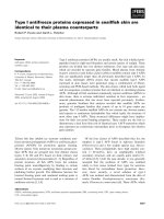
Hypergammaglobulinemia
Following pristane treatment, WT mice develop hypergamma-
globulinemia characterized by the production of high levels of
IgG as well as increased levels of IgM [39]. Importantly, pris-
tane induces the production of high levels of IgG2a, a patho-
genic isotype that preferentially binds the activating Fc
receptor, FcγRIV [40]. IFN-I induces B-cell maturation and pro-
motes isotype switching to all subclasses of IgG [41,42]. We
examined the production of immunoglobulin isotypes in pris-
tane-treated IFNAR2
-/-
mice (Figure 1). Consistent with the
known role of IFN-I in isotype switching, pristane-treated
IFNAR2
-/-
mice had significantly higher levels of total serum
IgM and significantly lower levels of total serum IgG when
compared with pristane-treated WT mice. In contrast to the
phenotype seen in IFNAR1
-/-
mice [33], pristane-treated
IFNAR2
-/-
mice developed significantly lower levels of the path-
ogenic isotype IgG2a as compared with pristane-treated WT
mice. There were no significant differences in the levels of
IgG1, IgG2b, or IgG3 between pristane-treated WT and
IFNAR2
-/-
mice.
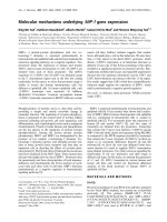
Autoantibody production
We have used autoantigen microarrays to profile the autoanti-
body response in murine models of SLE [32,43-45] and in
humans with rheumatic diseases [46,47]. We employed this
technique to systematically profile the autoantibody response
in pristane-treated WT and IFNAR2
-/-
mice. Serum from indi-
vidual mice was used to probe lupus autoantigen microarrays
that contained more than 50 candidate SLE autoantigens. A
table containing raw median pixel intensity minus background
values for all array antigens is provided [see Additional data file
2]. We used the SAM algorithm [34] to determine statistically
significant differences in array reactivity between pristane-
treated WT and IFNAR2
-/-
mice followed by hierarchical clus-
tering [35] to order individual mice on the basis of similarity of
autoantibody profiles directed against the significant antigens
identified by SAM. The results are displayed as a heatmap
(Figure 2). SAM identified reactivity to components of two
RNA-containing complexes as significantly different between
these two groups. Autoantibodies that recognize components
of the U1-snRNP complex (Sm/RNP, Sm, BB', U1-A, U1-C,
U1–70) and ribosomal P (RiboP) were present in pristane-
treated WT mice but were significantly decreased in pristane-
treated IFNAR2
-/-
mice. The two groups of mice separated into
completely distinct clusters based on autoantibody reactivity
to these autoantigens.
We frequently employ autoantigen microarrays as a screening
tool to identify autoantibody reactivities using a multiplex plat-
form and rely heavily on statistical algorithms to determine sig-
nificant differences. Reactivities to all autoantigens are then
validated using conventional techniques such as immunopre-
cipitation, ELISA, and Western blot. WT mice treated with
pristane develop high-titer autoantibodies capable of immuno-
precipitating the Sm/RNP complex from radiolabeled cell
extract [20]. As anticipated, serum autoantibodies from 7 of
10 (70%) WT mice treated with pristane immunoprecipitated
components of this complex; however, none of 10 (0%) pris-
tane-treated IFNAR2
-/-
mice developed these antibodies
(Table 2). These results confirm the specific lack of autoanti-
bodies directed against the Sm/RNP complex in serum from
Figure 1
Serum immunoglobulin levels in pristane-treated miceSerum immunoglobulin levels in pristane-treated mice. Total immu-
noglobulin levels were measured by enzyme-linked immunosorbent
assay in serum obtained 6 months after treatment with phosphate-buff-
ered saline (PBS) or pristane. Mean values with standard deviation are
shown for each group. P values were obtained using the Student t test
and are displayed above each plot.
IFNAR2: interferon-I receptor 2; n.s.: not significant; WT: wild-type.
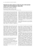
Figure 2
Autoantibody profiling of pristane-treated mice using autoantigen microarraysAutoantibody profiling of pristane-treated mice using autoantigen
microarrays. Individual arrays composed of over 50 recombinant or
purified antigens were incubated with diluted sera obtained 6 months
after pristane treatment. Pairwise significance analysis of microarrays
was used to determine antigen features with statistically significant dif-
ferences in array reactivity between pristane-treated wild-type (WT)
and pristane-treated IFNAR2
-/-
mice (false discovery rate < 0.05, fold
change > 3).
IFNAR2: interferon-I receptor 2; RiboP: ribosomal phosphoprotein P0;
Sm: Smith antigen; SmRNP: Smith antigen ribonucleoprotein.
Available online />Page 5 of 10
(page number not for citation purposes)
pristane-treated IFNAR2
-/-
mice, confirming the data obtained
using autoantigen microarrays.
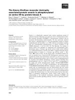
Our previous studies have demonstrated that the IFN-I down-
stream signaling molecule, IRF9, was required for the produc-
tion of IgG autoantibodies directed against the RNA-
associated targets, Sm/RNP and RiboP, as well as against the
DNA-associated targets, ssDNA and histones. Despite failing
to produce IgG autoantibodies, pristane-treated IRF9
-/-
mice
developed significantly higher titers of IgM autoantibodies
directed against the two RNA-associated complexes [32]. We
therefore examined the production of IgG and IgM autoanti-
bodies directed against these targets in IFNAR2
-/-
mice (Fig-
ure 3). Consistent with the microarray data, IFNAR2 is
absolutely required for the development of IgG anti-Sm/RNP
(Figure 3a, right panel) and anti-RiboP (Figure 3b, right panel)
autoantibodies. In contrast to the phenotype seen for IRF9
-/-
mice, however, pristane-treated IFNAR2
-/-
mice do not develop
significantly higher titers of IgM autoantibodies directed
against either of these targets as compared with pristane-
treated WT mice (Figures 3a and 3b, left panels). WT mice
treated with pristane develop high titers of IgG anti-ssDNA
(Figure 3c, right panel) and anti-histone (Figure 3d, right
panel) autoantibodies. Pristane-treated IFNAR2
-/-
mice
develop significantly lower titers of IgG autoantibodies
directed against these two targets (Figures 3c and 3d). There
are no significant differences in levels of IgM anti-ssDNA (Fig-
ure 3c, left panel) or anti-histone (Figure 3d, left panel)
between pristane-treated WT and IFNAR2
-/-
mice. These data
demonstrate that IFNAR2 is absolutely required for the devel-
opment of IgG autoantibodies directed against all of the major
antigenic targets in the pristane model of SLE: Sm/RNP,
RiboP, and the nucleosome.
Table 2
Immunoprecipitation of the Smith antigen/ribonucleoprotein
complex
Genotype Treatment Number Sm/RNP (percentage)
WT PBS 5 0 (0)
WT Pristane 10 7 (70)
IFNAR2
-/-
PBS 4 0 (0)
IFNAR2
-/-
Pristane 10 0 (0)
a
a
P < 0.005 versus wild-type (WT) pristane, Fisher exact test.
IFNAR2, interferon-I receptor 2; PBS, phosphate-buffered saline;
Sm/RNP, Smith antigen/ribonucleoprotein.
Figure 3
Autoantibody production in pristane-treated IFNAR2
-/-
miceAutoantibody production in pristane-treated IFNAR2
-/-
mice. Sera obtained 6 months after treatment with pristane or phosphate-buffered saline
(PBS) were analyzed for levels of IgM or IgG anti-Sm/RNP (a), anti-RiboP (b), anti-ssDNA (c), or anti-Histone (d) antibodies by enzyme-linked immu-
nosorbent assay. Data are plotted as absorbance values for individual animals minus background. P values were determined using the Mann-Whit-
ney t test for pristane-treated wild-type (WT) versus pristane-treated IFNAR2
-/-
mice and are displayed above each graph. Closed circles represent
serum from PBS-treated mice, and open circles represent serum from pristane-treated mice.
IFNAR2: interferon-I receptor 2; n.s.: not significant; OD: optical density; RiboP: ribosomal phosphoprotein P0; Sm/RNP: Smith antigen/ribonucleo-
protein; ssDNA: single-stranded DNA.
Arthritis Research & Therapy Vol 11 No 4 Thibault et al.
Page 6 of 10
(page number not for citation purposes)
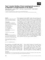
Toll-like receptor expression
PDC secretion of IFN-α has been shown to enhance the
expression of TLR7 in human naïve B cells [48]. In support of
this study, we have previously reported a critical role for the
IFN-I signaling components IRF9 and STAT1 in murine B-cell
expression of TLR7 as well as in the IFN-I-mediated induction
of TLR9 expression [32]. We examined the mRNA expression
levels of these TLRs in B cells from IFNAR2
-/-
mice. IFNAR2
-/-
B cells expressed lower basal levels of TLR7 when compared
with WT B cells; however, there was no significant difference
in the expression of TLR9 (Figure 4a). As demonstrated previ-
ously, the expression of TLR7 in B cells from WT mice was
induced more than 20-fold following treatment with IFN-α (Fig-
ure 4b). This induction of TLR7 expression was completely
dependent on IFNAR2 as there was no change in TLR7
expression in B cells from IFNAR2
-/-
mice following treatment
with IFN-α. The expression of TLR9 in WT B cells was upreg-
ulated approximately 3-fold upon treatment with IFN-α and this
upregulation was also completely dependent on IFNAR2 (Fig-
ure 4b). IFNAR2 is therefore required for the induction of TLR7
and TLR9 expression in B cells in response to IFN-α and for
normal basal levels of B-cell TLR7 expression.
Toll-like receptor activation
We next examined the functional ability of B cells from pris-
tane-treated IFNAR2
-/-
mice to respond to TLR7 and TLR9
agonists. B cells from pristane-treated WT and IFNAR2
-/-
mice
were cultured with the TLR7 agonist, Loxoribine, or the CpG
motif-containing TLR9 agonist, ODN1826. IFNAR2
-/-
B cells
proliferated significantly less (Figure 5a) and secreted signifi-
cantly less IL-6 (Figure 5b) versus WT B cells in response to
Loxoribine. Consistent with basal expression data, there were
no significant differences in proliferation (Figure 5a) or IL-6
secretion (Figure 5b) in response to the TLR9 agonist in B
cells from pristane-treated IFNAR2
-/-
mice.
Because IFN-α upregulated B-cell expression of TLR7 and
TLR9, we examined the ability of IFN-α to enhance B-cell acti-
vation by TLR ligands. B cells from WT mice pretreated with
IFN-α secreted significantly more IL-6 than untreated WT B
cells (P = 0.0001) in response to Loxoribine (Figure 5c). In
striking contrast, B cells from IFNAR2
-/-
mice secreted very low
levels of IL-6 in response to Loxoribine, and this was not
enhanced by pretreatment with IFN-α (P < 0.0001 versus IFN-
Figure 4
Expression of Toll-like receptors TLR7 and TLR9 in IFNAR2
-/-
B cellsExpression of Toll-like receptors TLR7 and TLR9 in IFNAR2
-/-
B cells.
(a) B cells were purified from wild-type (WT) or IFNAR2
-/-
mice using
magnetic beads. RNA was extracted and the relative mRNA expression
of TLR7 and TLR9 was measured. (b) Purified B cells were cultured in
the presence or absence of interferon-alpha (IFN-α). RNA was
extracted and the relative expression TLR7 and TLR9 was measured. P
values were determined using the Student t test.
IFNAR2: interferon-I receptor 2; n.s.: not significant.
Figure 5
Activation of Toll-like receptors TLR7 and TLR9 in IFNAR2
-/-
miceActivation of Toll-like receptors TLR7 and TLR9 in IFNAR2
-/-
mice.
(a) B cells were purified from pristane-treated wild-type (WT) or
IFNAR2
-/-
mice, and proliferation in response to Loxoribine or
ODN1826 was measured. Data are represented as the difference in
mean counts per minute (cpm) of stimulated and unstimulated triplicate
wells (Δ cpm) + standard error of the mean. (b) B cells were purified as
above and the concentration of interleukin-6 (IL-6) in the supernatant
was measured following stimulation with Loxoribine or ODN1826. (c) B
cells were purified as above and were cultured in the presence or
absence of interferon-alpha (IFN-α) for 24 hours before treatment with
Loxoribine or ODN1826. The concentration of IL-6 in the supernatant
was then measured. P values were determined using the Student t test.
IFNAR2: interferon-I receptor 2; n.s.: not significant; ODN: oligodeoxy-
nucleotide.
Available online />Page 7 of 10
(page number not for citation purposes)
α-treated WT B cells, Figure 5c). Although IFNAR2
-/-
B cells
responded normally to the TLR9 agonist in the absence of
exogenous IFN-α (Figure 5b), the IFN-α-mediated enhance-
ment of B-cell activation by ODN1826 was completely abol-
ished in B cells from IFNAR2
-/-
mice (Figure 5c). These studies
indicate that IFN-I signaling through IFNAR2 mediates both
the expression of, and activation through, nucleic acid-sensing
TLRs in B cells.
Discussion
Previously, we have demonstrated that the IFN-I signaling mol-
ecules, IRF9 and STAT1, were required for the production of
IgG autoantibodies in the pristane model and for the high
expression levels of TLR7 and TLR9 following treatment with
IFN-I in B cells [32]. Here, we describe the autoantibody pro-
file and TLR-dependent B-cell response in SLE mice geneti-
cally deficient in the IFNAR2 chain of the IFNAR. Autoantibody
profiling using autoantigen microarrays in combination with
conventional techniques to confirm the array results revealed
that, similar to the phenotype for IRF9
-/-
mice, pristane-treated
IFNAR2
-/-
mice specifically lacked IgG autoantibodies directed
against all of the major targets in the pristane model. These tar-
gets included components of the RNA-associated complexes
Sm/RNP and RiboP as well as the DNA-associated autoanti-
gens ssDNA and histones. B cells from IFNAR2
-/-
mice exhib-
ited defects in the expression of TLR7 as well as in responses
to TLR7 agonists in the absence of exogenous IFN-α. Upon
treatment with IFN-α, B cells from WT mice upregulated TLR7
expression over 20-fold, upregulated TLR9 expression approx-
imately 3-fold, and secreted significantly higher levels of IL-6 in
response to stimulation through either TLR7 or TLR9. In the
absence of IFNAR2, however, this IFN-α-mediated enhance-
ment of TLR7 and TLR9 expression and activation was com-
pletely abolished. TLR7 responses, in particular, were almost
undetectable. Taken together with our studies in IRF9
-/-
mice,
the results of these experiments demonstrate a critical role for
the IFN-I pathway in the activation of B cells and subsequent
autoantibody production in response to TLR agonists. We are
currently in the process of backcrossing the IRF9
-/-
, STAT1
-/-
,
and IFNAR2
-/-
genetic deletions onto the MRL/lpr background
in order to more carefully assess the role of this molecule in the
development of lupus nephritis and to determine whether
other major autoantigen classes are still targets of autoanti-
bodies.
There are three very important differences between the pheno-
types observed for IRF9
-/-
and IFNAR2
-/-
mice in the pristane
model. First, pristane-treated IRF9
-/-
mice developed signifi-
cantly higher titers of IgM autoantibodies directed against the
RNA-associated autoantigens Sm/RNP and RiboP [32]. This
phenotype was not observed in IFNAR2
-/-
mice as there was
no significant difference in levels of IgM autoantibodies
directed against these two targets versus WT mice treated
with pristane or versus PBS-treated IFNAR2
-/-
mice (Figures
3a and 3b). Second, although the expression of TLR7 was sig-
nificantly decreased in IRF9
-/-
B cells versus WT B cells follow-
ing treatment with IFN-α, TLR7 expression in IFN-α-treated
IRF9
-/-
B cells was actually significantly increased versus
untreated IRF9
-/-
B cells [32]. This was not the case for
IFNAR2
-/-
B cells as TLR7 expression was not induced, even
at lower levels, following treatment with IFN-α (not significant
versus untreated IFNAR2
-/-
B cells, Figure 5b). The small
increase in expression in IRF9
-/-
B cells has functional implica-
tions as IFN-α-treated IRF9
-/-
B cells secreted significantly
more IL-6 versus untreated IRF9
-/-
B cells in response to a
TLR7 agonist [32], whereas virtually no IL-6 was secreted by
IFNAR2
-/-
B cells in response to a TLR7 agonist, regardless of
whether they were treated with IFN-α (Figure 5). Our studies
therefore suggest that the IRF9-independent induction of
TLR7 by IFN-I may be sufficient to drive the partial activation of
B cells, which results in the production of high levels of IgM
autoantibodies directed against RNA-associated targets. It is
not sufficient, however, to drive the full activation of these cells
to differentiate into isotype-switched IgG-secreting plasma
cells. On the other hand, by inhibiting the IFN-I response fur-
ther upstream through the IFNAR2 chain of the receptor, we
observed a complete block in B-cell expression of TLR7, acti-
vation through TLR7, and autoantibody production directed
against RNA-associated targets. Third, IRF9
-/-
mice treated
with pristane developed fatal plasmacytomas as early as 6
months following pristane injection, whereas no IFNAR2
-/-
mice developed this phenotype. Because the majority of the
IRF9
-/-
mice developed this fatal condition prior to the conclu-
sion of the study, we were unable to accurately assess kidney
damage in this strain. As none of the IFNAR2
-/-
mice devel-
oped any signs of proteinuria over the course of the 12-month
study, we can now conclude that IFN-I signaling is crucial for
the development of end-organ pathogenesis in this model.
The block in isotype switching to IgG in IRF9
-/-
mice was
restricted to TLR-dependent antigens as IRF9
-/-
mice immu-
nized with ovalbumin (OVA) in complete Freund's adjuvant, a
strong stimulus, mounted an effective IgG anti-OVA response
[32]. Although higher levels of IgM-specific autoantibodies
were not observed in the pristane-treated IFNAR2
-/-
mice, total
serum levels of IgM were highly elevated in pristane-treated
IFNAR2
-/-
mice (Figure 1), suggesting that there may be
defects in isotype switching to IgG in these mice. Total serum
IgM levels were also increased in pristane-treated IFNAR1
-/-
mice [33]. Studies in IFNAR1
-/-
mice have revealed that IFN-I
promotes isotype switching to all subtypes of IgG [41,42],
although in the pristane model, total serum levels of IgG2a
were normal in IFNAR1
-/-
mice [33]. Future studies in IFNAR2
-
/-
mice are therefore aimed at investigating the role that
IFNAR2 plays in isotype switching in B cells in vitro and in
response to different TLR agonists in vivo.
Two key negative regulators of TLR responses have been
found to physically associate with the IFNAR1 chain of the
receptor: SOCS1 and the TAM receptors, which include Tyro,
Arthritis Research & Therapy Vol 11 No 4 Thibault et al.
Page 8 of 10
(page number not for citation purposes)
Axl, and Mer. The induction of the SOCS proteins by IFN-α is
dependent on the TAM receptors [29] and both SOCS1
-/-
and
TAM receptor triple-knockout mice develop spontaneous
lupus-like autoimmunity [49,50]. The expressions of the TAM
receptors themselves are upregulated by IFN and TLR signal-
ing. Both of these pathways require the presence of IFNAR1
and STAT1 [29]. Therefore, in addition to mediating signals ini-
tiated by IFN-α, IFNAR1 is critical for TAM receptor-mediated
negative regulation of pleiotropic TLR responses. The function
of TAM receptors has not been assessed in IFNAR2
-/-
mice,
although signals transduced by IFNAR2 are not influenced by
SOCS1 in vivo [30]. We hypothesize that the lack of negative
regulatory molecule function in IFNAR1
-/-
mice may result in
phenotypic differences between IFNAR1
-/-
and IFNAR2
-/-
mice
in models of autoimmunity. Such differences are notable in the
pristane model as IFNAR1
-/-
mice developed high serum titers
of the pathogenic IgG2a isotype and high ANA titers, although
the identity of the autoantigen driving this response is
unknown [33]. It will therefore be critical to assess the function
of TAM receptors and the differential roles of IFNAR1 and
IFNAR2 in the development of autoimmunity.
Unlike other murine models of SLE, such as the MRL/lpr and
the (NZB × NZW)F1 spontaneous models, the pristane model
is ideally suited for studying the IFN-I pathway in the develop-
ment of murine SLE. This is true for several reasons. First, pris-
tane injection has been shown to induce the accumulation of
an IFN-producing Ly6C-high monocyte population [51], which
drives the subsequent expression of IFN-I-inducible genes
[22]. These same genes are overexpressed in blood cells from
human lupus patients, and expression of these genes corre-
lates with the production of anti-nucleoprotein autoantibodies
[23,24,52-54]. In contrast, the expression of IFN-γ-regulated,
but not IFN-I-regulated, genes is enhanced in both splenocyte
subsets and kidneys of MRL/lpr mice, suggesting that the type
II IFN pathway rather than the IFN-I pathway plays the domi-
nant pathogenic role in the development of autoimmunity in
this model. Second, the spectrum of autoantibodies produced
upon pristane treatment represents several clinically assayed
specificities in human SLE patients [55]. The (NZB × NZW)F1
model is inadequate to study the anti-RNP response as these
animals do not develop autoantibodies directed against RNA-
associated autoantigens, although pristane treatment of (NZB
× NZW)F1 mice induces the production of these autoantibod-
ies [56]. Finally, pristane induces apoptosis both in vitro and
in vivo, providing a potential source of autoantigens [26].
Defects in clearance of apoptotic debris is a common feature
of human SLE [57]. Therefore, disease pathogenesis in the
pristane model recapitulates several key features of human
SLE, including kidney pathology, IFN-I pathway activation,
autoantibody production, and induction of apoptosis.
Conclusions
In summary, our data demonstrate a novel role for the IFNAR2
in TLR7- and TLR9-specific B-cell responses and in the gen-
eration of IgG autoantibody responses in vivo. We propose
that the production of IFN-I by DCs upon pristane treatment
[22] induces the expression of TLR7 and TLR9 in B cells,
resulting in the activation of autoreactive B cells and in autoan-
tibody production in vivo. This response is completely
dependent on signaling through IFNAR2. Our results provide
further support for the development of specific inhibitors of
TLR7, TLR9, and IFN-I signaling for the treatment of SLE in
human patients and suggest that patients may be selected for
such therapeutic approaches and monitored for response to
therapy based on the targeting of subsets of nucleic acid-
associated autoantigens. These studies are of particular
importance given that IFN-I and TLR inhibitors are already
being tested in SLE in early-phase human clinical trials. Our
studies provide a crucial link between the IFN-I system and
TLR signaling in vivo and suggest that IFN-I is upstream of
TLRs in the loss of B-cell tolerance to nucleic acid-associated
autoantigens in SLE.
Competing interests
In the past 5 years, PJU has served as a consultant to Cento-
cor, Inc. (Horsham, PA, USA), Biogen Idec (Cambridge, MA,
USA), Avanir Pharmaceuticals (Aliso Viejo, CA, USA), Amgen
(Thousand Oaks, CA, USA), UCB (Brussels, Belgium), Argos
Therapeutics, Inc. (Durham, NC, USA), AstraZeneca (London,
UK), CoMentis, Inc. (South San Francisco, CA, USA), Gilead
Sciences, Inc. (Foster City, CA, USA), REGiMMUNE Corpora-
tion (Mountain View, CA, USA), Johnson & Johnson (New
Brunswick, NJ, USA), and Genentech, Inc. (South San Fran-
cisco, CA, USA). PJU was a member of the scientific advisory
boards of Monogram Biosciences, Inc. (South San Francisco,
CA, USA) and XDx, Inc. (Brisbane, CA, USA) and is a
cofounder of and consultant to Bayhill Therapeutics (San
Mateo, CA, USA). DLT is currently an employee of Genentech,
Inc. The other authors declare that they have no competing
interests.
Authors' contributions
DLT conceived of the study idea, contributed to the experi-
mental design, performed experiments, participated in the writ-
ing of the manuscript and data interpretation, and helped to
perform array studies and conduct statistical analysis. KLG
contributed to the experimental design and assisted with ani-
mal studies. LYL monitored survival and proteinuria in the mice
and assisted with animal studies. IB helped to perform array
studies and conduct statistical analysis. PJH contributed to
the experimental design and supplied the IFNAR2
-/-
mice. PJU
assisted with conception of the study idea and participated in
its design, data analysis, and the writing of the manuscript. All
authors read and approved the final manuscript.
Available online />Page 9 of 10
(page number not for citation purposes)
Additional files
Acknowledgements
The authors thank Michael G Kattah, Alvina D Chu, Cindy Limb, Peggy
P Ho, and other members of the Utz laboratory for technical assistance
and helpful discussions. This work was supported by National Institutes
of Health (NIH) grants AI50854, AI50865, AR49328, and U19-
DK61934; NHLBI Proteomics contract N01-HV-28183; a grant from
the Northern California Chapter of the Arthritis Foundation; a Dana
Foundation grant; and a gift from the Floren Family Foundation to PJU.
DLT is the recipient of a National Science Foundation Graduate
Research Fellowship and a P.E.O. Sisterhood Scholar Award. KLG is
the recipient of an NIH National Research Service Award Fellowship (AI-
10663-02).
References
1. Banchereau J, Pascual V: Type I interferon in systemic lupus
erythematosus and other autoimmune diseases. Immunity
2006, 25:383-392.
2. Marshak-Rothstein A, Rifkin IR: Immunologically active autoanti-
gens: the role of toll-like receptors in the development of
chronic inflammatory disease. Annu Rev Immunol 2007,
25:419-441.
3. Savarese E, Chae OW, Trowitzsch S, Weber G, Kastner B, Akira
S, Wagner H, Schmid RM, Bauer S, Krug A: U1 small nuclear
ribonucleoprotein immune complexes induce type I interferon
in plasmacytoid dendritic cells through TLR7. Blood 2006,
107:3229-3234.
4. Lovgren T, Eloranta ML, Kastner B, Wahren-Herlenius M, Alm GV,
Ronnblom L: Induction of interferon-alpha by immune com-
plexes or liposomes containing systemic lupus erythemato-
sus autoantigen- and Sjogren's syndrome autoantigen-
associated RNA. Arthritis Rheum 2006, 54:1917-1927.
5. Kelly KM, Zhuang H, Nacionales DC, Scumpia PO, Lyons R, Aka-
ogi J, Lee P, Williams B, Yamamoto M, Akira S, Satoh M, Reeves
WH: "Endogenous adjuvant" activity of the RNA components
of lupus autoantigens Sm/RNP and Ro 60. Arthritis Rheum
2006, 54:1557-1567.
6. Vollmer J, Tluk S, Schmitz C, Hamm S, Jurk M, Forsbach A, Akira
S, Kelly KM, Reeves WH, Bauer S, Krieg AM: Immune stimula-
tion mediated by autoantigen binding sites within small
nuclear RNAs involves Toll-like receptors 7 and 8. J Exp Med
2005, 202:1575-1585.
7. Means TK, Latz E, Hayashi F, Murali MR, Golenbock DT, Luster
AD: Human lupus autoantibody-DNA complexes activate DCs
through cooperation of CD32 and TLR9. J Clin Invest 2005,
115:407-417.
8. Lau CM, Broughton C, Tabor AS, Akira S, Flavell RA, Mamula MJ,
Christensen SR, Shlomchik MJ, Viglianti GA, Rifkin IR, Marshak-
Rothstein A: RNA-associated autoantigens activate B cells by
combined B cell antigen receptor/Toll-like receptor 7 engage-
ment. J Exp Med 2005, 202:1171-1177.
9. Barrat FJ, Meeker T, Gregorio J, Chan JH, Uematsu S, Akira S,
Chang B, Duramad O, Coffman RL: Nucleic acids of mammalian
origin can act as endogenous ligands for Toll-like receptors
and may promote systemic lupus erythematosus. J Exp Med
2005, 202:1131-1139.
10. Hoffman RW, Gazitt T, Foecking MF, Ortmann RA, Misfeldt M, Jor-
genson R, Young SL, Greidinger EL: U1 RNA induces innate
immunity signaling. Arthritis Rheum 2004, 50:2891-2896.
11. Leadbetter EA, Rifkin IR, Hohlbaum AM, Beaudette BC, Shlomchik
MJ, Marshak-Rothstein A: Chromatin-IgG complexes activate B
cells by dual engagement of IgM and Toll-like receptors.
Nature 2002, 416:603-607.
12. Yasuda K, Richez C, Maciaszek JW, Agrawal N, Akira S, Marshak-
Rothstein A, Rifkin IR: Murine dendritic cell type I IFN production
induced by human IgG-RNA immune complexes is IFN regula-
tory factor (IRF)5 and IRF7 dependent and is required for IL-6
production. J Immunol 2007, 178:6876-6885.
13. Christensen SR, Shupe J, Nickerson K, Kashgarian M, Flavell RA,
Shlomchik MJ: Toll-like receptor 7 and TLR9 dictate autoanti-
body specificity and have opposing inflammatory and regula-
tory roles in a murine model of lupus. Immunity 2006,
25:417-428.
14. Savarese E, Steinberg C, Pawar RD, Reindl W, Akira S, Anders HJ,
Krug A: Requirement of Toll-like receptor 7 for pristane-
induced production of autoantibodies and development of
murine lupus nephritis. Arthritis Rheum 2008, 58:1107-1115.
15. Subramanian S, Tus K, Li QZ, Wang A, Tian XH, Zhou J, Liang C,
Bartov G, McDaniel LD, Zhou XJ, Schultz RA, Wakeland EK: A Tlr7
translocation accelerates systemic autoimmunity in murine
lupus. Proc Natl Acad Sci USA 2006, 103:9970-9975.
16. Pisitkun P, Deane JA, Difilippantonio MJ, Tarasenko T, Satter-
thwaite AB, Bolland S: Autoreactive B cell responses to RNA-
related antigens due to TLR7 gene duplication. Science 2006,
312:1669-1672.
17. Santiago-Raber ML, Kikuchi S, Borel P, Uematsu S, Akira S, Kotzin
BL, Izui S: Evidence for genes in addition to Tlr7 in the Yaa
translocation linked with acceleration of systemic lupus ery-
thematosus. J Immunol 2008, 181:1556-1562.
18. Fairhurst AM, Hwang SH, Wang A, Tian XH, Boudreaux C, Zhou
XJ, Casco J, Li QZ, Connolly JE, Wakeland EK: Yaa autoimmune
phenotypes are conferred by overexpression of TLR7. Eur J
Immunol 2008, 38:1971-1978.
19. Ehlers M, Fukuyama H, McGaha TL, Aderem A, Ravetch JV: TLR9/
MyD88 signaling is required for class switching to pathogenic
IgG2a and 2b autoantibodies in SLE. J Exp Med 2006,
203:553-561.
20. Satoh M, Reeves WH: Induction of lupus-associated autoanti-
bodies in BALB/c mice by intraperitoneal injection of pristane.
J Exp Med 1994, 180:
2341-2346.
21. Craft J: Antibodies to snRNPs in systemic lupus erythemato-
sus. Rheum Dis Clin North Am 1992, 18:311-335.
22. Nacionales DC, Kelly KM, Lee PY, Zhuang H, Li Y, Weinstein JS,
Sobel E, Kuroda Y, Akaogi J, Satoh M, Reeves WH: Type I inter-
feron production by tertiary lymphoid tissue developing in
response to 2,6,10,14-tetramethyl-pentadecane (pristane).
Am J Pathol 2006, 168:1227-1240.
23. Zhuang H, Narain S, Sobel E, Lee PY, Nacionales DC, Kelly KM,
Richards HB, Segal M, Stewart C, Satoh M, Reeves WH: Associ-
ation of anti-nucleoprotein autoantibodies with upregulation
of Type I interferon-inducible gene transcripts and dendritic
cell maturation in systemic lupus erythematosus. Clin Immu-
nol 2005, 117:238-250.
24. Kirou KA, Lee C, George S, Louca K, Papagiannis IG, Peterson
MG, Ly N, Woodward RN, Fry KE, Lau AY, Prentice JG, Wohlge-
muth JG, Crow MK: Coordinate overexpression of interferon-
alpha-induced genes in systemic lupus erythematosus. Arthri-
tis Rheum 2004, 50:3958-3967.
The following Additional files are available online:
Additional data file 1
A table providing a description of the autoantigens used
on the protein microarrays, which includes the source,
origin, tag information, and known disease associations
for each antigen.
See />supplementary/ar2771-S1.XLS
Additional data file 2
A table containing raw median pixel intensity minus
background values for all array antigens and samples
used in this study.
See />supplementary/ar2771-S2.XLS
Arthritis Research & Therapy Vol 11 No 4 Thibault et al.
Page 10 of 10
(page number not for citation purposes)
25. Lee PY, Kumagai Y, Li Y, Takeuchi O, Yoshida H, Weinstein J, Kel-
lner ES, Nacionales D, Barker T, Kelly-Scumpia K, van Rooijen N,
Kumar H, Kawai T, Satoh M, Akira S, Reeves WH: TLR7-depend-
ent and FcgammaR-independent production of type I inter-
feron in experimental mouse lupus. J Exp Med 2008,
205:2995-3006.
26. Calvani N, Caricchio R, Tucci M, Sobel ES, Silvestris F, Tartaglia
P, Richards HB: Induction of apoptosis by the hydrocarbon oil
pristane: implications for pristane-induced lupus. J Immunol
2005, 175:4777-4782.
27. Hardy MP, Owczarek CM, Trajanovska S, Liu X, Kola I, Hertzog PJ:
The soluble murine type I interferon receptor Ifnar-2 is present
in serum, is independently regulated, and has both agonistic
and antagonistic properties. Blood 2001, 97:473-482.
28. Owczarek CM, Hwang SY, Holland KA, Gulluyan LM, Tavaria M,
Weaver B, Reich NC, Kola I, Hertzog PJ: Cloning and character-
ization of soluble and transmembrane isoforms of a novel
component of the murine type I interferon receptor, IFNAR 2.
J Biol Chem 1997, 272:23865-23870.
29. Rothlin CV, Ghosh S, Zuniga EI, Oldstone MB, Lemke G: TAM
receptors are pleiotropic inhibitors of the innate immune
response. Cell 2007, 131:1124-1136.
30. Fenner JE, Starr R, Cornish AL, Zhang JG, Metcalf D, Schreiber
RD, Sheehan K, Hilton DJ, Alexander WS, Hertzog PJ: Suppres-
sor of cytokine signaling 1 regulates the immune response to
infection by a unique inhibition of type I interferon activity. Nat
Immunol 2006, 7:33-39.
31. van Boxel-Dezaire AH, Rani MR, Stark GR: Complex modulation
of cell type-specific signaling in response to type I interferons.
Immunity 2006, 25:361-372.
32. Thibault DL, Chu AD, Graham KL, Balboni I, Lee LY, Kohlmoos C,
Landrigan A, Higgins JP, Tibshirani R, Utz PJ: IRF9 and STAT1 are
required for IgG autoantibody production and B cell expres-
sion of TLR7 in mice. J Clin Invest 2008, 118:1417-1426.
33. Nacionales DC, Kelly-Scumpia KM, Lee PY, Weinstein JS, Lyons
R, Sobel E, Satoh M, Reeves WH: Deficiency of the type I inter-
feron receptor protects mice from experimental lupus. Arthritis
Rheum 2007, 56:3770-3783.
34. Tusher VG, Tibshirani R, Chu G: Significance analysis of micro-
arrays applied to the ionizing radiation response. Proc Natl
Acad Sci USA 2001, 98:5116-5121.
35. Eisen MB, Spellman PT, Brown PO, Botstein D: Cluster analysis
and display of genome-wide expression patterns.
Proc Natl
Acad Sci USA 1998, 95:14863-14868.
36. Saldanha AJ: Java Treeview – extensible visualization of micro-
array data. Bioinformatics 2004, 20:3246-3248.
37. Protocols – The Utz Lab – Stanford University School of Med-
icine [ />]
38. Satoh M, Kumar A, Kanwar YS, Reeves WH: Anti-nuclear anti-
body production and immune-complex glomerulonephritis in
BALB/c mice treated with pristane. Proc Natl Acad Sci USA
1995, 92:10934-10938.
39. Hamilton KJ, Satoh M, Swartz J, Richards HB, Reeves WH: Influ-
ence of microbial stimulation on hypergammaglobulinemia
and autoantibody production in pristane-induced lupus. Clin
Immunol Immunopathol 1998, 86:271-279.
40. Nimmerjahn F, Bruhns P, Horiuchi K, Ravetch JV: FcgammaRIV: a
novel FcR with distinct IgG subclass specificity. Immunity
2005, 23:41-51.
41. Jego G, Palucka AK, Blanck JP, Chalouni C, Pascual V,
Banchereau J: Plasmacytoid dendritic cells induce plasma cell
differentiation through type I interferon and interleukin 6.
Immunity 2003, 19:225-234.
42. Le Bon A, Schiavoni G, D'Agostino G, Gresser I, Belardelli F,
Tough DF: Type i interferons potently enhance humoral immu-
nity and can promote isotype switching by stimulating den-
dritic cells in vivo. Immunity 2001, 14:461-470.
43. Kattah MG, Alemi GR, Thibault DL, Utz PJ: A novel two-color Fab
labeling method for autoantigen protein microarrays. Nat
Methods 2006, 3:745-51.
44. Graham KL, Vaysberg M, Kuo A, Utz PJ: Autoantigen arrays for
multiplex analysis of antibody isotypes. Proteomics 2006,
6:5720-5724.
45. Sekine H, Graham KL, Zhao S, Elliott MK, Ruiz P, Utz PJ, Gilkeson
GS: Role of MHC-linked genes in autoantigen selection and
renal disease in a murine model of systemic lupus erythema-
tosus. J Immunol 2006, 177:7423-7434.
46. Robinson WH, DiGennaro C, Hueber W, Haab BB, Kamachi M,
Dean EJ, Fournel S, Fong D, Genovese MC, de Vegvar HE, Skriner
K, Hirschberg DL, Morris RI, Muller S, Pruijn GJ, van Venrooij WJ,
Smolen JS, Brown PO, Steinman L, Utz PJ: Autoantigen microar-
rays for multiplex characterization of autoantibody responses.
Nat Med 2002, 8:295-301.
47. Balboni I, Chan SM, Kattah M, Tenenbaum JD, Butte AJ, Utz PJ:
Multiplexed protein array platforms for analysis of autoim-
mune diseases. Annu Rev Immunol 2006, 24:391-418.
48. Bekeredjian-Ding IB, Wagner M, Hornung V, Giese T, Schnurr M,
Endres S, Hartmann G: Plasmacytoid dendritic cells control
TLR7 sensitivity of naive B cells via type I IFN. J Immunol
2005,
174:4043-4050.
49. Hanada T, Yoshida H, Kato S, Tanaka K, Masutani K, Tsukada J,
Nomura Y, Mimata H, Kubo M, Yoshimura A: Suppressor of
cytokine signaling-1 is essential for suppressing dendritic cell
activation and systemic autoimmunity. Immunity 2003,
19:437-450.
50. Lu Q, Lemke G: Homeostatic regulation of the immune system
by receptor tyrosine kinases of the Tyro 3 family. Science
2001, 293:306-311.
51. Lee PY, Weinstein JS, Nacionales DC, Scumpia PO, Li Y, But-
filoski E, van Rooijen N, Moldawer L, Satoh M, Reeves WH: A
novel type I IFN-producing cell subset in murine lupus. J
Immunol 2008, 180:5101-5108.
52. Hua J, Kirou K, Lee C, Crow MK: Functional assay of type I inter-
feron in systemic lupus erythematosus plasma and associa-
tion with anti-RNA binding protein autoantibodies. Arthritis
Rheum 2006, 54:1906-1916.
53. Bennett L, Palucka AK, Arce E, Cantrell V, Borvak J, Banchereau J,
Pascual V: Interferon and granulopoiesis signatures in sys-
temic lupus erythematosus blood. J Exp Med 2003,
197:711-723.
54. Baechler EC, Batliwalla FM, Karypis G, Gaffney PM, Ortmann WA,
Espe KJ, Shark KB, Grande WJ, Hughes KM, Kapur V, Gregersen
PK, Behrens TW: Interferon-inducible gene expression signa-
ture in peripheral blood cells of patients with severe lupus.
Proc Natl Acad Sci USA 2003, 100:2610-2615.
55. Tan EM: Antinuclear antibodies: diagnostic markers for
autoimmune diseases and probes for cell biology. Adv Immu-
nol 1989, 44:93-151.
56. Yoshida H, Satoh M, Behney KM, Lee CG, Richards HB, Shaheen
VM, Yang JQ, Singh RR, Reeves WH: Effect of an exogenous
trigger on the pathogenesis of lupus in (NZB × NZW)F1 mice.
Arthritis Rheum 2002, 46:2235-2244.
57. Gaipl US, Voll RE, Sheriff A, Franz S, Kalden JR, Herrmann M:
Impaired clearance of dying cells in systemic lupus erythema-
tosus. Autoimmun Rev 2005, 4:189-194.