Báo cáo y học: "Preclinical characterization of DEKAVIL (F8-IL10), a novel clinical-stage immunocytokine which inhibits the progression of collagen-induced arthritis" ppsx
Bạn đang xem bản rút gọn của tài liệu. Xem và tải ngay bản đầy đủ của tài liệu tại đây (2.81 MB, 15 trang )
Available online />
Research article
Vol 11 No 5
Open Access
Preclinical characterization of DEKAVIL (F8-IL10), a novel
clinical-stage immunocytokine which inhibits the progression of
collagen-induced arthritis
Kathrin Schwager1, Manuela Kaspar1, Frank Bootz1,2, Roberto Marcolongo3, Erberto Paresce4,
Dario Neri2 and Eveline Trachsel1
1Philochem
AG, c/o ETH Zurich, Institute of Pharmaceutical Sciences, Wolfgang-Pauli-Strasse 10 HCI E520, CH-8093 Zurich, Switzerland
of Pharmaceutical Sciences, ETH Zürich, Wolfgang-Pauli-Strasse 10, CH-8093 Zurich, Switzerland
3Centro Interdipartimentale Studio Biochimico-Clinico Patologie Osteoarticolari, Via Doninzetti 7, University of Siena, 53100 Siena, Italy
4Department of Rheumatology, Instituto Ortopedico Gaetano Pini, via Pini 9, 20122 Milan, Italy
2Institute
Corresponding author: Dario Neri,
Received: 9 Mar 2009 Revisions requested: 15 Apr 2009 Revisions received: 4 Sep 2009 Accepted: 25 Sep 2009 Published: 25 Sep 2009
Arthritis Research & Therapy 2009, 11:R142 (doi:10.1186/ar2814)
This article is online at: />© 2009 Schwager et al.; licensee BioMed Central Ltd.
This is an open access article distributed under the terms of the Creative Commons Attribution License ( />which permits unrestricted use, distribution, and reproduction in any medium, provided the original work is properly cited.
Abstract
Introduction In this article, we present a comparative
immunohistochemical evaluation of four clinical-stage
antibodies (L19, F16, G11 and F8) directed against splice
isoforms of fibronectin and of tenascin-C for their ability to stain
synovial tissue alterations in rheumatoid arthritis patients.
Furthermore we have evaluated the therapeutic potential of the
most promising antibody, F8, fused to the anti-inflammatory
cytokine interleukin (IL) 10.
Methods F8-IL10 was produced and purified to homogeneity in
CHO cells and shown to comprise biological active antibody
and cytokine moieties by binding assays on recombinant antigen
and by MC/9 cell proliferation assays. We have also
characterized the ability of F8-IL10 to inhibit arthritis progression
in the collagen-induced arthritis mouse model.
Results The human antibody F8, specific to the extra-domain A
of fibronectin, exhibited the strongest and most homogenous
staining pattern in synovial biopsies and was thus selected for
the development of a fully human fusion protein with IL10 (F8-
Introduction
The therapeutic potential of recombinant cytokines is often
limited by severe toxicities, even at low doses, thus preventing
dose escalation and the establishment of a sufficient concentration at target tissues. It is becoming increasingly clear that
IL10, also named DEKAVIL). Following radioiodination, F8-IL10
was able to selectively target arthritic lesions and tumor neovascular structures in mice, as evidenced by autoradiographic
analysis and quantitative biodistribution studies. The
subcutaneous administration route led to equivalent targeting
results when compared with intravenous administration and was
thus selected for the clinical development of the product. F8IL10 potently inhibited progression of established arthritis in the
collagen-induced mouse model when tested alone and in
combination with methotrexate. In preparation for clinical trials in
patients with rheumatoid arthritis, F8-IL10 was studied in
rodents and in cynomolgus monkeys, revealing an excellent
safety profile at doses tenfold higher than the planned starting
dose for clinical phase I trials.
Conclusions Following the encouraging preclinical results
presented in this paper, clinical trials with F8-IL10 will now
elucidate the therapeutic potential of this product and whether
the targeted delivery of IL10 potentiates the anti-arthritic action
of the cytokine in rheumatoid arthritis patients.
monoclonal antibodies could be used to deliver cytokines at
sites of disease, therefore increasing their potency and sparing normal tissues. This pharmacodelivery strategy has been
mainly investigated for cancer therapy applications, leading to
the preclinical [1-5] and clinical [6,7] investigation of several
ACR: American College of Rheumatology; BSA: bovine serum albumin; CIA: collagen-induced arthritis; DMSO: dimethylsulfoxide; EDA: extra-domain
A of fibronectin; EDB: extra-domain B of fibronectin; ELISA: enzyme linked immunosorbent assay; GLP: good laboratory practice; HyHel: antibody
specific to hen egg lysozyme; Ig: immunoglobulin; IL: interleukin; PBS: phosphate buffered saline; PCR: polymerase chain reaction; rhuIL10: recombinant human IL10; SAP: streptavidin-alkaline phosphatase; scFv: single chain variable fragment; SIP: small immunoprotein; TnC: tenascin C; TNF:
tumor necrosis factor.
Page 1 of 15
(page number not for citation purposes)
Arthritis Research & Therapy
Vol 11 No 5
Schwager et al.
antibody-cytokine fusion proteins. For example, our group has
brought immunocytokines based on human IL2 [8-11] and on
human TNF [11-13] to phase I and phase II clinical trials.
Recently, we have observed that antibody-based pharmacodelivery strategies can also be used in the non-oncological
setting [14,15]; for example, aiming at the targeted delivery of
anti-inflammatory cytokines at sites of inflammation. We have
reported that the L19 antibody, specific to the alternatively
spliced extra-domain B (EDB) of fibronectin [16,17], could be
fused to human IL10, thus generating an immunocytokine
capable of preferential accumulation at neovascular sites of
cancer and arthritis and capable of inhibiting the progression
of established collagen-induced arthritis (CIA) in the mouse
[18]. Our preclinical and clinical experience has shown that
recombinant antibody fragments (e.g., single chain variable
fragments (scFv) with long [19] or short [20] linkers) were particularly suited for the development of antibody-based therapeutics capable of selective accumulation at sites of disease,
while being rapidly cleared from other body locations [3,2126]. Furthermore, components of the modified extracellular
matrix, such as splice isoforms of fibronectin and tenascin-C
(TnC), were found to be ideal for antibody-based pharmacodelivery applications, in view of their abundant expression at
accessible sites of tissue remodeling, while being undetectable in most normal human tissues [27,28].
IL10 is a particularly attractive anti-inflammatory cytokine for
arthritis treatment, which has exhibited an excellent tolerability
profile in rodents, monkeys and patients at doses up to 25 μg/
kg [29,30]. Recombinant human IL10 (Tenovil TM) was shown
to inhibit paw swelling and disease progression in the mouse
CIA model. This product was also found to synergize with
TNF-blocking antibodies [31] and has been tested in clinical
trials in combination with methotrexate [32,33]. The clinical
development of Tenovil TM was discontinued because of
insufficient efficacy of the compound in humans. However, in
a placebo-controlled phase I/II study American College of
Rheumatology (ACR) 20 responses were 63% for the recombinant human IL10 (rhuIL10) groups, compared with 10% for
placebo [32,33]. Similar results were observed with TNF
blockers [34].
Encouraged by the promising results obtained with L19-IL10,
we have now performed a comparative immunohistochemical
analysis on synovial tissue biopsies obtained from rheumatoid
arthritis patients of four extensively validated human monoclonal antibodies generated in our laboratory. In addition to
L19, we studied F16 (specific to the extra-domain A1 of TnC;
[10,35]), G11 (specific to the extra-domain C of TnC; [36,37])
and F8 (specific to the extra-domain A (EDA) of fibronectin;
[38]). The observation of an intense and diffuse staining pattern with the anti-EDA antibody F8 led to the development of
F8-IL10, a fully-human recombinant immunocytokine which is
now entering clinical trials in patients with rheumatoid arthritis.
Page 2 of 15
(page number not for citation purposes)
In this article, we present an extensive in vitro and in vivo characterization of F8-IL10, including the ability of this therapeutic
protein to preferentially localize at sites of arthritis and to inhibit
disease progression in the CIA model. The clinical development plans for F8-IL10 are also justified by the excellent tolerability profile observed in rodents and monkeys.
Materials and methods
Immunohistochemical analysis
For immunohistochemistry on synovial tissue samples, 10 μm
cryostat sections were fixed in ice-cold acetone and stained
for FN-EDA, FN-EDB, TnC-A1 and TnC-C. These antibodies
do not work on freshly frozen paraffin-embedded specimens.
Primary antibodies in small immunoprotein (SIP) format were
added onto the sections in a final concentration of 2 μg/ml and
detected with rabbit anti-human IgE antibody (Dako, Glostrup,
Denmark) followed by biotinylated goat anti-rabbit IgG antibody (Biospa, Milan, Italy) and streptavidin-alkaline phosphatase (SAP) complex (Biospa, Milan, Italy). Fast Red TRSalt
(Sigma-Aldrich, St Louis, MO, USA) was used as the phosphatase substrate. Sections were counterstained with hematoxylin, mounted with glycergel mounting medium (Dako,
Glostrup, Denmark) and analyzed with an Axiovert S100 TV
microscope (Zeiss, Feldbach, Switzerland). In total, freshly frozen pathology specimens of seven patients were analyzed by
immunohistochemistry.
For immunofluorescence, a double staining for FN-EDA, FNEDB, TnC-A1 respectively TnC-C and von Willebrand factor
was performed. The following primary antibodies were used:
scFv(F8), scFv(L19), scFv(F16) resp. scFv(G11) and polyclonal rabbit anti-human von Willebrand factor (Dako, Glostrup,
Denmark). As secondary detection antibodies mouse anti-Myc
(9E10) monoclonal antibody followed by Alexa Fluor 594 goat
anti-mouse IgG (Molecular Probes, Leiden, The Netherlands)
was used for scFv and Alexa Fluor 488 goat anti-rabbit
(Molecular Probes, Leiden, The Netherlands) for von Willebrand factor. Slides were mounted and analyzed as described
before.
Cloning, expression and characterization of a scFv(F8)human IL10 fusion protein
The human IL10 gene was amplified from the previously
cloned fusion protein L19-IL10 using the following primer
sequences:
a
backward
antisense
primer,
5'
TAATGGTGATGGTGATGGTGGTTTCGTATCTTCATTGTCATGTAGGCTTC-3'; and a forward sense primer, 5'-TTTTCCTTTTGCGGCCGCTCATTAGTTTCGTATCTTCATTGTCATGTA-3', which appended part of a 15
amino acid linker (SSSSG)3 at its N-terminus and a stop
codon and NotI restriction site at its C-terminus.
The gene for the single-chain variable fragment (F8) was
amplified with a signal peptide using the following primer pair:
a backward antisense primer, 5'-CCCAAGCTTGTCGAC-
Available online />
CATGGGCTGGAGCC-3' and a forward sense primer, 5'GAGCCGGAAGAGCTACTACCCGATGAGGAAGATTTGATTTCCACCTTG-GTCCCTTG-3'. Using this strategy, a
HindIII restriction site was inserted at the N-terminus and a
complementary part of the linker sequence was inserted at the
C-terminus.
The single-chain Fv and IL10 fragments were then assembled
using PCR and cloned into the HindIII and NotI restriction sites
of the mammalian cell-expression vector pcDNA3.1(+) (Invitrogen, Basel, Switzerland).
Cloning of a TNF receptor fusion protein
TNF receptor (R) II extracellular domain was amplified using a
backward antisense primer, 5'-TTTTCCTTTTGCGGCCGCTCATTA-3';
and
a
forward
sense
primer,
5'GGGTAGTAGCTCTTCCGGCTCATCGTCCAGCGGCGTGCCCGCCAAGGTTG-3', which appended part of a 15
amino acid linker (SSSSG)3 at its N-terminus and a stop
codon and NotI restriction site at its C-terminus.
The gene for the single-chain variable fragment (F8) was
amplified with a signal peptide using the following primer pair:
a backward antisense primer, 5'-CCCAAGCTTGTCGACCATGGGCTGGAGCC-3' and a forward sense primer, 5'GAGCCGGAAGAGCTACTACCCGATGAGGAAGATTTGATTTCCACCTTG-GTCCCTTG-3'. Using this strategy, a
HindIII restriction site was inserted at the N-terminus and a
complementary part of the linker sequence was inserted at the
C-terminus. The resulting PCR assembly product was cloned
into the HindIII and NotI restriction sites of the mammalian cellexpression vector pcDNA3.1(+) expressed in CHO-S cells.
Expression and purification of F8-IL10
CHO-S cells were stably transfected with the previously
described plasmid and selection was carried out in the presence of G418 (0.5 g/l). Clones of G418-resistant cells were
screened for expression of the fusion protein by ELISA using
recombinant EDA of human fibronectin as antigen and protein
A horseradish peroxidase for detection (GE Healthcare, Chalfont St Giles, UK). Following generation of monoclonal cell
lines, the best expressing clone was adapted to growth in
PowerCHO-2 CD protein-free medium (Lonza, Basel, Switzerland) for large-scale production of F8-IL10. The fusion protein
could be purified from cell culture medium by protein A affinity
chromatography, because there is a staphylococcal protein A
binding site present on most VH3 subclasses [39-41]. The size
of the fusion protein was analyzed in reducing and nonreducing conditions on SDS-PAGE and in native conditions by fast
protein liquid chromatography gel filtration on a Superdex S200 size exclusion column (GE Healthcare, Chalfont St Giles,
UK).
Bioactivity assay
Biological activity of human IL10 was determined by its ability
to induce the IL-4-dependent proliferation of MC/9 cells [42]
using a colorimetric thiazole blue (MTT) dye-reduction assay
(Sigma-Aldrich, St Louis, MO, USA). In a 96-well microtitre
plate, 10,000 MC/9 (murine mast cell line) (ATCC-LGC,
Molsheim Cedex, France) cells/well in 200 μl of medium containing 5 pg (0.05 units)/ml of murine IL4 (eBiosciences, San
Diego, CA, USA) were treated for 48 hours with varying
amounts of human IL10. The human IL10 standard and fusion
proteins were used at a maximum concentration of 100 ng/ml
IL10 equivalents and serially diluted. To this, 10 μl of 5 mg/ml
MTT was added and the cells were incubated for three to five
hours. The cells were than centrifuged lysed with dimethylsulfoxide (DMSO) and read for absorbance at 570 nm.
Collagen induced arthritis mouse model
Male DBA/1 mice (8 to 10 weeks old) were immunized by
intradermal injection at the base of the tail with 150 μg of
bovine type II collagen (Chondrex, Inc., Redmond, WA, USA)
emulsified with equal volumes of Freund's complete adjuvant
(Chondrex, Inc., Redmond, WA, USA). The procedure was
repeated two weeks after the first immunization. Mice were
inspected daily and each mouse that exhibited erythema and/
or paw swelling in one or more limbs was assigned to an imaging or treatment study.
Arthritis was monitored defining a clinical score. Each limb
was graded daily in a nonblinded fashion (0 = normal, 1 =
swelling of one or more fingers of the same limb and 2 = swelling of the whole paw), with a maximum score of eight per animal [43].
Near infrared imaging of arthritic paws
The selective accumulation of SIP(F8) in arthritic mice was
tested by near-infrared imaging analysis, as described by
Birchler and colleagues [44]. Briefly, SIP(F8) was labeled
using Alexa750 (Molecular Probes, Leiden, The Netherlands),
according to the manufacturer's recommendations, and
injected into the tail vein of arthritic mice (n = 3). Mice were
anaesthetized using ketamin, 80 mg/kg body weight, and
medetomidine, 0.2 mg/kg body weight, and imaged in a near
infrared mouse imager 24 hours after injection.
Phosphorimage analysis of arthritic paws with
radiolabeled F8-IL10
For a more detailed targeting analysis of SIP(F8) and F8-IL10
the proteins were radio-iodinated and injected intravenously or
subcutaneously, respectively (150 μg protein, 7 μCi). Mice (n
= 2) were sacrificed 24 hours after injection, paws were
exposed to a phosphorimager screen (Fujifilm, Dielsdorf, Switzerland) for one hour and read in a PhosphorImager (Fujifilm
BAS-5000, Dielsdorf, Switzerland). Data were analyzed using
Aida Image Analyzer v.4.15 (Fujifilm, Dielsdorf, Switzerland).
Page 3 of 15
(page number not for citation purposes)
Arthritis Research & Therapy
Vol 11 No 5
Schwager et al.
Quantitative biodistribution studies in tumor mice
To compare the in vivo targeting performance after subcutaneous and intravenous injection quantitative biodistribution
analyses using radiolabeled antibody preparations were performed as described before. Briefly, purified F8-IL10 was radioiodinated with 125I and injected intravenously or
subcutaneously into 129Sv mice (n = 4) grafted with a subcutaneous F9 tumor (150 μg, 8 μCi per mouse). Mice were sacrificed 24, 48, 72, or 96 hours after injection. Organs were
weighed and radioactivity was counted using a Cobra γ counter (Packard, Meriden, CT, USA). Radioactivity content of representative organs was expressed as the percentage of the
injected dose per gram of tissue (%ID/g ± standard error).
Ex vivo immunohistochemical detection of F8-IL10 and
HyHel10-IL10 in arthritis paws
At the end of therapy, mice were killed and paws were embedded in cryombedding compound (Microm, Walldorf, Germany)
and stored at -80°C. Sections (10 μm) were cut and fixed in
acetone. F8-IL0 and HyHel10-IL10 were detected using a
biotinylated anti-human IL10 antibody (eBiosciences, San
Diego, CA, USA) followed by SAP complex (Biospa, Milan,
Italy). Fast Red TRSalt (Sigma-Aldrich, St Louis, MO, USA)
was used as the phosphatase substrate. Sections were counterstained with hematoxylin, mounted with glycergel mounting
medium (Dako, Glostrup, Denmark) and analyzed with an Axiovert S100 TV microscope (Zeiss, Feldbach, Switzerland).
In a similar experiment a comparison of targeted and systemic
application of IL10 was performed. The antibody specific to
hen egg lysozyme (HyHel) 10-IL10 and F8-IL10 were labeled
with 125I and intravenously injected into 129Sv mice (n = 4)
grafted with a subcutaneous F9 tumor (150 μg, 8 μCi per
mouse). Tumor and organ uptake was measured 24 hours
after injection, as described above. Experiments were performed in agreement with Swiss regulations and under a
project license granted by the Veterinäramt des Kantons
Zürich, Switzerland (169/2008).
Immunofluorescence studies of infiltrating cells
To evaluate the role of effector cell responses in vivo immunofluorescent staining of paw sections of therapy mice was performed using antibodies against the following antigens: rat
anti-mouse F4/80 (anti-macrophage; Abcam, Cambridge,
UK), rat anti mouse CD45 (BD Biosciences, San Jose, CA,
USA), rabbit anti-asialo GM1 (anti-NK; Wako Pure Chemical
Industries, Tokyo, Japan) and rat anti-mouse CD4 and rat antimouse CD8. Cryosections were thawed and fixed by immersion in cold acetone for 10 minutes. Blocking was performed
by incubating the sections with 20% donkey/goat serum in
PBS for one hour. Following washing with PBS twice for five
minutes at room temperature, sections were incubated with
the primary antibodies in 12% BSA in PBS over night at 4°C.
Sections were washed three times for five minutes with PBS
at room temperature and then incubated with fluorescent
Alexa 488- or 594-coupled secondary antibodies (BD Biosciences, San Jose, CA, USA) and Hoechst, Frankfurt, Germany (4,6-diamidino-2-phenylindole) in 12% BSA-PBS.
Finally, sections were washed three times for five minutes in
PBS and mounted with glycergel and a coverglass (VWR
International, Dietikon, Switzerland). Images were obtained
using the individual fluorescent channels using an Axioskop 2
mot plus (Carl Zeiss, Feldbach, Switzerland).
Combination therapy study with methotrexate
Each mouse that exhibited erythema and/or swelling of one or
more paws was randomly assigned to a treatment or control
group and therapy was started. Mice were given a subcutaneous or intravenous injection of F8-IL10 (3 × 200 μg), saline or
an intraperitoneal injection of methotrexate (3 × 100 μg). For
the combination study mice were given an intravenous injection of F8-IL10 (3 × 200 μg) followed by an intraperitoneal
injection of methotrexate (3 × 100 μg). Eight mice were analyzed per group. The arthritic score was evaluated daily in a
nonblinded fashion. The results are displayed as the mean ±
standard error for each group. Experiments were performed in
agreement with Swiss regulations and under a project license
granted by the Veterinäramt des Kantons Zürich, Switzerland
(171/2007).
Comparison of targeted and untargeted delivery of IL10
Cloning, expression and purification of an HyHel10-IL10
fusion protein has been described before [18]. Therapy was
performed as described above. Briefly, arthritis mice were
injected subcutaneously with saline, HyHel10-IL10 (200 μg),
TNFRII-fusion (100 μg) or F8-IL10 (200 μg). Six to seven mice
were analyzed per group. Experiments were performed in
agreement with Swiss regulations and under a project license
granted by the Veterinäramt des Kantons Zürich, Switzerland
(171/2007).
Page 4 of 15
(page number not for citation purposes)
Staining was quantified in representative 10 times microscopic images using ImageJ software [45] and expressed as
a percentage of measurement area.
Anti-bovine collagen-II antibodies
Levels of anti-bovine collagen-II antibodies at the termination
of experiments were determined using standard ELISA techniques as described before [46]. Microtiter plates were
coated with bovine collagen II solution (5 μg/ml) overnight at
4°C. After washing they were blocked for two hours at room
temperature with 2% BSA. Samples were tested in triplicates
at 1:800 dilution. Bound total IgG, IgG1 and IgG2a were
detected by incubation with horseradish peroxidase conjugated goat anti-mouse IgG/IgG1 or IgG2a antibodies (Santa
Cruz Biotechnology, Santa Cruz, CA, USA).
Available online />
Analysis of mouse plasma cytokine levels
Mouse plasma cytokine level analysis was performed at
Cytolab (Cytolab, Muelligen, Switzerland). A multiplexed particle-based flow cytometric cytokine assay was used [47]. MAP
Fluorokine cytokine kits were purchased from R&D (Oxon,
UK). The procedures closely followed the manufacturer's
instructions. The analysis was conducted using a conventional
flow cytometer (FC500 MPL, BeckmanCoulter, Nyon, Switzerland).
Toxicology studies in cynomolgus monkey
Preclinical toxicology studies were performed at Centre International de Toxicologie, Evreux, in accordance with good laboratory practice (GLP) guidelines (Study number 34975TSP).
During the study two groups (group 2 and 3) of three male and
three female cynomolgus monkeys received test Dekavil
(F8IL10) by subcutaneous injection in the dorsum at the doselevel of 180 μg/kg/administration, three times a week for eight
weeks. Another group (group 1) of three males and three
females received the formulation buffer for Dekavil (F8-IL10),
under the same experimental conditions, and acted as a control group.
Animals in group 3 were also administered methotrexate starting on day 4, as well as folic acid 24 hours after each methotrexate administration. Both these test items were
administered by oral gavage with capsules, once a week until
the end of the study.
Blood samples were taken from all the animals for determination of serum levels of Dekavil (F8IL10) on day 1 and on the
last day of dosing, at designated time-points.
Animals were checked daily for reaction to treatment and the
following investigations were performed: body weight, food
consumption, ophthalmoscopy, electrocardiography, blood
pressure, hematology and clinical chemistry.
On completion of the treatment period, animals were sacrificed and submitted to a complete macroscopic examination.
Single dose intravenous toxicity study in mice
Single dose toxicity study was performed at the Research Toxicology Center in Rome, Italy, in accordance with GLP guidelines (Study number 74250).
A single group of five male and five female mice (Hsd:ICR(CD1)) was intravenously injected with 20 mg/kg F8-IL10 followed
by a 14-day observation period. A control group of five male
and five female mice (Hsd:ICR(CD-1)) was injected with the
vehicle alone (saline). All animals were killed with carbon dioxide at the end of the observation period and subjected to
necropsy.
Statistical analysis
Data are expressed as the mean ± standard error of the mean.
Differences in the arthritis score between different groups
were compared using Mann-Whitney test.
Results
Immunohistochemical analysis of rheumatoid synovial
tissue specimens
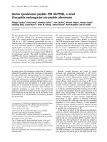
Figure 1 presents a comparative immunohistochemical and
immunofluorescence analysis of the human monoclonal antibodies L19, G11, F16 and F8. In total, pathology specimens
of seven patients were analyzed, four of which are shown in
Figure 1. Both F16 and F8 displayed a stronger staining pattern compared with L19 and G11. The F8 antibody sometimes
exhibited a diffuse stromal staining or a vascular staining pattern, but consistently reacted strongly with both human and
murine specimens of arthritis and was thus selected for pharmacodelivery applications. Furthermore, F8 and F16 exhibited
a prominent perivascular staining pattern in tissue specimens
from patients suffering from psoriatic arthritis and osteoarthritis. In tumor-bearing mice, the in vivo targeting potential of F8
and L19 was comparable when assessed by quantitative biodistribution studies [38].
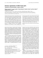
Cloning and in vitro characterization of F8-IL10
The immunocytokine F8-IL10 was cloned in a mammalian
expression vector by sequentially fusing the F8 in scFv format
[19,38] in frame with the human IL10 gene, using flexible aminoacid linkers (Figure 2a). The resulting plasmid pKS1 was linearized and used to stably transfect CHO-S cells. A short five
amino acid linker was used to bridge VH and VL domains
within the scFv antibody fragment moiety, thus driving the formation of a stable non-covalent homodimer (Figures 2b and
2c) [20]. F8-IL10 could be purified to homogeneity on protein
A (Figures 2b and 2c), retained full immunoreactivity when
tested by affinity chromatography on an EDA-sepharose resin
(data not shown) and displayed a biological activity comparable with that of recombinant human IL10 used in equimolar
amounts in a MC/9 cell proliferation assay (Figure 2d) [42]. In
a crossreactivity study on tissue microarray none of the healthy
tissue sections showed any staining with F8-IL10, except for
ovary (1/3), placenta (3/3) and uterus (2/3) [see Additional
data file 1]. This finding is in excellent agreement with the
known expression of oncofetal antigens in organs of the
female reproductive system [48].
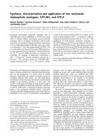
F8-IL10 selectively targets arthritic lesions and tumors in
mice
The in vivo targeting properties of the F8 antibody and of F8IL10 were tested in CIA mice, using both fluorescently labeled
and radioiodinated protein preparations. Figure 3a shows
near-infrared fluorescence images [44,49] of arthritic mice 24
hours after intravenous injection of 100 μg SIP(F8) [38,50]
labeled with Alexa750 dye. A preferential accumulation of the
F8 antibody could be detected in the inflamed extremities. A
Page 5 of 15
(page number not for citation purposes)
Arthritis Research & Therapy
Vol 11 No 5
Schwager et al.
Figure 1
Immunohistochemical analysis of rheumatoid arthritis specimens, psoriatic arthritis specimens and osteoarthritis specimens Immunohistochemistry
specimens.
with the small immunoproteins L19, G11, F16, and F8 was performed in different pathology specimens obtained from biopsies of patients with rheumatoid arthritis, psoriatic arthritis or osteoarthritis. In total, pathology specimens of seven patients were analyzed, four of them are shown above. Furthermore, immunofluorescence double staining with L19, G11, F16 and F8 (red) and von Willebrand factor (green) was performed on rheumatoid
synovial tissue specimens of one patient (rheumatoid arthritis (1)). Overall F8 exhibited the strongest staining of all tested antibodies. It showed a diffuse stromal staining in certain areas and a vascular staining pattern in others. For negative controls, the primary antibody was omitted. Scale bars =
100 μm. neg ctrl = negative control.
more detailed targeting analysis was obtained using 125Ilabeled preparations of SIP(F8) and of F8-IL10. Twenty-four
hours after intravenous or subcutaneous administration,
arthritic limbs were imaged on a PhosphorImager, revealing a
preferential protein accumulation at arthritic fingers and paws
compared with healthy control paws (Figures 3b and 3c). The
Page 6 of 15
(page number not for citation purposes)
ranges of lesion to nonaffected paw ratios measured by phosphorimaging were 7.4 to 13.9 for SIP(F8) intravenous and 5.0
to 6.8 for F8-IL10 subcutaneous. The administration of comparable amounts of antibodies of irrelevant specificity in the
mouse in recombinant SIP format did not exhibit any preferential uptake at sites of inflammation [51].
Available online />
Figure 2
Cloning, expression and purification of F8-IL10. (a) Schematic representation of the cloning strategy of the F8-IL10 fusion protein. (b) SDS-PAGE
F8-IL10
analysis of purified fusion proteins: lane 1, molecular-weight marker; lanes 2 and 3, F8-IL10 under nonreducing and reducing conditions, respectively. (c) Gel-filtration analysis of affinity-purified F8-IL10. The peak eluting at a retention volume of 12 ml corresponds to the noncovalent
homodimeric form of F8-IL10. (d) MC/9 cell proliferation assay. F8-IL10 displayed biological activity comparable with the one of recombinant human
IL10 used as a standard in the assay.
Page 7 of 15
(page number not for citation purposes)
Arthritis Research & Therapy
Vol 11 No 5
Schwager et al.
Figure 3
In vivo targeting of the small immunoprotein F8 and the fusion protein F8-IL10 in arthritic mice. (a) Near infrared fluorescence imaging. Arthritic mice
the small immunoprotein F8 and the fusion protein F8-IL10 in arthritic mice
(n = 3) were injected with small immunoprotein (SIP) (F8)-Alexa750. Near infrared fluorescence imaging analysis was performed 24 hours after
injection. Arrows indicate grade 2 swelling in the front paws of the mice. (b to c) Phosphorimaging. Arthritic mice (n = 2) were injected (b) intravenously with 125I-labelled SIP(F8) or (c) subcutaneously with 125I-labelled F8-IL10. Uptake of radio-iodinated antibodies was analyzed by phosphorimaging 24 hours after injection. The ranges of lesion to nonaffected paw ratios measured by phosphorimaging were 7.4 to 13.9 for SIP(F8)
intravenously and 5.0 to 6.8 for F8-IL10 subcutaneously.
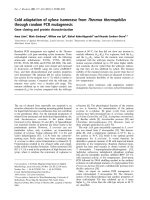
The subcutaneous administration of therapeutic proteins in
patients with arthritis is often preferable compared with the
intravenous administration route, which is typically performed
at the hospital. In order to investigate whether a selective in
vivo targeting of lesions could be obtained using F8-IL10 both
with subcutaneous and intravenous administrations, we performed a comparative biodistribution study in tumor-bearing
mice. We chose a cancer model rather than an arthritis model
for this analysis, because tumor-bearing mice provide a quantitative biodistribution analysis of therapeutic proteins. Figure
4a illustrates biodistribution results (expressed as a percentage of injected dose per gram of tissue) for a radioiodinated
preparation of F8-IL10, administered intravenously or subcutaneously. For both administration routes, a preferential tumor
uptake could be observed, with excellent tumor:organ ratios at
24 and 48 hours following injection. An antibody-IL10 fusion
protein of irrelevant specificity in the mouse [1,50,52] exhibited a reduced tumor uptake in the same animal model (Figure
4b). In order to quantitatively assess the residence time of F8IL10 on neovascular lesions following subcutaneous administration, a biodistribution study was performed sacrificing
tumor-bearing mice at 24, 48, 72 and 96 hours and correcting
for the tumor volume increase during the study period. Figure
4c shows that the immunocytokine efficiently and stably localized at the tumor site, while being cleared from all normal
organs. No statistically significant difference could be
observed in terms of tumor uptake between the 48 and 96
hour time points.
Page 8 of 15
(page number not for citation purposes)
Inhibition of arthritis progression in the collageninduced model of arthritis
The CIA model was used to assess the therapeutic potential
of F8-IL10 when used alone or in combination with methotrexate. Mice were allowed to reach an arthritic score of 1 to 3,
before receiving three injections (days 1, 4 and 7) of F8-IL10
(200 μg) and/or of methotrexate (100 μg). The F8-IL10 dose
for the mouse was calculated from the recommended equivalent dose of 20 μg/kg of recombinant human IL10 used in clinical trials using a body surface correction algorithm [53] and a
correction factor for the activity of human IL10 in mice [29].
Figure 5a shows that mice treated with methotrexate did not
exhibit any detectable reduction of arthritis, in line with previously published results where comparable doses of methotrexate in the same mouse model had no significant effect on
the onset of CIA [54]. Disease progression was substantially
inhibited for F8-IL10 with intravenous administration and with
subcutaneous administration. Both subcutaneous injections of
F8-IL10 and the combination treatment of methotrexate plus
intravenous F8-IL10 allowed the maintainence of an arthritic
score below 3 until the mice were sacrificed (18 days after the
beginning of pharmacological treatment). Similar to what has
previously been reported [18], the therapeutic performance of
an antibody-IL10 fusion protein of irrelevant specificity in the
mouse exhibited a worse therapeutic benefit, confirming the
contribution of selective targeting to therapeutic outcome (Figure 5b). We were not allowed by the local authorities (Veterinäramt des Kantons Zürich) to extend the duration of the
observation period for the mice in order to keep animal discomfort within an acceptable limit, but it would have obviously
Available online />
Figure 4
Biodistribution study in F9 tumor-bearing mice. In all biodistribution experiments four mice were analyzed per group. Radioactivity content of tumor or
F9 tumor-bearing mice
organs is expressed as percentage of the injected dose per gram of tissue (%ID/g) ± standard error. (a) Comparison of intravenous and subcutaneous injection. Tumor bearing mice were injected intravenously or subcutaneously with 125I-labelled F8-IL10 and sacrificed 24 or 48 hours after injection. (b) Comparison of targeted and untargeted IL10. Mice were injected intravenously with 125I-labelled F8-IL10 or 125I-labelled HyHel10-IL10
(HyHel10 is an antibody specific to hen egg lysozyme and is not recognizing any murine antigen). They were sacrificed 24 hours after injection. (c)
Residence time of F8-IL10 following subcutaneous administration. Mice were injected with 125I-labelled F8-IL10 and sacrificed 24, 48, 72, or 96
hours after injection.
Page 9 of 15
(page number not for citation purposes)
Arthritis Research & Therapy
Vol 11 No 5
Schwager et al.
Figure 5
Therapy studies of F8-IL10 in the CIA mouse model. (a) Combination with methotrexate. Arthritic mice were given injections with saline (black
model
squares), methotrexate 100 μg intraperitoneally (open circles), F8-IL10 200 μg subcutaneously (black triangles), F8-IL10 200 μg intravenously
(black circles), or a combination of F8-IL10 200 μg intravenously and methotrexate (MTX) 100 μg intraperitoneally (crosses). Injections were started
at day 1 after arthritis onset and then repeated every third day for three injections per animal, as indicated by the arrows. The arthritic score was evaluated daily and expressed as the mean ± standard error of the mean (SEM) of eight mice per group. * 1 P < 0.05 versus saline; * 2 P < 0.05 versus
F8-IL10 intravenously. (b) Comparison of targeted versus systemic application of IL10. Arthritic mice were injected subcutanously with saline (black
squares), HyHel10-IL10 200 μg (open circles), F8-TNFRII (crosses), or F8-IL10 200 μg (black circles) every third day for three injections, as indicated by arrows. Arthritic score is expressed as the mean ± SEM of six to seven mice per group. * P < 0.05 versus saline. (c) Ex vivo immunohistochemical detection of F8-IL10 and HyHel10-IL10 in arthritis paws. Analysis of the arthritis paws at the end of therapy (day 12 for F8-IL10 and day 10
for HyHel10-IL10) showed that F8-IL10 is still detectable by immunohistochemistry using an anti-human-IL10-antibody. (d) Analysis of plasma
cytokines levels at the end of therapy. F8-IL10-treated mice showed significantly decreased IL6 levels compared with the saline group. Furthermore,
IL1b serum levels of F8-IL10-treated mice were below the lower limit of detection. * P < 0.05 versus saline. (e) Anti type-II collagen antibodies. Titers
of bovine type II collagen-specific total IgG, IgG1 and IgG2a antibodies were determined by ELISA. A clear reduction of total IgG and IgG2a, but not
IgG1, antibody levels was observed in F8-IL10-treated mice. * P < 0.05 versus saline.
Page 10 of 15
(page number not for citation purposes)
Available online />
Figure 6
Immunofluorescence analysis of infiltrating cells. At termination of the therapy experiment a comparative immunofluorescence analysis of infiltrating
cells
cells from mice treated with saline or F8-IL10 was performed. (a) Representative immunofluorescence images of paw sections. Scale bars = 100
μm. (b) Sections were evaluated for area percentage positive staining and a significant decrease of infiltrating leukocytes was observed. * P < 0.05
versus saline.
been of scientific interest to monitor disease stabilization over
a longer period of time.
Paws and blood of mice were analyzed at the end of the therapy and we could demonstrate by immunohistochemistry that
F8-IL10 is still detectable in arthritic paws (Figure 5c). Analysis of plasma cytokines of sacrificed mice showed significantly
(P = 0.004) decreased IL6 levels for F8-IL10-treated mice
(Figure 5d). Furthermore, saline-treated mice showed elevated
IL1b levels compared with healthy control and F8-IL10-treated
mice. In our mouse model of CIA, the therapeutic activity of F8IL10 was found to be comparable with the one of a recombinant biopharmaceutical based on the extracellular part of
murine TNF receptor 2, administered with the same schedule
(Figure 5b).
Figure 6 shows a comparative immunofluorescence analysis
of infiltrating cells from mice treated with saline or F8-IL10.
Staining with an anti-CD45 antibody revealed that F8-IL10treated mice presented a significantly (P = 0.03) lower level of
infiltrating leukocytes in the paw compared with the saline
treatment group. In accordance with this finding, staining with
an anti-asialo-GM1 antibody, which preferentially stains natural killer cells, with the macrophage-specific antibody F4/80
and with CD4/CD8 antibodies, showed a decreased infiltration of these cells in paws of F8-IL10-treated mice.
Anti type-II collagen antibodies
Humoral immunity was followed by measurement of serum levels of anti-collagen II immunoglobulin (Ig) isotypes. Serum
samples were obtained from both control and F8-IL10-treated
animals at the termination of the experiment and total IgG antibody levels, as well as IgG1 and IgG2a isotype levels were
Page 11 of 15
(page number not for citation purposes)
Arthritis Research & Therapy
Vol 11 No 5
Schwager et al.
determined by ELISA (Figure 5e). Total IgG levels were significantly lower in F8-IL10-treated animals than in controls (P <
0.05). When analyzing specific isotypes, no significant differences were seen in the anti-collagen II IgG1 levels between
the two groups. However, the anti-collagen II IgG2a titers
were significantly lower (P < 0.05) in sera from F8-IL10treated mice, as seen for other anti-arthritic therapeutic interventions in the CIA model [55].
Safety pharmacology profile of F8-IL10
In preparation for a dose-finding, pharmacokinetic phase I
study of F8-IL10 in combination with methotrexate in patients
with active rheumatoid arthritis we performed a toxicity assessment of F8-IL10 in combination with methotrexate in cynomolgus monkeys. In this study, three groups of monkeys (each
group consisting of three female and three male animals)
received administrations of either F8-IL10 alone, F8-IL10 plus
methotrexate or saline. During the study F8-IL10 was injected
subcutaneously three times a week for eight weeks at a dosage of 180 μg/kg (60 μg/kg IL10 equivalents), which reflects
10 times the initial human dose intended for administration
during the phase I clinical study. Methotrexate was given on a
weekly basis at the standard dosage of 0.65 mg/kg.
There were no relevant findings in body weight evolution, food
consumption, quantitative electrocardiography parameters or
systolic and diastolic blood pressure values. No relevant ophthalmological findings were noted in any groups. A ventricular
premature complex was recorded in one female treated with
F8-IL10 alone in week 4, after treatment.
During the course of the study (week 4), a regenerative anemia
was observed, however a complete recovery was noted in
week 7. No toxicologically relevant findings were observed in
the blood biochemical parameters at the end of week 4 and at
the end of the treatment period in any groups.
Pharmacokinetic data were obtained during the toxicology
study. Blood samples were collected at pre-dose, 5 and 30
minutes, and 3 and 24 hours after the injection. The serum
concentration of F8-IL10 was measured using a validated
colorimetric ELISA. Many of the samples analyzed were found
to be below the level of quantification (< 0.25 ng/ml). However, for those samples in which a positive result was
obtained, maximum serum levels were generally observed
three hours after the subcutaneous injection of F8-IL10 with
serum levels of about 20 ng/ml. After 24 hours no more detection of F8-IL10 in serum was possible.
In conclusion, subcutaneous administration of F8-IL10 alone
or in combination with methotrexate was generally well tolerated.
The acute toxicity of F8-IL10 was investigated in mice after
intravenous administration of a single dose level of 20 mg/kg,
Page 12 of 15
(page number not for citation purposes)
corresponding to 300 times the human starting dose proposed for clinical trials [53], followed by a 14-day observation
period. Body weights were recorded weekly and necropsy
was performed on all animals. No mortality occurred and no
clinical signs were noted in both male and female animals.
Changes in body weight observed at the end of the study were
within the expected range for this strain and age of animals. No
changes of toxicological significance were observed in the
weight of organs. No abnormalities were detected in all
treated animals at the necropsy examination and no abnormalities were observed at the injection site.
These results indicate that F8-IL10 had no toxic effect on mice
following a single intravenous administration at a dose level of
20 mg/kg body weight. The product was well locally tolerated
when injected into the tail vein at the dose level tested.
Discussion
In this article, we have compared four human monoclonal antibodies specific to alternatively-spliced components of the
extracellular matrix and have identified F8 as a suitable candidate for pharmacodelivery applications in rheumatoid arthritis.
F8 recognizes the extra-domain EDA of fibronectin [38] and
consistently yielded stronger staining of arthritic specimens
compared with the L19, G11 and F16 antibodies. In analogy
to our previous work in this area [18], we fused F8 to human
IL10, generating the immunocytokine F8-IL10 (DEKAVIL),
which was shown to preferentially localize at sites of arthritis
in the collagen-induced murine model of the disease. F8-IL10
was able to stabilize clinical features of arthritis in this animal
model and was found to be well tolerated in monkeys at human
equivalent doses of 20 μg/kg [53].
Preclinical studies were facilitated by the fact that F8 binds
with comparable affinity to EDA of murine, monkey and human
origin [38].
The rationale behind the development of F8-IL10 as a novel
biopharmaceutical relies on the promising, yet not sufficiently
satisfactory, preclinical and clinical data reported for recombinant human IL10 (Tenovil TM). In controlled clinical trials in
patients with rheumatoid arthritis, Tenovil exhibited ACR20
values substantially higher than the ones in control groups and
comparable with the ACR20 values reported for TNF blockers.
However, the ACR50 values observed with Tenovil, while significantly better compared with the ones observed in patients
treated only with methotrexate, were not as good as those
reported for Humira (Adalimumab), Remicade (Infliximab) and
Enbrel (Etanercept) [32-34].
In spite of these observations, we and others have extensively
demonstrated in animal models that the antibody-based delivery of cytokines to sites of disease can substantially improve
the therapeutic index of these biopharmaceuticals. Indeed, our
group has developed fully human fusion proteins based on the
Available online />
pro-inflammatory cytokines IL2 and TNF (L19-IL2; L19-TNF;
F16-IL2) [3,8,10,11] which are currently being investigated in
phase I and in phase II clinical trials in patients with cancer. To
our knowledge, F8-IL10 will be the first anti-inflammatory
immunocytokine to be tested in the clinical setting and it will
be interesting to learn whether the improved performance and
selectivity documented in the mouse model of arthritis holds
true for patients with rheumatoid disease. Encouraged by the
excellent tolerability profile observed in cynomolgus monkeys,
we have submitted a request for clinical trials in Italy.
When developing F8-IL10 for industrial pharmaceutical programs, care was devoted to identifying a suitable formulation
which could be compatible with subcutaneous administration.
Indeed, we were not aware at the beginning of the study of any
quantitative biodistribution analysis performed with diseasetargeting antibody fragments following subcutaneous administration. Using radioiodinated protein preparations, we studied
the biodistribution properties of F8-IL10 both in mouse models
of arthritis and in tumor-bearing mice, where targeting performance can be expressed as percent injected dose per
gram. The conventional intravenous administration route
yielded tumor targeting results comparable with the ones
obtained following a subcutaneous administration, thus providing a robust rationale for the development of clinical trials
featuring subcutaneous injections. Experience gained with
TNF blocking antibodies suggests that subcutaneous administration may be better accepted by patients and may lead to
a better compliance, reducing the need to visit hospital sites
for each administration.
Conclusions
The data presented in this article provide a strong rationale for
the clinical investigation of F8-IL10 as a novel biopharmaceutical for the therapy of patients with rheumatoid arthritis who
have failed at least two lines of biological therapy. Clinical
studies will reveal whether the promising preclinical results
can be translated to the clinical setting and, potentially,
whether F8-IL10 could find a broader clinical applicability as a
targeted anti-inflammatory agent for diseases which overexpress the EDA domain of fibronectin.
experiments, were involved in data interpretation and prepared
the manuscript. All authors read and approved the final manuscript.
Additional files
The following Additional files are available online:
Additional file 1
A Figure showing crossreactivity of F8-IL10 study on
tissue microarray sections (Biochain, Hayward, USA).
Sections were blocked with FCS and then incubated
with 5 μg/ml of purified FITC-labeled F8-IL10 for one
hour. For amplification of the signal bound antibody was
detected using rabbit anti-FITC antibody and
subsequent AlexaFluor594 goat anti-rabbit IgG. Slides
were mounted with glycergel and analyzed with an
AxioScop 2MOT+ fluorescence microscope. None of
the healthy tissue sections showed any staining with F8IL10, except for ovary (1/3), placenta (3/3) and uterus (2/
3).
See />supplementary/ar2814-S1.PDF
Acknowledgements
Financial support from the ETH Zürich, the Swiss National Science
Foundation (grant # 3100A0-105919/1), the Swiss Cancer League
(Robert-Wenner-Award), the SWISSBRIDGE-Stammbach Foundation
and European Union Projects IMMUNO-PDT (grant # LSHC-CT-2006037489), DIANA (grant # LSHB-CT-2006-037681) and ADAMANT
(HEALT-F2-2008-201342) is gratefully acknowledged.
References
1.
2.
3.
Competing interests
DN is a cofounder and shareholder of Philogen SpA (Siena,
Italy), the company that owns DEKAVIL.
4.
Authors' contributions
5.
KS participated in designing the study, cloned, produced and
characterized the F8-IL10 fusion protein, performed the animal
experiments and assisted in preparing the manuscript. MK and
ET participated in characterizing the fusion proteins and
assisted in the animal experiments. FB set up the animal model
in our laboratory and contributed essentially to the animal
experiments. RM and EP provided the human arthritic specimens and gave helpful advice. DN and ET supervised the
6.
7.
8.
Halin C, Rondini S, Nilsson F, Berndt A, Kosmehl H, Zardi L, Neri
D: Enhancement of the antitumor activity of interleukin-12 by
targeted delivery to neovasculature. Nat Biotechnol 2002,
20:264-269.
Gafner V, Trachsel E, Neri D: An engineered antibody-interleukin-12 fusion protein with enhanced tumor vascular targeting properties. Int J Cancer 2006, 119:2205-2212.
Schliemann C, Palumbo A, Zuberbuhler K, Villa A, Kaspar M,
Trachsel E, Klapper W, Menssen HD, Neri D: Complete eradication of human B-cell lymphoma xenografts using rituximab in
combination with the immunocytokine L19-IL2. Blood 2009,
113:2275-2283.
Gillies SD, Lan Y, Wesolowski JS, Qian X, Reisfeld RA, Holden S,
Super M, Lo KM: Antibody-IL-12 fusion proteins are effective in
SCID mouse models of prostate and colon carcinoma metastases. J Immunol 1998, 160:6195-6203.
Huang TH, Chintalacharuvu KR, Morrison SL: Targeting IFNalpha to B cell lymphoma by a tumor-specific antibody elicits
potent antitumor activities. J Immunol 2007, 179:6881-6888.
Schrama D, Reisfeld RA, Becker JC: Antibody targeted drugs as
cancer therapeutics. Nat Rev Drug Discov 2006, 5:147-159.
King DM, Albertini MR, Schalch H, Hank JA, Gan J, Surfus J, Mahvi
D, Schiller JH, Warner T, Kim K, Eickhoff J, Kendra K, Reisfeld R,
Gillies SD, Sondel P: Phase I clinical trial of the immunocytokine EMD 273063 in melanoma patients. J Clin Oncol 2004,
22:4463-4473.
Carnemolla B, Borsi L, Balza E, Castellani P, Meazza R, Berndt A,
Ferrini S, Kosmehl H, Neri D, Zardi L: Enhancement of the antitumor properties of interleukin-2 by its targeted delivery to the
Page 13 of 15
(page number not for citation purposes)
Arthritis Research & Therapy
9.
10.
11.
12.
13.
14.
15.
16.
17.
18.
19.
20.
21.
22.
23.
24.
25.
Vol 11 No 5
Schwager et al.
tumor blood vessel extracellular matrix.
Blood 2002,
99:1659-1665.
Menrad A, Menssen HD: ED-B fibronectin as a target for antibody-based cancer treatments. Expert Opin Ther Targets 2005,
9:491-500.
Marlind J, Kaspar M, Trachsel E, Sommavilla R, Hindle S, Bacci C,
Giovannoni L, Neri D: Antibody-mediated delivery of interleukin-2 to the stroma of breast cancer strongly enhances the
potency of chemotherapy.
Clin Cancer Res 2008,
14:6515-6524.
Borsi L, Balza E, Carnemolla B, Sassi F, Castellani P, Berndt A,
Kosmehl H, Biro A, Siri A, Orecchia P, Grassi J, Neri D, Zardi L:
Selective targeted delivery of TNFalpha to tumor blood vessels. Blood 2003, 102:4384-4392.
Halin C, Gafner V, Villani ME, Borsi L, Berndt A, Kosmehl H, Zardi
L, Neri D: Synergistic therapeutic effects of a tumor targeting
antibody fragment, fused to interleukin 12 and to tumor necrosis factor alpha. Cancer Res 2003, 63:3202-3210.
Balza E, Mortara L, Sassi F, Monteghirfo S, Carnemolla B, Castellani P, Neri D, Accolla RS, Zardi L, Borsi L: Targeted delivery of
tumor necrosis factor-alpha to tumor vessels induces a therapeutic T cell-mediated immune response that protects the
host against syngeneic tumors of different histologic origin.
Clin Cancer Res 2006, 12:2575-2582.
Birchler M, Viti F, Zardi L, Spiess B, Neri D: Selective targeting
and photocoagulation of ocular angiogenesis mediated by a
phage-derived human antibody fragment. Nat Biotechnol
1999, 17:984-988.
Trachsel E, Kaspar M, Bootz F, Detmar M, Neri D: A human mAb
specific to oncofetal fibronectin selectively targets chronic
skin inflammation in vivo.
J Invest Dermatol 2007,
127:881-886.
Zardi L, Carnemolla B, Siri A, Petersen TE, Paolella G, Sebastio G,
Baralle FE: Transformed human cells produce a new fibronectin isoform by preferential alternative splicing of a previously
unobserved exon. Embo J 1987, 6:2337-2342.
Pini A, Viti F, Santucci A, Carnemolla B, Zardi L, Neri P, Neri D:
Design and use of a phage display library. Human antibodies
with subnanomolar affinity against a marker of angiogenesis
eluted from a two-dimensional gel. J Biol Chem 1998,
273:21769-21776.
Trachsel E, Bootz F, Silacci M, Kaspar M, Kosmehl H, Neri D: Antibody-mediated delivery of IL-10 inhibits the progression of
established collagen-induced arthritis. Arthritis Res Ther 2007,
9:R9.
Huston JS, Levinson D, Mudgett-Hunter M, Tai MS, Novotny J,
Margolies MN, Ridge RJ, Bruccoleri RE, Haber E, Crea R, et al.:
Protein engineering of antibody binding sites: recovery of specific activity in an anti-digoxin single-chain Fv analogue produced in Escherichia coli. Proc Natl Acad Sci USA 1988,
85:5879-5883.
Holliger P, Prospero T, Winter G: "Diabodies": small bivalent
and bispecific antibody fragments. Proc Natl Acad Sci USA
1993, 90:6444-6448.
Santimaria M, Moscatelli G, Viale GL, Giovannoni L, Neri G, Viti F,
Leprini A, Borsi L, Castellani P, Zardi L, Neri D, Riva P: Immunoscintigraphic detection of the ED-B domain of fibronectin, a
marker of angiogenesis, in patients with cancer. Clin Cancer
Res 2003, 9:571-579.
Borsi L, Balza E, Bestagno M, Castellani P, Carnemolla B, Biro A,
Leprini A, Sepulveda J, Burrone O, Neri D, Zardi L: Selective targeting of tumoral vasculature: comparison of different formats
of an antibody (L19) to the ED-B domain of fibronectin. Int J
Cancer 2002, 102:75-85.
Berndorff D, Borkowski S, Sieger S, Rother A, Friebe M, Viti F,
Hilger CS, Cyr JE, Dinkelborg LM: Radioimmunotherapy of solid
tumors by targeting extra domain B fibronectin: identification
of the best-suited radioimmunoconjugate. Clin Cancer Res
2005, 11:7053s-7063s.
Tijink BM, Neri D, Leemans CR, Budde M, Dinkelborg LM, Stigtervan Walsum M, Zardi L, van Dongen GA: Radioimmunotherapy
of head and neck cancer xenografts using 131I-labeled antibody L19-SIP for selective targeting of tumor vasculature. J
Nucl Med 2006, 47:1127-1135.
Sauer S, Erba PA, Petrini M, Menrad A, Giovannoni L, Grana C,
Hirsch B, Zardi L, Paganelli G, Mariani G, Neri D, Durkop H,
Menssen HD: Expression of the oncofetal ED-B containing
Page 14 of 15
(page number not for citation purposes)
26.
27.
28.
29.
30.
31.
32.
33.
34.
35.
36.
37.
38.
39.
40.
41.
42.
43.
44.
45.
fibronectin isoform in hematologic tumors enables ED-B targeted 131I-L19SIP radioimmunotherapy in Hodgkin lymphoma patients. Blood 2009, 113:2265-2274.
Spaeth N, Wyss MT, Pahnke J, Biollaz G, Trachsel E, Drandarov K,
Treyer V, Weber B, Neri D, Buck A: Radioimmunotherapy targeting the extra domain B of fibronectin in C6 rat gliomas: a preliminary study about the therapeutic efficacy of iodine-131labeled SIP(L19). Nucl Med Biol 2006, 33:661-666.
Neri D, Bicknell R: Tumour vascular targeting. Nat Rev Cancer
2005, 5:436-446.
Schliemann C, Neri D: Antibody-based targeting of the tumor
vasculature. Biochim Biophys Acta 2007, 1776:175-192.
Rosenblum IY, Johnson RC, Schmahai TJ: Preclinical safety evaluation of recombinant human interleukin-10. Regul Toxicol
Pharmacol 2002, 35:56-71.
Huhn RD, Radwanski E, O'Connell SM, Sturgill MG, Clarke L,
Cody RP, Affrime MB, Cutler DL: Pharmacokinetics and immunomodulatory properties of intravenously administered
recombinant human interleukin-10 in healthy volunteers.
Blood 1996, 87:699-705.
Walmsley M, Katsikis PD, Abney E, Parry S, Williams RO, Maini
RN, Feldmann M: Interleukin-10 inhibition of the progression of
established collagen-induced arthritis. Arthritis Rheum 1996,
39:495-503.
Maini R, Paulus H, Breedveld F, Moreland L, St Clair EW, Russell
A, Charles P, Davies D, Grint P, Wherry J, Feldmann M: rHuIL-10
in subjects with active rheumatoid arthritis (RA): A phase I and
cytokine response study. Arthritis Rheum 1997, 40(suppl):224.
Weinblatt M, St Clair E, Breedveld F, Moreland L, Keystone E, Lee
S, Robison L, Furst D, Bulpitt K, Veys E, Haverty T, Grint P, Wherry
J: rHUIL-10 (Tenovil) plus methotrexate (MTX) in active rheumatoid arthritis (RA): a phase I/II study. American College of
Rheumatology 63rd Annual Scienific Meeting: 12 to 17 November, 1999; Boston, MA 1999:Abstract 598.
Rau R: Adalimumab (a fully human anti-tumour necrosis factor
alpha monoclonal antibody) in the treatment of active rheumatoid arthritis: the initial results of five trials. Ann Rheum Dis
2002, 61(Suppl 2):ii70-73.
Brack SS, Silacci M, Birchler M, Neri D: Tumor-targeting properties of novel antibodies specific to the large isoform of
tenascin-C. Clin Cancer Res 2006, 12:3200-3208.
Carnemolla B, Castellani P, Ponassi M, Borsi L, Urbini S, Nicolo G,
Dorcaratto A, Viale G, Winter G, Neri D, Zardi L: Identification of
a glioblastoma-associated tenascin-C isoform by a high affinity recombinant antibody. Am J Pathol 1999, 154:1345-1352.
Silacci M, Brack SS, Spath N, Buck A, Hillinger S, Arni S, Weder
W, Zardi L, Neri D: Human monoclonal antibodies to domain C
of tenascin-C selectively target solid tumors in vivo. Protein
Eng Des Sel 2006, 19:471-478.
Villa A, Trachsel E, Kaspar M, Schliemann C, Sommavilla R, Rybak
JN, Rosli C, Borsi L, Neri D: A high-affinity human monoclonal
antibody specific to the alternatively spliced EDA domain of
fibronectin efficiently targets tumor neo-vasculature in vivo.
Int J Cancer 2008, 122:2405-2413.
Sasso EH, Silverman GJ, Mannik M: Human IgA and IgG F(ab')2
that bind to staphylococcal protein A belong to the VHIII subgroup. J Immunol 1991, 147:1877-1883.
Silacci M, Brack S, Schirru G, Marlind J, Ettorre A, Merlo A, Viti F,
Neri D: Design, construction, and characterization of a large
synthetic human antibody phage display library. Proteomics
2005, 5:2340-2350.
Hoogenboom HR, Winter G: By-passing immunisation. Human
antibodies from synthetic repertoires of germline VH gene
segments rearranged in vitro. J Mol Biol 1992, 227:381-388.
Thompson-Snipes L, Dhar V, Bond MW, Mosmann TR, Moore KW,
Rennick DM: Interleukin 10: a novel stimulatory factor for mast
cells and their progenitors. J Exp Med 1991, 173:507-510.
Williams RO, Marinova-Mutafchieva L, Feldmann M, Maini RN:
Evaluation of TNF-alpha and IL-1 blockade in collageninduced arthritis and comparison with combined anti-TNFalpha/anti-CD4 therapy. J Immunol 2000, 165:7240-7245.
Birchler M, Neri G, Tarli L, Halin C, Viti F, Neri D: Infrared photodetection for the in vivo localisation of phage-derived antibodies directed against angiogenic markers. J Immunol Methods
1999, 231:239-248.
ImageJ [ />
Available online />
46. Perez N, Plence P, Millet V, Greuet D, Minot C, Noel D, Danos O,
Jorgensen C, Apparailly F: Tetracycline transcriptional silencer
tightly controls transgene expression after in vivo intramuscular electrotransfer: application to interleukin 10 therapy in
experimental arthritis. Hum Gene Ther 2002, 13:2161-2172.
47. Vignali DA: Multiplexed particle-based flow cytometric assays.
J Immunol Methods 2000, 243:243-255.
48. Kaspar M, Zardi L, Neri D: Fibronectin as target for tumor therapy. Int J Cancer 2006, 118:1331-1339.
49. Neri D, Carnemolla B, Nissim A, Leprini A, Querze G, Balza E, Pini
A, Tarli L, Halin C, Neri P, Zardi L, Winter G: Targeting by affinitymatured recombinant antibody fragments of an angiogenesis
associated fibronectin isoform.
Nat Biotechnol 1997,
15:1271-1275.
50. Neri D, Momo M, Prospero T, Winter G: High-affinity antigen
binding by chelating recombinant antibodies (CRAbs). J Mol
Biol 1995, 246:367-373.
51. Trachsel E, Neri D: Antibodies for angiogenesis inhibition, vascular targeting and endothelial cell transcytosis. Adv Drug
Deliv Rev 2006, 58:735-754.
52. Smith-Gill SJ, Mainhart CR, Lavoie TB, Rudikoff S, Potter M: VLVH expression by monoclonal antibodies recognizing avian
lysozyme. J Immunol 1984, 132:963-967.
53. Reagan-Shaw S, Nihal M, Ahmad N: Dose translation from animal to human studies revisited. FASEB J 2008, 22:659-661.
54. Wunder A, Muller-Ladner U, Stelzer EH, Funk J, Neumann E,
Stehle G, Pap T, Sinn H, Gay S, Fiehn C: Albumin-based drug
delivery as novel therapeutic approach for rheumatoid arthritis. J Immunol 2003, 170:4793-4801.
55. Khoury M, Escriou V, Courties G, Galy A, Yao R, Largeau C, Scherman D, Jorgensen C, Apparailly F: Efficient suppression of
murine arthritis by combined anticytokine small interfering
RNA lipoplexes. Arthritis Rheum 2008, 58:2356-2367.
Page 15 of 15
(page number not for citation purposes)