Critical care medicine - part 2 ppsx
Bạn đang xem bản rút gọn của tài liệu. Xem và tải ngay bản đầy đủ của tài liệu tại đây (91.19 KB, 15 trang )
18 Discharge Note
Date and time:
Procedure:
Indications:
Patient Consent: Document that the indications, risks and alternatives to
the procedure were explained to the patient. Note that the patient was
given the opportunity to ask questions and that the patient consented to
the procedure in writing.
Lab tests: Relevant labs, such as the INR and CBC
Anesthesia: Local with 2% lidocaine
Description of Procedure: Briefly describe the procedure, including
sterile prep, anesthesia method, patient position, devices used, anatomic
location of procedure, and outcome.
Complications and Estimated Blood Loss (EBL):
Disposition: Describe how the patient tolerated the procedure.
Specimens: Describe any specimens obtained and labs tests which were
ordered.
Name of Physician: Name of person performing procedure and supervis-
ing staff.
Discharge Note
The discharge note should be written in the patient’s chart prior to discharge.
Discharge Note
Date/time:
Diagnoses:
Treatment: Briefly describe therapy provided during hospitalization,
including surgical procedures and antibiotic therapy.
Studies Performed: Electrocardiograms, CT scans.
Discharge medications:
Follow-up Arrangements:
Fluids and Electrolytes
Maintenance Fluids Guidelines:
70 kg Adult: D5 1/4 NS with 20 mEq KCI/Liter at 125 mL/hr.
Specific Replacement Fluids for Specific Losses:
Gastric (nasogastric tube, emesis): D5 ½ NS with 20 mEq/L KCL.
Diarrhea: D5LR with 15 mEq/liter KCI. Provide 1 liter of replacement for
each 1 kg or 2.2 lb of body weight lost.
Bile: D5LR with 25 mEq/liter (½ amp) of sodium bicarbonate.
Pancreatic: D5LR with 50 mEq/liter (1 amp) sodium bicarbonate.
Blood Component Therapy 19
Blood Component Therapy
A. Packed red blood cells (PRBCs). Each unit provides 250-400 cc of
volume, and each unit should raise hemoglobin by 1 gm/dL and
hematocrit by 3%. PRBCs are usually requested in two unit increments.
B. Type and screen. Blood is tested for A, B, Rh antigens, and antibodies
to donor erythrocytes. If blood products are required, the blood can be
rapidly prepared by the blood bank. O negative blood is used when type
and screen information is not available, but the need for transfusion is
emergent.
C. Type and cross match sets aside specific units of packed donor red
blood cells. If blood is needed on an urgent basis, type and cross should
be requested.
D. Platelets. Indicated for bleeding if there is thrombocytopenia or platelet
dysfunction in the setting of uncontrolled bleeding. Each unit of platelet
concentrate should raise the platelet count by 5,000-10,000. Platelets are
usually transfused 6-10 units at a time, which should increase the platelet
count by 40-60,000. Thrombocytopenia is defined as a platelet count of
less than 60,000. For surgery, the count should be greater than 50,000.
E. Fresh Frozen Plasma (FFP) is used for active bleeding secondary to
liver disease, warfarin overdose, dilutional coagulopathy secondary to
multiple blood transfusions, disseminated intravascular coagulopathy, and
vitamin K and coagulation factor deficiencies. Administration of FFP
requires ABO typing, but not cross matching.
1. Each unit contains coagulation factors in normal concentration.
2. Two to four units are usually required for therapeutic intervention.
F. Cryoprecipitate
1. Indicated in patients with Hemophilia A, Von Willebrand's disease, and
any state of hypofibrinogenemia requiring replacement (DIC), or
reversal of thrombolytic therapy.
2. Cryoprecipitate contains factor VIII, fibrinogen, and Von Willebrand
factor. The goal of therapy is to maintain the fibrinogen level above
100 mL/dL, which is usually achieved with 10 units given over 3-5
minutes.
Total Parenteral Nutrition
Infuse 40-50 mL/hr of amino acid dextrose solution in the first 24 hr; increase
daily by 40 mL/hr increments until providing 1.3-2 x basal energy requirement
and 1.2-1.7 gm protein/kg/d (see formula, page 142)
Standard Solution per Liter
Amino acid solution (Aminosyn) 7-10%
Dextrose 40-70%
Sodium
Potassium
Chloride
Calcium
Phosphate
Magnesium
Acetate
500 mL
500 mL
35 mEq
36 mEq
35 mEq
4.5 mEq
9 mMol
8.0 mEq
82-104 mEq
20 Enteral Nutrition
Multi-Trace Element Formula 1 mL/d
Regular insulin (if indicated) 10-20 U/L
Multivitamin 12 (2 amp) 10 mL/d
Vitamin K (in solution, SQ, IM) 10 mg/week
Vitamin B 12 1000 mcg/week
Fat Emulsion:
-Intralipid 20% 500 mL/d IVPB infused in parallel with standard solution at 1
mL/min x 15 min; if no adverse reactions, increase to 20-50 mL/hr. Serum
triglyceride level should be checked 6h after end of infusion (maintain
<250 mg/dL).
Cyclic Total Parenteral Nutrition
-12-hour night schedule; taper continuous infusion in morning by reducing rate
to half original rate for 1 hour. Further reduce rate by half for an additional
hour, then discontinue. Restart TPN in evening. Taper at beginning and
end of cycle. Final rate should be 185 mL/hr for 9-10h with 2 hours of
taper at each end, for total of 2000 mL.
Peripheral Parenteral Supplementation
-Amino acid solution (ProCalamine) 3% up to 3 L/d at 125 cc/h OR
-Combine 500 mL amino acid solution 7% or 10% (Aminosyn) and 500 mL
20% dextrose and electrolyte additive. Infuse at up to 100 cc/hr in
parallel with intralipid 10% or 20% at 1 mL/min for 15 min (test dose);
if no adverse reactions, infuse 500 mL/d at 20 mL/hr.
Special Medications
-Famotidine (Pepcid) 20 mg IV q12h or 40 mg/day in TPN OR
-Ranitidine (Zantac) 50 mg IV q6-8h.
-Insulin sliding scale or continuous IV infusion.
Labs
Baseline: Draw labs below. Chest x-ray, plain film for tube placement
Daily Labs: Chem 7, osmolality, CBC, cholesterol, triglyceride (6h after end
of infusion), serum phosphate, magnesium, calcium, urine specific gravity.
Weekly Labs: Protein, iron, TIBC, INR/PTT, 24h urine nitrogen and
creatinine. Pre-albumin, transferrin, albumin, total protein, AST, ALT,
GGT, alkaline phosphatase, LDH, amylase, total bilirubin.
Enteral Nutrition
General Measures: Daily weights, nasoduodenal feeding tube. Head of bed at
30 degrees while enteral feeding and 2 hours after completion. Record bowel
movements.
Continuous Enteral Infusion: Initial enteral solution (Osmolite, Pulmocare,
Jevity) 30 mL/hr. Measure residual volume q1h x 12h, then tid; hold feeding
for 1 h if residual is more than 100 mL of residual. Increase rate by 25-50
mL/hr at 24 hr intervals as tolerated until final rate of 50-100 mL/hr (1 cal/mL)
as tolerated. Three tablespoons of protein powder (Promix) may be added to
each 500 cc of solution. Flush tube with 100 cc water q8h.
Enteral Bolus Feeding: Give 50-100 mL of enteral solution (Osmolite,
Pulmocare, Jevity) q3h initially. Increase amount in 50 mL steps to max of
250-300 mL q3-4h; 30 kcal of nonprotein calories/d and 1.5 gm protein/kg/d.
Before each feeding measure residual volume, and delay feeding by 1 h if
>100 mL. Flush tube with 100 cc of water after each bolus.
Special Medications:
Radiographic Evaluation of Interventions 21
-Metoclopramide (Reglan) 10-20 mg PO, IM, IV, or in J tube q6h.
-Famotidine (Pepcid) 20 mg J-tube q12h OR
-Ranitidine (Zantac) 150 mg in J-tube bid.
Symptomatic Medications:
-Loperamide (Imodium) 24 mg PO or in J-tube q6h, max 16 mg/d prn OR
-Diphenoxylate/atropine (Lomotil) 5-10 mL (2.5 mg/5 mL) PO or in J-tube q4-
6h, max 12 tabs/d OR
-Kaopectate 30 cc PO or in J-tube q6h.
Radiographic Evaluation of Interventions
I. Central intravenous lines
A. Central venous catheters should be located well above the right atrium,
and not in a neck vein. Rule out pneumothorax by checking that the lung
markings extend completely to the rib cages on both sides. Examine for
hydropericardium (“water bottle” sign, mediastinal widening).
B. Pulmonary artery catheter tips should be located centrally and
posteriorly, and not more than 3-5 cm from midline.
II. Endotracheal tubes. Verify that the tube is located 3 cm below the vocal
cords and 2-4cm above the carina; the tip of tube should be at the level of
aortic arch.
III. Tracheostomies. Verify by chest x-ray that the tube is located halfway
between the stoma and the carina; the tube should be parallel to the long
axis of the trachea. The tube should be approximately 2/3 of width of the
trachea; the cuff should not cause bulging of the trachea walls. Check for
subcutaneous air in the neck tissue and for mediastinal widening secondary
to air leakage.
IV. Nasogastric tubes and feeding tubes. Verify that the tube is in the stomach
and not coiled in the esophagus or trachea. The tip of the tube should not be
near the gastroesophageal junction.
V. Chest tubes. A chest tube for pneumothorax drainage should be near the
level of the third intercostal space. If the tube is intended to drain a free-
flowing pleural effusion, it should be located inferior-posteriorly, at or about
the level of the eighth intercostal space. Verify that the side port of the tube
is within the thorax.
VI. Mechanical ventilation. Obtain a chest x-ray to rule out pneumothorax,
subcutaneous emphysema, pneumomediastinum, or subpleural air cysts.
Lung infiltrates or atelectasis may diminish or disappear after initiation of
mechanical ventilation because of increased aeration of the affected lung
lobe.
Arterial Line Placement
Procedure
1. Obtain a 20-gauge 1 ½-2 inch catheter over needle assembly (Angiocath),
arterial line setup (transducer, tubing and pressure bag containing
heparinized saline), arm board, sterile dressing, lidocaine, 3 cc syringe, 25-
gauge needle, and 3-O silk suture.
22 Central Venous Catheterization
2. The radial artery is the most frequently used artery. Use the Allen test to verify
the patency of the radial and ulnar arteries. Place the extremity on an arm
board with a gauze roll behind the wrist to maintain hyperextension.
3. Prep the skin with povidone-iodine and drape; infiltrate 1% lidocaine using a
25-gauge needle. Choose a site where the artery is most superficial and
distal.
4. Palpate the artery with the left hand, and advance the catheter-over-needle
assembly into the artery at a 30-degree angle to the skin. When a flash of
blood is seen, hold the needle in place and advance the catheter into the
artery. Occlude the artery with manual pressure while the pressure tubing is
connected.
5. Advance the guide wire into the artery, and pass the catheter over the guide
wire. Suture the catheter in place with 3-0 silk and apply dressing.
Central Venous Catheterization
I. Indications for central venous catheter cannulation: Monitoring of central
venous pressures in shock or heart failure; management of fluid status;
insertion of a transvenous pacemaker; administration of total parenteral
nutrition; administration of vesicants (chemotherapeutic agents).
II. Location: The internal jugular approach is relatively contraindicated in
patients with a carotid bruit, stenosis, or an aneurysm. The subclavian
approach has an increased risk of pneumothorax in patients with emphysema
or bullae. The external jugular or internal jugular approach is preferable in
patients with coagulopathy or thrombocytopenia because of the ease of
external compression. In patients with unilateral lung pathology or a chest
tube already in place, the catheter should be placed on the side of predomi-
nant pathology or on the side with the chest tube if present.
III. Technique for insertion of external jugular vein catheter
1. The external jugular vein extends from the angle of the mandible to
behind the middle of the clavicle, where it joins with the subclavian vein.
Place the patient in Trendelenburg's position. Cleanse skin with Betadine-
iodine solution, and, using sterile technique, inject 1% lidocaine to
produce a skin weal. Apply digital pressure to the external jugular vein
above the clavicle to distend the vein.
2. With a 16-gauge thin wall needle, advance the needle into the vein. Then
pass a J-guide wire through the needle; the wire should advance without
resistance. Remove the needle, maintaining control over the guide wire
at all times. Nick the skin with a No. 11 scalpel blade.
3. With the guide wire in place, pass the central catheter over the wire and
remove the guide wire after the catheter is in place. Cover the catheter
hub with a finger to prevent air embolization.
4. Attach a syringe to the catheter hub and ensure that there is free back-
flow of dark venous blood. Attach the catheter to an intravenous infusion.
5. Secure the catheter in place with 2-0 silk suture and tape. The catheter
should be replaced weekly or if there is any sign of infection.
6. Obtain a chest x-ray to confirm position and rule out pneumothorax.
IV. Internal jugular vein cannulation. The internal jugular vein is positioned
behind the stemocleidomastoid muscle lateral to the carotid artery. The
catheter should be placed at a location at the upper confluence of the two
bellies of the stemocleidomastoid, at the level of the cricoid cartilage.
Central Venous Catheterization 23
1. Place the patient in Trendelenburg's position and turn the patient's head
to the contralateral side.
2. Choose a location on the right or left. If lung function is symmetrical and no
chest tubes are in place, the right side is preferred because of the direct
path to the superior vena cava. Prepare the skin with Betadine solution
using sterile technique and placea drape. Infiltrate the skin and deeper
tissues with 1% lidocaine.
3. Palpate the carotid artery. Using a 22-gauge scout needle and syringe,
direct the needle lateral to the carotid artery towards the ipsilateral nipple
at a 30-degree angle to the neck. While aspirating, advance the needle
until the vein is located and blood flows back into the syringe.
4. Remove the scout needle and advance a 16-gauge, thin wall catheter-
over-needle with an attached syringe along the same path as the scout
needle. When back flow of blood is noted into the syringe, advance the
catheter into the vein. Remove the needle and confirm back flow of blood
through the catheter and into the syringe. Remove the syringe, and use a
finger to cover the catheter hub to prevent air embolization.
5. With the 16-gauge catheter in position, advance a 0.89 mm x 45 cm spring
guide wire through the catheter. The guidewire should advance easily
without resistance.
6. With the guidewire in position, remove the catheter and use a No. 11
scalpel blade to nick the skin.
7. Place the central vein catheter over the wire, holding the wire secure at all
times. Pass the catheter into the vein, remove the guidewire, and suture
the catheter with 0 silk suture, tape, and connect it to an IV infusion.
8. Obtain a chest x-ray to rule out pneumothorax and confirm position of the
catheter.
V. Subclavian vein cannulation. The subclavian vein is located in the angle
formed by the medial 1/3 of the clavicle and the first rib.
1. Position the patient supine with a rolled towel located between the
patient's scapulae, and turn the patient's head towards the contralateral
side. Prepare the area with Betadine iodine solution, and, using sterile
technique, drape the area and infiltrate 1% lidocaine into the skin and
tissues.
2. Advance the 16-gauge catheter-over-needle, with syringe attached, into
a location inferior to the mid-point of the clavicle, until the clavicle bone
and needle come in contact.
3. Slowly probe down with the needle until the needle slips under the
clavicle, and advance it slowly towards the vein until the catheter needle
enters the vein and a back flow of venous blood enters the syringe.
Remove the syringe, and cover the catheter hub with a finger to prevent
air embolization.
4. With the 16-gauge catheter in position, advance a 0.89 mm x 45 cm
spring guide wire through the catheter. The guide wire should advance
easily without resistance.
5. With the guide wire in position, remove the catheter, and use a No. 11
scalpel blade to nick the skin.
6. Place the central line catheter over the wire, holding the wire secure at all
times. Pass the catheter into the vein, and suture the catheter with 2-0
silk suture, tape, and connect to an IV infusion.
7. Obtain a chest x-ray to confirm position and rule out pneumothorax.
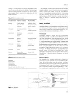
24 Pulmonary Artery Catheter Values
VI. Pulmonary artery catheterization
1. Using sterile technique, cannulate a vein using the technique above. The
subclavian vein or internal jugular vein is commonly used.
2. Advance a guide wire through the cannula, then remove the cannula, but
leave the guide wire in place. Keep the guide wire under control at all
times. Nick the skin with a number 11 scalpel blade adjacent to the guide
wire, and pass a number 8 French introducer over the wire into the vein.
Remove the wire and connect the introducer to an IV fluid infusion, and
suture with 2-0 silk.
3. Pass the proximal end of the pulmonary artery catheter (Swan Ganz) to
an assistant for connection to a continuous flush transducer system.
4. Flush the distal and proximal ports with heparin solution, remove all
bubbles, and check balloon integrity by inflating 2 cc of air. Check the
pressure transducer by quickly moving the distal tip and watching the
monitor for response.
5. Pass the catheter through the introducer into the vein, then inflate the
balloon with 1.0 cc of air, and advance the catheter until the balloon is in
or near the right atrium.
6. The approximate distance to the entrance of the right atrium is deter-
mined from the site of insertion:
Right internal jugular vein: 10-15 cm.
Subclavian vein: 10 cm.
Femoral vein: 35.45 cm.
7. Advance the inflated balloon, while monitoring pressures and wave forms
as the PA catheter is advanced. Advance the catheter through the right
ventricle into the main pulmonary artery until the catheter enters a distal
branch of the pulmonary artery and is stopped (as evidenced by a
pulmonary wedge pressure waveform).
8. Do not advance the catheter while the balloon is deflated, and do not
withdraw the catheter with the balloon inflated. After placement, obtain a
chest X-ray to ensure that the tip of catheter is no farther than 3-5 cm
from the mid-line, and no pneumothorax is present.
Normal Pulmonary Artery Catheter Values
Right atrial pressure 1-7 mm Hg
RVP systolic 15-25 mm Hg
RVP diastolic 8-15 mm Hg
Pulmonary artery pressure
PAP systolic 15-25 mm Hg
PAP diastolic 8-15 mm Hg
PAP mean 10-20 mm Hg
Acute Coronary Syndromes 25
Cardiovascular Disorders
Roham T. Zamanian, MD
Farhad Mazdisnian, MD
Michael Krutzik, MD
Acute Coronary Syndromes (Acute Myocardial
Infarction and Unstable Angina)
Acute myocardial infarction (AMI) and unstable angina are part of a spectrum
known as the acute coronary syndromes (ACS), which have in common a
ruptured atheromatous plaque. These syndromes include unstable angina,
non–Q-wave MI, and Q-wave MI. The ECG presentation of ACS includes ST-
segmentelevation infarction, ST-segment depression (including non–Q-waveMI
and unstable angina), and nondiagnostic ST-segment and T-wave abnormalities.
Patients with ST-segment elevation will usually developQ-wave MI. Patients with
ischemic chest discomfort who do not have ST-segment elevation will develop
Q-wave MI and non–Q-wave MI or unstable angina.
I. Clinical evaluation of chest pain and acute coronary syndromes
A. History. Chest pain is present in 69% of patients with AMI. The pain may
be characterized as a constricting or squeezing sensation in the chest.
Pain can radiate to the upper abdomen, back, either arm, either shoulder,
neck, or jaw. Atypical pain presentations in AMI include pleuritic, sharp or
burning chest pain. Dyspnea, nausea, vomiting, palpitations, or syncope
may be the only complaints.
B. Cardiac Risk factors include hypertension, hyperlipidemia, diabetes,
smoking, and a strong family history (coronary artery disease in early or
mid-adulthood in a first-degree relative).
C. Physical examination may reveal tachycardia or bradycardia, hyper- or
hypotension, or tachypnea. Inspiratory rales and an S3 gallop are
associated with left-sided failure. Jugulovenous distention (JVD),
hepatojugular reflux, and peripheral edema suggest right-sided failure. A
systolic murmur may indicate ischemic mitral regurgitation or ventricular
septal defect.
II. Laboratory evaluation of chest pain and acute coronary syndromes
A. Electrocardiogram (ECG). The initial ECG reveals diagnostic ST
elevations in only 40% of patients with a confirmed AMI. ST-segment
elevation (equal to or greater than 1 mV) in two or more contiguous leads
provides strong evidence of thrombotic coronary arterial occlusion and
makes the patient a candidate for immediate reperfusion by thrombolysis
or angioplasty.
B. Laboratory markers
1. Creatine phosphokinase (CPK) enzyme is found in the brain, muscle,
and heart. The cardiac-specific dimer, CK-MB, however, is present
almost exclusively in myocardium.
26 Acute Coronary Syndromes
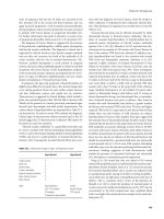
Common Markers for Acute Myocardial Infarction
Marker Initial Eleva-
tion After MI
Mean Time to
Peak Eleva-
tions
Time to Re-
turn to Base-
line
Myoglobin 1-4 h 6-7 h 18-24 h
CTnl 3-12 h 10-24 h 3-10 d
CTnT 3-12 h 12-48 h 5-14 d
CKMB 4-12 h 10-24 h 48-72 h
CKMBiso 2-6 h 12 h 38 h
CTnI, CTnT = troponins of cardiac myofibrils; CPK-MB, MM = tissue
isoforms of creatine kinase.
2. CK-MB subunits. Subunits of CK, CK-MB, -MM, and -BB, are
markers associated with a release into the blood from damaged cells.
Elevated CK-MB enzyme levels are observed in the serum 2-6 hours
after MI, but may not be detected until up to 12 hours after the onset
of symptoms.
3. Cardiac-specific troponin T (cTnT) is a qualitative assay and cardiac
troponin I (cTnI) is a quantitative assay. The cTnT level remains
elevated in serum up to 14 days and cTnI for 3-7 days after infarction.
4. Myoglobin is the first cardiac enzyme to be released. It appears
earlier but is less specific for MI than other markers. Myoglobin is most
useful for ruling out myocardial infarction in the first few hours.
III. Initial treatment of acute coronary syndromes
A. Continuous cardiac monitoring and IV access should be initiated.
Morphine, oxygen, nitroglycerin, and aspirin
("MONA") should be
administered to patients with ischemic-type chest pain unless contraindi-
cated.
B. Morphine is indicated for continuing pain unresponsive to nitrates.
Morphine reduces ventricular preload and oxygen requirements by
venodilation. Administer morphine sulfate 2-4 mg IV every 5-10 minutes
prn for pain or anxiety.
C. Oxygen should be administered to all patients with ischemic-type chest
discomfort and suspected ACS for at least 2 to 3 hours.
D. Nitroglycerin
1. Nitroglycerin is an analgesic for ischemic-type chest discomfort.
Nitroglycerin is indicated for the initial management of pain and
ischemia unless contraindicated by hypotension (SBP <90 mm Hg) or
RV infarction. Continued use of nitroglycerin beyond 48 hours is only
indicated for recurrent angina or pulmonary congestion.
2. Initially, give up to three doses of 0.4 mg sublingual NTG every five
minutes or nitroglycerine aerosol, 1 spray sublingually every 5
minutes. An infusion of intravenous NTG may be started at 10-20
mcg/min, titrating upward by 5-10 mcg/min every 5-10 minutes
Acute Coronary Syndromes 27
(maximum, 3 mcg/kg/min). Titrate to decrease the mean arterial
pressure by 10% in normotensive patients and by 30% in those with
hypertension. Slow or stop the infusion if the SBP drops below 100
mm Hg.
E. Aspirin
1. Aspirin should be given as soon as possible to all patients with
suspected ACS unless the patient is allergic to it. Aspirin therapy
reduces mortality after MI by 25%.
2. A dose of 160-325 mg of aspirin should be chewed and swallowed on
day 1 and continued PO daily thereafter. If aspirin is contraindicated,
clopidogrel (Plavix) 75 mg qd should be administered.
IV. Risk stratification, initial therapy, and evaluation for reperfusion in
the emergency department
Risk Stratification with the First 12-Lead ECG
Use the 12-lead ECG to triage patients into 1 of 3 groups:
1. ST-segment elevation
2. ST-segment depression(
$1 mm)
3. Nondiagnostic or normal ECG
A. Patients with ischemic-type chest pain and ST-segment elevation $1mm
in 2 contiguous leads have acute myocardial infarction. Reperfusion
therapy with thrombolytics or angioplasty is recommended.
B. Patients with ischemic-type pain but normal or nondiagnostic ECGs or
ECGs consistent with ischemia (ST-segment depression only) usually do
not have AMI. These patients should not be given fibrinolytic therapy.
C. Patients with normal or nondiagnostic ECGs usually do not have AMI, and
they should be evaluated with serial cardiac enzymes and additional tests
to determine the cause of their symptoms.
V. Management of ST-segment elevation myocardial infarction
A. Patients with ST-segment elevation have AMI should receive reperfusion
therapy with fibrinolytics or percutaneous coronary intervention.
B. Reperfusion therapy: Fibrinolytics
1. Patients who presentwith ischemic pain and ST-segment elevation (
$1
mm in
$2 contiguous leads) within 12 hours of onset of persistent pain
should receive fibrinolytic therapy unless contraindicated. Patients with
a new bundle branch block (obscuring ST-segment analysis) and
history suggesting acute MI should also receive fibrinolytics or
angioplasty.
28 Acute Coronary Syndromes
Treatment Recommendations for AMI
Supportive Care for Chest Pain
• All patients should receive supplemental oxygen, 2 L/min by nasal canula, for a
minimum of three hours
• Two large-bore IVs should be placed
Aspirin:
Inclusion
Clinical symptoms or suspicion of AMI
Exclusion
Aspirin allergy, active GI bleeding
Recommendation
Chew and swallow one dose of160-325 mg, then orally qd
Thrombolytics:
Heparin:
Inclusion
Exclusion
Recommendation
All patients with ischemic pain and ST-segment elevation ($
1 mm in $2 contiguous leads) within 12 hours of onset of
persistent pain, age <75 years.
All patients with a new bundle branch block and history
suggesting acute MI.
Active internal bleeding; history of cerebrovascular acci-
dent; recent intracranial or intraspinal surgery or trauma;
intracranial neoplasm, arteriovenous malformation, or aneu-
rysm; known bleeding diathesis; severe uncontrolled hyper-
tension
Reteplase (Retavase) 10 U IVP over 2 min x 2. Give sec-
ond dose of 10 U 30 min after first dose
OR
Tenecteplase (TNKase): <60 kg: 30 mg IVP; 60-69 kg: 35
mg IVP; 70-79 kg: 40 mg IVP; 80-89 kg: 45 mg IVP;
$90 kg:
50 mg IVP
OR
t-PA (Alteplase, Activase) 15 mg IV over 2 minutes, then
0.75 mg/kg (max 50 mg) IV over 30 min, followed by 0.5
mg/kg (max 35 mg) IV over 30 min.
Inclusion
Exclusion
Recommendation
Administer concurrently with thrombolysis
Active internal or CNS bleeding
Heparin 60 U/kg IVP, followed by 12 U/kg/hr continuous IV
infusion x 48 hours. Maintain aPTT 50-70 seconds
Acute Coronary Syndromes 29
Beta-Blockade:
Inclusion
All patients with the diagnosis of AMI. Within 12 hours of
diagnosis of AMI
Exclusion
Severe COPD, hypotension, bradycardia, AV block, pulmo-
nary edema, cardiogenic shock
Recommendation
Metoprolol (Lopressor), 5 mg IV push every 5 minutes for
three doses; followed by 25 mg PO bid. Titrate up to 100
mg PO bid
OR
Atenolol (Tenormin), 5 mg IV, repeated in 5 minutes, fol-
lowed by 50-100 mg PO qd.
Nitrates:
ACE Inhibitors:
Inclusion
Exclusion
Recommendation
All patients with ischemic-type chest pain
Nitrate allergy; sildenafil (Viagra) within prior 24 hours;
hypotension; caution in right ventricular infarction
0.4 mg NTG initially q 5 minutes, up to 3 doses or nitroglyc-
erine aerosol, 1 spray sublingually every 5 minutes. IV
infusion of NTG at 10-20 mcg/min, titrating upward by 5-10
mcg/min q 5-10 minutes (max 3 mcg/kg/min). Slow or stop
infusion if systolic BP <90 mm Hg
Inclusion
Exclusion
Recommendation
All patients with the diagnosis of AMI. Initiate treatment
within 24 hours after AMI
Bilateral renal artery stenosis, angioedema caused by
previous treatment
Lisinopril (Prinivil) 2.5-5 mg qd, titrate to 10-20 mg qd.
Maintain systolic BP >100 mm hg
C. Thrombolytics
1. ECG criteria for thrombolysis
a. ST Elevation (>1 mm in two or more contiguous leads), time to
therapy 12 hours or less, age younger than 75 years.
b. A new bundle branch block (obscuring ST-segment analysis) and
history suggesting acute MI.
2. Alteplase (t-PA, tissue-plasminogen activator, Activase) and
Reteplase (Retavase) convert plasminogen to plasmin. Both agents
are clot-specific and bind to new thrombus. Activase is superior to
streptokinase. The alteplase thirty-day mortality rate of 6.3% is the
lowest of the fibrinolytics, compared with 7.3% for streptokinase.
Alteplase provides the earliest and most complete reperfusion.
3. Streptokinase (SK, Streptase) provides greater benefit in older
patients with a smaller amount of myocardium
at risk who present
later and those with a greater risk of ICH. The dose of IV SK is 1.5
million units given over 60 minutes.
D. Reperfusion therapy: Percutaneous coronary intervention
30 Acute Coronary Syndromes
1. PCI is preferable to thrombolytic therapy if performed in a timely
fashion by individuals skilled in the procedure. Coronary angioplasty
provides higher rates of flow than thrombolytics and is associated with
lower rates of reocclusionand postinfarction ischemia than fibrinolytic
therapy.
2. Patients at high risk for mortality or severe LV dysfunction with signs
of shock, pulmonary congestion,heart rate >100 bpm, and SBP <100
mm Hg should be sent to facilities capable of performing cardiac
catheterization and rapid revascularization. When available within 90
minutes, PCI is recommended for all patients, particularly those who
have a high risk
of bleeding with fibrinolytic therapy.
E. Heparin is recommended in patients receiving selectivefibrinolytic agents
(tPA/Reteplase/tenectaplase). A bolus dose of 60 U/kg should be
followed by infusion at a rate of 12 U/kg/hour (a maximum bolus of 4000
U/kg and infusion of 1000 U/h for patients weighing <70 kg). An aPTT of
50 to 70 seconds is optimal.
F. Beta-blockade use during and after AMI reduces mortality by 36%.
Contraindications to beta-blockers include severe LV failure and
pulmonary edema, bradycardia (heart rate <60 bpm), hypotension (SBP
<100 mm Hg), signs of poor peripheral perfusion, second- or third-degree
heart block.
1. Metoprolol (Lopressor), 5 mg IV push every 5 minutes for three
doses; followed by 25 mg PO bid. Titrate up to 100 mg PO bid OR
2. Atenolol (Tenormin), 5 mg IV, repeated in 5 minutes, followed by 50-
100 mg PO qd.
G. ACE-inhibitors increase survival in patients with AMI. ACE-inhibitors
should be started between 6 to 24 hours after AMI and continued for 4-6
weeks, and indefinitely if ejection fraction <40%.
1. Captopril (Capoten) is given as a 6.25 mg initial dose and titrated up
to 50 mg po bid, or
2. Lisinopril (Prinivil) may be given as 2.5-5 mg qd, titrate to 10-20 mg
qd.
VI. Management Non–Q-wave MI and high-risk unstable angina with ST-
segment depression.
A. Non–Q-wave MI and unstable angina present with ST-segment
depression. Among patients with ST-segment
depression, fibrinolytic
therapy provides no benefit. Fibrinolytic therapy should not be used in
patients with ST-segment depression or nondiagnostic ECGs.
B. Aspirin (325 mg qd) and heparin, 60 U/kg IVP, followed by 12 U/kg/hr
continuous IV infusion x 48 hours (aPTT 50-70 seconds) should be given
to patients with ST-segment depression or T-wave inversion with
ischemic-type chest pain.
Acute Coronary Syndromes 31
Heparin and ST-Segment Depression and Non–Q-Wave MI/Unstable
Angina
! IV heparin therapy for 3 to 5 days is standard for high-risk and some
intermediate-risk patients. Treat for 48 hours, then individualized
therapy.
! LMWH is an acceptable alternative to IV unfractionated heparin.
-Enoxaparin (Lovenox) 1.0 mg/kg SQ q12h OR
-Dalteparin (Fragmin) 120 IU/kg (max 10,000 U) SQ q12h.
C. Nitrates should be given for recurrent angina. Sublingual nitroglycerin
(NTG) , initially, give up to three doses of 0.4 mg sublingual NTG every
five minutes or nitroglycerine aerosol, 1 spray sublingually every 5
minutes. Nitroglycerin patch 0.2 mg/hr qd. Allow for nitrate-free period to
prevent tachyphylaxis. Isosorbide dinitrate (Isordil) 10-60 mg PO tid -
[5,10,20, 30,40 mg], or isosorbide mononitrate (Imdur) 30-60 mg qd.
D. Beta-blockers. A beta-blocker should be initiated for patients with ST-
segment depression.
1. Beta-blockers reduce the size of the infarct in patients who do not
receive fibrinolytic therapy. A significant decrease in death and
nonfatal infarction has been observed in patients treated with beta-
blockers after infarction.Contraindications to beta-blockers: severe LV
failure and pulmonary edema, bradycardia (heart rate <60 bpm),
hypotension (SBP <100 mm Hg), signs of poor peripheral perfusion,
second- or third-degree heart block.
2. Metoprolol (Lopressor), 5 mg IV push every 5 minutes for three
doses; followed by 25 mg PO bid. Titrate up to 100 mg PO OR
3. Atenolol (Tenormin), 5 mg IV, repeated in 5 minutes, followed by 50-
100 mg PO qd.
E. Coronary angiography is recommended for high-risk patients with
recurrent ischemia, depressed LV function, widespread changes on the
ECG, or prior MI.
F. Glycoprotein IIb/IIIa inhibitors
1. The GP IIb/IIIa receptor blockers reduce platelet aggregation. The GP
IIb/IIIa inhibitors should be used for patients with non-ST-segment
elevation MI or high-risk unstable angina. These agents should be
used with aspirin or clopidogrel and unfractionated heparin. The dose
of unfractionated heparin should be reduced by
a when combined
with GP blockers.
2. Intravenous GP blocker dosages
a. Abciximab (ReoPro), 0.25 mg/kg IVP over 2 min, then 0.125
mcg/kg/min (max 10 mcg/min) for 12 hours.
b. Eptifibatide (Integrilin), 180 mcg/kg IVP over 2 min, then 2
mcg/kg/min for 24-72 hours. Use 0.5 mcg/kg/min if creatinine is
>2.0 mg/dL.
c. Tirofiban (Aggrastat), 0.4 mcg/kg/min for 30 min, then 0.1
mcg/kg/min IV infusion for 24-72 hours. Reduce dosage by 50% if
the creatine clearance is <30 mL/min.
32 Acute Coronary Syndromes
VII. Management of patients with a nondiagnostic ECG