General ultrasound In the critically ill - part 9 pot
Bạn đang xem bản rút gọn của tài liệu. Xem và tải ngay bản đầy đủ của tài liệu tại đây (1.43 MB, 20 trang )
The Neck 155
The Neck
The neck veins were studied in Chap. 12.
Carotid artery exploration can be useful in a
comatose patient. A traumatic dissection will be
sought, although the Doppler is the usual tech-
nique. Does the two-dimensional approach not
give already basic information in some or a major-
ity of
cases?
This could make the Doppler informa-
tion redundant in first-line analysis in these cases.
Another application of a two-dimensional scan-
ning can be the evaluation of vascular injury by
screening for calcifications at the carotid
arteries,
a
marker of
the
arterial system.
A retropharyngeal abscess can be sought [10].
In this
area,
traumatic hematomas, other abscesses
or cervicofacial cellulitis can be documented.
However,
CT
is preferred here.
The trachea is perfectly detectable at the cervi-
cal level: anterior and median with posterior
air artifacts. Applying pressure that is more than
very light can be very unpleasant in moderately
sedated patients. The trachea is quickly lost since
it takes a posterior direction when entering the
thorax. Via the anterior or lateral approach, one
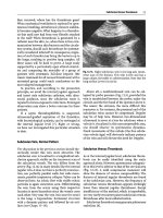
can study its external configuration (Fig.
21.8).
Its
anteroposterior and lateral diameters can be mea-
sured, at inspiration and expiration. Tracheomala-
cia may be detected this way. Since the tracheal
wall is fibrocartilaginous, nothing prevents the
ultrasound analysis of the tracheal content: the
ultrasound beam encounters the wall, then the
air, which stops beam progression. If the anterior
wall is thickened by a granuloma or other causes
of tracheal stenosis or obstruction, this obstacle
will be accurately detected and analyzed. Within
the lumen
itself,
secretions accumulated above
an inflated balloon can be detected (Fig. 21.9).
This finding may
have
clinical outcome. Of course,
fibroscopy will remain the reference test for tra-
cheal disorders, but the principle remains the
same: give the patient a first noninvasive, rapid
approach that can alter the usual management,
depending on the operator's skill. Some authors
use ultrasound for the guidance of percutaneous
tracheostomy
[11].
The intubation tube itself will
give a particular
signal,
whose clinical application
is under investigation.
The thyroid, especially the isthmus, can be use-
fully located before tracheostomy (Fig. 21.8). An
aberrant brachiocephalic artery can be located
[12],
but also the closeness of the innominate vein
or thyroid hypertrophy. Diagnostic ultrasound is
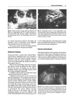
Fig.
21.8.
Transverse anterior cervical scan
at the
thyroid
isthmus. The two thyroid lobes (X) and the posterior
shadow of the trachea
(T)
are recognized. Since an air
barrier is visible immediately posterior to the anterior
wall of the trachea, it can be possible to conclude that
the anterior
wall,
at
this
level,
is
thin
Fig.
21.9.
As
opposed to
Fig.
21.8,
this
trachea is entirely
crossed
by the
ultrasound
beam.
There
is
accumulation
of secretions above the inflated balloon. This pattern
vanishes if the balloon is deflated, but the patient
coughs. In addition, the anterior tracheal wall can be
accurately
measured,
here
thickened
to 4
mm
contributive if an abnormal thyroid gland is
described in
a
patient with suspicion of severe dys-
thyroidism. In a young female admitted for acute
hypercalcemia, ultrasound immediately detected a
suspect mass evoking a parathyroid tumor. This
resulted in prompt surgery, which confirmed the
diagnosis.
Finally,
the rough integrity of the cervical verte-
brae can be assessed via the anterolateral cervical
156 Chapter
21 Head
and
Neck
References
Fig.
21.10.
Longitudinal paramedian scan of the neck.
Posterior to the internal jugular vein (V) and the
muscle, a thick hyperechoic line represents the anterior
wall of the cervical rachis, here straight without solu-
tion of continuity
(arrows).
Upright cervical rachis
approach (Fig.
21.10).
Why not use first-line ultra-
sound when there is suspicion of cervical rachis
fracture?
For these new fields, of immediate interest in the
ICU, high-frequency probes (7.5 or 10 MHz) may
be relevant.
The Nape of the Neck
Suboccipital puncture is sometimes performed in
patients with intracranial hypertension. Would
ultrasound guidance or location be useful in this
reputedly risky technique? We are currently inves-
tigating the possibilities in this area.
1.
Rouby
JJ,
Laurent
P,
Gosnach M, Cambau E, Lamas
G,
Zouaoui
A,
Leguillou
JL,
Bodin
L,
Khac
TD,
Mar-
sault
C,
Poete
P,
Nicolas
MH,
Jarlier V, Viars P (1994)
Risk factors and clinical relevance of nosocomial
maxillary sinusitis in the critically ill. Am
}
Respir
Grit Care Med 150:776-783
2.
Landmann MD (1986) Ultrasound screening for
sinus disease. Otolaryngol Head Neck Surg 94:157-
161
3.
Beuzelin
G,
Mousset
G,
FroehHch
P,
Senac
J,
Gory G,
Goursot
G,
Fombeur
JP
(1990) Evaluation de Techo-
graphie sinusienne dans le diagnostic des sinusites
maxillaires purulentes en reanimation. Rean Soins
Intens Med Urg
6:538
4.
Rippe
JM,
Irwin
RS,
Alpert
JS,
Fink
MP
(1991) Inten-
sive care medicine. Little Brown, Boston, p 709
5.
Lichtenstein D, Biderman P, Meziere G, Gepner A
(1998) The sinusogram: a real-time ultrasound sign
of maxillary sinusitis. Intensive Gare Med 24:1057-
1061
6. Berges 0, Torrent M (1986) Echographie de Foeil et
de
Forbite.
Vigot,
Paris
7.
Lichtenstein D Bendersky
N,
Meziere
G,
Goldstein I
(2002) Ultrasound diagnosis of cranial hypertensi-
on by measuring optic nerve caliper. Reanimation
11 [Suppl3]:170
8. Hamburger J (1977) Petite encyclopedie medicale.
Flammarion,
Paris,
pp 1377-1378
9. Gzosnyka
M,
Matta
BF,
Smielewski
P,
Kirkpatrick PJ,
Pickard JD (1998) Gerebral perfusion pressure in
head-injured patients: a noninvasive assessment
using transcranial Doppler ultrasonography.
J
Neu-
rosurg. 88:802-808
Rippe
JM,
Irwin
RS,
Alpert
JS,
Fink
MP
(1991) Inten-
sive care medicine. Little Brown, Boston, p 704
Sustic
A,
Kovac
D,
Zgaljardic
Z,
Zupan
Z,
Krstulovic
B (2000) Ultrasound-guided percutaneous dilata-
tional tracheostomy: a safe method to avoid cranial
misplacement of the tracheostomy tube. Intensive
Gare Med 26:1379-1381
12.
Hatfield
A,
Bodenham
A
(1999) Portable ultrasound
of the anterior neck prior to percutaneous dilatatio-
nal tracheostomy. Anesthesia 54:660-663
10
11
CHAPTER
22
Soft Tissues
Soft tissues are accessible to ultrasound. They can
be of interest in several instances.
Soft Tissue Abscess
The ultrasound signs include
hypoechoic,
heteroge-
neous mass and inconstant punctiform hyperechoic
areas indicating bacterial gas
(Fig.
22.1),
signs indi-
cating a
fluid
nature such as posterior enhancement
(which is inconstant) or changes in dimensions
under probe pressure (but such maneuvers can be
very harmful, not to say risky). In fact, abscess and
hematoma often
have
similar patterns, and
the
ultra-
sound-guided tap will make a definite diagnosis.
Necrotizing Cellulitis
The role that ultrasound can play is not well
known in necrotizing celluHtis. The diagnosis is
usually clinical. Surgical exploration alone speci-
fies the extension of the necrosis [1]. Ultrasound
may theoretically allow early diagnosis by showing
deep areas of emphysema before they become clin-
ically accessible. Ultrasound may also distinguish
between gangrenous cellulitis (which preserves the
muscle) and necrotizing fasciitis (with myonecro-
sis).
Hypoechoic areas dissociating the muscle
fibers would then be observed.
Deep Hematoma
A hematoma gives well-limited mass that is ane-
choic at the first stage and can quickly become
echoic and heterogeneous (Fig. 22.2). In case of
doubt, ultrasound-guided investigation can give
the diagnosis.
A hematoma can develop anywhere and give
distinctive signs. At the rectus abdominis muscle,
its extraperitoneal nature will be recognized since
the peritoneal sliding will be preserved, posterior
to the
mass.
In severe forms, it can be the source of
compression (bowel,bladder, etc.) [2].
Fig. 22.1.
Huge heterogeneous collection in the gluteal
area. With ultrasound guidance, the tap withdrew pus,
thus confirming the
abscess.
Young
patient with trauma
Fig. 22.2.
Thigh collection in another traumatized pa-
tient. The pattern is not far from that described in
Fig, 22.1
but here is a partially solid hematoma
158 Chapter
22
Soft Tissues
Parietal Emphysema
Parietal emphysema generates air comet-tail-type
artifacts. They usually conceal the deeper struc-
tures
(Fig.
22.3).
The presence of parietal emphyse-
ma is certainly one of the rare indications to
cancel ultrasound examination. However, it is
sometimes possible to hide the masses of gas by
gentle pressure. At the thoracic level, this is facili-
tated by the ribs, which remain solid under pres-
sure.
Lung sliding can then sometimes be analyzed
(see Chap. 16). Note that pneumothorax is not
always present.
Let us recall that comet-tail artifacts generated
by parietal emphysema can be a dangerous pitfall
for the beginner when they appear as
E
lines. This
pattern may be erroneously interpreted as
B
lines
or lung
rockets,
and genuine pneumothorax can be
missed (see
Fig.
16.11,
p
113).
The search for
the
bat
sign in this setting prevents this pitfall.
Fig.
22.3.
Parietal emphysema. The deep structures in
this thoracic view are unrecognizable since they are
hidden by numerous comet-tail artifacts.
This
aspect is
unusable.
These are
W
lines,
defined as comet-tail arti-
facts arising from different
levels
in the soft tissues
Edematous Syndromes
In cases of major hydric retention, the soft tissues
are enlarged by edema, with hypoechoic zones dis-
sociating the muscles. The analysis of the deeper
structures is not hindered, as water is a good con-
ductor for ultrasound beams.
In situations such as nephrotic syndrome with
massive hypoalbuminemia, more or less substan-
tial effusions can affect all of the anatomical com-
partments.
Parietal Vessels
Ultrasound can be useful to accurately locate the
epigastric or internal mammary vessels if a local
tap is considered (see
Fig.
5.12,
p 32).
Undernutrition
The nutritional status of
a
patient is usually moni-
tored by weighing the patient. This is a simple
parameter. However, the maneuver is demanding
for the paramedical team, and above all, the data
obtained is a rough result of inverse trends: in a
critically ill patient, the muscles and fat compart-
ments decrease whereas the water compartment
increases. Once more, ultrasound can potentially
Fig.
22.4.
Transverse scan of
the
paraumbilical abdomi-
nal
wall.
The
white arrows
sharply delimit the fat com-
partment
(17
mm),
the black arrows
the muscular com-
partment (9 mm for the muscle). These measures can
easily
be
repeated during the stay of
the
patient. Probe
with 7.5-MHz frequency
provide logic-based assistance.
A
differential analy-
sis of the fat
[3],
muscle and interstitial compart-
ments can in fact be carried out
(Fig.
22.4).
Accept-
ing that these variations are the same in any part of
the body, only one standardized area should be
investigated. An easy-to-access and reliable area is,
for instance,
a
transverse, paraumbilical scan of the
rectus abdominis muscle (Fig. 22.4) or, perhaps
better, a transverse scan of the crural muscle at
mid-thigh. Ultrasound may also detect interstitial
edema before clinical evidence, but this precise
issue has not yet been investigated.
References 159
Miscellaneous
References
Multiple disorders such as
cysts,
arterial aneurysms,
osteomas,
etc.
not related to the acute illness can be
detected in the soft tissues.
Traumatic Rhabdomyolysis
The muscular loges have increased volume, with-
out abscess or hematoma to explain the clinical
swelling.
A
hypoechoic pattern of the muscles with
disorganization of the normal muscular architec-
ture has been described
[4].
Another advantage of
ultrasound is ruling out associated venous throm-
bosis (with here a possible place for Doppler if the
compression maneuver is harmful).
Malignant Hyperthermia
A heterogeneous and grainy pattern of the mus-
cles,
with a hypoechoic pattern of the septa and
fascia is described by some
[5],
not found by others
[6].
The rarity of this syndrome in our ICU has
until now prevented us from forming an opinion.
1.
Offenstadt
G
(1991) Infections des parties moUes par
les germes anaerobies. Rev Prat 13:1211-1214
2.
Blum A, Bui P, Boccaccini H, Bresler L, Claudon M,
Boissel
P,
Regent
D
(1995) Imagerie des formes graves
de rhematome des grands droits sous anticoagu-
lants.
J
Radiol 76:267-273
3.
Armellini
F,
Zamboni
M,
Rigo
L,
Todesco
T,
Bergamo-
Andreis I
A,
Procacci
C,
Bosello 0 (1990) The contri-
bution of sonography to the measurement of intra-
abdominal fat.
J
Clin Ultrasound 18:563-567
4.
Lamminen
AE,
Hekali
PE,
Tiula
E,
Suramo
I,
Korhola
OA (1989) Acute rhabdomyolysis: evaluation with
magnetic resonance imaging compared with CT and
ultrasonography. Br
J
Radiol 62:326-331
5.
Von Rohden L, Steinbicker
V,
Krebs
P,
Wiemann D,
Koeditz
H
(1990) The value of ultrasound for the dia-
gnosis of malignant hyperthermia.
J
Ultrasound Med
9:291-295
6. Antognini
JF,
Anderson M, Cronan M, McGahan JP,
Gronert GA (1994) Ultrasonography: not useful in
detecting susceptibility
to
malignant hyperthermia.
J
Ultrasound Med 13:371-374
CHAPTER
23
Ultrasound
in
the
Surgical
Intensive
Care
Unit
An »echological« distinction between medical and
surgical patients should not make sense per
se,
but
some differences can be underlined.
General Issues
The surgical patient is often surrounded by a bar-
rage of acoustic
barriers:
wounds,
dressings,
ortho-
pedic material, cervical collar. This may limit the
use of ultrasound, but these obstacles can be over-
come.
The problems of asepsis are more important
than in the medical
setting,
and vigilance regarding
crossed infections must be reinforced.
The Abdomen
Dressings sometimes cover the entire abdominal
wall, but these limitations can be bypassed. The
dressings can be withdrawn, the probe can be in-
serted in sterile conditions, a sterile contact prod-
uct can be used, although these procedures may
seem overly
restrictive.
The
sterile protection of the
probe should conduct the ultrasound beam with-
out interference [1]. Fine transparent adhesive
dressings such as OpSite and Tegaderm offer the
advantage of being transparent to ultrasound.
Their use should therefore be encouraged. Some
thick dressings may appear impenetrable by ultra-
sound, but we have noted that ultrasound beams
occasionally are not stopped, and basic answers to
clinical questions can be obtained. In addition,
medical personnel should be taught to wisely apply
dressings, since critically ill postoperative patients
will unavoidably
have
ultrasound examinations.
Apart from the anomalies described in earlier
chapters, ultrasound can search for infected post-
operative collections [2] (Fig. 23.1). For some
authors, ultrasound sensitivity is high, whereas
specificity
is
low
[3].
It is true that noninfected col-
lections are most often encountered in this setting,
such as serous, lymph, urine, bile or digestive liq-
Fig.23.1.
Intra-abdominal abscess in
a
man operated on
for colic ischemia. Transverse scan of the right fossa
iliaca.
The
ultrasound-guided tap
was
particularly
rele-
vant here
uids.
These collections are usually anechoic. Their
observation alone is usually sufficient for diagno-
sis.
The increase in volume of a collection is one
criterion for reoperation in postoperative peritoni-
tis
[4].
We simplify the approach by adopting the
easy tap
policy.
At
the expense of useless taps (but
never deleterious if basic rules are respected), sep-
tic or hemorrhagic postoperative complications
will be promptly detected.
The classic subphrenic abscess is rare in our
observations.
Acute acalculous cholecystitis is probably a
complication particular to the surgical ICU.
Forgotten foreign bodies will easily be detected.
A compress gives a large image with a matrix-like
pattern and a massive acoustic
shadow.
A
metallic
instrument has a strikingly straight shape, with
typical posterior artifacts we call
S
lines.
Hematomas are first anechoic, then rapidly
become echo-rich and yield heterogeneous, solid
images. They can be observed in the retroperi-
toneum,
the
pelvis,
and the rectus abdominis muscle.
164 Chapter 23 Ultrasound In the Surgical Intensive Care Unit
Postoperative Abdominal Interventional
Ultrasound
A simple tap will confirm infected collections. Per-
cutaneous drainage under ultrasound guidance
deserves to be subsequently tried. The fluidity
helps in choosing the appropriate caliper of the
material [5]. This kind of procedure can preclude
subsequent surgery, which has higher morbidity
and mortality rates. This is the best procedure
for some [6], who reserve conventional surgery
for complex cases, or when a percutaneous route
appears dangerous (bowel obstacles, for instance).
Before inserting a large drain, it can be advanta-
geous to withdraw the maximum amount of pus
with a fine needle, which will in certain cases be
considered sufficient.
Postoperative Thoracic Ultrasound
Hemothorax, pneumothorax, tamponade, phrenic
paralysis, pneumomediastinum, some false aneu-
rysms (see Chap. 19) and sometimes mediastinitis
are accessible with ultrasound.
In the postoperative thoracic period, the inten-
sivist must promptly determine if the content of
the hemithorax is fluid or air. Ultrasound immedi-
ately provides the answer.
A periaortic collection can be detected and even
tapped with ultrasound guidance. Sepsis of the
prosthesis will thus sometimes be diagnosed. In
this severe setting, the current habit
is,
however, to
perform
CT,
despite its invasiveness.
Here again, appropriate information to the team
limits the extent of the dressings.
Thromboembolic Disorders
Lower Extremity Veins
Ultrasound is more laborious in surgical patients
than in medical patients, especially trauma patients,
as the dressings, surgical devices, pain and post-
contusion changes can decrease the potential of
ultrasound. Deep venous thrombosis, however,
seems more frequent in the surgical ICU, perhaps
because local trauma is a major cause for venous
thrombosis. It must be remembered that compres-
sion ultrasound can be painful, and Doppler may
have an interest here.
Fig.
23.2.
Massive thrombosis of the left internal jugular
vein in
a
patient
who
underwent venous catheterization.
Note that this thrombosis is completely occlusive and
extends at least 6 cm in the craniocaudal axis
Upper Extremity Veins
A frequent problem in the emergency setting is the
difficulty of inserting a central venous catheter. In
surgical ICUs, patients have already been man-
aged. Hypovolemia has been corrected. Therefore,
problems in inserting venous lines may not be as
critical as in the medical ICU.
In our experience, the frequency of internal
jugular venous thrombosis seems extremely high
in severely ill surgical ICU patients (Fig. 23.2, and
see Figs. 12.6,12.9,12.10,12.13, pp 72-74). Indepen-
dent factors may explain this, such as the possibly
more frequent use of cardiac catheterization in
certain surgical ICUs.
References
1.
Kox W, Boultbee J (1988) Abdominal ultrasound in
intensive care. In: Kox W, Boultbee J, Donaldson R
(eds) Imaging and labelling techniques in the criti-
cally
ill.
Springer-Verlag, London, pp 127-135
2.
Weill FS (1989) Echographie abdominale du post-
opere.
In:
Weill
FS
(ed) L'ultrasonographie en patho-
logie
digestive.
Vigot,
Paris,
pp 536-544
3.
Mueller
PR,
Simeone JF (1983) Intra-abdominal abs-
cesses: diagnostic by sonography and computerized
tomography Radiol Clin North Am 21:425-431
4.
Dazza FE (1985) Peritonites graves en reanimation:
modalites du traitement chirurgical. In: Reanima-
tion et medecine d'urgence. Expansion Scientifique
Fran^aise,
Paris,
pp 271-286
5.
Van Sonnenberg E, Mueller PR, Ferrucci JT (1984)
Percutaneous drainage of 250 abdominal abscesses
and fluid collections. Radiology 151:337-347
6. Pruett TL, Simmons RL (1988) Status of percuta-
neous catheter drainage of
abscesses.
Surg
Clin North
Am 68:89
CHAPTER
24
Ultrasound in Trauma
In the trauma context, ultrasound has a Hmited
place in patients who are lucky enough to arrive
alive at a hospital where a CT whole-body exami-
nation is readily available. CT in fact answers a
majority of questions at the head, thorax and
abdominal
levels.
However, the extreme handiness
of a
small,
autonomous ultrasound device makes it
possible to envisage a major role on site. In addi-
tion, it is undoubtedly useful to invest time in
ultrasound if in the future CT has limited access
for reasons of irradiation. All abdominal and tho-
racic and even cephalic disorders have ultrasound
expression.
Thoracic Trauma
On site, ultrasound detects disorders requiring
immediate management: hemothorax, pneumoth-
orax, and selective intubation. A tamponade can
be found easily as well as aortic rupture provided
there is a favorable morphotype. Early signs of
lung contusion are available. This is useful since
early radiograph misdiagnoses these alveolar-inter-
stitial disorders in 63% of cases [1]. Myocardial
contusion can also give signs in two-dimensional
ultrasound.
Abdominal Trauma
In this context, detection of peritoneal effusion is
such a basic step that it sums up the role of ultra-
sound in pre-hospital use
[2].
Fluid detected in the
peritoneal cavity is usually blood, but urine, bile
or digestive fluids can give effusions in trauma
patients.
The rupture of a hollow organ gives pneu-
moperitoneum.
The other findings should be dealt with sepa-
rately. Analysis of the various parenchymas
depends on the patient's morphotype and diges-
tive
gas.
A
parenchymatous contusion
(liver,
spleen,
or kidney) gives a heterogeneous, rather hypo-
echoic than hyperechoic image
(Fig.
24.1).
Fracture
of a parenchyma can yield a fine hyperechoic
line (Fig.
24.2).
A
pancreas trauma gives the same
patterns as acute pancreatitis. A subcapsular
hematoma gives a hypoechoic image in a bicon-
vex lens. The diagnosis of vascular pedicle rup-
Diaphragmatic Rupture
A diagnosis of diaphragmatic rupture creates a
challenge that CT and MRI are far from solving.
Ultrasound has no precise place here. Lacking
experience, we cannot assess this area. The only
comment to be made is that the diaphragm is
almost always detectable using ultrasound in criti-
cally ill patients (see Figs. 4.9, p 22,15.5 and 15.7,
pp 98 and 17.2 and
17.15,
pp 117 and 126).
Fig.
24.1.
Liver contusion. Heterogeneous ragged image
within the liver parenchyma in a patient with abdomi-
nal trauma.
V,
inferior vena cava
166 Chapter 24 Ultrasound
in
Trauma
On-site checking for this accurate vertebra pile
can provide vital information before CT on rachis
stability. A traumatic dissection of the carotid
artery can be detected using two-dimensional
ultrasound
alone,
but
we
lack data to confirm this.
The hemosinus, cranial dish-pan fracture and
many other points will undoubtedly be document-
ed in the future.
Bone
and
Soft Tissue Trauma
Fig.
24.2.
Kidney fracture. The clear line
(white arrow)
indicates
a
virtual space at the level of
the
fracture. The
black arrowheads
delineate the hematoma of the renal
space
Fig. 24.3.
Displaced fracture of the femoral diaphysis.
The proximal and distal segments are 20 mm distant,
without overriding
(arrows).
Real-time analysis clearly
depicts this type of lesion
ture,
especially at the kidney, is usually better
approached by Doppler and other imaging modal-
ities (CT or angiography).
Cervicocephalic Trauma
The brain is not really accessible to ultrasound, but
optic nerve analysis can give information on
a
pos-
sible
brain
edema.
Eyeball integrity can be checked
using ultrasound. A solution of cervical vertebra
continuity
is
also accessible to ultrasound from CI
toC7.
Ultrasound can, if necessary, detect long-bone
fractures
(Fig.
24.3).
Bones have a complex geome-
try,
but at certain levels such as femoral diaphysis,
ultrasound can analyze the cortex with
accuracy.
A
minimal solution of continuity can be detected by
scanning.
Ultrasound makes no pretense of replac-
ing radiography, inasmuch as the probe can be
harmful.
However,
in the sedated patient, this is no
longer a problem, and the field of ultrasound is
again broadened.
Indeed,
a
very wide-ranging domain needs to be
created, with an investment in bone ultrasound
that intensivists may not wish to undertake. On the
other hand, it is not excluded that the coming
decades will see the emergence of a new type of
specialist who
will
be able to considerably simplify
numerous situations where only radiography or
CT supplied the answers, and in the radiology
department.
Let us imagine a few situations: recognition of
a
cranial dish-pan fracture, a displacement of the
cervical rachis (see Fig. 21.10, p 156), a long bone
fracture (femur, tibia, fibula, humerus, radius,
cubitus, fingers, etc.), even a rib fracture all give
specific ultrasound signs. Multiple cases can be
imagined from the most vital (odontoid) to the
most functional (scaphoid). For each of these
cases,
radiography can provide solutions, but
we are sure that ultrasound holds surprises in
reserve.
With swelling of a limb, ultrasound can settle
between hematoma, muscular contusion and
venous thromboses.
Whole-Body
Exploration: CT
or
Ultrasound?
Many authors highlight the role of
CT
in the initial
assessment of the polytraumatized patient
[3,
4].
CT provides a complete study of the deep organs,
the skeleton (especially the cervical spine), a func-
References 167
tional study by iodine injection that shows vascu-
lar ruptures or parenchymal lesions at the liver,
spleen, kidneys, etc. CT is more easily accepted
(once the patient is on the table) since ultrasound
can be harmful here.
However, CT is reserved for the most stable
patients, i.e., the least severely traumatized. Un-
stable patients are those who will definitely benefit
from an immediate on-site ultrasound scanning
(see Chap. 25). Let us recall that 20% of thoracic
trauma cases do not arrive alive at the hospital
References
1.
Schild HH, Strunk H, Weber W, Stoerkel S, Doll G,
Hein
K,
Weitz
M
(1989) Pulmonary contusion: CT vs
plain
radiograms.
J
Computed Assist Tomogr 13:417-
420
2.
Rose
JS,
Levitt
MA,
Porter
J
et al (2001) Does the pre-
sence of ultrasound really affect computed tomogra-
phic scan use? A prospective randomized trial of
ultrasound in trauma.
J
Trauma 51:545-550
3.
Societe de Reanimation de Langue Fran^aise (1989)
Echographie abdominale en
urgence,
apports
et
limi-
tes.
In:
Van Gansbeke
D,
Matos
C,
Askenasi
R,
Braude
P,
Tack
D,
Lalmand
B,
Avni EF (eds) Reanimation et
medecine d^urgence. Expansion Scientifique Fran-
^aise,
Paris,
pp 36-53
4.
Societe de Reanimation de Langue Fran^aise (2000)
Strategic des examens complementaires dans les
traumatismes du thorax.
In:
Leone
M,
Chaumoitre K,
Ayem
ML,
Martin C (eds) Actualites en reanimation
et urgences
2000.
Elsevier,
Paris,
pp 329-346
CHAPTER
25
Emergency Ultrasound Outside the Intensive Care Unit
The intensive care unit is only the first step for
practicing and developing emergency ultrasound.
Ultrasound in the Emergency Room
The development of ultrasound in the emergency
room can solve many situations. For the moment,
the critically ill patient admitted to the ER will be
quickly taken in charge by the intensivist. Respira-
tory distress, circulatory shock, coma, acute renal
failure, drug poisoning, pneumothorax and others
are situations where the patient is usually man-
aged directly by the intensivist.
On the other hand, countless situations that do
not depend on intensive care medicine and are
managed by the emergency physician will be sim-
plified by the use of ultrasound. Pneumonia, renal
colic,
venous thrombosis, rib fracture, an impres-
sive number of situations can be quickly diag-
nosed or quickly ruled
out.
It is to be expected that
a rational use of ultrasound in the emergency
room can solve the problem of the accumulation of
patients at the emergency
room,
an important part
of the public image of the hospital.
The surgeon called at the ER considers ultra-
sound a beneficial tool that will reinforce her
clinical sense. Acute appendicitis [1], intestinal
obstruction, pneumoperitoneum are some exam-
ples among many others.
Note that ultraportable units are a false solution
to a real problem: in the ER, there is enough room
for the
1978
technology units such
as
the ADR-4000.
The place for non-ICU emergency ultrasound
will not be limited to the ER alone.
Pre-hospital Ultrasound
In a helicopter or an airplane, room is a true
concern, and ultraportable units may be advanta-
geous.
The first experiment in emergency extra-
hospital ultrasound was, to our knowledge, prac-
ticed with the described logistics [2]. Using an
ultraportable ultrasound unit, the emergency
physician directly answered vital clinical questions
on
site.
The aim of this experiment was to analyze
the percentage of clinical questions ultrasound
answered. Some items such as pneumothorax,
hemothorax, hemopericardium, and acute hypov-
olemia (inferior vena cava caliper) were investigat-
ed and provided the answer to 90.6% of the
questions. Therefore and without error, the first
pre-hospital ultrasound emergency diagnosis was
given in the desert, was for pneumothorax, and
was done in January 1996.
Air Medicine
This first experience of pre-hospital ultrasound
came in fact from the air [2]. It was performed
from
a
helicopter over Africa. This small helicopter
had enough room for our ultraportable ultrasound
unit, which in fact fit in a small bag. The local
conditions (vibrations, possible interferences) in
no way affected the ultrasound examination. In
many countries with low-density population,
physicians (flying doctors) willingly use the air
route, and may feel reinforced by this supplemen-
tary tool.
Physician-Attended Ambulances
What
was
possible in a small helicopter
is
also pos-
sible in an ambulance. Should one be destitute in
the full arid desert of Mauritania or highly med-
icalized on the road in the heart of
Paris,
one may
feel the need for an immediate diagnosis. When the
far-reaching possibilities of ultrasound are consid-
ered, it is hard to believe that this will not be part
of the future.
A
traumatized patient will be confi-
dently approached, pneumothorax or hemothorax
immediately detected, a central venous access
promptly inserted in extreme emergency, a dysp-
References 169
neic patient or even a comatose patient properly
guided.
A pilot's license, so to speak, will be indispens-
able,
more than ever. On the other hand, the devel-
opment of sophisticated echocardiography in the
ambulance without having first provided the
teams with general emergency ultrasound (which
includes the main heart emergencies) would be a
suboptimal
way to
exploit ultrasound possibilities.
Pediatric and Neonatal Intensive Care Unit
The use of ultrasound will be highly contributive
in pediatric and neonatal ICUs. First, the neonate
will benefit from high frequency probes, which
means higher diagnostic precision. In fact, the
higher the frequency, the better the image. Since
the deleterious effects of
the
ionizing radiation are
now established in the child, any noninvasive rou-
tine method should be carefully studied [3].
Monitoring the respiratory and cardiac func-
tions,
mastering the central
veins,
the transfontanel
route are some of the many points of impact to be
investigated. An entire chapter will be devoted to
the child in the next edition.
Ultrasound of the World
Ultrasound will be as useful in the wealthy
ICUs
of
the affluent world as in the numerous disadvan-
taged regions of the world where
CT
is lacking - or
even a simple radiography
unit.
In this very partic-
ular setting, a small unsophisticated
device,
with a
solid padlock,
will
act as a terminal to make advis-
edly therapeutic decisions using extremely simpli-
fied logistics.
References
1.
Puylaert
JBCM (1986) Acute
appendicitis:
ultrasound
evaluation using graded compression. Radiology
158:355-360
2.
Lichtenstein
D,
Courret
JP
(1998) Feasibility of ultra-
sound in the
helicopter.
Intensive Care
Med
24:1119
3.
Brenner
DJ,
Elliston
CD,
Hall
EJ,
Berdon
WE
(2001)
Estimated risks of radiation-induced fatal cancer
from pediatric
CT.
Am J
Roentgenol 176:289-296
CHAPTER
26
Interventional Ultrasound
The ICU is a privileged arena for practicing inter-
ventional ultrasound. It allows therapeutic man-
agement at the bedside of untransportable critical-
ly ill patients. It remains, in experienced hands, a
safe method [1]. As the patient is, by definition,
under high surveillance, early detection of the
main complications (hemorrhage or sepsis) is
guaranteed.
As
the procedures are usually done on
sedated patients, stress-induced complications [2]
are also bypassed.
Interventional ultrasound
is
indicated for almost
every
organ.
Its positive findings are as valuable as
its negative ones.
As regards interventional imaging, CT is pre-
ferred by
some
to ultrasound
[3].
Here again, ultra-
sound offers overwhelming advantages: a bedside
procedure, permanent control of the
procedure,
no
irradiation for the patient or for the operator's
hands;
it is the method of choice for others [4].
Procedures regarding pleural or peritoneal effu-
sions,
gallbladder, central
veins,
etc.
were described
in the corresponding chapters.
Diagnostic Procedures
The following sites have been routinely investigat-
ed at the bedside of our
patients:
pleura (including
patients on mechanical ventilation), pericardium,
peritoneum, gallbladder, hepatic abscess, splenic
abscess, retroperitoneal hematoma, soft tissue
abscess, and mediastinitis through sternal dis-
union.
Following basic rules and using ultrasound
guidance, compUcations caused by the procedure
were absent. Diagnostic relevance was signifi-
cant, and a retrospective study will confirm this
utility.
Therapeutic Procedures
The following percutaneous procedures are rou-
tinely used: drainage of purulent pleurisy and
pericardial tamponade, aspiration of abdominal
abscesses (liver, spleen, pancreas or peritoneum),
and percutaneous nephrostomy. Percutaneous
cholecystostomy
is
recommended
by some
authors.
Patient Management
Patient management can be greatly facilitated by
ultrasound:
• Insertion of central venous catheter:
• Internal jugular
• Subclavian
• Femoral (when there is no arterial pulse)
• Insertion of suprapubic catheter
• Right heart catheterization
• Insertion of
a
Blakemore probe
• Caval filter insertion and percutaneous gastro-
stomy, although these remain theoretical
Other Diagnostic Procedures
A parenchymal biopsy may provide emergency
documentation of tuberculous miliary, hepatic
metastases.
Basic Technique for an Ultrasound-Guided
Procedure
For the rather large collections (i.e., projection of
4 cm^ or more), an ultrasound-guided landmark
can be established, followed by the puncture. Once
the landmark has been determined, the patient
must remain strictly in the same position, the
ultrasound unit is
switched-off,
the skin is disin-
fected and the needle is inserted. This procedure
Targeting 171
concerns the large majority of pleural or peri-
toneal effusions. It has the advantage of great sim-
pUcity and can be done in a few instants without
help.
For smaller targets such as central veins, the
procedure is performed under permanent ultra-
sound guidance. There are probably several proto-
cols.
We
will describe our technique, carried out by
a single operator. We prefer this procedure, since
the maneuvers and encounters between the probe
and the needle are coordinated by one person
alone.
The operator puts on sterile clothes and installs
the operative field.
If real asepsis is not fully controlled, interven-
tional ultrasound is impossible. The probe as well
as
a
long part of the cable should
be
protected. This
was a major problem. We were not able to find a
system dedicated to these applications on the mar-
ket. A sterile glove is not a viable solution. The
solution came from the combination of a sheath
initially dedicated to a video camera and a trans-
parent adhesive dressing (OpSite type), which is
not an obstacle for ultrasound beams and closes
the opened end of the sheath. This solution is as
elegant as it is efficient. It requires less than 2 min
to set up.
The procedure can begin. General anesthesia
should not exempt from local anesthesia. Betadine
proves to be an effective contact product (sterile
gel can again be used). The operator holds the
probe in one hand, the needle in the
other,
and pro-
ceeds to the tap.
Once the needle is in the target, the probe can be
released. It is laid down on the field, because it is
sometimes necessary to use it again.
In rare and delicate
cases,
a
second operator will
help in aspiring the syringe, whereas the needle
and the probe are firmly held by the first operator.
Targeting
Any procedure must be planned: where should the
probe and the needle be applied? Which anatomi-
cal structures will be pierced? How far will the
needle enter? This can be rapidly checked.
The Target
The probe must be applied so as to settle the target
comfortably in the sights. Small needle-angle
errors are more easily corrected. In other words.
the target should not disappear from the screen if
small movements are applied to the probe.
The target must sometimes remain motionless
(perfect precision is needed in lesions near the
diaphragm). One major advantage of our setting
is that perfect apnea is extremely easy to obtain:
the ventilator can be disconnected a few seconds,
which results in the target being strictly motion-
less.
In a spontaneously breathing, tired patient,
such apnea would be hard to obtain.
Relation Between Probe and Needle
Servo-control systems
exist.
The needle is inserted
through a device fixed on the
probe.
We
do not use
this kind of system but instead use one hand to
hold the probe, one hand to insert the needle, and
visually direct the needle. This has the following
advantages: very simple material, great flexibility,
as the operator is free to make slight changes in
needle inclination, for instance, and above all, the
use of the same device as in the 25 previous chap-
ters.
The operator must invest, however, in under-
standing the position of objects in space. The
needle must follow the sole plane of the
probe.
The
needle can be more or less parallel to the probe,
more or less far, but the needle must remain in the
probe's plane.
Skin-to-Target Distance and Needle Length
The distance that will be covered by the needle to
reach the target can be measured (see Fig. 15.10,
p 100). In fact, the distance the needle is inserted
must be slightly longer than the distance measured
on the screen. One explanation is that the probe
pulls the soft tissues and brings the skin nearer the
target,
which the needle does not
do.
To
be precise,
the distance read on the screen should be multi-
plied by a correction factor (of roughly
1.2-1.4)
that depends on the patient's adiposity and the
pressure that must be exerted on the probe to have
an image.
Location of the Point of Needle Insertion
and Needle Angulation
With the probe firmly held and the target fully on
the
screen,
the
needle penetrates exactly
in
the sec-
tion plane of the probe, which is defined by the lat-
eral landmark.
Thus,
the needle remains visible on
the screen for the entire length of its trajectory (see
Fig.
12.17,
p 77).
172 Chapter
26
Interventional Ultrasound
The correct angle between the probe long axis
and the needle long axis, the correct distance
between the probe head and the needle tip can be
extremely
variable.
After making complex calcula-
tions,
we now use a more intuitive method. It is
simplest to first apply the probe is just over the
target. The angle between needle and probe long
axes is 45°. The distance d between the point of
needle insertion and the probe head is equal to
the distance between the probe and the target (ver-
tical route). The needle length L must be at least
equal to:
L>dV2
X
correction rate
i.e.
roughly
L
>
2
d
If the needle does not
go
straight toward the target,
it should be withdrawn as far as possible and
inserted again with a corrected angle. Making
angulations without withdrawing the needle could
lacerate the soft tissues.
Visualization of the Needle Within the Soft Tissues
A minor problem, as we will
see,
is that the needle
is not always perfectly visible on the screen when
it crosses the superficial tissues, which concerns
small targets such as a subclavian vein. During
venous catheterization, the needle is perfectly vis-
ible in two-thirds of
cases,
but it
is
more difficult to
detect it in the last third. This situation comes up
whether the needle is thin or thick, the gain is low
or high, the patient is thin or overweight, whether
the patient is on corticotherapy, or whether an
anesthetizing product has been locally injected.
When there is evidence of a hardly visible nee-
dle,
our attitude is in fact very simple. The pro-
gression of the needle through the soft tissues is
followed, in the exact continuation of the plane of
the
probe.
At
a precise moment, the tip of the nee-
dle is seen when the proximal wall of the vein (in
venous procedures) is reached, and when the nee-
dle enters the lumen. The problem is solved. Small
maneuvers can help meanwhile. The operator can
give fine to-and-fro movements to the needle,
which can help visuaUze the needle. It is also
possible to hold the needle directly, without it
being mounted on a syringe, with the critical
condition that there is no risk of gas embolism (see
Chap.
12).
This
maneuver provides fine-tuned con-
trol of the procedure.
To sum
up,
this situation should not be a source
of complications. There are indeed many solu-
tions.
Some manufacturers produce grainy nee-
dles,
which can be detected more easily in these
circumstances. Some inject small quantities of air
in order to locate the end of the needle. This may
rapidly render the area impossible to interpret.
Color Doppler has been described as an aid
[5],
as
well as the cabled transmission of an electric
source at the end of
the
probe
[6,7].
Calling on CT
in this particular situation remains of question-
able value. This would result in transportation,
greater cost, and irradiation reaching both the
patient and the operator.
Penetration of the Target
When a needle crosses a parenchyma, visuaUzing
it is usually easy (see the case of
a
subclavian vein.
Fig. 12.17, or the pericardium, Fig. 20.17). When
the target
is
an encapsulated collection whose deep
wall is concave toward the
probe,
such as a urinary
or gallbladder target, echo ghosts can sometimes
be generated. These echoes can be unsettling since
they mislead the operator, who may risk blindly
inserting the
needle.
A
moderate gap between the
middle and the end of the needle can be seen with
certain probes. Experience is required to distin-
guish real from artifactual echoes.
The needle always shifts the proximal
wall
of the
vein,
the gallbladder,
etc.
slightly before piercing it.
Some recommend inserting the needle roughly. We
never
like
to be
rough.
If done,
however,
this
proce-
dure requires very careful monitoring of
the
struc-
tures located posterior to the target.
Equipment
for
Percutaneous Drainage
Whenever possible, we use the simplest material.
Experience shows that the majority of emergency
procedures require very simple material. For with-
drawing pleural effusions,
we
find it elegant to use
a very fine, 16-gauge catheter, which is withdrawn
at the end of the procedure. The sophisticated
materials used in the radiology department are
rarely indicated.
Just one word will be said about these tech-
niques. There are a great number materials on the
market. The catheter should be rigid enough to
avoid plications during skin and aponeurosis
crossing. Its caliper should be adapted to the type
of collection. The material used is in fact complex
to understand, since the caliper is expressed either
in F (for French), where number and caliper
increase in parallel, or it is expressed in G (for
General Precautions Before Any Puncture 173
gauge),
and here it is just the opposite: the smaller
the caliper the greater the gauge. It would have
been simpler to measure all instruments in mil-
limeters. The catheter ends straight or with a pig-
tail.
It can be multiperforated. It is introduced
using the trocar or the Seldinger method.
Whenever possible,
we
use a 16-gauge Cathlon,
60 mm in length, inserted according to the trocar
principle. The important point is to try to avoid
subcutaneous bayonet-shaped routes, which
would end the procedure. Prolongations are wel-
come since the distal end of the Cathlon will gain
in freedom. This material is sufficient for the
majority of pleural and peritoneal effusions, and,
in thin
patients,
pericardial and gallbladder proce-
dures.
In other cases where very thick liquid is sus-
pected, a Pleurocath (a thin chest tube) may be
preferred. We have also been able to successfully
drain hepatic abscesses using an 8-F catheter pig-
tail with lateral holes (Fig.
26.1).
With these mate-
rials,
it is wise to check that the pieces are perfect-
ly adapted before the procedure. Cathlon, guide,
dilatator and catheter are sometimes furnished in
kits - not the cheapest solution.
General Precautions Before Any Puncture
All precautions are a part of normal procedure.
They include:
1.
The nature of the target. Vascular, hydatid,
endocrine (pheochromocytoma) masses must
be suspected before the puncture makes the
diagnosis,
in a rather dramatic
way.
In our expe-
rience,
the vascular nature of
a
mass can be sus-
pected if it is possible to detect an echoic flow,
with, for instance, whirling movement within a
false aneurysm (see Fig. 19.12 and correspond-
ing text, p
137).
A
slow movement without real
flow can result from the passive mixing in a
closed collection (plankton sign, see pp
101).
In
case of doubt, a Doppler study can be done.
2.
The right indication. It is not possible to detail
this vast domain. Experience plays a large role.
Rules can change. Let us recall, however, that in
a critically ill patient with pleural or peritoneal
effusion for instance, an easy puncture or easy
tap policy will always be beneficial. It is impor-
tant to recognize that a collection is fluid, which
makes it possible to use minimally traumatizing
equipment.
Fig.
26.1.
Pigtail catheter inserted within the hepatic
abscess shown in
Fig.
75y p
42
3.
The areas crossed. If the pleura is crossed when
an abdominal collection is punctured via the
intercostal route, it can be contaminated. At the
middle axillary line, the pleura can reach the
tenth rib [8]. It should be remembered that
ultrasound is an excellent tool for detecting the
lower aspect of the pleura.
Pleural effusion must not be punctured if there
is interposition of the lung.
The internal mammary (thoracic level) or epi-
gastric (abdominal level) vessels should be
avoided. Ultrasound can detect precisely the
location of these vessels using a
5-MHz
probe.
When the gallbladder is punctured, a transhep-
atic approach limits the risk of biliary leakage in
the peritoneum (the technique is detailed in
Chap.
8).
4.
The necessary apnea. If
a
mobile target must be
reached in a spontaneously breathing patient,
the procedure must be done under apnea, which
is difficult to obtain. If the patient has to breathe
again, the needle should be released, in order
not to lacerate the soft tissues by a firmly held
needle [9].
5.
Precautions regarding hemostasis. Hemostasis
is usually impaired in very critically ill patients.
If the basic rules described above are respected,
such troubles are never an obstacle to an ultra-
sound-guided procedure. In 14 years experi-
ence,
the only side effect induced was the
transfusion of two blood units in a patient in
whom a postprocedure compression was not
performed.
174 Chapter 26 Interventional Ultrasound
References
1.
Nolsoe C, Nielsen L, Torp-Pedersen S, Holm HH
(1990) Major complications and deaths due to inter-
ventional ultrasonography: a review of 8000 cases.
J
Clin Ultrasound 18:179-184
2.
Barth KH, Matsumoto AH (1991) Patient care in
interventional radiology: a perspective. Radiology
178:11-17
3.
Dondelinger RF, Kurdziel JC (1993) Drainage per-
cutane des collections abdominales guide par Tima-
gerie. In: Actualites en Reanimation et Urgences.
Arnette,pp3-15
4.
O'Moore
PV,
Mueller PR, Simeone JF, Saini S, Butch
RJ,
Hahn
PF,
Steiner
E,
Stark
DD,
Ferrucci
JT
Jr (1987)
Sonographic guidance in diagnostic and therapeutic
interventions in the pleural space. Am J Roentgenol
149:1-5
5.
Hamper UM, Savader BL, Sheth S (1991) Improved
needle-tip visualization by color Doppler sonogra-
phy.
Am J
Roentgenol 156:401-402
6. Winsberg F, Mitty HA, Shapiro RS, Hsu-Chong Y
(1991) Use of
an
acoustic transponder for ultrasound
visualization of biopsy needles. Radiology 180:877-
878
7.
Perella RR, Kimme-Smith
C,
Tessler
FN,
Ragavendra
N,
Grant EG (1992) A new electronically enhanced
biopsy
system:
value in improving needle-tip visibili-
ty during sonographically guided interventional
procedures.
Am J
Roentgenol 158:195-198
8. Nichols DM, Cooperberg
PL,
Golding RH, Burhenne
HJ (1984) The safe intercostal approach? Pleural
complications in abdominal interventional radiolo-
gy.
Am J
Roentgenol 141:1013-1018
9. Duvauferrier R, Carnos C, Delperrier-de Korvin B,
Guibert JL (1986) Guidage sous echographie. In:
Duvauferrier R, Ramee A, Guibert JL (eds) Radiolo-
gie et echographie interventionnelles. Axone, Mont-
pellier,
pp.
39-45
CHAPTER
27
Emergency Ultrasound and Antibiotic Therapy
The study of infectious diseases is a basic field of
intensive
care.
We
will not insist on the necessity
of
an
early,
accurate and noninvasive diagnosis of the
infectious concerns that surround the critically
ill patient. In septic shock, it has been demonstrat-
ed that early and adapted antibiotic therapy is a
priority as compared to symptomatic measures
(fluid therapy, vasoactive
drugs,
etc.),
and delays in
this antibiotic therapy result in an increased death
rate
[1,2].
General ultrasound plays a first-line role in this
step.
It allows a diagnosis of the sepsis site at the
bedside and, at the same time, provides an ultra-
sound-guided sample, which is promptly sent to
the laboratory. This authorizes immediate treat-
ment, not probabilistic but adapted.
Many sites are suitable for this two-step
approach. We will detail the example of pleural
effusion. It is extremely frequent at admission of
a patient with acute respiratory failure, but it is
rarely recognized on the usual radiographs. It is
usually detected on subsequent radiographs or
on CT, in a patient already on probabilistic anti-
biotic therapy. When recognized at admission,
pleural effusion is not often taken into account
by the intensivist who desires not to cause damage
by a hazardous tap. If one considers all patients
with pleural effusion, with or without antibiotic
therapy, the diagnostic tap identifies a microor-
ganism with an extremely high frequency, 16% in
our data. If care is taken to perform this tap at
admission, this rather high rate will dramatically
increase.
Another eloquent example is pneumonia. Pneu-
monia
is
a compact mass swarming with microbes.
The usual procedures (plugged telescopic catheter)
result in false-positives and false-negatives, and an
additional risk of pneumothorax. A transcuta-
neous tap of certain alveolar consolidations, pro-
vided rules are respected, generally withdraws a
pure culture of the responsible germ. A random-
ized study
is
planned on this very point.
Mediastinitis will be immediately diagnosed if
the collection extends lateral to the sternum. It
should be remembered that the internal mamma-
ry vessels can be avoided using ultrasound loca-
tion.
Bacterial pericarditis can be recognized in the
same way.
At the abdominal level, the detection of peri-
toneal effusion, particularly when
small,
should be
followed by exploratory tap as soon as there
is
clin-
ical suspicion of infectious process.
A
parenchyma abscess of the liver or spleen can
be recognized and punctured at admission.
A collection developing in a patient suffering
from acute pancreatitis raises the old question of
whether it is an abscess or a simple
necrosis.
In the
favorable cases, an ultrasound-guided tap answers
this question.
The gallbladder is a classic target in the ICU,but
we
have seen that the ultrasound data alone are not
solid enough for ordering surgery or deciding on
abstention. Ultrasound-guided bile aspiration is
not
a
risky procedure when basic rules are respect-
ed, although we lack sufficient experience to say
whether this procedure is contributive or not.
A
septic syndrome occurring around
a
retroperi-
toneal hematoma can be a superinfection. An
ultrasound-guided tap is possible in some cases,
and can avoid the need for CT.
Occasionally, ultrasound detects an abscess of
the soft
tissues,
which will be punctured following
the same logic.
Last, still within the idea of searching for a sep-
tic
site,
maxillary
sinusitis,
endocarditis and sever-
al other diagnoses are accessible to two-dimen-
sional ultrasound.
All these applications have several features in
common. They can be achieved at the bedside,
promptly, with simple equipment and without irra-
diation. Maximum safety is ensured using the per-
manent visual control that ultrasound provides.
Clearly, the whole body can benefit from the diag-
nostic and interventional approach of ultrasound.
1
l(i Chapter 27 Emergency Ultrasound and Antibiotic Tlierapy
References
1.
Cariou A, Marchal
F,
Dhainaut JF (2000) Traitement
du choc septique: objectifs therapeutiques. In:
ActuaUtes en reanimation et urgences
2000.
Elsevier,
pp 213-223
2.
Natanson
C,
Danner
RL,
Reilly
JM,
Doerfler
ML,
Hoff-
man
WD,
Akin
GL,
Hosseini JM, Banks SM, Elin RJ,
MacVittie TJ et al (1990) Antibiotics versus cardio-
vascular support in a canine model of human septic
shock.
Am J
Physiol 259:H1440-H1147
CHAPTER
28
Analytic Study
of
Frequent and/or Severe Situations
Our small, compact ultrasound device allows for
whole-body
exploration.
How
does it work in prac-
tice?
Exploration
of
Septic Shocic
or
Febrile State
at Admission
Ultrasound can rapidly give basic information,
detecting pleural effusion, pneumonia, peritoneal
collection, rupture of hollow organ with pneu-
moperitoneum, acute cholecystitis, biliary or uri-
nary obstacle, and acute disorders of the liver,
spleen, kidney and pancreas. Cardiac vegetation,
mediastinal collection or soft tissues are some-
times recognized as the source of
sepsis.
Nearly all
of these findings can benefit from ultrasound-
guided diagnostic tap.
The Bowel
Many items are accessible:
• Peristalsis
• Wall thickening
• Loop caliper
• Liquid contents
• Intraparietal gas
• Intrahepatic gas
• Gastric repletion
They contribute to the following diagnoses:
• Mesenteric infarction
• Occlusion
• Pseudomembranous colitis
• (Theoretical) bullous pneumatosis
• (Theoretical) complicated gastroduodenal ulcer
• Acute gastric dilatation
Ultrasound
of
an
Abdominal Disorder
Nearly all major painful abdominal syndromes
give ultrasound signs. Ultrasound gives more
information than plain radiography and is often
able to replace CT when the surgery is indicated.
A methodic analysis should be carried out of the
following structures.
The Wall
A parietal hematoma or abscess can sometimes
simulate intra-abdominal emergencies.
The Peritoneum
A search for gut sUding and peritoneal analysis
can detect pneumoperitoneum, peritonitis or
hemoperitoneum, all disorders usually requiring
surgery.
Other Hollow Organs
Cholecystitis, angiocholitis, obstacle of the upper
urinary tract and bladder distension are common
diagnoses.
Plain Organs
Liver, spleen and sometimes kidney abscesses are
usually rapidly diagnosed. Acute pancreatitis gives
signs in the best cases.
Vessels
• Leakage of abdominal aortic aneurysm
• Mesenteric venous thrombosis
Retroperitoneum (Aorta
and
Kidneys Excluded)
Retroperitoneal hematoma