Insulin Action and Its Disturbances in Disease - part 9 docx
Bạn đang xem bản rút gọn của tài liệu. Xem và tải ngay bản đầy đủ của tài liệu tại đây (660.04 KB, 62 trang )
480 INSULIN RESISTANCE, HYPERTENSION AND ENDOTHELIAL DYSFUNCTION
26. Vallance, P., Collier, J. and Moncada, S. (1989) Effects of endothelium-derived nitric
oxide on peripheral arteriolar tone in man. Lancet 2, 997–1000.
27. Yki-Jarvinen, H. and Utriainen, T. (1998) Insulin-induced vasodilatation: Physiology or
pharmacology? Diabetologia 41, 369–379.
28. Ueda, S., Petrie, J. R., Cleland, S. J., Elliott, H. L. and Connell, J. M. C. (1998) The
vasodilating effect of insulin is dependent on local glucose uptake: A double blind,
placebo-controlled study. J Clin Endocrinol Metab 83, 2126–2131.
29. Cleland, S. J., Petrie, J. R., Ueda, S., Elliott, H. L. and Connell, J. M. C. (1999) Insulin-
mediated vasodilation and glucose uptake are functionally linked in humans. Hypertension
33, 554–558.
30. Ueda, S., Petrie, J. R., Cleland, S. J., Elliott, H. L. and Connell, J. M. C. (1998) Insulin
vasodilatation and the ‘arginine paradox’. Lancet 351, 959–960.
31. Zeng, G. and Quon, M. J. (1996) Insulin-stimulated production of nitric oxide is inhibited
by Wortmannin: Direct measurement in vascular endothelial cells. J Clin Invest 98,
894–898.
32. Aljada, A. and Dandona, P. (2000) Effect of insulin on human aortic endothelial nitric
oxide synthase. Metab Clin Exp 49, 147–150.
33. Kuboki, K., Jiang, Z Y., Takahara, N., Ha, S W., Igarashi, M., Yamauchi, T., Feener,
E. P., Herbert, T. P., Rhodes, C. J. and King, G. L. (2000) Regulation of constitutive
nitric oxide synthase gene expression in endothelial cells and in vivo. A specific vascular
action of insulin. Circulation 101, 676–681.
34. Sobrevia, L., Nadal, A., Yudilevich, D. L. and Mann, G. E. (1996) Activation of L-
arginine transport (system y+) and nitric oxide synthase by elevated glucose and insulin
in human endothelial cells. J Physiol 490, 775–781.
35. Petrie, J. R., Ueda, S., Webb, D. J., Elliott, H. L. and Connell, J. M. C. (1996)
Endothelial nitric oxide production and insulin sensitivity: A physiological link with
implications for pathogenesis of cardiovascular disease. Circulation 93, 1331–1333.
36. Cleland, S. J., Petrie, J. R., Small, M., Elliott, H. L. and Connell, J. M. C. (2000) Insulin
action is associated with endothelial function in hypertension and type 2 diabetes.
Hypertension 35(1), 507–511.
37. Abe, H., Yamada, N., Kuwaki, T., Shimada, M., Osuga, J., Shionoiri, F., Yahagi, N.,
Kadowaki, T., Tamemoto, H., Ishibashi, S., Yazaki, Y. and Makuuchi, M. (1998)
Hypertension, hypertriglyceridemia, and impaired endothelium-dependent vascular
relaxation in mice lacking insulin receptor substrate-1. J Clin Invest 101, 1784–1788.
38. Jiang, Z. Y., Lin, Y W., Clemont, A., Feener, E. P., Hein, K. D., Igarashi, M.,
Yamauchi, T., White, M. F. and King, G. L. (1999) Characterization of selective
resistance to insulin signaling in the vasculature of obese Zucker (fa/fa) rats. J Clin
Invest 104, 447–457.
39. Pinkney, J. H., Stehouwer, C. D. A., Coppack, S. W. and Yudkin, J. S. (1997) Endothe-
lial dysfunction: Cause of the insulin resistance syndrome. Diabetes 46, S9–S13.
40. Baron, A. D. (1994) Hemodynamic actions of insulin. Am J Physiol – Endocrinol Metab
267, E187–E202.
41. Utriainen, T., Nuutila, P., Takala, T., Vicini, P., Ruotsalainen, Ronnemaa, T., Tolva-
nen, T., Raitakari, M., Haaparanta, M., Kirvela, O., Cobelli, C. and Yki-Jarvinen, H.
(1997) Intact insulin stimulation of skeletal muscle blood flow, its heterogeneity and
redistribution, but not of glucose uptake in non-insulin-dependent diabetes mellitus. J
Clin Invest 100, 777–785.
42. Natali, A., Bonadonna, R., Santoro, D., Galvan, A. Q., Baldi, S., Frascerra, S., Palombo,
C., Ghione, S. and Ferrannini, E. (1994) Insulin resistance and vasodilation in essential
hypertension. Studies with adenosine. J Clin Invest 94, 1570–1576.
REFERENCES 481
43. Nuutila, P., Raitakari, M., Laine, H., Kirvela, O., Takala, T., Utriainen, T., Makimat-
tila, S., Pitkanen, O P., Ruotsalainen, U., Iida, H., Knuuti, J. and Yki-Jarvinen, H. (1996)
Role of blood flow in regulating insulin-stimulated glucose uptake in humans: Stud-
ies using bradykinin, [15O]water, and [18F]fluoro-deoxy-glucose and positron emission
tomography. J Clin Invest 97, 1741–1747.
44. Shankar, R. R., Wu, Y., Shen, H Q., Zhu, J S. and Baron, A. D. (2000) Mice with
disruption of both endothelial and neuronal nitric oxide synthase exhibit insulin resistance.
Diabetes 49, 684–687.
45. Hayashi, T., Wojtaszewski, J. F. P. and Goodyear, L. J. (1997) Exercise regulation
of glucose transport in skeletal muscle. Am J Physiol – Endocrinol Metab 273,
E1039–E1051.
46. Young, M. E., Radda, G. K. and Leighton, B. (1997) Nitric oxide stimulates glucose
transport and metabolism in rat skeletal muscle in vitro. Biochem J 322, 223–228.
47. Collison, M., Glazier, A. M., Graham, D., Morton, J. J., Dominiczak, M. H., Ait-
man, T. J., Connell, J. M., Gould, G. W. and Dominiczak, A. F. (2000) Cd36 and molec-
ular mechanisms of insulin resistance in the stroke-prone spontaneously hypertensive rat.
Diabetes 49, 2222–2226.
48. Williams, S. B., Cusco, J. A., Roddy, M A., Johnstone, M. T. and Creager, M. A. (1996)
Impaired nitric oxide-mediated vasodilation in patients with non-insulin-dependent
diabetes mellitus. J Am Coll Cardiol 27, 567–574.
49. Steinberg, H. O., Chaker, H., Leaming, R., Johnson, A., Brechtel, and Baron, A. D.
(1996) Obesity/insulin resistance is associated with endothelial dysfunction. Implications
for the syndrome of insulin resistance. J Clin Invest 97, 2601–2610.
50. Hogikyan, R. V., Galecki, A. T., Pitt, B., Halter, J. B., Greene, D. A. and Supiano, M. A.
(1998) Specific impairment of endothelium-dependent vasodilation in subjects with type
2 diabetes independent of obesity. J Clin Endocrinol Metab 83, 1946–1952.
51. Nitenberg, A., Paycha, F., Ledoux, S., Sachs, R., Attali, J R. and Valensi, P. (1998)
Coronary artery responses to physiological stimuli are improved by deferoxamine but not
by L-arginine in non-insulin-dependent diabetic patients with angiographically normal
coronary arteries and no other risk factors. Circulation 97, 736–743.
52. Makimattila, B., Liu, M L., Vakkilainen, J., Schlenzka, A., Lahdenpera, S., Syvanne, M.,
Mantysaari, M., Summanen, P., Bergholm, R., Taskinen, M R. and Yki, Jarvinen, H.
(1999) Impaired endothelium-dependent vasodilation in type 2 diabetes: Relation to LDL
size, oxidized LDL, and antioxidants. Diabetes Care 22, 973–981.
53. Tooke, J. E. and Goh, K. L. (1999) Vascular function in Type 2 diabetes mellitus and
pre-diabetes: The case for intrinsic endotheliopathy. Diabet Med 16, 710–715.
54. Caballero, A. E., Arora, S., Saouaf, R., Lim, S. C., Smakowski, P., Park, J. Y.,
King, G. L. L., Horton, E. S. and Veves, A. (1999) Microvascular and macrovas-
cular reactivity is reduced in subjects at risk for type 2 diabetes. Diabetes 48, 1856–
1862.
55. Valverde, A. M., Teruel, T., Navarro, P., Benito, M. and Lorenzo, M. (1998) Tumor
necrosis factor-alpha causes insulin receptor substrate-2-mediated insulin resistance and
inhibits insulin-induced adipogenesis in fetal brown adipocytes. Endocrinology 139,
1229–1238.
56. Feinstein, R., Kanety, H., Papa, M. Z., Lunenfeld, B. and Karasik, A. (1993) Tumor
necrosis factor-alpha supresses insulin-induced tyrosine phosphorylation of insulin
receptor and its substrates. JBiolChem268, 26 055–26 058.
57. Halse, R., Pearson, S. L., McCormack, J. G., Yeaman, S. J. and Taylor, R. (2001) Effects
of tumor necrosis factor-α on insulin action in cultured human muscle cells. Diabetes 50,
1102–1109.
482 INSULIN RESISTANCE, HYPERTENSION AND ENDOTHELIAL DYSFUNCTION
58. Wang, P., Ba, Z. E. and Chaudry, I. H. (1994) Administration of tumour necrosis
factor-alpha in vivo depresses endothelium-dependent relaxation. Am J Physiol 266,
H2535–H2541.
59. Ruan, H., Hacohen, N., Golub, T. R., Van Parijs, L. and Lodish, H. F. (2002) Tumor
necrosis factor-α suppresses adipocyte-specific genes and activates expression of
preadipocyte genes in 3T3-L1 adipocytes: nuclear factor-κB activation by TNF-α is
obligatory. Diabetes 51, 1319–1336.
60. Dandona, P., Aljada, A., Mohanty, P., Ghanim, H., Hamouda, W., Assian, E. and
Ahmad, S. (2001) Insulin inhibits intranuclear nuclear factor κB and stimulates IκBin
mononuclear cells in obese subjects: evidence for an anti-inflammatory effect? J Clin
Endocrinol Metab 86, 3257–3265.
61. Peraldi, P., Xu, M. and Spiegelman, B. M. (1997) Thiazolidinediones block tumor
necrosis factor-alpha-induced inhibition of insulin signaling. J Clin Invest 100,
1863–1869.
62. Hundal, R. S., Petersen, K. F., Mayerson, A. B., Randhawa, P. S., Inzucchi, S., Shoel-
son, S. E. and Shulman, G. I. (2002) Mechanism by which high-dose aspirin improves
glucose metabolism in type 2 diabetes. J Clin Invest 109, 1321–1326.
63. Yuan, M., Konstantopoulos, N., Lee, J., Hansen, L., Li, Z. W., Karin, M. and
Shoelson, S. E. (2001) Reversal of obesity- and diet-induced insulin resistance with
salicylates or targeted disruption of Ikkbeta. Science 293, 1673–1677.
64. Ventre, J., Doebber, T., Wu, M., MacNaul, K., Stevens, K., Pasparakis, M., Kollias, G.
and Moller, D. E. (1997) Targeted disruption of the tumor necrosis factor-alpha gene:
metabolic consequences in obese and nonobese mice. Diabetes 46, 1526–1531.
65. Ofei, F., Hurel, S., Newkirk, J., Sopwith, M. and Taylor, R. (1996) Effects of an
engineered human anti-TNF-a antibody (CPD571) on insulin sensitivity and glycemic
control in patients with NIDDM. Diabetes 45, 881–885.
66. Stears, A. J. and Byrne, C. D. (2001) Adipocyte metabolism and the metabolic syndrome.
Diabetes Obes Metab 3, 129–142.
67. Saltiel, A. R. and Kahn, R. C. (2001) Insulin signalling and the regulation of glucose and
lipid metabolism. Nature 414, 799–806.
68. Shulman, G. I. (2000) Cellular mechanisms of insulin resistance. J Clin Invest 106,
171–176.
69. Bermudez, E. A., Rifai, N., Buring, J., Manson, J. E. and Ridker, P. M. (2002)
Interrelationships among circulating interleukin-6, C-reactive protein, and traditional
cardiovascular risk factors in women. Arterioscleros Thrombos Vasc Biol 22, 1668–1673.
70. Han, T. S., Sattar, N., Williams, K., Gonzalez-Villalpando, C., Lean, M. E. J. and
Haffner, S. M. (2002) Prospective study of C-reactive protein in relation to the
development of diabetes and metabolic syndrome in the Mexico City Diabetes Study.
Diabetes Care 25, 2016–2021.
71. Festa, A., D’Agostino, R., Tracy, R. P. and Haffner, S. M. (2002) Elevated levels of
acute-phase proteins and plasminogen activator inhibitor-1 predict the development of
type 2 diabetes: the Insulin Resistance Atherosclerosis Study. Diabetes 51, 1131–1137.
72. Bastard, J P., Maachi, J., Van Nhieu, J. T., Jardel, C., Brucker, E., Grimaldi, A.,
Robert, J J., Capeau, J. and Hainque, B. (2002) Adipose tissue IL-6 content correlates
with resistance to insulin activation of glucose uptake both in vivo and in vitro. J Clin
Endocrinol Metab 87, 2084–2089.
73. Mantzoros, C. S. (1999) The role of leptin in human obesity and disease: a review of
current evidence. AnnInternMed130, 671–680.
74. Shimomura, I., Hammer, R. E., Ikemoto, S., Brown, M. S. and Goldstein, J. L. (1999)
Leptin reverses insulin resistance and diabetes mellitus in mice with congenital
lipodystrophy. Nature 401, 73–76.
REFERENCES 483
75. Steppan, C. M., Brown, E. J., Wright, C. M., Bhat, S., Banerjee, R. R., Dai, C. Y.,
Enders, G. H., Silberg, D. G., Wen, X., Wu, G. D. and Lazar, M. A. (2001) A family
of tissue-specific resistin-like molecules. Proc Natl Acad Sci USA 98, 502–506.
76. Nagaev, I. and Smith, U. (2001) Insulin resistance and type 2 diabetes are not related to
resistin expression in human fat cells or skeletal muscle. Biochem Biophys Res Commun
285, 561–564.
77. Yamauchi, T., Kamon, J., Waki, H., Terauchi, Y., Kubota, N., Hara, K., Mori, Y.,
Ide, T., Murakami, K., Tsuboyama-Kasaoka, N., Ezaki, O., Akanuma, Y., Gavrilova, O.,
Vinson, C., Reitman, M. L., Kagechika, H., Shudo, K., Yoda, M., Nakano, Y., Tobe, K.,
Nagai, R., Kimura, S., Tomita, M., Froguel, P. and Kadowaki, T. (2001) The fat-derived
hormone adiponectin reverses insulin resistance associated with both lipoatrophy and
obesity. Nature Med 7, 941–946.
78. Lindsay, R. S., Funahashi, T., Hanson, R. L., Matsuzawa, Y., Tanaka, S., Tataranni,
P. A., Knowler, W. C. and Krakoff, J. (2002) Adiponectin and development of type
2 diabetes in the Pima Indian population. Lancet 360, 57–8.
79. Adamczak, M., Wiecek, A., Funahashi, T., Chudek, J., Kokot, F. and Matsuzawa, Y.
(2003) Decreased plasma adiponectin concentration in patients with essential
hypertension. Am J Hypertens 16, 72–75.
80. Kumada, M., Kihara, S., Sumitsuji, S., Kawamoto, T., Matsumoto, S., Ouchi, N.,
Arita, Y., Okamoto, Y., Shimomura, I., Hiraoka, H., Nakamura, T., Funahashi, T. and
Matsuzawa, Y. (2003) Osaka CAD Study Group. Coronary artery disease. Association
of hypoadiponectinemia with coronary artery disease in men. Arterioscleros Thrombos
Vas cr Bio l 23, 85–9.
81. Steinberg, H. O., Tarshoby, M., Monestel, R., Hook, G., Cronin, J., Johnson, A.,
Bayazeed, B. and Baron, A. D. (1997) Elevated circulating free fatty acid levels impair
endothelium-dependent vasodilation. J Clin Invest 100, 1230–1239.
82. deKreutzenberg, S. V., Crepaldi, C., Marchetto, S., Calo, L. and Tiengo, A. (2000)
Plasma free fatty acids and endothelium-dependent vasodilation: Effect of chain-length
and cyclooxygenase inhibition. J Clin Endocrinol Metab 85, 793–798.
83. Davda, R. K., Stepniakowski, K. T., Lu, G., Ullian, M. E., Goodfriend, T. L. and
Egan, B. M. (1995) Oleic acid inhibits endothelial nitric oxide synthase by a protein
kinase C-independent mechanism. Hypertension 26, 764–770.
84. Moller, D. E. (2001) New drug targets for type 2 diabetes and the metabolic syndrome.
Nature 414, 821–827.
16
Insulin Resistance and Polycystic
Ovary Syndrome
Neus Potau
16.1 Introduction
Polycystic ovary syndrome (PCOS) is a common endocrine condition that affects
women of reproductive age. In a broad sense PCOS may be considered to be
synonymous with chronic unexplained hyperandrogenaemia, which accounts for
approximately 95 per cent of hyperandrogenism in women.
1
The most frequent forms of hyperandrogenism are premature pubarche (de-
fined as the appearance of pubic hair before 8 years) in the pre-pubertal period
and PCOS in the post-pubertal period, which affects approximately 5–10 per
cent of women of reproductive age.
2
Insulin resistance and compensatory hyperinsulinaemia are prominent fea-
tures of many women with PCOS. The aetiology of this condition is unknown,
but recent evidence suggests that the principal underlying disorder is insulin
resistance and that the resulting hyperinsulinaemia stimulates excess ovarian
androgens.
3
Associated with insulin resistance, these women exhibit hyperlip-
idaemia and have a high risk of type 2 diabetes and cardiovascular disease in
later life.
4, 5
The new concept arising from the link with insulin resistance intro-
duces a concept that not only has major implications for the health of affected
women but also offers a potential for new treatments.
Nowadays, the current treatment mainly with antiandrogens has been associ-
ated with insulin sensitizers such as metformin or thiazolidinediones. The results
obtained with these drugs seem to confirm their efficacy in reversing metabolic
and ovarian abnormalities in these women and adolescent girls.
Insulin Resistance. Edited by Sudhesh Kumar and Stephen O’Rahilly
2005 John Wiley & Sons, Ltd ISBN: 0-470-85008-6
486 INSULIN RESISTANCE AND POLYCYSTIC OVARY SYNDROME
16.2 Definition of polycystic ovary syndrome (PCOS)
and diagnostic criteria
PCOS is probably the most common endocrine disorder in women. Although
not universally accepted, the 1990 point Conference of the National Institute of
Health/National Institute of Child Health and Human Development established
the diagnostic criteria on PCOS.
PCOS is defined as a metabolic condition characterized by hyperandrogenism
(hirsutism, acne, androgenic alopecia) and chronic anovulation (irregular menses
with menses every 6 weeks to 6 months or amenorrhea) with the exclusion of spe-
cific disorders, such as non-classical adrenal hyperplasia due to 21-hydroxylase
deficiency, hyperprolactinaemia, androgen-secreting tumours and thyroid dis-
eases. Thus, the most widely accepted definition of PCOS is the association of
clinical and/or biochemical evidence of androgen excess with chronic anovulation
(having excluded specific underlying disorders of the pituitary or adrenals).
6
This syndrome as a form of functional ovarian hyperandrogenism is a preva-
lent disorder affecting approximately 5–10 per cent of reproductive women.
2
The prevalence of polycystic ovaries increases throughout puberty, reaching
about 26 per cent by the age of 15. The prevalence of PCOS among teenage
girls is not known but is clearly common.
7
Ethnic differences in the prevalence of PCOS have not been explored but
not significant differences between white and black women in the USA have
been observed.
2
Similar prevalence (6.8 and 6.5 per cent) was reported in two
European countries.
8, 9
Insulin resistance, a common feature of PCOS, can be characterized as im-
paired action of insulin in the uptake and metabolism of glucose. Impaired
insulin action leads to elevated insulin levels, which causes a decrease in the
synthesis of two important binding proteins: insulin-like growth factor binding
protein (IGFBP1) and sex hormone binding globulin (SHBG). IGFBP1 binds
IGF I and IGF II and SHBG binds to sex steroids, especially androgens. Obesity,
which is seen in 50–65 per cent of PCOS patients, may increase the insulin
resistance and hyperinsulinaemia.
3
Acanthosis nigricans, a dark and hyperpigmented hyperplasia of the skin typ-
ically found at the nape of the neck and axila, is a marker of insulin resistance.
Acanthosis nigricans is usually found in about 30 per cent of hyperandrogenic
women. The triad of hyperandrogenism, insulin resistance and acanthosis nigri-
cans (HAIR-AN) syndrome appears in a subgroup of patients with PCOS.
10
Chronically elevated luteinizing hormone (LH) and insulin resistance are two
of the most common endocrine aberrations seen in PCOS. The genetic cause of
high LH is not known. In vitro and in vivo evidence offers support that high LH
and hyperinsulinemia work synergistically, causing ovarian growth, androgen
production and ovarian cyst formation.
1
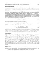
Figure 16.1 shows the multiple factors
that can contribute to the development of PCOS.
DEFINITION OF POLYCYSTIC OVARY SYNDROME (PCOS) 487
INSULIN
OBESITY
INSULIN
RESISTANCE
PITUITARY
LH
ACTH
ADRENAL
GLANS
P
450c17
ANDROGENS
DHEAS
PCOS
OVARY
P
450c17
ANDROGENS
TESTOSTERONE
ANDROSTENEDIONE
AND
FOLLICLE ARREST
ANOVULATION
OTHER FACTORS
Genes
Puberty
Premature
pubarche
Low
birthweight
IGFI
Figure 16.1 Development of PCOS and the multiple factors that affect steroid dysregu-
lation. Synergic role of insulin, LH and IGFI in androgen production. Other factors such
as genes or functional abnormalities in prenatal, childhood or pubertal periods must be
considered
The diagnosis of polycystic ovary syndrome is usually made on the basis of
a combination of clinical and biochemical criteria (Table 16.1).
The degree of hirsutism can be assessed by the Ferriman–Gallwey score, a
simple, semiquantitative method for recording the distribution and severity of
excess body hair.
11
The classic anatomical pattern of polycystic ovaries can be identified by
ultrasound assessment as increased number of subcapsular follicular cysts and
increased intervening stroma.
12
These ultrasound features are consistent with, but
not essential for, the diagnosis of the syndrome.
13
Serum levels of testosterone
and androstenedione are usually increased. DHEAS dehydroepiandrosterone sul-
fate levels are increased by up to 50 per cent in women with PCOS. Elevated
free testosterone activity, defined by the free androgen index, represents the
most sensitive biochemical marker supporting the diagnosis. Prolactin is usually
488 INSULIN RESISTANCE AND POLYCYSTIC OVARY SYNDROME
Table 16.1 Clinical and biochemical evaluation of PCOS
Menstrual disturbances
Ferriman–Gallwey score
Clinical Pelvic ultrasonography
Obesity (BMI)
Testosterone, androstenedione, DHEAS
SHBG
Biochemical FSH, LH, prolactin
Fasting glucose and insulin
OGTT
Test GnRH agonist (nafarelin, leuprolide acetate)
Dexamethasona suppression
normal, although it has been reported that approximately 15 per cent of PCOS
patients have mild elevations.
14
No single test is diagnostic of the syndrome, but choice should be guided
by clinical presentation. Serum LH levels are typically elevated in PCOS but
up to 50 per cent of the young women with other clinical and biochemical
features of the syndrome may have normal serum LH levels. Measurement of
LH is therefore of limited diagnostic value; it is quite specific that raised LH
and normal FSH essentially occur only in PCOS, but this is not very sensitive.
1
To assess insulin resistance with compensatory hyperinsulinism, fasting blood
glucose and insulin could be useful and simple to detect a primary abnormal-
ity. With a standard oral glucose tolerance test, a hyperinsulinaemic response,
impaired glucose tolerance or type 2 diabetes could be documented.
The abnormal response of 17 α-hydroxyprogesterone after an agonist ana-
logue of gonadotrophin-releasing hormone (GnRH) challenge has been described
in women and adolescents.
15, 16
Short-term leuprolide acetate (500 µgsc)isa
reliable tool for identification of the ovary source of hyperandrogenaemia. The
response was considered supranormal if the peak plasma 17 α-hydroxyprogester-
one 24 h postestimulation was greater than 4.75 nmol/l (160 ng/dl).
16
Hyperandrogenism in PCOS may therefore represent an intrinsic abnormality
of ovarian theca-interstitial cell function. This conclusion is supported by clinical
studies suggesting that the ovary is the primary abnormality site.
The response observed in women with PCOS in the above mentioned test
(GnRH agonist) could not be explained on the basis of LH hyper-responsiveness.
Women with PCOS given an hCG challenge test produce more androstenedione
and 17 α-hydroxyprogesterone than normal subjects and this difference remains
evident after suppression of endogenous LH secretion by GnRH.
17, 18
As many hyperandrogenic anovulatory women have significantly increased
ovarian steroidogenic responses to stimulation with GnRH analogues, Rosenfield
and colleagues have coined the term ‘functional ovarian hyperandrogenism,’ as
an alternative to PCOS.
19
HYPERANDROGENISM AND HYPERINSULINISM 489
16.3 Hyperandrogenism and hyperinsulinism
The earliest description of ‘diabete des femmes a barbe’ pointed out the rela-
tionship between androgen excess in women and disturbances in carbohydrate
metabolism.
20
The coexistence of severe insulin resistance and acanthosis nigri-
cans in three lean adolescent women confirmed the association between hyper-
androgenism and hyperinsulinism.
21
Insulin resistance associated with PCOS was also reported some years later by
Chang and colleagues in 1983.
22
This resistance, which is independent of obesity,
causes hyperinsulinaemia
23
and more than 50 per cent of the obese women with
PCOS are insulin resistant compared with age and weight-matched controls.
24
Hyperinsulinaemia is shown to be a characteristic finding in women with
ovarian androgen excess, even in the absence of diabetes. Nowadays, it has
become evident that insulin resistance is a cardinal feature of PCOS that could
serve as the pathogenic link between hyperandrogenism and hyperinsulinism.
Because insulin resistance is related to many manifestations of PCOS, there
tends to be substantial overlap between the PCOS phenotype and the so-called
‘metabolic syndrome’ or ‘syndrome X’: obesity, glucose intolerance, hyperten-
sion, macrovascular disease and dyslipidaemia, which are seen in both syn-
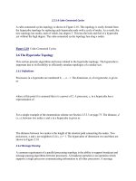
dromes. Figure 16.2 shows the metabolic and endocrine disorders associated
with PCOS and insulin resistance.
It is generally accepted that women with PCOS are predisposed to type 2
diabetes and that the development of diabetes cannot be attributed solely to the
obesity that typically accompanies PCOS. The prevalence of impaired glucose
tolerance in PCOS is between 30 and 40 per cent and that of type 2 diabetes
is between 5 and 10 per cent.
4, 5
These prevalences approximate those in Pima
indians, who have one of the highest rates of diabetes in the world. In addition,
suggesting some genetic risk factor in this process, most women and adolescents
with PCOS have a family history of type 2 diabetes.
25, 26
Elevated serum androgens may at times cause mild insulin resistance but it is
unlikely that the insulin resistance of PCOS occurs as a result of hyperandrogen-
ism.
27
Insulin resistance persists in women with PCOS in whom both ovaries
have been removed surgically or in women whose ovarian androgen produc-
tion has been suppressed with the use of long-acting gonadotrophin-releasing
hormone (GnRH) agonist.
1
Pre-pubertal women with acanthosis nigricans are hyperinsulinaemic, yet ele-
vated serum androgen levels do not appear until several years following the
diagnosis of insulin resistance. In the same way, some women with point muta-
tions in the insulin receptor gene causing hyperinsulinaemic insulin resistance
have been shown to have PCOS. Collectively, the genetic syndromes of severe
insulin resistance secondary to mutations in the insulin receptor gene (leprechau-
nism, Rabson–Mendenhall syndrome and type A insulin resistance syndrome)
have a common phenotype characterized by hyperandrogenism, insulin resis-
tance with hyperinsulinism and acanthosis nigricans. These observations support
490 INSULIN RESISTANCE AND POLYCYSTIC OVARY SYNDROME
PCOS
Insulin resistance
Glucose intolerance
Type 2 diabetes
Dyslipidaemia
Metabolic syndrome
Cardiovascular disorders
Hyperinsulinaemia
Liver Ovary
SHBG
IGFBP
1
Androgen
production
Metabolic
disorders
Endocrine
disorders
Obesity
Figure 16.2 Endocrine and metabolism disorders in PCOS
the idea that the hyperinsulinaemia of PCOS is a causal factor in the accompa-
nying hyperandrogenism.
3
It has been suggested that insulin, as IGFI, is capable of enhancing a variety of
steroidogenic pathways, not only in ovarian thecal cells, but in ovarian granulosa
cells, adrenocortical cells and the periphery. Furthermore, insulin seems to be
capable of exerting such effects directly, at elevations too modest to invoke such
action via interaction with the IGFI receptor.
28
Hyperinsulinaemia appears to be a major factor in the ovarian dysfunction of
PCOS. Any treatment that lowers insulin levels produces a decrease in androgen
levels and improves ovarian function.
The increase in insulin levels common in PCOS may precipitate hyper-
androgenaemia in genetically vulnerable individuals by acting through latent
abnormalities in steroidogenesis regulation, although it probably has only a
minimal effect on ovarian function in many individuals.
Paradoxically, hyperinsulinaemia is capable of exerting systemic effects in
patients moderately resistant to the effects of insulin on glucose metabolism.
Thus, it is capable of lowering IGFBP1 and SHBG concentrations and stimu-
lating ovarian steroidogenesis.
ASSESSMENT OF INSULIN RESISTANCE IN PCOS 491
One plausible hypothesis that tries to explain the relationships between hyper-
insulinaemia and hyperandrogenaemia is the unified serine activity in both
insulin receptors and cytochrome P450c17. Hormonally regulated serine phos-
phorylation of adrenal P450c17 by a c-AMP-dependent kinase accounts for a
large increase in 17–20 lyase activity and has been proposed as the mecha-
nism for normal adrenarche.
29
Phosphorylation studies of the insulin receptors
in fibroblasts from PCOS patients have shown that around half of the PCOS
women have an increase in serine phosphorylation, which produces an inhibi-
tion of tyrosine phosphorylation and a reduction of insulin signal transduction.
30
This means that abnormal serine phosphorylation, possibly associated with a
single kinase, may be responsible for excessive serine phosphorylation of the
insulin receptors and P450c17, leading to insulin resistance and adrenal/ovarian
hyperandrogenism. Even though the responsible kinase has not been identified
and the explained theory has not been confirmed, recent results suggest that a
serine kinase-mediated pathway may be involved in the insulin resistance of
PCOS patients.
31
At the moment, there is no unified theory to explain a heterogeneous dis-
ease such as PCOS, but the key role of insulin in this process is not ques-
tioned (Figure 16.1). The onset may occur in late childhood since many of the
metabolic and endocrine features of the disorder mimic puberty. Associated
with this are increases in the pulse, an amplitude of luteinizing hormone (LH),
increasing androgen concentrations, hyperinsulinism and irregular menses. Mul-
tiple, small ovarian cysts are seen on ultrasound examination and are a common
and normal feature of puberty. It is therefore possible that women genetically
predisposed to polycystic ovarian syndrome fail to resume normal insulin sensi-
tivity and continue to express metabolic and endocrine features usually confined
to puberty.
32, 33
16.4 Assessment of insulin resistance in PCOS
The euglycaemic–hyperinsulinaemic clamp technique
34
is the gold standard for
assessing insulin sensitivity and it is often combined with the hyperglycaemic
clamp to determine the adequacy of compensatory β-cell hypersecretion.
35
Insulin resistance and β-cell responsiveness can also be assessed by the fre-
quently sampled intravenous glucose tolerance test.
36
However, they cannot be used on a routine clinical basis or epidemiological
studies because they are too laborious, time consuming and invasive, especially
in children and adolescents. Surrogates based on fasting glucose and insulin and
on insulin and glucose responses to oral glucose have often been used.
Although assessment of either fasting or peak insulinaemia after OGTT could
provide sufficient data to classify individuals into normal, mild to moderate and
severe insulin resistance, the results of this test must be interpreted in the context
of plasma glucose levels, because the presence of any degree of hyperglycaemia
492 INSULIN RESISTANCE AND POLYCYSTIC OVARY SYNDROME
suggests the existence of defects in insulin secretion, which invalidates the
degree of insulinaemia as an index of insulin resistance. Fasting insulin lev-
els above 50–70 µU/ml or insulin peak in post-oral-glucose challenge above
350 µU/ml suggest severe insulin resistance, in contrast to the fasting insulin
levels below 20 µU/ml or OGTT peak insulin below 150 µU/ml observed in
normal individuals.
37
Various indices have been derived from the basis data provided by the oral
glucose tolerance test (OGTT) which allow quantitative estimation of β-cell
function, such as mean serum insulin index (MSI).
38, 39
Measures of insulin sensitivity based on fasting glucose and insulin include
the homeostasis model assessment (HOMA),
40
fasting insulin resistance index
(FIRI),
41
fasting glucose insulin ratio,
24
and quantitative insulin sensitivity check
index (QUICKI)
42
and others.
Determining the fasting glucose insulin ratio could be a good screening test
in that it is simple, quick and relatively inexpensive to obtain a single blood
sample, and it has been validated against ‘gold standard’ methodology.
43, 44
However, the glucose insulin ratio is most useful in a purely insulin-resistant
population, before overt β-cell dysfunction develops.
A fasting glucose–insulin ratio of less than seven in girls with premature
pubarche or obesity may be helpful in the early identification of those at risk
for complications of insulin resistance
43
and this finding was recently validated
44
by a stepwise regression analysis showing that the fasting glucose–insulin ratio
was significantly predictive of insulin sensitivity. A ratio of less than seven is
a cut-off for diagnosis of insulin resistance in adolescents with PCOS, and in
adult women with PCOS the ratio is less than 4.5.
24
As mentioned, measures
of insulin sensitivity can also be obtained from the OGTT.
Fasting insulin sensitivity and post-oral-glucose compensatory hyperinsuli-
naemia are closely related, although they do reflect distinct aspects of glucose
regulation. Fasting insulin levels reflect hepatic insulin sensitivity and the abil-
ity of insulin to suppress hepatic glucose production.
45
Post-oral-glucose insulin
excursions, on the other hand, in part reflect the need to suppress hepatic glucose
production and also the requirement to increase peripheral glucose disposal.
45
The high prevalence of impaired glucose tolerance and type 2 diabetes melli-
tus found in adult women with PCOS was also found in adolescents with PCOS
by means of 2 h glucose levels after 75 g glucose challenge.
46
To predict these
abnormalities the OGTT would be the choice and it was finally recommended
that adolescents with PCOS should undergo periodic screening for abnormal
glucose tolerance using 2 h post-challenge plasma glucose levels.
47
16.5 Gene studies on PCOS
Several reports have stressed that PCOS is a familial disorder; however, the
genetic basis of the syndrome remains controversial.
48
GENE STUDIES ON PCOS 493
It is difficult to determine the mode of inheritance of this heterogeneous
syndrome and there is an absence of an equivalent male phenotype. Some studies
have revealed an autosomal dominant mode of inheritance considering premature
balding in men as the primary male phenotype.
49, 50
On the other hand, there are studies of families with high prevalence of PCOS
in which the Mendelian autosomal dominant mode of inheritance cannot explain
the mode of inheritance of the syndrome,
51
while in another study an X-linked
model was postulated.
52
As a result, the mode of inheritance remains unclear
and more than one gene defect seems to participate in the pathogenesis of the
syndrome. Thus, PCOS appears to be an oligogenic disorder and several genes
may be involved in its aetiology.
The presence of insulin resistance and compensatory hyperinsulinaemia led
to the assumption that genes involved in the secretion and action of insulin may
play a role in the pathogenesis of PCOS.
Molecular studies of the coding region of the insulin receptor gene in women
with PCOS have shown a large number of silent polymorphisms, mainly in
intronic regions. The majority of these polymorphisms have also been iden-
tified in normal subjects and are considered to be common polymorphisms,
which do not lead to remarkable molecular disturbance in the insulin receptor
gene.
53
There is, however, evidence of a stable abnormality in insulin receptor phos-
phorylation in cells from women with PCOS. Increased insulin-dependent serine
phosphorylation of the insulin receptor β-subunit in skin fibroblast and skele-
tal muscle from 50 per cent of the women with PCOS was found compared
with controls.
30
The serine-phosphorylated insulin receptor had reduced ability
to phosphorylate tyrosine, suggesting that it may impair signal transduction.
A single-nucleotide polymorphism in the exon 17 C/T of the insulin receptor
was most frequently found in lean patients with PCOS compared with lean
controls, but the role of this susceptibility needs to be determined.
54
The minisatellite of the insulin gene INS VNTR (insulin gene variable number
tandem repeats) has been investigated since this region is directly implicated in
the regulation of insulin secretion.
The INS VNTR is a functional polymorphism, so it regulates the transcription
of the insulin gene and probably the expression of the IGF-II gene, which is
adjacent to the insulin gene.
55
An association between PCOS and allelic variation
at the INS VNTR locus has been reported. It was shown that class III alleles
and especially III/III genotypes are associated with PCOS and are most strongly
associated with anovulatory PCOS. The group of women with one or two class
III alleles had significantly higher fasting insulin levels and higher mean body
mass index than women with the I/genotype.
56
This finding was confirmed
in another study.
57
Conversely, in another European population of girls who
presented with precocious pubarche, hyperinsulinaemia and dyslipidaemia were
related to both birth weight and INS VNTR class I alleles.
58
494 INSULIN RESISTANCE AND POLYCYSTIC OVARY SYNDROME
Other candidate genes in the pathogenesis of PCOS are the encoding genes
of steroidogenic enzymes, such as CYP17, CYP11α and CYP19.
Recent studies have shown that PCOS may be the result of overfunction of the
enzyme that catalyses androgen production (cytochrome P450c17α). Cytochrome
P450c17α is an enzyme with two functions, since it has both 17 α-hydroxylase
and 17,20-lyase activities. In the thecal cells P450c17α converts progesterone
to 17 α-hydroxyprogesterone through its 17 α-hydroxylase activity, and then it
converts 17 α-hydroxyprogesterone to androstendione through its 17,20-lyase
activity.
15
Clinical studies have shown an abnormality in the regulation of 17 α-hydroxy-
lase/17,20-lyase (the rate-limiting step in androgen biosynthesis in the ovaries
and the adrenals) in women presenting with PCOS, as evidenced by increased
17 α-hydroxylase and to a lesser extent 17,20-lyase activity, since in these
women there is an exaggerated serum 17α-hydroxyprogesterone to stimulation
by gonadotrophin-releasing hormone agonists, as already mentioned.
19
The other gene involved in the steroidogenic pathway is CYP11α, which
encodes P450scc, the enzyme for cholesterol side chain cleavage that catalyses
the conversion of cholesterol to pregnenolone, which is the initial and rate-
limiting step at the start of the steroid hormone biosynthetic pathway. It has
been hypothesized that up-regulation of this enzyme could lead to increased
androgen production.
59
After some contradictory results, no association was found between any of
the alleles of the CYP11α and the presence of PCOS.
60, 61
The enzyme aromatase encoded by CYP19 catalyses the conversion of andro-
gens to oestrogens. It has been found that granulosa cells from anovulatory
polycystic ovaries are hyper-responsive to follicle-stimulating hormone (FSH)
in vitro, displaying significantly greater oestradiol production than granulosa
cells from normal ovaries.
62
So far, there is no evidence of any association of
alleles of this gene with PCOS.
63
The androgen receptor, through which all androgens act, has also been inves-
tigated, especially the polymorphic CAG repeat within exon 1, which encodes a
polyglutamine chain in the N-terminal transactivation domain.
64
The length of
the polymorphic CAG repeat sequence is inversely correlated to the androgen
receptor transcriptional activity.
An association between increased hirsutism and decreased CAG repeat length
has been demonstrated.
65, 66
However, further studies need to be conducted to
analyse the role of androgen receptors in the pathogenesis of PCOS.
Another gene studied to analyse the possible genetic origin of PCOS is LH
β-subunit gene; since about 50 per cent of women with PCOS have hyper-
secretion of LH associated with anovulation, an adverse role of LH gene may
be suspected.
One polymorphic variant seems to protect obese women from developing
symptomatic PCOS,
67
but another LH variant has been identified as a result of
PREMATURE PUBARCHE, HYPERINSULINISM AND PCOS 495
a single missense mutation in exon 3 of the LH β-subunit gene. This variant
seems to play a role in female infertility but further studies are required to
determine the pathological significance of this variant.
68
Recent investigations have shown an association between PCOS and follistat-
in,
60
but the contribution of follistatin gene in the development of PCOS has
not been confirmed.
69, 70
16.6 Premature pubarche, hyperinsulinism and PCOS
Premature pubarche is defined as the early appearance of pubic hair, before 8
years in girls and 9 years in boys, independently of the appearance of axillary
hair and apocrine secretion, and of pubertal development. The incidence of
premature pubarche is almost tenfold higher in girls than in boys. In most cases,
premature pubarche is due to an exaggerated variant of normal maturation of
adrenal gland function being a most frequent form of hyperandrogenism in the
pre-pubertal period.
71
Enzymatic defects of steroidogenesis are pathological causes of premature
pubarche, with a reported frequency around seven per cent in these girls.
72
Genetic defects in the CYP21 gene, which encodes the 21 hydroxylase en-
zyme, have been investigated, and the incidences of molecular defects were com-
parable in the premature pubarche and control groups. There is no relationship
between the presence of carrier status and endocrine–metabolic abnormalities.
73
Prospective studies of larger cohorts of premature pubarche girls are needed to
ascertain the long-term clinical relevance of CYP21 heterozygosity.
In the absence of an adrenal enzymatic defect, premature pubarche has been
associated with an acceleration of statural growth and bone maturation, with-
out affecting the timing of the onset or the progression of puberty or the
final height.
74
Re-evaluation of adrenal function in young women with a history of prema-
ture pubarche revealed an increased incidence of so called ‘idiopathic functional
adrenal hyperandrogenism’. A pattern of adrenal secretion that affects 50 per
cent of these girls gives rise to a suprahormonal response to ACTH test. Idio-
pathic functional adrenal hyperandrogenism has been attributed to a dysregula-
tion of adrenal cytochrome P450c17, prominently in the
5
pathway.
75
Post-pubertal follow-up of girls with premature pubarche has documented
more than tenfold prevalence of ‘functional ovarian hyperandrogenism’ (45
versus 3 per cent in the normal adolescent population), a form of PCOS at
adolescence, which is usually associated with hyperinsulinaemia and dyslipidae-
mia.
46, 76
This sequence seems to occur more frequently in girls with elevated
DHEAS and or androstenedione at diagnosis of premature pubarche.
77
Assessment of ovulatory function in girls with a history of precocious pub-
arche revealed that the fractions of ovulating girls and ovulatory cycles in
late post-menarche were strikingly higher (P<0.001) in the non-premature
496 INSULIN RESISTANCE AND POLYCYSTIC OVARY SYNDROME
pubarche than in the premature pubarche subgroup (91 versus 20 and 47 versus
12 per cent), with no differences in early post-menarche.
78
It could be assumed
that the development of ovarian hyperandrogenism after premature pubarche is
preceded by an apparently normal phase, with regular cycles lasting for about
3–5 years after menarche.
In general, puberty is associated with increasing fasting and glucose-
stimulated insulin concentrations and a decrease in insulin sensitivity.
79, 33
The
insulin resistance during puberty is restricted to peripheral glucose metabolism
and is associated with concomitant increases in growth hormone and insulin-
like growth factor (IGFI) secretion and a decrease in IGFBP1 and SHBG
concentrations.
80
The hyperinsulinaemia and increased IGFI activity during puberty have
been proposed as inducing factors in the development of PCOS in susceptible
subjects.
32, 81
In girls with premature pubarche, hyperinsulinism is already detectable before
puberty and throughout all states of pubertal development. It is often accom-
panied by an increased early insulin response to glucose, by an elevated free
androgen index and by decreased IGFBP1 and SHBG concentrations.
46
In addi-
tion to hyperinsulinaemia, girls with premature pubarche display supranormal
triglyceride levels, very low density lipoprotein cholesterol, and very low density
lipoprotein triglyceride concentrations.
76
Both hyperinsulinaemia and altered lipid profile support the concept that the
cluster of highly atherogenic abnormalities may already start by childhood, in
agreement with other studies pointing towards an early development of the patho-
physiological events leading to type 2 diabetes and cardiovascular disease.
82
The frequent association of premature pubarche with functional ovarian hyper-
androgenism and hyperinsulinism could have in common early origin rather than
being the result of a direct inter-relationship later in life. Reduced foetal growth
was first related to type 2 diabetes in older adults
83
and also was found to be
associated with insulin resistance in pre-pubertal and post-pubertal children born
small for gestational age.
84, 85
Girls with premature pubarche have lower birth-weight standard deviation
(SD) scores than control girls.
86
Those girls with premature pubarche who sub-
sequently develop functional ovarian hyperandrogenism have even lower birth
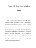
weights. Finally, the lowest birth weights were found in girls with – in addi-
tion – pronounced hyperinsulinism
86
(Figure 16.3).
The precise mechanism governing the aforementioned relationship is currently
unknown, but the results seem to suggest that premature pubarche and hyperinsuli-
naemia may precede the development of ovarian hyperandrogenism, and possibly
PCOS, and that this sequence may have a common early origin (low birth weight
serving as a marker). These data support the early life hypothesis that disease in
post-natal life may have its origin in the foetal environment, and that this process
can be attributed to changes in the programming of foetal endocrine axes.
83
TREATMENT APPROACH WITH ANTIANDROGENS 497
Precocious pubarche
Hyperinsulinism
Ovarian hyperandrogenism
Birth-weight SDS
–
n
= 31
–
–
+
n
= 25
–
–
n
= 11
+
+
+
+
n
= 12
–
+
∗
∗
∗
−2
−1
0
1
−3
Mean
p
≤ 0.01
∗
± Std. dev.
± Std. err.
Figure 16.3 Birth-weight scores of post-menarcheal control girls (−, − and −) and post-
menarcheal girls with a history of premature pubarche without ovarian hyperandrogenism and
without hyperinsulinaemia (+, − and −), with ovarian hyperandrogenism and without hyper-
insulinaemia (+, + and −) and with both ovarian hyperandrogenism and hyperinsulinaemia
(+, + and +)(J Clin Endocrinol Metab 1998, 83, 3558–3562, (with permission)
The follow-up related findings in girls with premature pubarche suggest that
this process should no longer be considered a normal variant of development but
rather a clinical marker of endocrine–metabolic disorders associated with reduced
foetal growth.
16.7 Treatment approach with antiandrogens
Current treatments until now have been addressed to reduce the main presenting
features such as irregular menses, hirsutism or infertility. Oral contraceptives are
commonly used to regulate menses and decrease ovarian androgen production.
Increasing levels of sex-hormone-binding globulin while decreasing ovarian
androgen production reduces the circulating free testosterone and subsequently
androgen activity; however, the combined pill exacerbates insulin resistance,
mainly in obese patients, in whom this treatment may be unsuitable.
Hirsutism may be addressed by the use of antiandrogens, cyproterone acetate
or spironolactone. Their principal mode of action is the inhibition of the binding
of dihydrotestosterone to its receptors at the hair follicle. Beneficial effects can be
seen after some months of treatment, but excessive hair growth returns soon after
cessation of treatment. Cyproterone acetate may exacerbate irregularity of the
menstrual cycle, and both drugs are unsuitable for use in those trying to conceive.
Another alternative approach to be used is finasteride, an inhibitor of the type 2
isoenzyme of 5α-reductase, the enzyme responsible for conversion of testosterone
498 INSULIN RESISTANCE AND POLYCYSTIC OVARY SYNDROME
to the active metabolite dihydrotestosterone. The other one is flutamide, the most
common antiandrogen used as a therapeutic regime in the treatment of hirsutism.
Flutamide is a non-steroidal compound that seems to act only at the receptor
site and is therefore considered a pure antiandrogen. Liver toxicity is a rare but
potentially severe side-effect of flutamide, which is dose dependent.
Several recent papers have been published evaluating treatment results of PCOS
with flutamide, spironolactone, cyproterone acetate, ketoconazole and finaster-
ide.
87–89
Those drugs employed currently constitute a satisfactory alternative
therapeutic regime in the treatment of hyperandrogenism. However, a long treat-
ment period is always required to improve hirsutism and prevent or delay its
relapse.
87
The reduction of androgen levels by flutamide restores normal ovarian
regulation of GnRH secretion in PCOS and may have a place in the therapeutic
regime aimed at establishing cyclic ovulation in women with PCOS.
89
According to our results,
88
flutamide treatment was accompanied by a marked
decrease in hirsutism score, free androgen index, testosterone and androstene-
dione levels and by an increase in sex-hormone-binding globulin concentrations.
However, there were no substantial changes in the pattern of menstrual cycles,
gonadotropin, oestradiol or dehydroepiandrosterone sulfate concentrations, and
there were no detectable effects on the 17-hydroxyprogesterone response to GnRH
agonist. Serum triglycerides, total cholesterol and low-density lipoprotein choles-
terol levels decreased markedly during flutamide therapy, whereas high-density
lipoprotein cholesterol, fasting glycaemia–insulinaemia and the insulin response
to a glucose load remained unchanged.
In conclusion, low dose flutamide treatment was found to be an effective and
safe approach to reduce hirsutism and circulating androgen, low-density lipopro-
tein cholesterol and triglyceride levels in girls with functional ovarian hyper-
androgenism after premature pubarche.
88
However, flutamide failed to increase
high-density lipoprotein cholesterol levels or decrease hyperinsulinaemia, these
being two major risk factors for subsequent cardiovascular disease.
16.8 Treatment approach with insulin sensitizers
(metformin)
Taking into consideration the aforementioned, the administration of insulin sensi-
tizer drugs such as metformin or thiazolidinediones could potentially reverse the
metabolic process and restore the ovarian function.
Several reports have been published in recent years addressing evaluation of
the effect of insulin sensitizer agents in PCOS women, such as biguanides and
thiazolidinediones (metformin and troglitazone).
Most of the metabolic abnormalities of PCOS can be reversed by metformin,
with the additional benefit of enough normalization of the endocrine milieu to
allow regular menstrual cycles, reversal of infertility and spontaneous pregnancy.
Thus, one report,
90
despite the short treatment period (8 weeks), was able to show
TREATMENT APPROACH WITH INSULIN SENSITIZERS (METFORMIN) 499
an improvement in insulin sensitivity associated with decrease in serum LH and
androgens. In contrast, it has been shown
91
that the administration of metformin in
24 obese women, presenting with hirsutism according to the criteria of Ferriman
and Gallwey had no additional benefit over the effect of low caloric diet in improv-
ing hyperinsulinaemia and hyperandrogenaemia. Moreover, in this study there
was no control group for the weight loss intervention. Recently, a study has been
published that supported the Vel
´
azquez et al. results, in which administration of
metformin in obese women with PCOS reduces ovarian cytochrome P450c17
activity and ameliorates hyperandrogenism and hirsutism by decreasing insulin
concentrations. In these women the exaggerated serum 17α-hydroxyprogesterone
response to stimulation by gonadotropin-releasing hormone agonist was reduced
after metformin treatment.
92
On the other hand, contradictory results were obtained in a study with a limited
number of PCOS women with moderate to extreme obesity. The study concluded
that hyperinsulinaemia and androgen excess in obese non-diabetic women with
PCOS were not improved by the administration of metformin.
93
Subsequently, one further study has been published to assess menstrual cyclic-
ity in 40 oligoamenorrheic women with PCOS. After a six-month course of
metformin an improvement in menstrual cyclicity and fertility was seen.
94
Another aspect that could be modified by metformin is the ovulatory response
to clomiphene. The frequency of spontaneous ovulation and ovulation induced by
clomiphene can be increased in obese women with PCOS by decreasing serum
insulin concentration with metformin.
95
An improvement in menstrual pattern
after metformin treatment has also been described and confirmed that, by reducing
hyperinsulinism, metformin determines a reduction in intraovarian androgens.
96
This leads to a reduction in oestradiol levels and favours orderly follicular growth
in response to exogenous gonadotropins.
97
In PCOS women with abdominal obesity, long-term treatment induced reduc-
tion in body mass index associated with a significant improvement of hirsutism
and menses abnormalities.
98
Moreover, the 17-hydroxyprogesterone response to
human chorionic gonadotropin was lower after metformin treatment,
99
giving a
direct demonstration that metformin leads to a reduction in stimulated ovarian
cytochrome P450c17 activity, concomitantly with a reduction in basal insulin and
testosterone levels and a significant increase in SHBG and IGFBP1.
100
In conclusion, metformin reduced hyperinsulinaemia and hyperandrogenaemia,
independently of changes in body weight. In a large number of subjects these
changes were associated with striking sustained improvements in menstrual abnor-
malities and resumption of ovulation.
101
Although not reported by all investigators, metformin seems to cause a decline
in insulin levels and reverses metabolic and ovarian abnormalities. Many of these
changes occur even in the absence of changes in body mass index.
The action of metformin is not fully known. It inhibits hepatic glucose
production and increases peripheral tissue sensitivity to insulin. In vitro
500 INSULIN RESISTANCE AND POLYCYSTIC OVARY SYNDROME
therapeutic concentrations of metformin have been shown to stimulate the tyrosine
kinase activity of the intracellular portion of the β-subunit of the human insulin
receptors.
102
We reported the results of 10-month treatment with metformin (850 mg twice
daily) in a lean girl aged 13 years and 6 months with severe hirsutism, acne, clitoral
hypertrophy, acanthosis nigricans and primary amenorrhea. Hormonal assessment
Metformin – + –
–
+
–
0
40
80
120
NS
NS
0
10
20
***
**
0
50
100
150
*** ***
0
50
100
******
0
10
***
**
0
25
50
75
****
LDL (mg/dL) HDL (mg/dL)
Glucose (mg/dL) MSI (mU/L)
Metformin
–
+
––
+–
Metformin – +
––
+
–
Ferriman & Gallwey Free Androgen Index
5
Figure 16.4 Clinical, endocrine and metabolic values before metformin treatment (−)after
6 months of treatment (+) and 3 months after treatment (−) in adolescent girls with hirsutism,
hyperandrogenism, oligomenorrhea, dyslipidaemia and hyperinsulinism after precocious pub-
arche. The top panel displays fasting glucose and mean serum insulin (MSI) during OGTT.
The middle panel shows changes in hirsutism score and FAI. The bottom panel shows changes
in serum LDL and HDL cholesterol (J Clin Endocrinol Metab 2000, 85, 3526–3530, with
permission)
TREATMENT APPROACH WITH INSULIN SENSITIZERS 501
showed a severe insulin resistance with hyperinsulinaemia and hyperandroge-
naemia. Molecular analysis of the insulin receptor gene showed a heterozygous
missense mutation (Val 1028) in exon 17 of the insulin receptor, abolishing
autophosphorylation of the insulin receptor β-subunit. Basal androgens and fasting
insulin concentrations decreased significantly during treatment, whereas SHBG
concentration increased. Breast development progressed and menarche occurred
in the fifth month of therapy. No side-effects were documented.
103
The results commented on above encourage the use of metformin in hyperinsuli-
naemic and hyperandrogenic women, but at present few studies have addressed the
use of metformin in children as a treatment for either insulin resistance PCOS.
104
In non-obese adolescent girls with hirsutism, hyperinsulinism, hyperandrogenism
and dyslipidaemia, metformin therapy tends to normalize these abnormalities in
concert.
105
Thus, in non-obese girls with an adolescent variant of PCOS, insulin-
sensitizing treatment reduces hyperinsulinism, dyslipidaemia and hyperandro-
genism and restores eumenorrhea and also induced ovulation
106
(Figure 16.4).
In conclusion, metformin was found to be an effective approach to reverse
metabolic and ovarian abnormalities even in adolescent girls. Prolonged treatment
with metformin has been proved to be safe in type 2 diabetes mellitus and in a
pregnant hyperandrogenic woman.
107
The most common morbidity associated
with its use is gastrointestinal distress, specifically diarrhoea and abdominal pain,
which is often transient and seems to be lessened if the dose is gradually increased.
16.9 Treatment approach with insulin sensitizers
(thiazolidinediones)
Another class of insulin-sensitizing agents, the thiazolidinediones, have been used
to improve PCOS abnormalities. These drugs require the presence of insulin, but
they do not stimulate insulin secretion. They mainly activate a nuclear receptor
called PPARγ (peroxisome proliferator-activated receptor gamma), which is most
strongly expressed in adipose tissue. Activated PPARγ increases transcription
of certain insulin-sensitive genes, including those that code for GLUT4 glucose
transporters and enzymes for lipogenesis.
The first thioglitazonedione, troglitazone, was introduced in Japan and the USA
in 1997 and withdrawn in 2000 due to reports of fatal idiosyncratic hepatotoxi-
city.
108
Other thiazolidinediones such as rosiglitazone and pioglitazone have
little evidence of hepatotoxicity, except two non-fatal cases of hepatocellular
damage observed with the initiation of rosiglitazone therapy. Thus, monitoring
of serum alanine transaminase should be performed before starting and during
therapy.
109, 110
Dunaif et al.
111
evaluated 21 PCOS subjects who received either 200 or
400 mg/day troglitazone for 12 weeks in a randomized, double-blind study.
Treatment with troglitazone resulted in significant improvement in insulin action.
Increases in insulin sensitivity were significant at both doses of troglitazone
502 INSULIN RESISTANCE AND POLYCYSTIC OVARY SYNDROME
but were more marked at 400 mg than at 200 mg. This was accompanied by
decreases in circulating insulin levels, both basally and after glucose load,
which were accounted for almost entirely by changes at 400 mg troglitazone
dose. In this report, insulin sensitivity was improved independent of weight
loss and hyperandrogenism was ameliorated. The author claimed
111
that this
observation is consistent with the hypothesis that hyperinsulinaemia contributes to
hyperandrogenism in PCOS. However, the apparent dose-related effect suggests
that these changes were troglitazone mediated.
Recently it could also be demonstrated that troglitazone improves the ovulatory
dysfunction, hirsutism, hyperandrogenemia and insulin resistance of PCOS in a
dose-related fashion, with a minimum of adverse effects.
112
It should be noted that
none of the insulin-sensitizing drugs have Food and Drug Administration (FDA)
approval for use in PCOS, hirsutism or hyperandrogenism with insulin resistance.
Considering that women with PCOS may have insulin resistance secondary
to a deficiency of D-chiro-inositol-containing phosphoglycans that mediate
insulin action, the administration of this substance could improve insulin
sensitivity. According to this hypothesis D-chiro-inositol increased insulin action
in patients with PCOS, thereby improving ovulatory function and decreasing
serum androgen concentrations, hirsutism, blood pressure and plasma triglyceride
concentrations.
113
The aforementioned results using insulin sensitizers give grounds for consider-
ing them a therapeutic approach for PCOS, alone or combined with antiandrogenic
drugs or oral contraceptives.
16.10 Conclusion
The therapeutical interventions with insulin-sensitizing agents corroborate the idea
that insulin resistance with hyperinsulinaemia may indeed be a prime factor under-
pinning the metabolic and hormonal disorders affecting anovulatory and ovarian
hyperandrogenic women and adolescents.
Randomized, controlled trials with safe insulin sensitizers will have to be con-
ducted, especially in young women and adolescents with hyperinsulinism and
anovulatory hyperandrogenism in an attempt to normalize insulin sensitivity and
ovarian function.
Considering that insulin sensitizers have less effect on hirsutism than antian-
drogens, these drugs could be combined with an antiandrogen such as flutamide at
low doses. Again, collaborative randomized trials in wide populations should be
conducted to assess treatment results of the clinical and metabolic abnormalities
in women and adolescents.
References
1. Franks, S. (1995) Medical progress article: polycystic ovary syndrome. N Engl J Med
333, 853–861.
REFERENCES 503
2. Knochenhauer, E. S., Key, T. J., Kahsar-Miller, M., Waggoner, W., Boots, L. R. and
Azziz, R. (1998) Prevalence of the polycystic ovary syndrome in unselected Black and
White women of the Southeastern United States: a prospective study. J Clin Endocrinol
Metab 83, 3078–3082.
3. Dunaif, A. (1997) Insulin resistance and the polycystic ovary syndrome: mechanism and
implications for pathogenesis. Endocr Rev 18, 774–800.
4. Ehrmann, D. A., Barnes, R. B., Rosenfield, R. L., Cavaghan, M. K. and Imperial, J.
(1999) Prevalence of impaired glucose tolerance and diabetes in women with polycystic
ovary syndrome. Diabetes Care 22, 141–146.
5. Legro, R. S., Kunselman, A. R., Dodson, W. C. and Dunaif, A. (1999) Prevalence and
predictors of risk for type 2 diabetes mellitus and impaired glucose tolerance in
polycystic ovary syndrome: a prospective, controlled study in 254 affected women.
J Clin Endocrinol Metab 84, 165–169.
6. Zawadski, J. K. and Dunaif, A. (1992) Diagnostic criteria for polycystic ovary
syndrome: towards a rational approach. In: Dunaif, A., Givens, J. R., Haseltine, F.
and Merriman, G. R., eds. Polycystic Ovary Syndrome. Boston, MA: Blackwell,
377–384.
7. Bridges, N. A., Cooke, A., Healy, M. J. R., Hindmarsh, P. C. and Brook, C. G. (1993)
Standard for ovarian volume in childhood and puberty. Fertil Steril 60, 456–460.
8. Diamanti-Kandarakis, E., Kouli, C. R., Bergiele, A. T., Filandra, F. A., Tsianateli, C.,
Spina, G. G., Zapanti, E. P. and Bartzis, M. I. (1999) A survey of the polycystic ovary
syndrome in the Greek island of Lesbos: hormonal and metabolic profile. J Clin
Endocrinol Metab 84, 4006–4011.
9. Asunci
´
on, M., Calvo, R. M., San Millan, J. L., Sancho, J., Avila, S. and Escobar-
Morreale, H. F. (2000) A prospective study of the prevalence of the polycystic ovary
syndrome in unselected Caucasian women from Spain. J Clin Endocrinol Metab 85,
2434–2438.
10. Barbieri, R. L. (1994) Hyperandrogenism, insulin resistance and acanthosis nigricans:
10 years of progress. J Reprod Med 39, 327–336.
11. Ferriman, D. and Gallwey, J. D. (1961) Clinical assessment of body hair growth in
women. J Clin Endocrinol Metab 21, 1440–1447.
12. Adams, J., Franks, S., Polson, D. W., Mason, H. D., Abdulwahid, N., Tucker, M.,
Morris, D. V., Price, J. and Jacobs, H. S. (1985) Multifollicular ovaries: clinical and
endocrine features and response to pulsatile gonadotropin-releasing hormone. Lancet 2,
1375–1379.
13. Polson, D. W., Wadsworth, J., Adams, J. and Franks, S. (1988) Polycystic ovaries: a
common finding in normal women. Lancet 1, 870–872.
14. Lucciano, A. A., Chapler, F. K. and Serman, B. M. (1984) Hyperprolactinemia in the
polycystic ovary syndrome. Fertil Steril 41, 719–725.
15. Rosenfield, R. L., Barnes, R. B., Cara, J. F. and Lucky, A. W. (1990) Dysregulation of
cytochrome P450c17α as the cause of polycystic ovarian syndrome. Fertil Steril 53,
785–791.
16. Ib
´
a
˜
nez, L., Potau, N., Zampolli, M., Prat, N., Gussiny
´
e, M., Saenger, P., Vicens-
Calvet, E. and Carrascosa, A. (1994) Source localization of androgen excess in adolescent
girls. J Clin Endocrinol Metab 79, 1778–1784.
17. Ib
´
a
˜
nez, L., Hall, J., Potau, N., Carrascosa, A., Prat, N. and Taylor, A. (1996) Ovarian 17-
hydroxyprogesterone hyperresponsiveness to gonadotropin-releasing hormone (GnRH)
agonist challenge in women with polycystic ovary syndrome is not mediated by
luteinizing hormone hypersecretion: evidence from GnRH agonist and human chorionic
gonadotropin stimulation testing. J Clin Endocrinol Metab 81, 4103–4107.
504 INSULIN RESISTANCE AND POLYCYSTIC OVARY SYNDROME
18. Gilling-Smith, C., Story, H., Rogers, V. and Franks, S. (1997) Evidence for a primary
abnormality of thecall cell steroidogenesis in the polycystic ovary syndrome. Clin
Endocrinol 47, 93–99.
19. Rosenfield, R. L., Barnes, R. B. and Ehrmann, D. A. (1994) Studies of the nature of 17-
hydroxyprogesterone hyperresponsiveness to gonadotropin-releasing agonist challenge
in functional ovarian hyperandrogenism. J Clin Endocrinol Metab 79, 1686–1692.
20. Archard, C. and Thiers, J. (1921) Le virilisme filaire et son association a l’insuffisance
glycolytique (diabete des femmes a barbe). Bull Acad Natl Med 86, 51–64.
21. Kahn, C. R., Fliers, J. S., Bar, R. S., Archer, J. A., Gorden, P., Martin, M. M. and
Roth, J. (1976) The syndromes of insulin resistance and acanthosis nigricans. N Engl J
Med 294, 739–745.
22. Chang, R. J., Nakamura, R. M., Judd, H. L. and Kaplan, S. A. (1983) Insulin resistance
in nonobese patients with polycystic ovarian disease. J Clin Endocrinol Metab 57,
356–359.
23. Dunaif, A., Segal, K. R., Futterweit, W. and Dobrjansky, A. (1989) Profound peripheral
insulin resistance, independent of obesity, in polycystic ovary syndrome. Diabetes 38,
1165–1174.
24. Legro, R. S., Finegood, D. and Dunaif, A. (1998) A fasting glucose to insulin ratio is a
useful measure of insulin sensitivity in women with polycystic ovary syndrome. J Clin
Endocrinol Metab 83, 2694–2698.
25. Sir-Peterman, T., Angel, B., Maliqueo, M., Carvajal, F., Santos, L. and P
´
erez-Bravo, F.
(2002) Prevalence of type II diabetes mellitus and insulin resistance in parents of women
with polycystic ovary syndrome. Diabetologia 45, 959–964.
26. Ib
´
a
˜
nez, L., Castell, C., Tresserras, R. and Potau, N. (1999) Increased prevalence of type
2 diabetes mellitus and impaired glucose tolerance in first-degree relatives of girls with
a history of precocious pubarche. Clin Endocrinol 51, 395–401.
27. Speiser, P. W., Serrat, J., New, M. I. and Gertner, J. M. (1992) Insulin insensitivity
in adrenal hyperplasia due to nonclassical steroid 21-hydroxylase deficiency. J Clin
Endocrinol Metab 75, 1421–1424.
28. Rosenfield, R. L. (1999) Ovarian and adrenal function in polycystic ovary syndrome.
Endocrinol Metab Clin North Am 28, 265–293.
29. Zhang, L. H., Rodriguez, H., Ohno, S. and Miller, W. L. (1995) Serine phosphorylation
of human P450c17α increases 17,20-lyase activity: implications for adrenarche and
polycystic ovary syndrome. PNAS 92, 10 619–10 623.
30. Dunaif, A., Xia, J., Book, C. B., Schenker, E. and Tang, Z. (1995) Excessive insulin
receptor serine phosphorylation in cultured fibroblasts and in skeletal muscle. A potential
mechanism for insulin resistance in the polycystic ovary syndrome. J Clin Invest 96,
801–810.
31. Li, M., Youngren, J. F., Dunaif, A., Goldfine, I. D., Maddux, B. A., Zhang, B. B. and
Evans, J. (2002) Decreased insulin receptor (IR) autophosphorylation in fibroblast from
patients with PCOS: Effects of serine kinase inhibitors and IR activators. J Clin
Endocrinol Metab 87, 4088–4093.
32. Nobels, F. and Dewailley, D. (1992) Puberty and polycystic ovary syndrome: The
insulin/insulin-like growth factor hypothesis. Fertil Steril 58, 655–663.
33. Potau, N., Ib
´
a
˜
nez, L., Riqu
´
e, S. and Carrascosa, A. (1997) Pubertal changes in insulin
secretion and peripheral insulin sensitivity. Horm Res 48, 219–226.
34. DeFronzo, R. A., Tobin, J. D. and Andres, R. (1979) Glucose clamp technique: a method
for quantifying insulin secretion and resistance. Am J Physiol 237, E214–E223.
35. Polonsky, K. S., Given, B. D., Hirsch, L., Shapiro, E. T., Tillil, H., Beebe, C.,
Galloway, J. A., Frank, B. H., Karrison, T. and Van Cauter, E. (1988) Quantitative study
of insulin secretion and clearance in normal and obese subjects. J Clin Invest 81, 435–441.