PATHOLOGY OF VASCULAR SKIN LESIONS - PART 8 doc
Bạn đang xem bản rút gọn của tài liệu. Xem và tải ngay bản đầy đủ của tài liệu tại đây (1.67 MB, 34 trang )
224 Sangüeza and Requena / Pathology of Vascular Skin Lesions
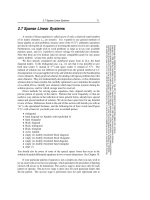
Fig. 7. AIDS-associated Kaposi’s sarcoma involving the oral mucosa.
adnexae and blood vessels, producing the so-called promontory sign (71) (Fig. 9). In
other areas, the blood vessels infiltrate between collagen bundles of the dermis, giving
the appearance that they are “dissecting” the stroma. In rare instances, the newly formed
blood vessels form clusters that resemble small hemangiomas (71,74). The inflammatory
cells present are predominantly lymphocytes and plasma cells. The presence of plasma
cells around newly formed irregular blood vessels is a helpful clue in the histopathologic
diagnosis of the patch stage of Kaposi’s sarcoma (71). Another characteristic feature of
patch stage lesions of Kaposi’s sarcoma is the presence of scattered necrotic endothelial
cells, a feature that has been emphasized at both the conventional microscopy (77) and
ultrastructural level (41). Hemosiderin-laden macrophages are another feature frequently
found in early lesions of Kaposi’s sarcoma. The histopathologic features just described
for the patch lesions can also be seen in clinically normal areas of skin in patients who
have Kaposi’s sarcoma elsewhere, which supports the notion of the diffuseness of the
process from its inception (79,80).
Plaque lesions of Kaposi’s sarcoma tend to involve the entire dermis and even the
upper part of the subcutaneous fat. At this stage, there is an increased number of spindle
cells arranged in short fascicles between collagen bundles centered around proliferating
vascular channels. The spindle cells line irregularly shaped, slit-like vascular spaces that
contain isolated erythrocytes. They display minimal or no atypia, with only a few to no
mitotic figures.
09/Sangüeza/217-274/F 01/16/2003, 10:12 AM224
Chapter 9 / Malignant Neoplasms 225
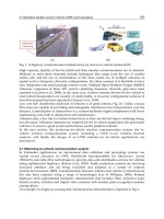
Fig. 8. Histopathologic features of patch stage Kaposi’s sarcoma. (A) At scanning magnification,
the lesions consists of increased numbers of jagged spaces at different levels of the dermis. (B)
Higher magnification shows that the jagged spaces are lined by thin endothelial cells. (C) At this
point it is possible to see inflammatory infiltrate with some plasma cells and extravasated eryth-
rocytes.
When the number of spindle cells increases, lesions of Kaposi’s sarcoma become
nodular. Then the spindle cells are arranged in interwoven fascicles with erythrocytes
scattered in the interstices (Fig. 10). Nuclear atypia, pleomorphism, and mitotic figures
may be seen but are usually not very prominent. In rare instances, however, especially
09/Sangüeza/217-274/F 01/16/2003, 10:12 AM225
226 Sangüeza and Requena / Pathology of Vascular Skin Lesions
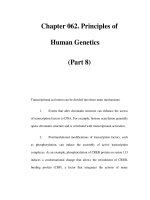
Fig. 9. Histopathologic features in plaque lesions of Kaposi’s sarcoma. (A) Scanning power shows
involvement of the upper part of the dermis by the neoplastic process. (B) Higher magnification
shows irregular vascular spaces and inflammatory infiltrate. (C) The “promontory” sign is evident
around preexisting capillaries. (D) Numerous plasma cells are present in the stroma surrounding
the areas of “promontory” sign.
09/Sangüeza/217-274/F 01/16/2003, 10:12 AM226
Chapter 9 / Malignant Neoplasms 227
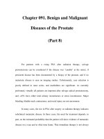
Fig. 10. Histopathologic features in nodular lesions of Kaposi’s sarcoma. (A) Scanning magnifi-
cation shows a well-circumscribed cellular nodule in the dermis. (B) Higher magnification dem-
onstrates that the nodule is composed of fascicles of spindle cells. Some congestive vascular
lumina are also seen. (C) Spindle cells are monomorphous, and nuclear atypia, pleomorphism, and
mitotic figures are not usually prominent.
09/Sangüeza/217-274/F 01/16/2003, 10:12 AM227
228 Sangüeza and Requena / Pathology of Vascular Skin Lesions
in the African variant (81), a significant number of mitotic figures and atypical cells may
be seen in lesions of Kaposi’s sarcoma. A rather characteristic, but probably not specific,
finding is the presence of the so-called hyaline globules. Although these are most
common in the plaque and nodular lesions of Kaposi’s sarcoma, they can be present at
any stage of the disease. These globules are periodic acid-Schiff (PAS)-positive and
diastase-resistant, consist of eosinophilic spherules measuring between 1 and 10 µm, and
are located both intra- and extracellularly. Most likely these hyaline globules represent
degenerated erythrocytes that are phagocytized and confined to the phagolysosomes of
the neoplastic cells (82–85). These globules have been also described in other vas-
cular proliferations such as angiosarcomas, pyogenic granulomas, and granulation
tissue (84).
In rare instances, lesions of Kaposi’s sarcoma may present clinically as bullous lesions
with histopathologic features of a lymphangioma. These lesions show irregular vascular
channels lined by a single layer of flattened endothelial cells devoid of erythrocytes
within the dermis (86–94). The absence of hemosiderin deposits and the scarcity of
spindle cells also contribute to a lymphangiomatous appearance of the lesions. Occasion-
ally, the lymphangiomatous pattern can be seen focally within an otherwise stereotypical
lesion of Kaposi’s sarcoma. There have been patients with classic Kaposi’s sarcoma in
which some lesions showed a lymphangioma-like pattern, whereas other lesions of the
same patient exhibited the typical findings associated with Kaposi’s sarcoma, with abun-
dant extravasated erythrocytes and hemosiderin deposits. Chronic lymphedema (86) and/
or the use of electron beam therapy (94) on the involved extremity may be responsible
for the formation of the lymphangioma-like lesions in Kaposi’s sarcoma.
From a histogenetic point of view, current evidence suggests that Kaposi’s sarcoma is
a proliferative process. Ultrastructural and immunohistochemical studies have shown that
the spindle cell component shows endothelial differentiation (95,96). Whether this repre-
sents endothelium of blood vessels or lymphatics remains to be determined (97–107). Well-
formed vessels in lesions of Kaposi’s sarcoma are lined by cells that are positive for factor
VIII-related antigen, but studies for this marker in neoplastic spindle cells have provided
conflicting results (96,99,102–109). In contrast, Ulex europaeus I lectin has consistently
been detected in the spindle cells (99,108). Rutgers et al. (97) concluded that spindle cells
in Kaposi’s sarcoma are blood vascular endothelial cells, because they stain with mono-
clonal antibodies OKM5, anti-E92, and HCl, which react with blood capillary endothelium,
but not with lymphatic endothelium. Identical conclusions were reported by Scully et al.
(109) on the basis of the immunoreactivity of spindle cells with the antibody B721.
Russell Jones et al. (99,108) noted that immunoreactivity of the spindle cells varies
with the stage and type of the lesions. Early patch stage lesions have the profile of a
lymphatic lesion because the cells are positive for the antibody EN-4, which stain all
types of endothelial cells, but are negative with the antibody PAL-E, which is specific for
blood vessel endothelium. Nodular lesions of Kaposi’s sarcoma stain with EN-4 and
express variable immunoreactivity with PAL-E (99,108). Beckstead et al. (101) also
favored lymphatic endothelial differentiation because of the lack of HLA-DR/Ia and
alkaline phosphatase and the intense staining with 5' nucleotidase. In contrast, the
presence of abundant laminin and type IV collagen surrounding many of the individual
spindle cells has been interpreted as evidence favoring a blood vascular endothelium
rather than lymphatic endothelium differentiation (110–112). More recently, Weich et al.
09/Sangüeza/217-274/F 01/16/2003, 10:12 AM228
Chapter 9 / Malignant Neoplasms 229
(113) have suggested a close relationship between the Kaposi’s sarcoma cell and the
vascular smooth muscle cell, since the tumors express mRNA for α-smooth muscle actin.
Different investigators have found that the Kaposi’s sarcoma cells express the CD34
antigen, also known as the human progenitor cell antigen. This is a 105–120-kDa single-
chain transmembrane glycoprotein expressed constitutively by endothelial cells of small
blood, but not lymphatic, vessels in several tissues (114–118).
A nonvascular origin for the spindle cells of the Kaposi’s sarcoma has also been
suggested, based on the presence of many factor XIIIa-positive spindle cells in the lesions,
suggesting that the factor XIIIa-expressing dermal dendrocyte (a member of the mono-
nuclear phagocytic system) may be the cell of origin for the spindle-shaped cells of
Kaposi’s sarcoma (119,120). Other authors, however, believe that these factor XIIIa-
positive dendritic cells represent reactive hyperplasia of dermal dendrocytes, rather than
neoplastic cells (107). More recently, vascular endothelial growth factor receptor-3
(VEGFR-3) (121–124) and podoplanin (123,125), two relatively sensitive and specific
markers for lymphatic endothelium, have been identified in most Kaposi’s sarcomas,
supporting a lymphatic differentiation for this neoplasm.
Ultrastructural studies have documented that under electron microscopy most of the
spindle cells exhibit characteristics of endothelial cells, although a few of them also show
features of pericytes and fibroblasts (126). The cells surrounding vascular spaces show
few intercellular junctions, with gaps between them. A fragmented basal lamina encircles
the luminal cells in absence of pericytes. These ultrastructural features seem to be more
compatible with lymphatic than blood vascular differentiation, but it is possible that their
absence may be the result of the dilation of blood vessels (41).
The differential diagnosis of Kaposi’s sarcoma from pseudo-Kaposi’s sarcoma lesions
is usually straightforward. In both the acroangiodermatitis and arteriovenous malforma-
tion variants of pseudo-Kaposi’s sarcoma lesions, the blood vessels of the papillary
dermis are involved, demonstrating a lobular proliferation of round, dilated, thick-walled
capillaries, with plump endothelial cells (127). This vascular proliferation is superim-
posed on a background of dermal fibrosis, erythrocyte extravasation, and abundant
hemosiderin. The irregular jagged vascular channels with slit-like lumina surrounding
preexisting capillaries, found in early lesions of Kaposi’s sarcoma, are not seen.
Benign lymphangioendothelioma can be also mistaken for early lymphangioma-like
lesions of Kaposi’s sarcoma. Similarities include thin-walled, endothelium-lined vascu-
lar spaces between collagen bundles that appear to “dissect” the dermis. However, these
vascular spaces of benign lymphangioendothelioma are usually arranged horizontally in
the dermis and show no tendency to surround preexisting blood vessels as in Kaposi’s
sarcoma. Furthermore, the absence of extravasated erythrocytes, hemosiderin, and plasma
cells is also helpful. The clinical appearance of the lesion is helpful, too, because benign
lymphangioendothelioma presents as a solitary lesion.
Hobnail hemangioma also shares common histopathologic features with Kaposi’s
sarcoma, especially at the periphery of the lesion. In these areas, there are irregular
angulated vascular lumina that appear to be dissecting collagen bundles and abundant
hemosiderin, raising the possibility of early lesions of Kaposi’s sarcoma. However, in the
central areas of hobnail hemangioma, there are widely dilated vascular lumina with
intraluminal papillary projections, prominent endothelial cells, and frequent fibrin
thrombi. These features are not seen in Kaposi’s sarcoma.
09/Sangüeza/217-274/F 01/16/2003, 10:12 AM229
230 Sangüeza and Requena / Pathology of Vascular Skin Lesions
Spindle cell hemangioma is frequently confused with nodules of Kaposi’s sarcoma
because in both entities there are fascicles of spindle cells with slit-like vascular spaces
containing erythrocytes. However, spindle cell hemangioma shows dilated blood vessels
and areas of epithelioid cells, with prominent cytoplasmic vacuoles, which are not seen
in nodules of Kaposi’s sarcoma.
Kaposiform hemangioendothelioma bears a striking resemblance to the nodular lesions
of Kaposi’s sarcoma; however, the clinical settings are different. Kaposi’s sarcoma is
uncommon in infancy, and kaposiform hemangioendothelioma is a solitary neoplasm.
Furthermore, histopathologically, kaposiform hemangioendothelioma shows a lobulated
growth pattern and hemangioma-like areas, especially at the periphery of the lobules.
The biologic behavior of Kaposi’s sarcoma depends on the epidemiologic type of the
disease and the immune status of the host. There have been diverse opinions as to whether
Kaposi’s sarcoma represents a reactive vascular proliferation or a true neoplastic prolif-
eration. Currently, there is a consensus that Kaposi’s sarcoma does not produce metasta-
sis in the manner of conventional sarcomas, but rather it develops in a multifocal fashion
(128). This notion is based on the finding of changes in internal organs such as the lymph
nodes, gastrointestinal tract, lung, and kidney similar to those seen in the lesions of the
patch stage of Kaposi’s sarcoma in the skin. Despite the lack of metastatic potential,
patients can succumb to the effects of Kaposi’s sarcoma. Immunocompetent individuals
affected with the classic variant of Kaposi’s sarcoma have a mortality rate between 10 and
20% after 10 years, whereas Kaposi’s sarcoma in AIDS patients has a far more aggressive
course; the overall mortality rate is 41% and death occurs within a relatively short period
of time (129).
T
REATMENT
The treatment of Kaposi’s sarcoma includes local and/or systemic therapy. The appro-
priate selection of therapy in each case depends on the epidemiologic variant of the
disease, the number of lesions, and the immune status of the patient. In patients with
AIDS, new antiretroviral therapies, in particular the protease inhibitors, appear to be
changing the clinical course of Kaposi’s sarcoma. Local therapies include liquid nitrogen
cryotherapy, radiation therapy, laser therapy, and intradermal therapy with cytotoxic
chemotherapy drugs or interferon. Systemic therapies include limited intervention with
interferon, with or without zidovudine, and more aggressive intervention with single or
multiagent chemotherapy modalities. Therapeutic options for the different clinical set-
tings of Kaposi’s sarcoma have been recently reviewed by Tappero et al. (10). The most
recent alternatives consist of topical treatment with alitretinoin gel (130), and the admin-
istration of liposomal doxorubicin (131) or vinorelbine (132).
References
1. Kaposi M. Idiopathisches multiples Pigmentsarkom der Haut. Arch Dermatol Syph 1872;4:265–73.
2. DiGiovanna JJ, Safai B. Kaposi’s sarcoma: retrospective study of 90 cases with particular emphasis on
the occurrence, ethnic background, and prevalence of other diseases. Am J Med 1981;71:779–83.
3. Oettle AG. Geographical and racial differences in the frequency of Kaposi’s sarcoma as evidence of
environmental or genetic causes. Acta Unio Int Cancer 1962;18:330–63.
4. Gange RW, Wilson Jones E. Kaposi’s sarcoma and immunosuppressive therapy: an appraisal. Clin Exp
Dermatol 1978;3:135–46.
5. Centers for Disease Control. Follow-up on Kaposi’s sarcoma and Pneumocystis pneumonia. MMWR
1981;30:409–10.
09/Sangüeza/217-274/F 01/16/2003, 10:12 AM230
Chapter 9 / Malignant Neoplasms 231
6. Friedman-Kien L, Lauberstein L, Marmor M, et al. Kaposi’s sarcoma and Pneumocystis pneumonia
among homosexual men—New York and California. MMWR 1981;30:305–8.
7. Gottlieb GJ, Ragaz A, Vogel JV, et al. A preliminary communication on extensively disseminated
Kaposi’s sarcoma in young homosexual men. Am J Dermatopathol 1981;3:111–4.
8. Gottlieb GJ, Ackerman AB. Kaposi’s Sarcoma: A Text and Atlas. Philadelphia, Lea & Febiger, 1988.
9. Friedman-Kien AE, Saltzman BR. Clinical manifestations of classical, endemic African, and epidemic
AIDS-associated Kaposi’s sarcoma. J Am Acad Dermatol 1990;22:1237–50.
10. Tappero JW, Conant MA, Wolfe SF, Berger TG. Kaposi’s sarcoma. Epidemiology, pathogenesis,
histology, clinical spectrum, staging criteria and therapy. J Am Acad Dermatol 1993;28:371–95.
11. Chor PJ, Santa Cruz DJ. Kaposi’s sarcoma. A clinicopathologic review and differential diagnosis. J Cutan
Pathol 1992;19:6–20.
12. Wabinga HR, Parkin DM, Wabwire-Mangen F, Mugerwa J. Cancer in Kampala, Uganda, in 1989-91:
changes in incidence in the era of AIDS. Int J Cancer 1993;54:24–36.
13. Olweny CLM. Epidemiology and clinical features of Kaposi’s sarcoma in tropical Africa. In: Fried-
man-Kien AE, Laubenstin LJ, eds. AIDS: The Epidemic of Kaposi’s Sarcoma and Opportunistic
Infections. New York, Masson, 1984:35–40.
14. Myers BD, Kessler E, Levi J, et al. Kaposi’s sarcoma in kidney transplant recipients. Arch Intern Med
1974;133:307–11.
15. Harwood AR, Osoba D, Hofstader SL, et al. Kaposi’s sarcoma in recipients of renal transplants. Am
J Med 1979;67:759–65.
16. Penn I. Kaposi’s sarcoma in organ transplant recipients. Transplantation 1979;27:8–11.
17. Klein MB, Pereira FA, Kantor I. Kaposi’s sarcoma complicating systemic lupus erythematosus treated
with immunosuppression. Arch Dermatol 1974;110:602–4.
18. Kapadia SB, Krause JR. Kaposi’s sarcoma after long-term alkylating agent therapy for multiple
myeloma. South Med J 1977;70:1011–3.
19. Leung F, Fam AG, Osoba D. Kaposi’s sarcoma complicating corticosteroid therapy for temporal
arteritis. Am J Med 1981;71:320–2.
20. Hoshaw RA, Schwartz RA. Kaposi’s sarcoma after immunosuppressive therapy with prednisone. Arch
Dermatol 1980;116:1280–2.
21. Zisbrod Z, Haimov M, Schanzer H, et al. Kaposi’s sarcoma after kidney transplantation: report of
complete remission of cutaneous and visceral involvement. Transplantation 1980;30:383–4.
22. Brooks JJ. Kaposi’s sarcoma: a reversible hyperplasia. Lancet 1986;2:1309–11.
23. Beral V, Peterman TA, Berkelman RL, et al. Kaposi’s sarcoma among persons with AIDS: a sexually
transmitted infection? Lancet 1990;335:123–8.
24. Couturier E, Ancelle-Park RA, de Vicenzi I, et al. Kaposi’s sarcoma as a sexually transmitted disease.
Lancet 1990;335:1105.
25. Dictor M, Bendsoe N. Transmissible agent of Kaposi’s sarcoma. Lancet 1990;335:797.
26. Kitchen VS, French MAH, Dawkins RL. Transmissible agent of Kaposi’s sarcoma. Lancet 1990;
335:797–8.
27. Barry M, Vittecoq D, Liotier JY. Heterosexual transmission of the etiological agent of Kaposi’s sar-
coma. Lancet 1991;337:234.
28. Eggers HJ, Weyer J. Linkeage and independence of AIDS and Kaposi’s disease: the interaction of
human immunodeficiency virus and some coagents. Infection 1991;19:115–22.
29. Archibald CP, Schecter MT, Craib KJP, et al. Risk factors for Kaposi’s sarcoma in the Vancouver
lymphadenopathy-AIDS study. J Acquir Immuno Defic Syndr 1990;3(suppl 1):S18–S23.
30. Jacobson LP, Muñoz A, Phair JP, et al. Incidence of Kaposi’s sarcoma in a cohort of homosexual
men infected with the human immunodeficiency virus type I. J Acquir Immuno Defic Syndr
1990;3(suppl 1):S24–S31.
31. Giraldo G, Beth E, Haguenau F. Herpes-type virus particles in tissue culture of Kaposi’s sarcoma from
different geographic regions. J Natl Cancer Inst 1972;49:1409–26.
32. Giradlo G, Beth E, Kourilsky FM, et al. Antibody patterns of herpesvirus in Kaposi’s sarcoma: sero-
logic associations of European Kaposi’s sarcoma with cytomegalovirus. Int J Cancer 1975;15:839–48.
33. Giraldo G, Beth E, Henle W, et al. Antibody patterns to herpesviruses in Kaposi’s sarcoma. II. Serological
association of American Kaposi’s sarcoma with cytomegalovirus. Int J Cancer 1978;22:126–31.
34. Giraldo G, Beth E, Huang ES. Kaposi’s sarcoma and its relationship to cytomegalovirus (CMV). III.
CMV DNA and early antigens in Kaposi’s sarcoma. Int J Cancer 1980;26:23–9.
09/Sangüeza/217-274/F 01/16/2003, 10:12 AM231
232 Sangüeza and Requena / Pathology of Vascular Skin Lesions
35. Kestens L, Melbye M, Bigger RJ, et al. Endemic African Kaposi’s sarcoma is not associated with
immunodeficiency. Int J Cancer 1985;36:49–54.
36. Ambinder RF, Newman C, Hayward GS, et al. Lack of association of cytomegalovirus with endemic
African Kaposi’s sarcoma. J Infect Dis 1987;156:193–7.
37. Fiala M, Payme JE, Berne TV, et al. Epidemiology of cytomegalovirus infection after transplantation
and immunosuppression. J Infect Dis 1975;132:421–33.
38. Drew WL, Minitz L, Miner RC. Prevalence of cytomegalovirus infection in homosexual men. J Infect
Dis 1981;143:188–92.
39. Drew WL, Conant MA, Miner RC, et al. Cytomegalovirus and Kaposi’s sarcoma in young homosexual
men. Lancet 1982;2:125–7.
40. Friedman-Kien AE, Laubestein LJ, Rubenstein P, et al. Disseminated Kaposi’s sarcoma in homosexual
men. Ann Intern Med 1982;96:693–700.
41. McNutt NS, Fletcher V, Conant MA. Early lesions of Kaposi’s sarcoma in homosexual men: an
ultrastructural comparison with other vascular proliferations in skin. Am J Pathol 1983;111:62–77.
42. Minitz L, Drew L, Miner RC, et al. Cytomegalovirus in homosexual men: an epidemiologic study. Ann
Intern Med 1983;99:326–9.
43. Drew WL. Sexual transmission of CMV and its relationship to Kaposi’s sarcoma in homosexual men.
Birth Defects 1984;20:121–9.
44. Quinnan GV, Masur H, Rook AH, et al. Herpesvirus infections in the acquired immune deficiency
syndrome. JAMA 1984;252:72–7.
45. Civantos F, Penneys NS, Haines H. Kaposi’s sarcoma: absence of cytomegalovirus antigens. J Invest
Dermatol 1982;79:79–80.
46. Hashimoto H, Muller H, Muller F, et al. In situ hybridization analysis of cytomegalovirus lytic infection
in Kaposi’s sarcoma associated with AIDS. Virchows Arch A 1987;411:441–8.
47. Drew WL, Mills J, Hauer LB, et al. Declining prevalence of Kaposi’s sarcoma in homosexual AIDS
patients paralleled by fall in cytomegelovirus transmission. Lancet 1988;1:66.
48. Boldogh I, Beth E, Huang ES, Kyalwazi SK, Giraldo K. Kaposi’s sarcoma. IV. Detection of CMV
DNA, CMV RNA and CMNA in tumor biopsies. Int J Cancer 1981;28:469–74.
49. Siegal B, Levinton-Kriss S, Schiffer A, et al. Kaposi’s sarcoma in immunosuppression. Possibly the
result of a dual viral infection. Cancer 1990;65:492–8.
50. McDougal JK, Nelson JA. Myerson D, Beckmann AM, Galloway DA. HSV, CMV and HPV in human
neoplasia. J Invest Dermatol 1984;83:72S–76S.
51. Huang YQ, Li JJ, Rush MG, et al. HPV-16-related DNA sequences in Kaposi’s sarcoma. Lancet
1992;339:515–8.
52. Nickoloff BJ, Huang YQ, Li JJ, et al. Immunohistochemical detection of papillomavirus antigens in
Kaposi’s sarcoma. Lancet 1992;339:548–9.
53. Chang Y, Cesarman E, Pessin MS, Lee F, Knowles DM, Moore PS. Identification of herpesvirus-like
DNA sequences in AIDS-associated Kaposi’s sarcoma. Science 1994;266:1865–9.
54. Neipel F, Albraecht J-C, Fleckenstein B. Human herpes virus 8—the first human Rhadinovirus. Monogr
Natl Cancer Inst 1998;23:73–7.
55. Cathomas G, Stalder A, McGandy CE, Mihatsch MJ. Distribution of human herpesvirus 8 DNA in
tumorous and nontumorous tissue of patients with acquired immunodeficiency syndrome with and
without Kaposi’s sarcoma. Mod Pathol 1998;11:415–20.
56. Dictor M, Rambech E, Way D, et al. Human herpesvirus 8 (Kaposi’s sarcoma-associated herpes virus)
DNA in Kaposi’s sarcoma lesions, AIDS Kaposi’s sarcoma cell lines, endothelial Kaposi’s sarcoma
simulators, and the skin of immunosuppressed patients. Am J Pathol 1996;148:2009–16.
57. Herman PS, Shogreen LMR, White WL. The evaluation of human herpesvirus 8 (Kaposi’s sarcoma-
associated herpesvirus) in cutaneous lesions of Kaposi’s sarcoma: a study of formalin-fixed paraffin-
embedded tissue. Am J Dermatopathol 1998;20:7–11.
58. Jin YT, Tsai ST, Yan JJ, et al. Detection of Kaposi’s sarcoma-associated herpesvirus-like DNA sequence
in vascular lesions: a reliable diagnostic marker for Kaposi’s sarcoma. Am J Clin Pathol 1996;105:360–3.
59. Li JJ, Huang YQ, Cockerell CJ, Friedman-Kien AE. Localization of human herpes-like virus type 8 in
vascular endothelial cells and perivascular spindle shaped cells of Kaposi’s sarcoma lesions by in situ
hybridization. Am J Pathol 1996;148:1741–8.
60. Huang YQ, Li JJ, Kaplan MH, et al. Human herpesvirus-like nuclei acid in various forms of Kaposi’s
sarcoma. Lancet 1995;345:759–61.
61. Moore PS, Chang Y. Detection of herpesvirus-like DNA sequences in Kaposi’s sarcoma in patients
with and without HIV infection. N Engl J Med 1995;332:1181-5.
09/Sangüeza/217-274/F 01/16/2003, 10:12 AM232
Chapter 9 / Malignant Neoplasms 233
62. de Lellis L, Fabris M, Cassai E, et al. Herpesvirus-like DNA sequences in non-AIDS Kaposi’s sarcoma.
J Infect Dis 1995;172:1605–7.
63. Dupin N, Grandadam M, Calvez V, et al. Herpesvirus-like DNA sequences in patients with Mediter-
ranean Kaposi’s sarcoma. Lancet 1995;345:761–2.
64. Chang Y, Zielger J, Wabinga H. Kaposi’s sarcoma-associated herpesvirus and Kaposi’s sarcoma in
Africa. Arch Intern Med 1996;156:202–4.
65. Boshoff C, Whitby D, Hatziionnou T, et al. Kaposi’s sarcoma-associated herpesvirus in HIV-negative
Kaposi’s sarcoma. Lancet 1995;345:1043–4.
66. Lebbé C, Agbalika F, de Crémoux P, et al. Detection of human herpesvirus 8 and human T-cell
lymphotropic virus type 1 sequences in Kaposi’s sarcoma. Arch Dermatol 1997;133:25–30.
67. Cesarman E, Chang Y, Moore PS, Said JW, Knowles DM. Kaposi’s sarcoma-associated herpesvirus-like
DNA sequences in AIDS-related body-cavity-based lymphomas. N Engl J Med 1995;332:1186–91.
68. Ansari MQ, Dawson DB, Nador R, et al. Primary body cavity-based AIDS-related lymphomas. Am J
Clin Pathol 1996;105:221–9.
69. Soulier J, Grollet L, Oksenhendler E, et al. Kaposi’s sarcoma associated herpesvirus-like DNA sequences
in multicentric Castleman’s disease. Blood 1995;86:1276–80.
70. Rady PL, Yen A, Rollefson JL, et al. Herpesvirus-like DNA sequences in non-Kaposi’s sarcoma skin
lesions of transplants patients. Lancet 1995;345:1339–40.
71. Gottlieb GJ, Ackerman AB. Kaposi’s sarcoma: an extensively disseminated form in young homosexual
men. Hum Pathol 1982;13:882–92.
72. Ficarra G, Berson AM, Silverman S, et al. Kaposi’s sarcoma of the oral cavity: a study of 134 patients
with a review of the pathogenesis, epidemiology, clinical aspects, and treatment. Oral Surg Oral Med
Oral Pathol 1988;66:543–50.
73. McKenzie R, Travis WD, Dolan SA, et al. The causes of death in patients with human immunodefi-
ciency virus infection: a clinical and pathologic study with emphasis on the role of pulmonary diseases.
Medicine 1991;70:326–43.
74. Francis ND, Parkin JM, Weber J, Boylston AW. Kaposi’s sarcoma in acquired immune deficiency
syndrome (AIDS). J Clin Pathol 1986;39:469–74.
75. Bergfeld WF, Zemtsov A, Lang RS. Differentiation between AIDS-related and non-AIDS related
Kaposi’s sarcoma. Cleve Clin J Med 1987;54:315–9.
76. Santucci M, Pimpinelli N, Moretti S, Gianotti B. Classic and immunodeficiency-associated Kaposi’s
sarcoma: clinical, histologic, and immunologic correlations. Arch Pathol Lab Med 1988;12:1214–20.
77. Niedt GW, Myskowski PL, Urmacher C, Niedzwiecki D, Chapman D, Safai B. Histology of early
lesions of AIDS associated Kaposi’s sarcoma. Mod Pathol 1990;3:64–70.
78. Ackerman AB. Subtle clues to diagnosis by conventional microscopy: the patch stage of Kaposi’s
sarcoma. Am J Dermatopathol 1979;1:165–72.
79. Schwartz JL, Muhlbauer JE, Steiqbigel RT. Pre-Kaposi’s sarcoma. J Am Acad Dermatol 1984;11:377–80.
80. DeDobbeler G, Godfrine S, Andre J, et al. Clinically uninvolved skin in AIDS: evidence of atypical
dermal vessels similar to early lesions observed in Kaposi’s sarcoma. Ultrastructural study of four
patients. J Cutan Pathol 1987;14:154–7.
81. Enzinger FM, Weiss SW. Malignant vascular tumors. In: Soft Tissue Tumors, 3rd ed. St. Louis, MO,
Mosby, 1995:641–77.
82. Massarelli G, Scott CA, Mura A, et al. Hyaline bodies in Kaposi’s sarcoma: an immunocytochemical
and ultrastructural study. Appl Pathol 1989;7:26–33.
83. Kao GF, Johnson FB, Sulica VI. The nature of hyaline (eosinophilic) globules and vascular slits of
Kaposi’s sarcoma. Am J Dermatopathol 1990;12:256–67.
84. Furunaga M, Silverberg SG. Hyaline globules in Kaposi’s sarcoma: a light microscopic and immuno-
histochemical study. Mod Pathol 1991;4:187–90.
85. Aziz DC, Srolovitz HD, Brisson ML, Begin LR. Characterization of eosinophilic hyaline bodies in
Kaposi’s sarcoma. Lab Invest 1995;52:4A.
86. Ronchense F, Kern AB. Lymphangioma-like tumors in Kaposi’s sarcoma. Arch Dermatol 1957;75:418–28.
87. Gange RW, Wilson Jones E. Lymphangioma-like tumors in Kaposi’s sarcoma. Br J Dermatol
1979;100:327–34.
88. Leibowitz MR, Dagliotti M, Smith E, et al. Rapidly fatal lymphangioma-like Kaposi’s sarcoma. His-
topathology 1980;4:559–66.
89. Recht B, Nickoloff BJ, Wood GS. A bullous variant of Kaposi’s sarcoma in an elderly female. J Derm
Surg Oncol 1986;12:1192–7.
09/Sangüeza/217-274/F 01/16/2003, 10:12 AM233
234 Sangüeza and Requena / Pathology of Vascular Skin Lesions
90. Bossuyt L, Van den Oord J, Degreef H. Lymphangioma-like variant of AIDS associated Kaposi’s
sarcoma with pronounced edema formation. Dermatology 1995;190:324–6.
91. Noel J, deThier F, deDobbeleer G, Heenen M. Demonstration of herpesvirus 8 in a lymphangioma-like
Kaposi’s sarcoma occurring in a non-immunosuppressed patient. Dermatology 1997;194:90–1.
92. Cossu S, Satta R, Cottoni F, Massarelli G. Lymphangioma-like variant of Kaposi’s sarcoma: clinico-
pathologic study of seven cases with review of the literature. Am J Dermatopathol 1997;19;16–22.
93. Borroni G, Brazzelli V, Vignoli GP, Gaviglio MR. Bullous lesions in Kaposi’s sarcoma: case report.
Am J Dermatopathol 1997;19:379–83.
94. Davis DA, Scott DM. Lymphangioma-like Kaposi’s sarcoma: etiology and literature review. J Am
Acad Dermatol 2000;43:123–7.
95. Russell Jones R, Wilson Jones E. The histogenesis of Kaposi’s sarcoma. Am J Dermatopathol
1986;8:369–70.
96. Nadji M, Morales AR, Ziegles-Weissman J, Penneys NS. Kaposi’s sarcoma. Immunohistologic evi-
dence for an endothelial origin. Arch Pathol Lab Med 1981;105:274–5.
97. Rutgers JL, Wieczorek R, Bonetti F, et al. The expression of endothelial cell surface antigens by AIDS-
associated Kaposi’s sarcoma. Evidence for a vascular endothelial cell origin. Am J Pathol
1986;122:493–9.
98. Holden CA. Histogenesis of Kaposi’s sarcoma and angiosarcoma of the face and the scalp. J Invest
Dermatol 1989;93:119S–24S.
99. Russell Jones R, Spaull J, Spry C, Wilson Jones E. Histogenesis of Kaposi’s sarcoma in patients with
and without acquired immune deficiency syndrome (AIDS). J Clin Pathol 1986;39:742–9.
100. Witte MH, Stuntz M, Witte CL. Kaposi’s sarcoma. A lymphologic perspective. Int J Dermatol
1989;28:561–70.
101. Beckstead JH, Wood GS, Fletcher V. Evidence for the origin of Kaposi’s sarcoma from lymphatic
endothelium. Am J Pathol 1985;119:294–300.
102. Millard PR, Heryet AR. An immunohistochemical study of factor VIII related antigen and Kaposi’s
sarcoma using polyclonal and monoclonal antibodies. J Pathol 1985;146:31–8.
103. Sehested M, Jou-Jensen K. Factor VIII related antigen as an endothelial cell marker in benign and
malignant disease. Virchow Arch Pathol Anat 1981;391:217–25.
104. Burgdorf W, Mukai K, Rosai J. Immunohistochemical identification of factor VIII related antigen in
endothelial cells of cutaneous lesions of alleged vascular nature. Am J Clin Pathol 1981;75:167–71.
105. Guarda LG, Silva EG, Ordoñez NG, Smith JL. Factor VIII in Kaposi’s sarcoma. Am J Clin Pathol
1981;76:197–200.
106. Modlin RL, Hofman FM, Kemp FRA, Taylor CR, Conant MA, Rea TH. Kaposi’s sarcoma in homo-
sexual men: an immunohistochemical study. J Am Acad Dermatol 1983;8:620–7.
107. Gray MH, Trimble CL, Zirn J, McNutt S, Smoller BR, Varghese M. Relationship of factor XIIIa-
positive dermal dendrocytes to Kaposi’s sarcoma. Arch Pathol Lab Med 1991;115:791–6.
108. Russell-Jones R, Wilson-Jones E. The histogenesis of Kaposi’s sarcoma. Am J Dermatopathol
1986;8:369–70.
109. Scully PA, Steinman HK, Kennedy C, et al. AIDS-related Kaposi’s sarcoma displays differential
expression of endothelial surface antigens. Am J Pathol 1988;130:244–51.
110. Bendelac A, Kanitakis J, Chouvet B, et al. Basement membrane in Kaposi’s sarcoma: an immunohis-
tochemical and ultrastructural study. Pathol Res Pract 1985;180:626–32.
111. Kramer RH, Fuh GM, Hwang CBC, et al. Basement membrane and connective tissue proteins in early
lesions of Kaposi’s sarcoma associated with AIDS. J Invest Dermatol 1985;86:516–20.
112. Penneys NS, Bernstein H, Leonardi C. Confirmation of early Kaposi’s sarcoma by polyclonal antibody
to type IV collagen. J Am Acad Dermatol 1988;19:447–50.
113. Weich HA, Salahuddin SZ, Gill P, et al. AIDS-associated Kaposi’s sarcoma-derived cells in long-term
culture express and synthesize smooth muscle alpha-actin. Am J Pathol 1991;139:1251–8.
114. Sankey E, More L, Dhillon A. QBEND/10: a new immunostain for the routine diagnosis of Kaposi’s
sarcoma. J Pathol 1990;161:267–71.
115. Kraffert C, Planus L, Penneys NS. Kaposi’s sarcoma: further immunohistochemical evidence of a
vascular endothelial origin. Arch Dermatol 1991;127:1734–5.
116. Nickoloff B. The human progenitor cell antigen (CD34) is localized on endothelial cells, dermal
dendritic cells and perifollicular cells in formalin-fixed normal skin, and on proliferating endothelial
cells and stromal spindle-shaped cells in Kaposi’s sarcoma. Arch Dermatol 1991;127:523–9.
09/Sangüeza/217-274/F 01/16/2003, 10:12 AM234
Chapter 9 / Malignant Neoplasms 235
117. Cohen P, Rapini R, Farhood A. Expression of the human hematopoietic progenitor cell antigen CD34
in vascular and spindle cell tumors. J Cutan Pathol 1993;20:15–20.
118. Kanitakis J, Narvaez D, Claudy A. Expression of the CD34 antigen distinguishes Kaposi’s sarcoma
from pseudo-Kaposi’s sarcoma (acro angiodermatitis). Br J Dermatol 1996;134:44–6.
119. Nickoloff BJ, Griffiths CEM. Factor XIIIa-expressing dermal dendrocytes in AIDS-associated cuta-
neous Kaposi’s sarcoma. Science 1989;243:1736–7.
120. Nickoloff BJ, Griffiths CEM. The spindle-shaped cells in cutaneous Kaposi’s sarcoma. Am J Pathol
1989;135:793–800.
121. Lymboussaki A, Partanen TA, Olofsson B, et al. Expression of vascular endothelium growth factor C
receptor VEGFR-3 in lymphatic endothelium of the skin and in vascular tumors. Am J Pathol
1998;153:395–403.
122. Jussila L, Valtola R, Partanen TA, et al. Lymphatic endothelium and Kaposi’s sarcoma spindle cells
detected by antibodies against the vascular endothelium growth factor receptor 3. Cancer Res
1998;58:1599–604.
123. Weninger W, Partanen TA, Breiteneder-Geleff S et al. Expression of vascular endothelial growth factor
receptor-3 and podoplanin suggests a lymphatic endothelial cell origin of Kaposi’s sarcoma tumor
cells. Lab Invest 1999;79:243–51.
124. Folpe AL, Veikkola T, Valtola R, Weiss SW. Vascular endothelial growth factor receptor-3 (VEGFR-3):
a marker of vascular tumors with presumed lymphatic differentiation, including Kaposi’s sarcoma,
kaposiform and Dabska-type hemangioendotheliomas, and a subset of angiosarcomas. Mod Pathol
2000;13:180–5.
125. Breiteneder-Geleff S, Soleiman A, Kowalski H, et al. Angiosarcomas express mixed endothelial phe-
notypes of blood and lymphatic capillaries. Podoplanin as a specific marker for lymphatic endothelium.
Am J Pathol 1999;154:385–94.
126. Ruszczak Z, Mayer da Silva A, Orfanos CE. Kaposi’s sarcoma in AIDS: multicentric angioneoplasia
in early skin lesions. Am J Dermatopathol 1987;9:388–98.
127. LeBoit PE. Lobular capillary proliferation: the underlying process in diverse benign cutaneous vascular
neoplasms and reactive conditions. Semin Dermatol 1989;8:298–310.
128. Dorfman RF. Kaposi’s sarcoma with special reference to its manifestations in infants and children and
to the concepts of Arthur Purdy Stout. Am J Surg Pathol (suppl) 1986;1:68–77.
129. Mitsuyasu RT. Clinical variants and staging of Kaposi’s sarcoma. Semin Oncol 1987;14 (suppl 3):13–23.
130. Duvic M, Friedman-Kein AE, Looney DJ, et al. Topical treatment of cutaneous lesions of acquired
immunodeficiency syndrome-related Kaposi sarcoma using alitretinoin gel: results of phase 1 and 2
trials. Arch Dermatol 2000;136:1461–9.
131. Cheung TW, Remick SC, Azarnia N, Proper JA, Berrueco JR, Dezube BJ. AIDS-related Kaposi’s
sarcoma: a phase II study of liposomal doxorubicin. The TLC D-99 Study Group. Clin Cancer Res
1999;5:3432–7.
132. Nasti G, Errante D, Talamini R, et al. Vinorelbine is an effective and safe drug for AIDS-related
Kaposi’s sarcoma: results of a phase II study. J Clin Oncol 2000;18:1550–7.
09/Sangüeza/217-274/F 01/16/2003, 10:12 AM235
236 Sangüeza and Requena / Pathology of Vascular Skin Lesions
2. EPITHELIOID HEMANGIOENDOTHELIOMA
The term hemangioendothelioma is used for vascular neoplasms that histopathologi-
cally appear to be intermediate between hemangiomas and angiosarcomas (1). The prob-
lem with this term is that it has been used for various lesions with completely different
biologic behaviors. Thus, it has been applied to benign lesions such as spindle cell
hemangioendothelioma and kaposiform hemangioendothelioma, but it was also used for
low-grade angiosarcomas, including epithelioid hemangioendothelioma and retiform
hemangioendothelioma (2,3). To avoid confusion, more logical denominations should be
used for some of the vascular proliferations, currently covered by this term (3) (Table 2).
C
LINICAL FEATURES
Epithelioid hemangioendothelioma is a low-grade angiosarcoma first described by
Weiss and Enzinger in 1982 (4). It usually appears as a solitary, slightly painful soft tissue
tumor (5), although similar lesions have also been reported in the skin (6–13) and oral
cavity (14–16). In some cases the lesions consist of ulcerated nodules (17). Epithelioid
hemangioendotheliomas may occur at any age, but they are rare during childhood (18).
They affect both sexes in approximately equal proportion. At least one-half of the cases
are closely associated with or arise within a vessel, usually a vein (5). In some of these
cases, the occlusion of the vessel accounts for most of the symptoms, such as edema and/
or thrombophlebitis. Occasional associations with other neoplasms have been reported
including epithelioid dermatofibroma and a Spitz nevus, suggesting an underlying “epi-
thelioid” predisposition that could be responsible for the epithelioid appearance in all
three tumors (19). Multiple eruptive lesions involving the right arm and humerus have
also been described (20) (Fig. 11). A case of an epithelioid hemangioendothelioma in
association with a spindle cell hemangioendothelioma has also been published (21).
Before a diagnosis of primary cutaneous epithelioid hemangioendothelioma is estab-
lished, the possibility of cutaneous metastasis from a visceral epithelioid hemangioen-
dothelioma should be excluded (22,23).
Table 2
Proposed Terminology for Neoplasms Currently Named Hemangioendothelioma
Conventional terminology Proposed terminology
Benign vascular proliferations
Infantile hemangioendothelioma Infantile hemangioma
Spindle cell hemangioendothelioma Spindle cell hemangioma
Kaposiform hemangioendothelioma Kaposiform hemangioma
Malignant vascular proliferations
Epithelioid hemangioendothelioma Well-differentiated
epithelioid angiosarcoma
Endovascular papillary hemangioendothelioma Well-differentiated endovascular
papillary angiosarcoma
of children (Dabska’s tumor)
Retiform hemangioendothelioma Well-differentiated retiform angiosarcoma
Composite hemangioendothelioma Well-differentiated
composite angiosarcoma
09/Sangüeza/217-274/F 01/16/2003, 10:12 AM236
Chapter 9 / Malignant Neoplasms 237
HISTOPATHOLOGIC FEATURES
Cutaneous epithelioid hemangioendothelioma presents as a circumscribed dermal
nodule with an overlying acanthotic epidermis (Fig. 12). In some cases there is a striking
acrosyringeal proliferation, which resembles an eccrine syringofibroadenoma (13). The
neoplasm is composed of cords, strands, and nests of plump, epithelioid cells embedded
in a fibromyxoid or sclerotic stroma. Many of the neoplastic cells contain vacuoles in
their cytoplasm as a sign of primitive vascular differentiation. Slight cellular pleomor-
phism and occasional mitotic figures may be seen. Rarely, large, distinct vascular chan-
nels are present mainly in the central areas of the neoplasm. Occasionally, the stroma may
show foci of osseous metaplasia (24). Lesions that develop within a vessel extend
centrifugally from the lumen of the vascular structure to the adjacent soft tissue, preserving
the architecture of the involved vessel. The lumen is filled with necrotic debris and dense
collagen.
In many cases epithelioid hemangioendotheliomas are difficult to differentiate from
metastatic adenocarcinomas. The latter usually contain vacuoles within the neoplastic
cells, while in the former there may be erythrocytes within the vacuoles (4,5,13,25).
Immunohistochemistry is also useful in this differential diagnosis. The neoplastic cells
of epithelioid hemangioendothelioma express immunoreactivity for factor VIII-related
antigen, Ulex europaeus I lectin, CD31, and CD34 (1,5–10,13,25), but they may also stain
with cytokeratins (25,26) and α-smooth muscle actin (13,25). The epithelioid forms of an-
giosarcoma involving the skin and superficial soft tissues can also mimic epithelioid heman-
gioendothelioma. However, epithelioid angiosarcoma is composed of solid sheets of
neoplastic cells, many of them atypical, with abundant mitotic figures and with necrosis
occurring in both individual cells and large areas of the neoplasm (necrosis en masse). Elec-
tron microscopic studies in epithelioid hemangioendothelioma have demonstrated that the
neoplastic cells show characteristics of endothelial cells, with well-developed basal lamina,
Fig. 11. Eruptive multiple epithelioid hemangioendothelioma involving the anterior chest of an
adult man.
09/Sangüeza/217-274/F 01/16/2003, 10:12 AM237
238 Sangüeza and Requena / Pathology of Vascular Skin Lesions
09/Sangüeza/217-274/F 01/16/2003, 10:12 AM238
Chapter 9 / Malignant Neoplasms 239
Fig. 12. Histopathologic features of epithelioid hemangioendothelioma (A) Low power shows a
relative well-circumscribed nodule in the dermis. (B) The cellular nodule is mostly solid, but
vascular channels are also present. (C) Cytoplasmic vacuolization is prominent in many neoplastic
cells. (D) Higher magnification demonstrates that neoplastic cells show an epithelioid appearance,
and many of them contain vacuoles in their cytoplasm. Numerous erythrocytes are seen both in
the vacuoles and extravasated.
pinocytotic vesicles, and occasional Weibel-Palade bodies. They differ from normal endot-
helial cells by the presence of abundant intermediate filaments that fill the cytoplasm (27).
T
REATMENT
Treatment of superficial forms of epithelioid hemangioendothelioma includes wide
excision and clinical evaluation of the regional lymph nodes, since this is the most
common metastatic site. Metastases occur more frequently in the histopathologically
malignant forms. Fewer than half the patients who developed metastases died of their
disease, because most of the metastases occur in regional lymph nodes and excision of
these structures is curative, or provides at least long-term disease-free survival (1).
However, the follow-up of a recent series of 30 patients with epithelioid hemangioendot-
helioma of skin and soft tissues demonstrated systemic metastases in 21% of the cases,
and 17% of the patients died because of the tumor (25). Therefore, epithelioid heman-
gioendothelioma of the soft tissues should be regarded as a fully malignant, rather than
borderline, vascular neoplasm, although the prognosis is better than in conventional
angiosarcoma.
09/Sangüeza/217-274/F 01/16/2003, 10:12 AM239
240 Sangüeza and Requena / Pathology of Vascular Skin Lesions
References
1. Enzinger FM, Weiss SW. Hemangioendothelioma: Vascular tumors of intermediate malignancy. In:
Soft Tissue Tumors, 3rd ed. St Louis, MO, Mosby, 1995:627–40.
2. Mentzel T, Kutzner H. Hemangioendotheliomas: heterogeneous vascular neoplasms. Dermatopathol
Pract Concept 1999:5:102–9.
3. Requena L, Ackerman AB. Hemangioendothelioma? Dermatopathol Pract Concept 1999;5:110–2.
4. Weiss SW, Enzinger FM. Epithelioid hemangioendothelioma. A vascular tumor often mistaken for a
carcinoma. Cancer 1982;50:970–81.
5. Weiss SW, Ishak KG, Dial DH, Sweet DE, Enzinger FM. Epithelioid hemangioendothelioma and
related lesions. Semin Diagn Pathol 1986;3:259–87.
6. Ellis GL, Kratochvill FJ. Epithelioid hemangioendothelioma of the head and neck: a clinicopathologic
report of twelve cases. Oral Surg Oral Med Oral Pathol 1986;61:61–8.
7. Tyring S, Guest P, Lee P, Little W, Jaffe K, Pritchett R. Epithelioid hemangioendothelioma of the skin
and femur. J Am Acad Dermatol 1989;20:362–6.
8. Resnik KS, Kantor GR, Spielvogel RL, Ryan E. Cutaneous epithelioid hemangioendothelioma without
systemic involvement. Am J Dermatopathol 1993;15:272–6.
9. Diaz Cascajo C, Rey López A. Hemangioendotelioma de células fusiformes. Estudio de un caso. Actas
Dermosifiliogr 1994;85:221–4.
10. Malane SL, Sau P, Benson PM. Epithelioid hemangioendothelioma associated with reflex sympathetic
dystrophy. J Am Acad Dermatol 1992;26:325–8.
11. McKenzie ML. Epithelioid hemangioendothelioma of the wrist. Plast Reconstr Surg 1985;76:781–3.
12. Polk P, Webb JM. Isolated cutaneous epithelioid hemangioendothelioma. J Am Acad Dermatol
1997;36:1026–8.
13. Quante M, Patel NK, Hill S, et al. Epithelioid hemangioendothelioma presenting in the skin. A clinico-
pathologic study of eight cases. Am J Dermatopathol 1998;20:541–6.
14. De Araujo VC, Marcucci G, Sesso A, de Araujo NS. Epithelioid hemangioendothelioma of the gingiva:
case report and ultrastructural study. Oral Surg Oral Med Oral Pathol 1987;63:472–7.
15. Marrogi AJ, Boyd D, el-Mofty S, Waldron C. Epithelioid hemangioendothelioma of the oral cavity:
report of two cases and review of literature. J Oral Maxillofac Surg 1991;49:633–8.
16. Orsini G, Fioroni M, Rubini C, Piatelli A. Epithelioid hemangioendothelioma of the oral cavity: report
of case. J Oral Maxillofac Surg 2001;59:334–7.
17. Grezard P, Balme B, Ceruse P, Bailly C, Dujardin T, Perrot H. Ulcerated cutaneous epithelioid heman-
gioendothelioma. Eur J Dermatol 1999;9:487–90.
18. Roh HS, Kim YS, Suhr KB, Yoon TY, Lee JH, Park JK. A case of childhood epithelioid hemangioen-
dothelioma. J Am Acad Dermatol 2000;42:897–9.
19. Zelger BG, Wambacher B, Steiner H, Zelger B. Cutaneous epithelioid hemangioendothelioma, epithe-
lioid cell histiocytoma and Spitz nevus. Three separate epithelioid tumors in one patient. J Cutan Pathol
1997;24:641–7.
20. Kanik H, Hall JD, Bhawan J. Eruptive epithelioid hemangioendothelioma with spindle cells. Am J
Dermatopathol 1995;17:612–7.
21. Zoltie N, Roberts PF. Spindle cell hemangioendothelioma in association with epithelioid hemangioen-
dothelioma. Histopathology 1989;15:544–6.
22. Vignon-Pennamen MD, Varroud-Vial C, Jannsen F, Degott C, Verola O, Cottenot F. Metastases cutanées
d’un hémangioendothéliome épithelioide hépatique. Ann Dermatol Venereol 1989;116:864–6.
23. Vignon-Pennamen MD, Rybojad M, Verola A, Morel P. Hémangioendothéliome épithelioide: evolu-
tion dans 3 cas. Ann Dermatol Venereol 1997;124:165–6.
24. Kiryu H, Hashimoto H, Hori Y. Ossifiying epithelioid hemangioendothelioma. J Cutan Pathol
1996;23:558–61.
25. Mentzel T, Beham A, Calonje E, Katenkamp D, Fletcher CD. Epithelioid hemangioendothelioma of skin
and soft tissues: clinicopathologic and immunohistochemical study of 30 cases. Am J Surg Pathol
1997;21:363–74.
26. Gray MH, Rosenberg AE, Dicersin GR, Bhan AK. Cytokeratin expression in epithelioid vascular
neoplasms. Hum Pathol 1990;21:212–7.
27. Vasquez M, Ordoñez NG, English GW, Mackay B. Epithelioid hemangioendothelioma of soft tissue:
report of a case with ultrastructural observations. Ultrastruct Pathol 1998;22:73–8.
09/Sangüeza/217-274/F 01/16/2003, 10:12 AM240
Chapter 9 / Malignant Neoplasms 241
3. ENDOVASCULAR PAPILLARY ANGIOENDOTHELIOMA (DABSKA’S
TUMOR OR PAPILLARY INTRALYMPHATIC ANGIOENDOTHELIOMA)
CLINICAL FEATURES
Maria Dabska first described this rare but distinctive variant of low-grade angiosar-
coma in 1969 (1). In her original report, she described six children ranging in age from
4 months to 15 years affected with the disease. Clinically, the lesions presented as enlarg-
ing cutaneous lesions that occurred either as a diffuse swelling or intradermal tumors.
Affected sites included the head, neck, and extremities (Fig. 13). After the original report,
only a few reports of endovascular papillary angioendothelioma have appeared (2–10).
In most cases the neoplasms were located in the skin and subcutaneous fat and preferen-
tially affected infants and young children. However, adult cases have also been reported
(2,10,11), and identical lesions have been described in both the spleen (12) and bone (13).
A case of Dabska’s tumor developed within a preexisting vascular malformation in a
patient with angiomatosis (7), another patient with Dabska’s tumor showed an associated
lymphedema of the involved extremity (8), and still another patient developed a Dabska’s
tumor within a preexisting deep intramuscular vascular malformation (9).
H
ISTOPATHOLOGIC FEATURES
Histopathologically, the neoplasm is composed of interconnecting vascular channels
lined by atypical endothelial cells. The vascular spaces vary in size and shape ranging
from narrow channels to large vascular structures. The most characteristic histopatho-
logic feature consists of papillary plugs of atypical endothelium, with a central sclerotic
core of connective tissue, projecting into the lumina and producing a glomeruloid appear-
ance (Fig. 14). The endothelial cells are round to polyhedral with an atypical, hyperchro-
matic and eccentrically placed nuclei, located in the luminal border of the cell, producing
a surface bulge, accounting for the term “hobnail” or “matchstick.” Because of to the
striking presence of these endothelial cells, some authors have proposed the name hobnail
Fig. 13. Dabska’s tumor involving the posterior aspect of the leg of a boy with multiple vascu-
lar malformations.
09/Sangüeza/217-274/F 01/16/2003, 10:12 AM241
242 Sangüeza and Requena / Pathology of Vascular Skin Lesions
09/Sangüeza/217-274/F 01/16/2003, 10:12 AM242
Chapter 9 / Malignant Neoplasms 243
Fig. 14. (Opposite page) Histopathologic features of Dabska’s tumor. (A) Scanning power shows
an exophytic lesion. (B) Higher magnification shows irregular vascular spaces at different levels
of the dermis. (C) Some of these vascular channels show intraluminal papillary projections lined
by endothelial cells. (D) Many of these endothelial cells are polyhedral, with atypical hyperchro-
matic nuclei. The cytoplasm of some of the cells contains vacuoles, as an expression of primitive
lumen formation.
hemangioendothelioma to group two closely related neoplasms, Dabska’s tumor and
retiform hemangioendothelioma (see the next section) (14). The cytoplasm of some
hobnail endothelial cells contains vacuoles as an expression of primitive lumen forma-
tion (3). Mitotic figures may be seen, but only a few. In some vessels, intraluminal
lymphocytes are intermingled with the endothelial cells, indicating an intimate associa-
tion between the endothelial cells and the stromal lymphocytic component (4,8). Some
of the lesions described as Dabska’s tumor such as the one described by de Dulanto and
Armijo (2) in an adult, are somewhat different. This particular lesion consisted of mul-
tiple dilated vascular channels involving the dermis with their lumina mostly filled with
solid proliferations of endothelial cells and without prominent papillary projections (2).
Immunohistochemical studies in Dabska’s tumor have demonstrated that the neoplas-
tic cells express immunoreactivity for factor VIII-related antigen, Ulex europaeus I
lectin, C 3.1 antibody (an endothelial marker), CD31, CD34, and muscle-specific actin,
but they do not stain with S-100 protein, cytokeratins, epithelial membrane antigen, Leu-
M1, HLA-DR, α1-antichymotrypsin, or leukocyte common antigen (4,6–8,10,11).
VEGFR-3, a recently introduced marker for lymphatic endothelium, was positive in the
cases studied (11,15), supporting a lymphatic differentiation for this neoplasm. On the
basis of these findings, some authors have proposed the term papillary intralymphatic
angioendothelioma as the best name for this neoplasm (11). Flow cytometry studies have
shown diploid cells (8).
Other authors believe that endovascular papillary angioendothelioma is not a distinc-
tive entity but a histopathologic pattern that may be seen in different vascular lesions such
as angiosarcoma, retiform hemangioendothelioma, and glomeruloid hemangioma (16).
Retiform hemangioendothelioma is another variant of low-grade angiosarcoma, also
characterized by the presence of hobnail endothelial cells, that occurs mostly in adults
and is histopathologically characterized by long retiform vessels lined by a single layer
of hobnail endothelial cells (see the next section) (17). Some lesions of retiform heman-
gioendothelioma show overlapping histopathologic features, with some areas of intra-
vascular papillary structures having hyaline collagenous cores and lined by hobnail
endothelial cells, and other areas composed of long arborizing blood vessels arranged in
a retiform pattern. These cases support the notion that Dabska’s tumor and retiform
hemangioendothelioma are two closely related neoplasms.
Ultrastructurally, neoplastic cells of Dabska’s tumor show irregular nuclei, abundant
cytoplasmic filaments condensed in the perinuclear area, and abundant pinocytotic
vesicles. Weibel-Palade bodies have been also identified in some cells. The hyaline
globules are composed of abundant electron-dense basement membrane material (3,4,6).
T
REATMENT
Treatment of malignant endovascular papillary angioendothelioma consists of wide
excision accompanied by regional lymphadenectomy when these structures appear to
be clinically involved. Only two cases of Dabska’s original description had regional
09/Sangüeza/217-274/F 01/16/2003, 10:12 AM243
244 Sangüeza and Requena / Pathology of Vascular Skin Lesions
lymph node metastases, and one of these patients later died of widespread pulmonary
metastases (18).
References
1. Dabska M. Malignant endovascular papillary angioendothelioma of the skin in childhood. Clinicopatho-
logic study of 6 cases. Cancer 1969;24:503–10.
2. de Dulanto F, Armijo Moreno M. Malignant endovascular papillary hemangioendothelioma of the skin.
Acta Derm Venereol 1973;53:403–8.
3. Patterson K, Chandra RS. Malignant endovascular papillary angioendothelioma. A cutaneous border-
line tumor. Arch Pathol Lab Med 1985;109:671–3.
4. Manivel JC, Wick MR, Swanson PE, Patterson K, Dehner LP. Endovascular papillary angioendothelioma
of childhood: a vascular lesion possibly characterized by “high” endothelial cell differentiation. Hum
Pathol 1986;17:1240–4.
5. Magnin PH, Schroh RG, Barquin MA. Endovascular papillary angioendothelioma in children. Pediatr
Dermatol 1987;4:332–5.
6. Morgan J, Robinson MJ, Rosen LB, Unger H, Niven J. Malignant endovascular papillary angio-
endothelioma (Dabska tumor). A case report and review of the literature. Am J Dermatopathol
1989;11:64–8.
7. Quecedo E, Martínez Escribano J, Febrer I, Oliver V, Velasco M, Aliaga A. Dabska tumor developing
within a preexisting vascular malformation. Am J Dermatopathol 1996;18:302–7.
8. Fukunaga M, Ushigome S, Shishikura Y, Yokoi K, Ishikawa E. Endovascular papillary angio-
endothelioma-like tumor associated with lymphoedema. Histopathology 1995;27:243–9.
9. Argani P, Athanasian E. Malignant endovascular papillary angioendothelioma (Dabska tumor) arising
within a deep intramuscular hemangioma. Arch Pathol Lab Med 1997;121:992–5.
10. Yamada A, Uematsu K, Yasoshima H, et al. Endovascular papillary angioendothelioma (Dabska tumor)
in an elderly woman. Pathol Int 1998;48:164–7.
11. Fanburg-Smith JC, Michal M, Partanen TA, Alitalo K, Mietinen M. Papillary intralymphatic
angioendothelioma (PILA): a report of twelve cases of a distinctive vascular tumor with phenotypic
features of lymphatic vessels. Am J Surg Pathol 1999;23:1004–10.
12. Katz JA, Mahoney DH, Shukla LW, Smith CW, Gresik MV, Hawkins HK. Endovascular papillary
angioendothelioma in the spleen. Pediatr Pathol 1988;8:185–93.
13. McCarthy EF, Lietman S, Argani P, Frassica FJ. Endovascular papillary angioendothelioma (Dabska
tumor) of bone. Skeletal Radiol 1999;28:100–3.
14. Weiss SW, Goldblum JR. Hobnail (Dabska-retiform) hemangioendothelioma. In: Enzinger and Weiss’s
Soft Tissue Tumors, 4th ed., St. Louis, MO, Mosby, 2001:906–13.
15. Folpe AL, Veikkola T, Valtola R, Weiss SW. Vascular endothelial growth factor receptor-3 (VEGFR-3):
a marker of vascular tumors with presumed lymphatic differentiation, including Kaposi’s sarcoma,
kaposiform and Dabska-type hemangioendotheliomas, and a subset of angiosarcomas. Mod Pathol
2000;13:180–5.
16. Fukunaga M. Endovascular papillary angioendothelioma (Dabska tumor). Pathol Int 1998;48:840–1.
17. Calonje E, Fletcher CDM, Wilson-Jones E, Rosai J. Retiform hemangioendothelioma. A distinctive
form of low-grade angiosarcoma delineated in a series of 15 cases. Am J Surg Pathol 1994;18:115–25.
18. Schwartz RA, Dabski C, Dabska M. The Dabska tumor: a thirty-year retrospect. Dermatology
2000;201:1–5.
09/Sangüeza/217-274/F 01/16/2003, 10:12 AM244
Chapter 9 / Malignant Neoplasms 245
4. RETIFORM HEMANGIOENDOTHELIOMA
Calonje et al. (1) have recently reported 15 cases of a distinctive variant of low-grade
angiosarcoma of the skin for which they coined the term retiform hemangioendothelioma
(1). The lesions described were located preferentially on the lower and upper limbs,
although isolated lesions were also present on the scalp, trunk, and penis. The age range
of the reported patients was from 9 to 78 years. Retiform hemangioendothelioma arose
in the setting of chronic lymphedema in one patient and in another following radiotherapy
for carcinoma of the uterine cervix. Additional cases have recently been added to the
literature (2–8). Most of the patients were adults, but cases in children have also been
reported (1,5).
C
LINICAL FEATURES
Clinically, lesions of retiform hemangioendothelioma present as slowly growing
exophytic masses or plaque-like dermal and subcutaneous nodules preferentially located
on the extremities (Fig. 15). In two cases, multiple lesions developed in different anatomic
sites (3,6).
H
ISTOPATHOLOGIC FEATURES
Retiform hemangioendothelioma consists of elongated, arborizing blood vessels
involving the dermis, arranged in an architectural pattern reminiscent of that of the
normal rete testis (Fig. 16). Monomorphic hobnail endothelial cells line the vessels compos-
ing the neoplasm. Cytologic atypia is minimal in the hobnail cells of retiform hemangioen-
dothelioma, and few or no mitotic figures are seen. In some areas, the retiform pattern is
obscured by the presence of a dense inflammatory infiltrate of mature lymphocytes.
In addition to the retiform pattern, there are also more solid areas composed of epithe-
lioid or spindle cells and some dilated vascular channels with intraluminal papillary
projections similar to those seen in Dabska’s tumor. As previously stated, it is probable
that Dabska’s tumor and retiform hemangioendothelioma are closely related neoplasms.
Examples combining features of these two hemangioendotheliomas in the same lesion
have been described (1,5). The term composite hemangioendothelioma has recently been
proposed to designate vascular neoplasms that show a combination of benign, low-grade
malignant and malignant components of hemangioendothelioma (9). In these composite
hemangioendotheliomas, the predominant histologic components are those of epithe-
lioid and retiform hemangioendothelioma, but areas with spindle cells and angiosar-
coma-like elements were also identified (see the next section).
Immunohistochemically, the neoplastic endothelial cells lining vascular spaces show
immunoreactivity for factor VIII-related antigen, Ulex europaeus I lectin, CD31, and
CD34 (1,2,6,8). The spindle cells of the solid areas also express Ulex europaeus I lectin
and CD31 but are negative for factor VIII-related antigen and CD34. Cytokeratins and
smooth muscle actin are negative. The lymphocytic infiltrate usually shows a mixture of
B (CD20+) and T (CD3+) cells (1,6), although the intraluminal lymphocytes are pre-
dominantly T-cells (1). As in Dabska’s tumor, VEGFR-3, the recently introduced marker
for lymphatic endothelium, has been detected in the hobnail cells of retiform heman-
gioendothelioma (10,11), supporting the possibility of a lymphatic differentiation for this
neoplasm.
One case of retiform hemangioendothelioma studied by flow cytometry was shown to
be diploid (2), and DNA sequences of HHV-8 were detected in an example of retiform
09/Sangüeza/217-274/F 01/16/2003, 10:12 AM245
246 Sangüeza and Requena / Pathology of Vascular Skin Lesions
hemangioendothelioma in a 73-year-old woman with no evidence of immunodeficiency
or HIV infection (8).
T
REATMENT
All patients affected with retiform hemangioendothelioma were initially treated with
surgical excision. With a median follow-up of 7.25 years in 14 cases, retiform heman-
gioendothelioma has proved to be a low-grade angiosarcoma that recurs frequently but
has a very low metastatic rate, since a single regional lymph node developed in only one
patient and there have been no tumor-related deaths.
Fig. 15. Clinical features of retiform hemangioendothelioma. (A) Multiple violaceous nodules
grouped in a plaque. (B) Close-up view shows the angiomatous appearance of some of the nodules.
09/Sangüeza/217-274/F 01/16/2003, 10:12 AM246
Chapter 9 / Malignant Neoplasms 247
Fig. 16. Histopathologic features of retiform hemangioendothelioma. (A) Scanning power shows
a lesion involving diffusely the entire dermis. (B) Many elongated vascular channels are present
throughout the lesion. (Continued)
References
1. Calonje E, Fletcher CDM, Wilson Jones E, Rosai J. Retiform hemangioendothelioma. A distinctive form
of low-grade angiosarcoma delineated in a series of 15 cases. Am J Surg Pathol 1994;18:115–25.
2. Fukunaga M, Endo Y, Masui F, Yoshikawa T, Ishikawa E, Ushigome S. Retiform haemangio-
endothelioma. Virchows Arch 1996;428:301–4.
09/Sangüeza/217-274/F 01/16/2003, 10:12 AM247
248 Sangüeza and Requena / Pathology of Vascular Skin Lesions
3. Duke D, Dvorak AM, Harris TJ, Cohen LM. Multiple retiform hemangioendotheliomas. A low-grade
angiosarcoma. Am J Dermatopathol 1996;18:606–-10.
4. Dufau JP, de Saint Maur PP, Bellavoir A, Gros P. Hemangioendotheliome retiforme. Ann Pathol
1997;17:47–51.
5. Sanz-Trelles A, Rodrigo-Fernández I, Ayala-Carbonero A, Contreras-Rubio F. Retiform hemangioen-
dothelioma. A new case in a child with diffuse endovascular papillary endothelial proliferation. J Cutan
Pathol 1997;24:440–4.
Fig. 16. (C) These elongated vascular channels are lined by plump endothelial cells. (D) Many of
the endothelial cells lining the elongated vascular channels show epithelioid appearance. (E) In
some areas of the stroma there are dense aggregations of mature lymphocytes.
09/Sangüeza/217-274/F 01/16/2003, 10:12 AM248