Vital Signs and Resuscitation - part 6 docx
Bạn đang xem bản rút gọn của tài liệu. Xem và tải ngay bản đầy đủ của tài liệu tại đây (287.29 KB, 18 trang )
82 Vital Signs and Resuscitation
5
from confusion to lethargy. Visual changes, seizures and focal neurological
changes may occur. The physical exam often reveals papilledema and/or retinal
hemorrhages. Pressures may exceed 250/130 mmHg. Treatment:
nitroprusside (Nipride) 0.5 ug/kg/min IV is a fast acting arterial and venous
dilator. Labetalol (Normodyne) in 20mg IV increments may also be used.
Aortic Dissection
Aortic dissection is a tear of the thoracic aorta at the arch. Blood dissects
through the tunica intima into the tunica media. The typical patient is an
older hypertensive male with sudden onset of severe chest pain of a “tearing”
quality, radiating to the back. A proximal dissection affecting the aortic valve
and heart results in the diastolic murmur of aortic insufficiency, and pos-
sible pericardial effusion and tamponade. Involvement of the carotid arter-
ies may result in signs of stroke. Paraplegia may be present if the vertebral
and spinal arteries are involved. Pulse differences are aften present in the
extremities. Tachycardia and signs of inadequate organ perfusion such as
clammy skin and delayed capillary refill may be present. A chest x-ray often
shows a widened mediastinum. Treatment: a thoracic surgeon should be
immediately notified. A beta-blocker such as labetalol (Normodyne) 20 mg
IV is administered, followed by nitroprusside (Nipride), beginning at 0.5
µg/kg/min, to maintain the systolic pressure at about 120 mmHg.
Hypertension and Cerebrovascular Accident
In hypertension accompanying a cerebrovascular accident, it is some-
times difficult to determine whether hypertension is the cause or the result
of the problem. Increased blood pressure is frequently a response to stroke,
although the patient with a thrombotic or embolic stroke usually has only
a small elevation. As with an ischemic stroke, hypertension may contribute
to an intracerebral hemorrhage, or be the result of it. Subarachnoid hem-
orrhage is seen in a younger population and is the result of a ruptured cere-
bral aneurysm or a bleeding arteriovenous malformation. Treatment: blood
pressure management is seldom required for an ischemic stroke. If the dias-
tolic rises over 130 mmHg, increments of labetolol (Normodyne) 10 mg IV
every 20 minutes may be given to reduce the diastolic to slightly above
prestroke levels for both hemorrhagic and ischemic strokes (for further in-
formation, as well as the treatment for increased intracranial pressure, see
Chapter 6 and Fig. 6.3).
Hypertension and Cardiac Emergency
As with an acute cerebrovascular accident, it is often difficult to deter-
mine whether the hypertension caused the angina, myocardial infarction or
pulmonary edema, or was the result of an alteration in left ventricular per-
formance secondary to increased afterload that raised the blood pressure.
83Vital Sign #4: Blood Pressure
5
Treatment: nitroglycerine, a dilator of coronary arteries, is begun at 10 µg/
min IV. Further control may be needed by nitroprusside 0.5 µg/kg/min.
Treatment for acute pulmonary edema is discussed in Chapter 4 (see Fig. 4.12).
Secondary Hypertension
In 5% of cases the cause of hypertension is known (secondary hyperten-
sion). An important cause is hypertension related to pregnancy, which includes
gestational hypertension, pre-eclampsia and eclampsia. Gestational hyper-
tension is a blood pressure of 140/90 mmHg or greater after the 20th week
of pregnancy. Pre-eclampsia, often seen in primagravidas, consists of hyper-
tension, proteinuria and sometimes edema (eclampsia is pre-eclampsia with
seizures, which may occur if pre-eclampsia is untreated). Headache and visual
disturbances are common. Treatment for pre-eclampsia is hospitalization,
fetal monitoring, and intravenous magnesium sulfate for seizure control.
For a diastolic pressure of 110 mmHg or greater, hydralazine (Apresoline)
5 mg IV may be given. After the 36th week, induction of labor is the pre-
ferred treatment.
Renovascular hypertension is a common cause of secondary hyper-
tension. Treatment: in renal failure, nitroprusside is begun at 0.5 µg/kg/
min IV with the goal of maintaining the diastolic pressure at about 100
mmHg. Dialysis may be required.
Other causes of secondary hypertension are hypersecretion of steroid
hormones from the adrenal cortex in Cushing’s disease and Conn’s syn-
drome (primary aldosteronism), and pheochromocytoma, which is an
adrenaline-secreting tumor of the adrenal medulla causing episodic spells of
headache, sweating and heart palpitations from surges of epinephrine and
norepinephrine. Treatment: if the cause of Cushing’s disease, Conn’s syn-
drome or pheochromocytoma is a tumor, removal is the therapy.
A rare cause of episodic hypertension in a young person is coarctation of
the aorta, a congenital narrowing of the thoracic aorta near the left subcla-
vian artery causing high pressures in the upper body and low in the lower.
Blood pressure must be determined in the arms and legs to detect the condi-
tion. Treatment: aortic resection or balloon angioplasty.
Drugs or drug withdrawal may cause a hypertensive crisis. Cocaine and
amphetamines stimulate the adrenergic nervous system. Treatment: most of
the time the cocaine abuser with hypertension responds to diazepam (Valium)
5 mg IV or lorazepam (Ativan) 2 mg IV. In severe cases nitroprusside is added.
Occasionally a person taking one of the monoamine oxidase inhibitor
antidepressants indulges in Chianti wine, beer, cheese, or pickled herring
containing the amino acid tyramine. Tyramine releases norepinephrine from
sympathetic nerve endings normally inactivated by monoamine oxidase.
Suppression of the mechanism causes excessive norepinephrine release and a
hypertensive crisis. Treatment: nitroprusside 0.5 µg/kg/min.
84 Vital Signs and Resuscitation
5
Withdrawal from alcohol, opiates, or noncompliance with antihyperten-
sive drugs such as clonidine (Catapres), may also cause a hypertensive event.
Treatment: the hypertension of alcohol withdrawal usually responds to diaz-
epam (Valium) 5-10 mg IV or lorazepam (Ativan) 2 to 4 mg IV followed by
diazepam 10 mg or chlordiazepoxide (Librium) 50 mg PO every 6 hours. The
abrupt cessation of antihypertensive drugs such as clonidine may cause rebound
hypertension. Treatment consists of reinstitution of the drug and tapering.
Low Blood Pressure (Hypotension)
Shock represents inadequate circulatory perfusion to meet metabolic
demands. A significant sign is hypotension. Types of shock are hypovolemic
(i.e., hemorrhage, dehydration), cardiogenic (myocardial infarction, cardiac
tamponade), septic, neurogenic and anaphylactic. The systolic pressure is
usually low (below 70 mmHg; however, a hypertensive patient may be in
shock at a pressure of 120/80). The body’s automatic responses are acti-
vated. The prototype is seen in hypovolemic shock:
1. The baroreceptor mechanism is stimulated.
2. Arterioles in skin, muscles, kidneys and the GI tract containing
alpha receptors constrict. Blood is shunted to vital organs such as
the heart and brain. The result is cool, clammy skin, decreased
urinary output and fluid retention.
3. Oxygen is low, carbon dioxide is high and chemoreceptors are
stimulated, assisting in elevated blood pressure.
4. The sympathetic nervous system is stimulated and norepineph-
rine is released from sympathetic nerve endings.
5. Stimulation of the sympathetic system causes release of epineph-
rine and norepinephrine from the adrenal medulla, resulting in
further vasoconstriction and tachycardia.
6. Hypotension causes vasopressin (antidiuretic hormone, ADH) re-
lease from the posterior pituitary gland. In addition to its antidi-
uretic effect on the kidney, ADH is a vasoconstrictor.
7. The kidney secretes the enzyme renin, which acts in the lungs form
angiotensin II, a potent vasoconstrictor.
Baroreceptor and chemoreceptor control mechanisms occur in seconds.
Hormonal and kidney mechanisms require several minutes.
Hypovolemic Shock
Common causes of hypovolemic shock are hemorrhage from trauma and
gastrointestnal bleeding (i.e., ulcers). A less common cause is dehydration
from vomiting, diarrhea or low fluid intake. A 15% blood/fluid loss causes
tachycardia. A 15-30% loss causes tachycardia, tachypnea, decreased pulse
pressure and prolonged capillary refill. Only when the loss is about 30-40%
85Vital Sign #4: Blood Pressure
5
does the systolic pressure begin to drop. The skin is cool and clammy, ac-
companied by restlessness and anxiety. Treatment:
1. External hemorrhage is controlled by pressure;
2. Two large bore IV’s are placed and 2 liters (peds: 20ml/kg x 3) of
normal saline or lactated Ringer’s solution is infused wide open to
maintain a urine output of 30 cc/h (peds: 1 cc/kg/h);
3. A loss of 30% of blood volume requires the administration of blood
(type-specific packed cells or O-negative in an emergency);
4. Possible traumatic abdominal hemorrhage requires a diagnostic
peritoneal lavage and
5. A surgical consultation is required.
A normal heart-rate or bradycardia rather than tachycardia is sometimes
seen in hemorrhagic shock (5-50%) of cases). It is called paradoxical or
relative bradycardia, although the heart rate is normal (60-100) in most
cases (the median rate is 80, although a few cases are <60). A more precise
term would be hemorrhagic non-tachycardia. Originally thought to be a
vagal response to blood in the peritoneal cavity (i.e., abdominal trauma,
splenic rupture, bleeding ovarian cyst, ruptured ectopic pregnancy), it was
later discovered in thoracic and extremity trauma as well.
The reason for the response is the following: at a loss of about 15% body
fluid/blood, sympathetic activity is increased and vasoconstriction and ta-
chycardia occur. As blood loss approaches 30% and the systolic pressure
decreases the left ventricle is now contracting around a reduced volume.
This triggers stimualtion of unmyelinated afferent vagal fibers in the left
ventricle and bradycardia occurs. This reflex (vago-vagal reflex) prevents fur-
ther sympathetic stimulation and reduction of left ventricular volume, pre-
serving organ perfusion. Evidence: the efferent response is abolished by
atropine. As blood volume and pressure decline further, the baroreceptor
response overrides the reflex and tachycardia resumes, continuing until ter-
minal bradycardia and cardiac arrest occur.
The reflex does not seem to be rate-dependent ,and it is not consistently
seen. Current thinking is that the reflex is often overridden by the sympathetic/
baroreceptor response. It is not present in hypovolemia from dehydration.
In summary, an increase in heart-rate is a useful parameter for the assess-
ment of bleeding, but its absence does not rule out severe hemorrhage. Pro-
found shock may occur with a normal heart rate or bradycardia. It is
thus an unreliable assessment tool. Hypotension and an alteration in
behavior are more reliable signs. Treatment: fluid/blood resuscitation
at all stages, as noted previously.
Orthostatic Vital Signs
Confusion exists with this topic, sometimes for conceptual reasons but also
because of ambiguous terminology. Orthostatic vital signs are heart-rate and
86 Vital Signs and Resuscitation
5
blood-pressure. The word “orthostatic” means assuming an erect position.
When a person stands upright the heart-rate increases slightly (about 10
beats per minute), the systolic pressure decreases slightly and the diastolic
increases slightly (compensatory baroreceptor activity). As the body loses about
a liter of fluid, gravity begins to have an effect. The first sign of hypovolemia is
an increase in heart-rate, followed by a slight decrease in pulse pressure.
Orthostatic tachycardia is the correct but rarely used term for “posi-
tively orthostatic”. In a patient with blood loss or dehydration, an increase
in pulse-rate of 30 beats per minute on standing represents a blood/fluid loss
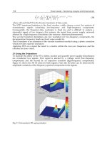
of about a liter. Figure 5.3 indicates that this is about a 20% loss. When
about 35% of the blood volume is lost (about 2 liters in the average adult), the
systolic pressure begins to drop (orthostatic tachycardia and hypotension).
Being “positively orthostatic” may also mean having orthostatic (pos-
tural) hypotension, or a drop in blood-pressure (>20/10 mmHg) on stand-
ing without an increase in heart-rate. This is usually not a sign of hypovolemia
but occurs in patients on beta-blockers, alpha-blockers, calcium-channel
blockers, nitrates, phenothiazines, with alcohol ingestion (impairment of
vasoconstriction) and in the rare person with autonomic dysfunction. It may
be seen with hypovolemia in the occasional elderly patient with a weak barore-
ceptor response, and as mentioned in the previous section, with blood loss
resulting in paradoxical bradycardia. Paradoxical bradycardia is not seen in
hypovolemia from dehydration.
Fig. 5.3. Evaluation of Fluid/blood Loss. Modified with permission from American
College of Surgeons: Committee on Trauma, Advanced Trauma Life Support for
Doctors, Student Course Manual, 6th ed., p. 98. ©1997 American College of Surgeons.
87Vital Sign #4: Blood Pressure
5
Orthostatic vitals are recorded as follows: the patient lies for three min-
utes and the blood-pressure and heart-rate are recorded. He then stands (or
sits up) for one minute and they are retaken. The patient has orthostatic
tachycardia (positively orthostatic) if the heart-rate increases by 30 beats per
minute, or if he becomes dizzy or light-headed with a lesser increase. Ortho-
static hypotension is present if only the blood pressure decreases >20/10
mmHg. Both conditions are considered “positively orthostatic”.
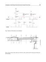
Orthostatics are often reported by using a stick-figure lying, sitting or
standing, with the appropriate heart-rate and blood-pressure readings adja-
cent to the figure.
An older term, the tilt test, is occasionally encountered in the literature.
The test was originally designed with the patient lying, then the patient was
tilted to a sitting position and the heart-rate and blood-pressure retested.
The test was positive if the pulse rate increased 15 to 20 beats, or the systolic
blood pressure decreased 15 to 20 mmHg. The test is no longer considered
valid (orthostatics from lying to sitting will not detect a 1000 cc blood/fluid
loss). However, if the heart-rate increases 30 or more beats per minute or the
person becomes dizzy or light-headed from lying to sitting, this is a positive
test. Results were written as “tilt positive” or “tilt negative”. When practitio-
ners today speak of a tilt test, they mean (hopefully) current orthostatics.
The elderly and and occasionally others do not always follow the rules. Car-
diac syncope and poor autonomic functioning may result in false positives or
negatives. An entity in some elderly, postural orthostatic tachycardia
Fig. 5.4. Orthostatic Vital Signs
88 Vital Signs and Resuscitation
5
syndrome, is a fall in blood pressure, tachycardia, near-syncope and symp-
toms of a transient ischemic attack (TIA), thought to be caused by auto-
nomic dysfunctioning and not hypovolemia.
The accuracy of orthostatic vital signs is frequently challenged. A recent
study showed that some normal subjects had a heart-rate increase from 5 to
39 beats per minute (with a mean of 17) from supine to standing. Both
systolic and diastolic pressures rose. The cause was related to baroreceptor
stimulation with both a-adrenergic and b-adrenergic effects. The conclusion
was that a wider than previously thought variability exists in the physiologic
response to standing.
To conclude, orthostatics are of value if the procedure is accurately per-
formed and accompanied by a careful history. Excluding other factors, light-
headedness or dizziness on standing or sitting upright is a positive test
regardless of the heart-rate. Patients are located within a bell-curve. At the
edges are rare false positives and negatives. Treatment: Fluid/blood resusci-
tation as previously described.
Capillary Refill
The capillary refill is the time it takes for blanching of the skin to return
to normal when the nailbed or hypothenar emminence is quickly squeezed.
It is a simple measurement of blood/fluid loss. The upper limit of normal in
males is 2 seconds; in females 2.9 seconds. A delay greater than this implies
a fluid deficit of about 100 cc/kg. The test has been challenged. Recent
studies indicate that its value as an isolated test for mild-to-moderate hypo-
volemia is minimal and even misleading. However, if orthostatics are abnor-
mal, the validity increases. The test is more sensitive in children (see Chapter
7). Treatment: Fluid/blood resuscitation as previously described.
Fig. 5.5. Orthostatic Figures
89Vital Sign #4: Blood Pressure
5
Abdominal Aortic Aneurysm (AAA)
A rare but potentially catastrophic cause of hypovolemic shock is a leak-
ing abdominal aortic aneurysm. Because of a weakened tunica media from
atherosclerosis, the abdominal aorta may slowly balloon out. A common
presentation is an older male with sudden onset of severe lower abdominal
and back pain, sometimes radiating to the groin and accompanied by hy-
potension. This represents a leaking aneurysm. Lower extremity pulses may
or may not be weak compared to upper ones. The physical exam reveals a
tender abdominal pulsatile mass. Treatment: a call is made to the surgeon
for immediate surgery, while 2 IV’s are started and blood is typed and crossed
for 10 units.
Cardiogenic Shock
Cardiogenic shock is pump failure, usually caused by an acute myocar-
dial infarction (involving about 40% of the myocardium), but occasionally
from cardiomyopathies, drugs, toxins, pulmonary embolism, cardiac tam-
ponade and some arrhythmias.
Cardiogenic Shock from Myocardial Infarction
The patient may migrate into a shock state from a heart attack or present
in shock. The main symptom is chest pain, although in the elderly, and
occasionally in others it is absent. In shock, the skin is cool and moist. Neck
veins are distended. Auscultation of the heart may reveal an S-3 gallop, a
new murmur, tachycardia or bradycardia. The EKG usually shows a pattern
of acute injury. Rales may be present. A chest x-ray may show pulmonary
edema. Serum markers (i.e., CK-MB and troponin) are usually positive. Treat-
ment for myocardial infarction:
1. The ABCs of resuscitation are followed (i.e., high flow oxygen,
pulse oximetry, intubation, IV access);
2. Aspirin 160 mg is chewed and swallowed;
3. Nitroglycerine 10 µg/min is given for pain, and also reduces preload
and afterload. If pain does not resolve, morphine sulfate 2-5 mg is
used;
4. For a systolic pressure less than 100 mmHg, a vasopressor is ad-
ministered (see next section) (however, if a right ventricular infarc-
tion is present, a fluid challenge of normal saline is used);
5. Heparin 80 units/kg IV bolus and 18µ/kg/hr is administered;
6. A beta-blocker such as metoprolol (Lopressor) 5 mg q for 5 min is
given for three doses (assuming no bradycardia or hypotension)
and
7. A thrombolytic agent such as alteplase (t-PA) (Activase) 100 mg
over 1.5 h or tenecteplase (TNKase) 40 mg over 5 seconds is
administered.
90 Vital Signs and Resuscitation
5
Treatment for cardiogenic shock: the patient in shock (systolic BP<90
mmHg, pulmonary edema) should be transferred as soon as posible to a
facility with the capability for intra-aortic balloon pump placement and per-
cutaneous transluminal coronary angioplasty (PTCA). A balloon-tipped
catheter is maneuvered into the blocked coronary artery; the balloon is in-
flated, dilating the narrowed artery and disrupting the atheromatous plaque.
A stent may be placed.
If that is not possible, thrombolytic therapy is begun (see above section).
For a systolic BP of 90-100 mmHg, accompanied by pulmonary edema, furo-
semide (Lasix) 80 mg and dobutamine 5 µg/kg/min IV are administered. If the
systolic BP is 70-90 mmHg, dopamine is begun at 5 µg/kg/min. Norepineph-
rine 2 µg/min is used for a systolic BP of <70 mmHg (see Fig. 4.12).
Shock from Cardiac Tamponade/Tension Pneumothorax
Other causes of cardiogenic shock are cardiac (pericardial) tamponade
and tension pneumothorax. In these cases, the heart is compressed—in
tension pneumothorax by air in the pleural cavity pressing against the heart,
and in cardiac tamponade by bleeding into the the pericardial sac. Com-
monly, cardiac tamponade is seen after a stab wound to the heart (which
usually nicks a vessel), but occasionally after blunt trauma. The condition
may also result from accumulated fluid secondary to metastatic disease (ma-
lignant pericardial effusion). Pulsus paradoxus may be present (see earlier
section) A familiar scenario is a young male seen in a trauma center for a stab
wound to the anterior chest who wants to leave, the wound being barely
visible. He is later found slumped on the cart, barely conscious, with a faint
pulse, decreased blood pressure and jugular venous distension (Beck’s triad).
Treatment: treatment for tension pneumothorax is needle decompression,
followed by chest tube placement, discussed in Chapter 4.Treatment for
cardiac tamponade is intravenous fluid infusion and immediate
pericardiocentesis.
Septic Shock
Bacteremia is an invasion of the bloodstream by infectious micro-organ-
isms. Severe bacteremia is sepsis. Release of toxins by microorganisms (i.e.,
gram negative bacteria) causes host macrophages to produce defense sub-
stances such as kinins, cytokines, complement and prostaglandins causing
vasodilation, increased capillary permeability, leaking of plasma into tissues
and a fall in blood pressure (septic shock). Disordered temperature regula-
tion, myocardial depression and multiple organ system failures occur. Com-
plications are disseminated intravascular coagulation (DIC) and adult
respiratory distress syndrome (ARDS). Frequent sites are the urinary tract,
GI tract and lungs. Extremes of age are particularly susceptible, as are burn
91Vital Sign #4: Blood Pressure
5
victims, diabetics, cancer patients and those having undergone recent inva-
sive procedures.
The common septic patient is an elderly person with a urinary tract in-
fection that has progressed to a systemic problem. The patient is warm and
flushed, mildly agitated, and the temperature is increased (“warm shock”).
This often progresses to “cold shock” from peripheral vasoconstriction and
hypotension. Obtundation is often present, accompanied by hyperventila-
tion because of metabolic acidosis. A widened pulse pressure is frequently
present. Treatment:
1. The ABCs of resuscitation are followed;
2. Two large bore IV’s are placed and Ringers lactate solution is in-
fused wide open to maintain a urine output of 30 cc/h (peds:
1 cc/kg/h);
3. If the blood pressure does not improve with fluid, a pressor such as
dopamine 5 ug/kg/min is added, and
4. An intravenous antibiotic relevant to the source of infection is
started, such as IV ceftriaxone 1 gm + gentamycin 1.5 mg/kg (peds:
ceftriaxone 50 mg/kg).
Neurogenic Shock
Neurogenic shock results from trauma to the spinal cord. Muscle flaccid-
ity and loss of reflexes below the injury (spinal shock) is followed by mild
hypotension and bradycardia (neurogenic shock) from damage to descend-
ing sympathetic pathways in the cord. Treatment consists of:
1. The ABCs of resuscitation;
2. Proper spinal immobilization;
3. Intravenous normal saline to maintain a systolic pressure above
70 mmHg;
4. If unable to maintain the blood pressure, dopamine 5 ug/kg/min is
added;
5. Bradycardia may be treated with atropine 1 mg IV every 5 minutes
to a total dose of 3 mg or a pacemaker if needed;
6. Methylprednisolone 30 mg/kg over 15 min is administered, fol-
lowed by an infusion of 5.4 mg/kg per hour (controversial) and
7. The patient is transferred to an appropriate spinal-cord facility.
Anaphylactic Shock
Anaphylaxis, or anaphylactic shock, is a severe allergic reaction. Com-
mon allergens are penicillin and bee/wasp venom. Mediators (i.e., histamine,
leukotrienes, prostaglandins) are released from mast cells. Flushing and an
itchy skin, a red rash (urticaria), shortness of breath and hypotension are
present. Sometimes swelling of the tongue, pharynx and larynx (laryngeal
edema) and wheezing (bronchospasm) occur. Treatment:
92 Vital Signs and Resuscitation
5
1. Immediate intravenous normal saline is begun to raise the blood
pressure;
2. High flow oxygen by mask is monitored by pulse oximetry;
3. Epinephrine 0.4 cc subq (peds: 0.01 cc/kg) (1:1000), or for severe
shock 1 to 10 cc (1:10,000) slow IV push;
4. An antihistamine such as diphenhydramine 50 mg IV (peds:
1 mg/kg) is given;
5. Methylprednisolone 125 mg IV (peds: 1 mg/kg)is administered;
6. A histamine receptor blocker such as ranitidine 50 mg IV (peds:
0.5 mg/kg) is given over 5 min, and
7. A nebulized beta-agonist (i.e., albuterol) is administered for bron-
chospasm.
Other
A low pressure is sometimes seen in myxedema, adrenal crisis, DKA, renal
failure, as well as with the use of drugs such as beta-blockers, calcium-channel
blockers, diuretics, opioids and sedative hypnotics.
Special Cases
Hypotension in Pregnancy
Cardiac output increases by 40% at the end of the first trimester. The
heart rate increases 10-15 beats per minute. Systolic and diastolic pressures
decrease about 10 mmHg in the second trimester. In late pregnancy, the
supine position compresses the inferior vena cava causing a further decrease
in blood pressure. The left lateral position relieves this effect.
Vaginal bleeding and hypovolemic shock in early pregnancy may occur
from spontaneous abortion or ectopic pregnancy, and in late pregnancy
from abruptio placentae or placenta previa. A postpartum hemorrhage
may also occur. If a spontaneous abortion becomes inevitable with heavy
vaginal bleeding, treatment is fluid resuscitation with normal saline,
followed by dilatation and curettage.
An unstable ectopic pregnancy, indicated by abdominal pain, vaginal
bleeding, hypotension and a positive pregnancy test, is diagnosed by culdo-
centesis (the stable patient is diagnosed by ultrasound). Treatment: 2 IV
lines are started, packed red cells are given for hemodynamic instability,
and the patient is prepared for surgery.
Abruptio placentae, or premature separation of the placenta in late preg-
nancy, is indicated by abdominal pain and vaginal bleeding. Treatment
includes fetal monitoring, IV normal saline and packed red cells as needed.
Placenta previa is painless bleeding in late pregnancy. Because the pla-
centa lies over the cervical os, a cesarean section may be required. Treat-
93Vital Sign #4: Blood Pressure
5
ment: fetal monitoring, IV normal saline and packed red cells are adminis-
tered as needed.
Postpartum hemorrhage results from uterine atony and sometimes from
retained fetal tissue. Treatment consists of IV normal saline, packed red cells
as needed and oxytocin or methylergonovine 0.2 mg IM to contract the
uterus and control bleeding. Curettage is performed to remove retained tissue.
The Dialysis Patient
Hypotension during or after dialysis is frequently seen because of fluid
loss. Treatment consists of placing the patient in the Trendelenburg position
and giving a fluid challenge of 200 cc of normal saline.
Practical Points
•First, the ABC’s of resuscitation are followed (see Chapter 8).
•When taking the blood pressure, if the muffling of diastolic pres-
sure is greater than 10 mmHg, two values should be noted: the
beginning and disappearance of muffling.
Example:
BP 210/110/80 right arm, supine
•Orthostatic tachycardia is an increase in heart rate from lying to
standing. If the heart rate increases 30 beats and/or neurologic
changes occur (i.e., dizziness), hypovolemia is present.
Example #1:
Pulse 101, BP 130/90, right arm lying
Pulse 132, BP 134/94, right arm standing
Example #2:
Pulse 106, BP 142/94, right arm lying
Pulse 124, standing dizzy—terminated
•If the blood pressure lowers from lying to standing without a heart
rate increase, this is orthostatic hypotension.
Example:
Pulse 76, BP 116/60, left arm lying
Pulse 78, BP 100/56, left arm standing
•In an acutely ill or trauma patient, always take BP in both arms.
The pressure in one may be compromised.
References
1. American Heart Association and the International Liaison Committee on Resusci-
tation (ILCOR). Guidelines 2000 for cardiopulmonary resuscitation and emer-
gency cardiovascular care. Baltimore: Lippincott, Williams & Wilkins, 2000.
2. Bakris G. Severe hypertension in a young patient. Hosp Pract 1993; 28:10.
3. Barach P. Pulsus paradoxus. Hosp Phys 2000; 36:49.
4. Baraff L, Schriger D. Orthostatic vital signs: Variation with age, specificity, and
sensitivity in detecting a 450 ml blood loss. Am J Emerg Med 1992; 10:2.
94 Vital Signs and Resuscitation
5
5. Bruce C et al. The effect of cocaine on the physiologic response to hemorrhagic
shock. Surgery 1993; 114:429.
6. Califf R, Bengtson J. Cardiogenic shock. N Engl J Med 1994; 330:24.
7. Chen H et al. Enhancement of vagal restraint on systemic blood pressure during
hemorrhage. Am J Physiol 1978; 234:192.
8. Cohn L. Aortic dissection: New aspects of diagnosis and treatment. Hosp Pract
1994; 29:3.
9. Craver J, Connolly M. The percutaneous intraaortic balloon pump and ventricular
assist devices. In: Schlant R, Alexander R. The Heart. New York: McGraw-Hill, 1994.
10. Deakin C, Low J. Accuracy of the advanced trauma life support guidelines for
predicting systolic blood pressure using carotid, femoral, and radial pulses. BMJ
2000; 321:673.
11. Demetriades D et al. Relative bradycardia in patients with traumatic hypotension.
J Trauma 1998; 45:534.
12. Enselberg C. Measurement of diastolic blood pressure by palpation. N Eng J Med
1961; 265:6.
13. Evans R et al. Does the hemodynamic response to acute central hypovolaemia de-
pend on the rate of fall of cardiac output? Clin Exp Pharmacol Physiol 1992; 19:657.
14. Grubb et al. The postural orthostatic tachycardia syndrome: A neurocardiogenic
variant identified during head-up tilt table testing. Pacing Clin Electrophysiol 1997;
20:2205.
15. Hals G, Carleton S. Pericardial disease and tamponade. Em Med Rep 1996; 17:161.
16. Hals G, Crump T. The pregnant patient: Guidelines for management of common
life-threatening medical disorders in the emergency department. Em Med Rep 2000;
21:53.
17. Hartmann A et al. Measurement of blood pressure in the brachial and posterior
tibial arteries using the Doppler method. J Ped 1973; 82:3.
18. Jackson R. Cardiogenic shock. In: Tintinalli J et al. Emergency Medicine: A Com-
prehensive Study Guide. New York: McGraw-Hill, 2000.
19. Jacob G et al. The neuropathic postural tachycardia syndrome. N Engl J Med 2000;
343:1008.
20. Jensen K. Heart and endocrine changes during central hypovolemia in man. Dan-
ish Medical Bulletin 1991; 38:443.
21. Kaplan N. Establishing control of refractory hypertension. Hosp Pract 1994; 29:5.
22. Knopp R et al. Use of the tilt test in measuring acute blood loss. Ann Emerg Med
1980; 9:29.
23. Koziol-McLain J et al. Orthostatic vital signs in emergency department patients.
Ann Emerg Med 1991; 20:6.
24. Kumar A et al. Hypertensive crisis. J Emerg Med 2000; 19:369.
25. Kussmaul A. Ueber schwielige Mediastino-Pericarditis und den paradoxen Puls.
Berl Klin Wochenschr 1873; 10:37-39.
26. Lewinter J et al. Vital sign measurement procedures. In: RobeRTS J, Hedges J, eds.
Clinical Procedures in Emergency Medicine. Philadelphia: WB Saunders, 1998.
27. Malhotra A, Townsend R. Clinical significance of systolic and pulse pressure. Em
Med 2000; 32:52.
28. Mansoor G, White W. Ambulatory blood pressure monitoring: A clinically rel-
evant tool for the diagnosis and management of hypertension. Res & Staff Phys
1999; 45:10.
29. Mansoor G, White W. Usefulness of home blood pressure monitoring in clinical
practice. Res & Staff Phys 2000; 46:21.
30. McGregor M. Pulsus paradoxus. N Engl J Med 1979; 301:480.
95Vital Sign #4: Blood Pressure
5
31. Pahwa R, Dellinger R. Cerebral hypoperfusion with “normal” blood pressure. J
Crit Illness 2000; 15:567.
32. Park M, Guntheroth W. Direct blood pressure measurement in brachial and femo-
ral arteries in children. Circul 1970; 41:231.
33. Pascarelli E, Bertrand C. Comparison of blood pressures in the arms and legs. N
Eng J Med 1964; 270:14.
34. Ram C. Secondary hypertension: workup and correction. Hosp Pract 1994; 29:4.
35. Rigolin V et al. Update on aortic dissection. Emerg Med 1993; 25:13.
36. Rodbard S, Margolis J. The auscultatory gap in arteriosclerotic heart disease. Circ
15:June, 1957.
37. Sander-Jensen K et al. Vagal slowing of the heart during haemorrhage: Observa-
tions from 20 consecutive hypotensive patients. Brit Med J 1986; 292:364.
38. Schmidt R. Shock. In: Markovchick V, Pons P. Emergency Medicine Secrets. Phila-
delphia: Hanley & Belfus, 1999.
39. Schriger D et al. Capillary refill—Is it a useful predictor of hypovolemic states? Ann
Emerg Med 1991; 20:6.
40. Secher N et al. Bradycardia during reversible hypovolaemic shock: Associated neu-
ral reflex mechanisms and clinical implications. Clin Exp Pharmacol Physiol 1992;
19:773.
41. Shabetai R et al. Pulsus paradoxus. J Clin Investig 1965; 44:11.
42. Shabetai R et al. The hemodynamics of cardiac tamponade and constrictive peri-
carditis. Am J Cardiol 1970; 26:480.
43. Thompson D et al. Relative bradycardia in patients with isolated penetrating ab-
dominal trauma and isolated extremity trauma. Ann Em Med 1990; 19:268.
44. Vayer J et al. Absence of a tachycardic response to shock in penetrating intraperito-
neal injury. Ann Em Med 1988; 17:227.
45. Witting M et al. Defining the positive tilt test: A study of healthy adults with
moderate acute blood loss. Ann Emerg Med 1994; 23:6.
46. Wo C et al. Unreliability of blood pressure and heart rate to evaluate cardiac output
in emergency resuscitation and critical illness. Crit Care Med 1993; 21:2.
47. Wynn R. Obstetrics and Gynecology. Philadelphia: Lea & Febiger, 1988.
48. Yutaka I et al. Clinical evaluation of semiautomatic and automatic devices for home
blood pressure measurement: Comparison between cuff-oscillometric and micro-
phone methods. J Hypertension 1989; 7:983.
96 The Vital Signs and Resuscitation
6
CHAPTER 6
Vital Sign #5: Level of Consciousness
The Glasgow Coma Scale, as well as AVPU (Alert, responds to Verbal
stimuli, responds to Painful stimuli, Unresponsive) have been rapid neu-
rological assessment tools for prehospital and hospital personnel for many
years, and to that extent level of consciousness has been a vital sign for
over three decades.
The brain is quite sensitive to body changes, and an alteration in mental
status often precedes abnormalities in other vital signs. Altered mentation
may range from bizarre behavior and confusion to coma, a state of unre-
sponsiveness from which the patient cannot be aroused. Psychotic behav-
ior is usually lucid and not confused. The person with delirium is confused,
and the onset is fairly sudden. Common causes of delirium are hypoglyce-
mia in the diabetic, drugs, alcohol, inappropriate meds in the elderly, infec-
tion, withdrawal syndromes and hypoxemia. Recently, the serotonin
syndrome, a complication of new antidepressant drugs, may cause agitation
and confusion from increased central serotonin neurotransmission. Delirium
is reversible. Untreated, it may progress to coma. Dementia, on the other
hand, is a gradual loss of mental capacity, is primarily a disorder of the eld-
erly and is rarely reversible. Alzheimer patients make up 70% of dementia
cases, with multi-infarct dementia accounting for 15-20% of others. The
three conditions may co-exist, particularly in the elderly. Overlaps and mis-
interpretations are frequent. Psychotic depression, for example, may be mis-
diagnosed as dementia. On the other hand, delirium from medicines may be
misdiagnosed as depression. Therefore, it is important to rule out an organic
cause for altered mentation before classifying behavior as a functional disorder.
Common causes of an altered level of consciousness are alcohol and drug
abuse (30%), hypoglycemia from insulin reactions (30%) and stroke (30%).
In some centers the percentages vary (i.e., trauma centers, hospitals that see
many elderly).
Anatomy and Physiology
The cerebral hempispheres are two large areas of white matter surrounded
by an outer thin mantel of nerve cell bodies, the grey matter, or cortex.
Association areas constitute the majority of the cortex and are involved with
higher learning or intelligence. Both cerebral hemispheres must be affected
for an altered level of consciousness to occur. Common causes of bilateral
Vital Signs and Resuscitation, by Joseph V. Stewart. ©2003 Landes Bioscience.
97Vital Sign #5: Level of Consciousness
6
cortical involvement are drugs, and oxygen or glucose deficits (metabolic
etiology).
The brainstem consists of the thalamus, hypothalamus, midbrain, pons
and medulla, and contains centers for vital functions. The reticular forma-
tion, an inner core of neurons in the spinal cord and brainstem, regulates
respiration, blood presssure, heart-rate, endocrine secretion, conditioned
reflexes, learning and consciousness. Incoming stimuli are integrated by the
reticular formation. A portion of it, the reticular activating system (RAS),
is responsible for the arousal reaction. Most patients with lesions of the RAS
are comatose. Common causes of brainstem involvement are trauma and
stroke (structural etiology). Some drugs affect the brainstem. For example,
epinephrine, amphetamines and cocaine stimulate RAS conduction; opiates
and barbiturates depress it.
In summary, a decrease in level of consciousness occurs if:
1. Both cerebral hemispheres are involved, or
2. The RAS brainstem is affected.
Fig. 6.1. The Reticular Formation.
98 The Vital Signs and Resuscitation
6
The vast majority of patients with decreased level of consciousness (about
85%) are in the metabolic category.
Glasgow Coma Scale (GCS)
Teasdale and Jennett developed their scale in 1974 not only to assess
changes in levels of consciousness in brain-damaged patients in prolonged
comas, but also to avoid imprecise terms such as lethargy, semicomatose,
stupor, etc. when describing the mental status of a patient (see Chapter 1).
An important point about the GCS is that it can be used as a continuum for
changing levels of consciousness. At about a GCS score of 8 the definition of
coma is fulfilled. The GCS is an integral part of trauma scales (see Fig. 6.3).
Fig. 6.2. Glasgow Coma Scale (GCS).
99Vital Sign #5: Level of Consciousness
6
The GCS is sometimes bypassed in the field for AVPU, an easier method of
evaluating level of consciousness because no numbering system is required.
A—alert
V—responds to verbal stimuli
P—responds to painful stimuli
U—unresponsive
The GCS is preferred over AVPU since both take about the same amount
of time and a tenuous area exists between V and P in terms of airway protec-
tion. Sometimes it is difficult to assess a gag reflex. Airway protection (en-
dotracheal intubation) to avoid aspiration of vomitus is required when
the gag reflex is lost or the GCS is 8 (Fig. 6.2).
Fig. 6.3. Revised Trauma Score (RTS).