Vital Signs and Resuscitation - part 7 potx
Bạn đang xem bản rút gọn của tài liệu. Xem và tải ngay bản đầy đủ của tài liệu tại đây (240.12 KB, 18 trang )
100 The Vital Signs and Resuscitation
6
Management of Altered Level of Consciousness
In contrast to the traditional approach in medicine, the comatose patient
or the patient with a significant alteration in level of consciousness requires
immediate management before completing the physical exam and acquiring
the history.
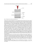
The ABCs of resuscitation are followed (Fig. 6.4). When an immobilized
patient arrives in the emergency department, the cervical collar and backboard
Fig. 6.4. Management of the Comatose Patient.
101Vital Sign #5: Level of Consciousness
6
are left in place until a cause is found for the decrease in level of conscious-
ness. Naloxone (Narcan) 2 mg and thiamine (vitamin B-1) 100 mg are
administered intravenously. If a fingerstick blood sugar is low or unavail-
able, glucose (50 cc of 50% dextrose) is administered after thiamine to reverse
hypoglycemia.
Naloxone reverses the effects of a narcotic by competitive inhibition at
the opioid receptor site. Thiamine prevents Wernicke’s Encephalopathy, a
rare neurological condition caused by thiamine deficiency seen in alcoholics
with poor nutrition. Signs and symptoms include nystagmus, occular nerve
palsy, ataxia and confusion. Thiamine functions as a coenzyme in the break-
down of glucose. Glucose given before thiamine depletes what little thia-
mine is available for glucose metabolism and may precipitate the syndrome.
Glucose and thiamine may be administered at the same time.
Naloxone, thiamine and glucose were referred to in the past as a “coma
cocktail” and were often automatically administered. If a fingerstick glucose
is normal, administering glucose is not indicated. The same applies to thia-
mine in the pediatric population. If a drug overdose is suspected, activated
charcoal is administered by gastric tube after endotracheal intubation
(Fig. 6.4).
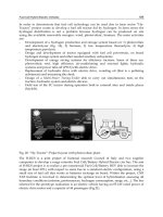
Increased Intracranial Pressure
Increased intracranial pressure is a life-threatening event and must be
dealt with immediately. Common causes are a head injury with intracranial
bleeding and a hemorrhagic stroke. Signs include papilledema, loss of spon-
taneous venous pulsations, an increase in systolic pressure, bradycardia, an
abnormal respiratory pattern and a fixed dilated pupil.
Carbon dioxide is a potent vasodilator in the brain and hyperventilation
blows off carbon dioxide and reduces pressure. Mannitol is an osmotic diuretic
that removes excess fluid from the brain. Increased intracranial pressure blocks
blood flow to the brain, and the hypoxia triggers an increase in systolic pres-
sure to re-establish flow. The increased blood pressure causes a baroreceptor
decrease in heart rate, and pressure against the RAS of the pons and medulla
decreases the heart and respiratory rates. The triad of increased blood pres-
sure, decreased heart rate and irregular breathing is the Cushing reflex. In
adults, often only the blood pressure rises. The triad occurs more often in
pediatrics. Intracranial pressure may cause the brain to push against the third
cranial nerve on that side causing a fixed dilated pupil, indicating compres-
sion of the lower part of the temporal lobe (uncus) against the tentorium
cerebelli with impending herniation (Fig. 6.5).
Treatment: intubation, hyperventilation, the head of the bed is raised
30˚ (except in the trauma patient with a cervical collar), furosemide 40 mg
102 The Vital Signs and Resuscitation
6
IV and/or mannitol 1 gm/kg IV is administered in consultation with a neu-
rosurgeon.
Neurological Examination
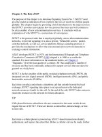
Signs of Metabolic Injury
Signs of metabolic injury, implying an intact brainstem, are roving eye
movements, a pupillary reaction to light (pinpoint pupils suggest opiates or
a pontine lesion. Dilated reactive pupils are seen with adrenergic or anticho-
linergic drugs), a normal oculocephalic reflex (doll’s eyes) consisting of
abruptly rotating the head to one side while the eyes deviate in the opposite
direction (this test should not be used in the trauma patient unless the c-
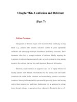
spine has been cleared), a normal oculovestibular reflex (instilling 50 ml of
cold water into the auditory canal causes deviation of the eyes toward the
water) and hyporeflexia.
Fig. 6.5. Increased Intracranial Pressure (ICP).
103Vital Sign #5: Level of Consciousness
6
Signs of Structural Injury
Signs of structural injury from trauma or stroke are fixed pupils, either
large or pinpoint (pinpoint pupils suggest a pontine hemorrhage. Fixed mid-
position pupils implies brainstem damage. One fixed dilated pupil suggests
impending uncal herniation), no extra-ocular movements, loss of oculocephalic
and oculovestibular reflexes, differences in movements of arms and legs, asym-
metry and increased deep tendon reflexes with upgoing toes (Babinski’s
Fig. 6.6. Metabolic vs. Structural Signs of Coma.
reflex) and decorticate or decerebrate posturing (arm flexion and leg ex-
tension in decorticate posturing represents injury to both cerebral hemi-
spheres; extension of the arms and legs in decerebrate posturing represents
injury to the brainstem). Decerebrate and decorticate posturing may occur
in metabolic derangements, but more commonly are seen with structural
damage. Fixed ocular deviation is toward a cortical lesion (Figs 6.7, 6.8).
Physical Examination
Vital signs may give a clue to the etiology. Hypothermia (including meta-
bolic causes such as hypothyroidism, hypoadrenalism, hypoglycemia and
sepsis) and hyperthermia may all cause a decreased level of consciousness.
104 The Vital Signs and Resuscitation
6
Fig. 6.7. Oculovestibular Reflex.
Fig. 6.8. Decerebrate vs. Decorticate Posturing.
Tachyarrhythmias and bradyarrhythmias suggests cardiac disease. Hyper-
ventilation is seen in diabetic ketoacidosis, uremia and cirrhosis.
Hypoventilation is common in opiate drug overdoses and in pulmonary
disease. Hypotension requires searching for the etiology of shock (see Chap-
ter 5). Hypertension suggests hypertensive encephalopathy or drugs such as
cocaine (Chapter 5).
105Vital Sign #5: Level of Consciousness
6
Breath: alcohol on the breath is noted, as is the fruity odor of diabetic
ketoacidosis. A petroleum or garlic odor is sometimes seen in organophos-
phate pesticide poisoning.
HEENT: evidence of trauma should be sought, such as bruising or lac-
erations of the head and face. A basilar skull fracture may cause cerebrospi-
nal fluid to leak from the nose or ear, or the extravasation of blood in the
middle ear (hemotympanum) into the skin around the eyes (raccoon eyes)
or over the mastoid process (Battle’s sign).
Neck: neck stiffness, Kernig and Brudzinki’s signs may indicate meningi-
tis (see next section). Jugular venous distention is noted, as is the size of the
thyroid gland.
Chest: signs of trauma should be sought, as above. The heart and lungs
are evaluated. Decreased breath sounds on one side may indicate a pneu-
mothorax or hemothorax. Jugular venous distention plus low blood pressure
may suggest cardiac tamponade (see Chapter 5).
Abdomen: jaundice and a distended abdomen is seen in alcoholic liver
disease. Abrasions, decreased bowel sounds, tenderness and rigidity suggest
trauma with possible internal hemorrhage.
Neurologic: (see previous section) the Glasgow Coma Scale is assessed
frequently for changes.
Skin: abrasions suggest trauma, jaundice suggests sequelae of liver dis-
ease, needle-tracks suggest drug abuse. Profuse sweating is seen with
organophosphate pesticide poisoning. Cold sweats are present in a patient
suffering a heart attack. A petechial or purpuric rash should alert one to
meningococcemia.
Causes and Treatments of Coma
Since one cannot question the patient, other avenues for history are uti-
lized. Medical tags and bracelets are sought. The questioning of EMTs is
vital. Were empty medicine bottles present in the house? If so, they should
be brought to the emergency department. Family and friends and neighbors
and bystanders should be questioned. A handy mnemonic device for
remembering the multiple causes of coma is “TIPS” and “AEIOU”. Treat-
ment for coma is supportive until the cause is found. The ABC’s of resusci-
tation are strictly followed (Fig. 6.9).
"TIPS”:
Trauma, Temperature
Trauma: in addition to head trauma, shock from hemorrhage, pericar-
dial tamponade, myocardial contusion and tension pneumothorax may cause
a decreased level of consciousness (see Chapter 5). A concussion is a tran-
sient loss of consciousness with no brain damage. A contusion, or bruising
of the brain with small hemorrhages and tissue tears, usually causes a loss of
106 The Vital Signs and Resuscitation
6
consciousness, sometimes briefly, sometimes for a long period (diffuse ax-
onal injury). A traumatic subarachnoid hemorrhage from injury to vessels
in the pia causes bleeding into cerebrospinal fluid in the subarachnoid space,
sometimes producing headache and stiff neck. An epidural hematoma is a
collection of blood between bone and dura from a laceration of the middle
meningeal artery. A subdural hematoma is blood between the dura and
arachnoid from tears in bridging dural veins. An intracerebral hemorrhage is
the accumulation of blood within brain substance. A CT will not show con-
cussions or in many cases contusions, but does reveal epidural, subdural and
intracerebral hemorrhages. Treatment: increased intracranial pressure (ICP)
is treated as previously described. Epidural and subdural hematomas require
surgical evacuation (Fig. 6.10).
Temperature: hypo- and hyperthermia are discussed in Chapter 2.
Infection
Common infections causing decreased levels of consciousness are sepsis
and bacterial meningitis (viral meningitis usually does not cause coma,
except in the pediatric population). Neurological findings in sepsis range
from lethargy or agitation to coma. Inflammatory mediators cause multi-
Fig. 6.9. Common Causes
of Coma.
107Vital Sign #5: Level of Consciousness
6
Fig. 6.10. Subdural and Epidural Hematomas.
organ-system failure and hypotension (septic shock) with inadequate perfu-
sion to the brain. Sepsis and septic shock are discussed in Chapter 5.
Bacterial meningitis is seen primarily in pediatrics and the elderly, with
sporadic outbreaks in other populations. With the advent of the H. influenzae
vaccine, the main organism is Strep. pneumoniae, not only in peds but in all
age groups. Seeding is from bacteremia, otitis media and sinusitis. Fever,
headache, altered mental status and HIV+ are important historical items.
Seizures may occur. The physical exam in infants may show hypothermia, a
bulging fontanelle, lethargy, dehydration and otitis media (see Chapter 7).
Older children and adults usually have nuchal rigidity, pain on extension of
the legs (Kernig sign) and passive neck flexion producing flexion of the hips
(Brudzinski sign). In meningococcal meningitis the skin may show pete-
chiae and purpura. Treatment: when meningitis is suspected, IV antibiotic
therapy (ceftriaxone or cefotaxime 2 gm, 50mg/kg in peds) is begun before
lumbar puncture. The presence of papilledema and loss of spontaneous venous
pulsations indicate increased intracranial pressure, and therapy for ICP should
be begun immediately (see earlier section). When ICP is suspected, a CT
should be done before an LP.
Stroke, Shock, Seizures
A patient with a cortical ischemic stroke involving one side of the brain,
with a profound sensory and motor loss on the opposite side of the body
along with aphasia (left brain) or inattention and unconcern (right brain)
and sometimes confusion, experiences no loss of consciousness unless mas-
sive ischemia causes brain edema. The less common brainstem ischemic
stroke (basal artery) causes coma from involvement of the RAS (see earlier
section). A hemorrhagic stroke begins with a headache and alteration in
consciousness that progresses to coma because of a severe global mass effect
108 The Vital Signs and Resuscitation
6
with increased intracranial pressure, compression of the brainstem and her-
niation. Diagnosis is made by CT. Treatment:
1. The ABCs are followed (fig. 6.4);
2. Blood pressure over 220/120 is treated with increments of labetalol
20 mg IV;
3. Increased intracranial pressure is controlled (Fig. 6.5);
4. Neurosurgical consultation is obtained and
5. The thrombolytic t-PA 0.9mg/kg IV (maximum 90mg) may be
given over an hour for an ischemic stroke if the time of onset is
known to be less than three hours (and no contraindications exist).
A subarachnoid hemorrhage is caused by rupture of a congenital (berry)
aneurysm in the Circle of Willis at the base of the brain, either at rest or
during exercise. The patient describes a sudden severe headache. Bleeding
into the subarachnoid space and ventricles produces a mild to severe de-
crease in level of consciousness. Preliminary diagnosis by CT or lumbar punc-
ture may be supplemented by angiography. Treatment:
1. Blood pressure is controlled by labetalol 20mg IV increments in
pre-hemorrhage levels;
2. The calcium-channel blocking agent nimodipine 60mg PO every
6 hours reduces vasospasm;
3. Seizures are prevented with fosphenytoin (cerebyx) 15mg/kg IV as
a loading dose and
4. Neurosurgical consultation is obtained.
Shock is discussed in Chapter 5.
Seizures: the most common cause of a seizure is failure to take anti-
convulsive medicine. Decreased level of consciousness is transient and the
person gradually awakens (post-ictal state). A rapid blood sugar is checked
and glucose is administered as needed. if the sezure continues, it is stopped
with lorazepam (Ativan) 4 mg IV over 2 minutes (Peds: 0.1 mg/kg) (or
midazolam ((Versed)) 0.2 mg/kg IM). For the persistent seizure (status
epilepticus), a second dose of fosphenytoin (Cerebyx) 20 mg/kg IV. If no
response occurs, phenobarbitol 18 mg/kg IV is used, and intubation may be
required. The continuous seizure may require a neuromuscular blocking agent
(i.e., vecuroium 0.1 mg/kg) or general anesthesia.
Other causes of seizures are congenital/genetic disorders, brain tumors,
eclampsia (discussed in Chapter 5), drugs such as theophylline, phenothiaz-
ines, lithium, cocaine and antidepressants, opportunistic cerebral infections
in AIDS patients and febrile seizures (discussed in Chapter 7).
109Vital Sign #5: Level of Consciousness
6
"AEIOU”:
Alcohol/Drugs
Alcohol: wide variability exists in each person’s response to alcohol,
depending on whether one is a chronic alcoholic or an occasional drinker.
This results in various degrees of intoxication, physical dependency (with-
drawal symptoms on stopping the drug) and tolerance (increased amounts
of drug for the same effect). In the emergency setting, it is not uncommon
to see an alert and oriented alcoholic with a blood alcohol level of 400 mg/dL,
while a nonalcoholic may be comatose at that level. A level of 100 mg/dL is
legal intoxication in most states. The nontolerant person usually shows a
decrease in level of consciousness at a level of about 300 mg/dL. Coma (GCS
of 8) may occur at about 400 mg/dL (often requiring intubation), and death
from respiratory depression may occur at 500 mg/dL (LD-50).
The alcoholic is at increased risk for a subdural hematoma, and a search
for bruises and abrasions should be sought. A rectal temperature is required.
A low threshold should exist for a head CT, as well as a diagnostic peritoneal
lavage to rule out abdominal injuries. Labs should include, in addition to a
serum ethanol level, a drug screen (cocaine is a common accompanying drug)
and a serum ammonia to rule out hepatic encephalopathy. Alcohol with-
drawal is seen about 48 hours after the last drink and exhibits a wide variety of
manifestations, including anxiety, tremors, visual hallucinations and seizures,
but usually does not show a decreased level of consciousness. Withdrawal
may be seen in the intoxicated patient. Treatment for alcohol withdrawal:
one liter of D5NS with MgSO
4
2 gm, folate 1 mg and an ampule of multi-
vitamins is administered for both intoxication and withdrawal since glyco-
gen, magnesium and vitamins are usually depleted. Gastric decontamination
with lavage and charcoal is indicated only in the rare case of an acute ingestion
of a large amount of alcohol over a short period of time in a nonalcoholic.
For withdrawal, lorazepam (Ativan) 2-4 mg IV is administered, followed by
2 mg every 30 minutes as needed.
The alcoholic may present with liver failure and coma from hepatic en-
cephalopathy, a condition in which nitrogenous and other compounds (i.e.,
ammonia, gamma-aminobutyric acid, mercaptans) normally removed by the
liver accumulate and gain access to the central nervous system, causing
neuroinhibition and cerebral edema. The serum ammonia is elevated, elec-
trolytes are often abnormal and asterixis (“liver flap” = hand tremor) is some-
times present. Treatment for hepatic encephalopathy: fluid and electrolyte
abnormalities are corrected. Lactulose may be given via nasogastric tube (30
cc TID). Lactulose is a nonabsorbable disaccharide when in contact with
colonic bacteria traps ammonia in the colon as nondiffusible ammonium
ions. Neomycin (1 gm via NG q8h) suppresses bacteria responsible for the
production of ammonia and other nitrogenous compounds.
110 The Vital Signs and Resuscitation
6
Drugs: it is not appropriate to list the multitude of drugs and toxins that
may cause an altered level of consciousness. Common drugs causing coma
are sedative/hypnotics and narcotics. An altered level of consciousness is seen
occasionally with carbon monoxide poisoning. Narcotics (i.e., morphine,
heroin) bind to opioid receptors in the brain (mu receptors). Sedative-
hypnotics (alcohol, benzodiazepines and barbiturates) facilitate gamma-
aminobutyric acid (GABA), the major inhibitory neurotransmitter of the
central nervous system. Benzodiazepines and barbiturates bind to GABA
receptors. No receptor has been identified for alcohol, but alcohol enhances
the action of GABA receptors. Treatment: the treatment for an overdose of
a sedative/hypnotic or narcotic is gastric lavage within the first hour. After
the first hour activated charcoal 50 gm PO or by gastric tube is adminis-
tered to prevent absorption of the drug. If the gag reflex is lost, the patient is
intubated and gastric lavage and/or charcoal is administered by gastric tube.
The antidote for opiate ingestion is naloxone, a competitive antagonist at
the mu opioid receptor site. The person awakens quickly. Naloxone may
then be administered 1 mg in 250 cc NS at 250 cc per hour. Flumazenil
(Romazicon) 0.2 mg IV is a competitive antagonist of the benzodiazepine
receptor and will reverse a benzodiazepine overdose. However, since seizures
have been reported with Flumazenil, it should be used for diagnostic rather
than therapeutic purposes.
A common inhalation injury is carbon monoxide poisoning from fires,
faulty gas heaters and in machine-shops where ventilation is poor. Carbon
monoxide (CO) is odorless and binds to hemoglobin 210 times more readily
than oxygen. Early symptoms are headache, dizziness, weakness and nausea.
Consciousness is affected at CO levels of 20-30%, confusion and syncope
occur at 40%, coma and seizures take place at 50%, and death occurs at
60%. Treatment: 100% oxygen by nonrebreather mask until the level is 0.
Use of hyperbaric oxygen therapy (HBO) is controversial, but is indicated
for comatose patients, those with major illnesses such as coronary artery
disease and the pregnant patient.
Endocrine, Electrolytes
Endocrine: severe hypothyroidism (myxedema coma) and acute adre-
nal insufficiency (from an exacerbation of Addison’s disease) are occasional
endocrine causes of decreased level of consciousness. Signs of myxedema are
hypothermia (75%), obesity, a surgical neck scar, cool dry skin, delayed DTRs,
anemia, electrolyte disturbances, and occasionally respiratory distress, brady-
cardia and CHF. Treatment: hydrocortisone 100 mg and levothyroxine
4 µg/kg IV. Acute adrenal insufficiency is sometimes seen when steroids
are withdrawn in a patient with Addison’s disease. Patients present with con-
fusion, lethargy, nausea, vomiting, hyperpigmentation and hypotension. Lab
studies show hyperkalemia, hyponatremia and hypoglycemia. Treatment:
111Vital Sign #5: Level of Consciousness
6
hydrocortisone 100 mg and 2 liters of D5NS to restore blood pressure and
correct hypoglycemia. Hyperkalemia is treated with an ampule of bicarbon-
ate over 5 minutes. If the K+ level is >8 meq, 10 cc of a 10% calcium gluconate
solution is administered over 10 minutes.
Electrolyte imbalances rarely cause a decrease in level of consciousness.
Occasionally severe hyponatremia may cause decreased mentation from the
syndrome of inappropriate antidiuretic hormone (SIADH) secretion from a
previous intracranial lesion or tumor. Treatment: water restriction. For severe
hyponatremia (< 115 mEq), 3% saline is administered at 100 cc per h.
Insulin
The most common disorder causing an altered and/or decreased level of
consciousness in all categories is hypoglycemia (glucose level < 50 mg) in
the insulin-dependent diabetic patient with decreased food intake (insulin
reaction) or the occasional patient on oral medication. Confusion is present,
and if sustained for any length of time a decreased level of consciousness
occurs. Treatment: IV glucose (D50) quickly reverses the condition. If an
IV is difficult to obtain, glucagon 1-2 mg may be administered IM (gluca-
gon catabolizes stored glycogen). Diabetic ketoacidosis (DKA), indicated
by glucose >250 mg, bicarb <15 mEq and pH <7.3, is treated with normal
saline, 5 L at 500 cc per hour, insulin 0.2 units/kg initially, then 0.1 unit/kg/
h IV infusion, potassium 20 mEq per liter and bicarbonate 1 to 2 amps if
pH is <6.9. The patient with hyperosmolar hyperglycemic nonketotic
coma (HHNC) (glucose >600 mg, negative ketoacidosis) is lethargic and
confused but rarely comatose. Fluid and potassium treatment is similar to
DKA, except that less insulin and more fluid is required.
Oxygen
Oxygen deficits are discussed in Chapter 4. An insidious cause of decreased
mentation is hypercapnia (carbon-dioxide narcosis) from hypoventilation
from many causes: two common ones are acute pulmonary edema and a
tiring asthmatic. The pCO
2
level in these cases is elevated above 50 mmHg
(sometimes to 100 mmHg) and the pH is decreased (respiratory acidosis).
Pulse oximetry may show a normal oxygen saturation. Treatment: increased
ventilatory support, requiring intubation and mechanical ventilation.
Uremia
Uremia (chronic renal failure) is caused by the accumulation of toxic
products of protein metabolism in the bloodstream. The blood urea nitro-
gen (BUN) and creatinine are increased. An altered mental status is believed
to be related to loss of the kidney’s capacity to synthesize vitamin D, leading
112 The Vital Signs and Resuscitation
6
to hypocalcemia and secondary hyperparathyroidism. Definitive treatment
is dialysis.
Practical Points
•First, the ABCs of resuscitation are followed (see Chapter 8).
•Everyone who deals with critically ill patients should be familiar
with the Glasgow Coma Scale. With the exception of AVPU, it is
the simplest of all the scores to remember, and thus it is the most
useful. A GCS of 8 is the definition of coma.
•In the critical patient, and particularly the trauma patient, serial
vitals and GCSs are important.
Examples:
1. LOC: rollover MVA with prolonged extrication. No eye open-
ing to pain, incomprehensible sounds, no response to painful
stimuli: GCS 4. En route 5 minutes later—eye opening to voice,
confused, withdraws from pain: GCS 11.
2. LOC: possible overdose. Eyes open to verbal, confused, obeys
commands: GCS 13.
References
1. American College of Surgeons. Advanced trauma life support (ATLS). Chicago,
1997.
2. Braakman R et al. Prognosis and prediction of outcome in comatose head injured
patients. Acta Neurochir Suppl 1986; 36:112.
3. Charness M et al. Ethanol and the nervous system. N Engl J Med 1989; 321:7.
4. Chuidian F. The unconscious patient: Evaluation and first-line interventions. J Crit
Ill 2000; 15:14.
5. Diamond I. Alcohol neurotoxicity. In: Asbury A et al. Clinical Neurobiology.
Philadelphia:WB Saunders, 1992.
6. Hemphill R et al. Delayed presentation after head injury: Is a computed tomogra-
phy necessary? Acad Emerg Med 1999; 6:957.
7. Huff J. Altered mental status and coma. In: Tintinalli J et al. Emergency Medicine:
A Comprehensive Study Guide. New York: McGraw-Hill, 2000.
8. Jackimczyk K. Altered mental status and coma. In: Markovchick V ed. Emergency
Medicine Secrets, Philadelphia: Mosby, 1999.
9. Jennett B. Clinical assessment of consciousness. Acta Neurochir Suppl 1986; 36:90.
10. Negrini B et al. Cerebrospinal fluid findings in aseptic versus bacterial meningitis.
Pediatrics 2000; 105:316.
11. Plant J, MacLeod D. Response of a promethazine-induced coma to Flumazenil.
Ann Emerg Med 1994; 24:5.
12. Plum F et al. Disorders of consciousness and higher brain function. In: Andreoli T
et al. Cecil Essentials of Medicine. Philadelphia: WB Saunders, 1997.
13. Scheinkstel C et al. Hyperbaric or normobaric oxygen for acute carbon monoxide
poisoning. Med J Aust 1999; 170:203.
14. Starkman S. Altered mental status. In: Hamilton G, ed. Emergency Medicine—An
Approach to Clinical Problem-Solving. Philadelphia: WB Saunders, 1991.
15. Starmark J, Lindgren S. Is it possible to define a general “conscious level”? Acta
Neurochir Suppl 1986; 36:103.
16. Sternbach G. The Glasgow Coma Scale. J Emerg Med 2000; 19:67.
17. Teasdale G, Jennett B. Assessment of coma and impaired consciousness: A practical
scale. Lancet 1974; 2:81.
113Pediatric Vitals
7
CHAPTER 7
Pediatric Vitals
The APGAR Score
The first vitals are recorded at birth. In 1952, Virginia Apgar, an anesthe-
siologist at the Sloane Hospital for Women in New York City, after assisting
in the delivery of >17,000 babies, developed a scoring system to assess as-
phyxia and predict neurologic outcomes in newborns. The scale, named after
her, consists of five parameters: heart-rate, respirations, reflex irritability,
muscle tone and color. A score is assigned at one minute and five minutes
after birth. Most newborns have a score between 7 and 10; from 4-6 is mod-
erately depressed, and 0-3 is severely depressed requiring resuscitative mea-
sures. If the 5 minute score is less than 7, scores are done every 5 minutes for
20 minutes. Apgar’s last name has been used as an acronym for remember-
ing the system:
A—Appearance (color)
P—Pulse
G—Grimace (reflex irritability)
A—Activity (muscle tone)
R—Respirations
Resuscitative measures for low APGAR scores is discussed in Chapter 8
(Fig. 7.1).
Vital Signs and Resuscitation, by Joseph V. Stewart. ©2003 Landes Bioscience.
Fig. 7.1. APGAR Score.
114 Vital Signs and Resuscitation
7
Temperature
The Newborn
The temperature of the newborn is normally the same as that of the
mother. Infants, particularly newborns, are prone to hypothermia. The high
ratio of body surface area to body mass causes 4 times more heat loss by
radiation and evaporation than in the adult. A contributing factor is the
sparse insulation against heat loss from the developing keratinization of skin
and subcutaneous fascia. Babies do not shiver. Instead they respond by
secreting catecholamines which constrict vessels and mobilize brown fat.
The mobilization of brown fat, which contains mitochondria that hydrolyze
and oxidize free fatty acids for energy, increases the metabolic rate by two-
fold or more. Impaired tissue perfusion from the cold may result in metabolic
acidosis, shock and cardiac arrest. The incubator or overhead heater main-
tains the infant at a temperature of 97.7-98.6 F˚(36.5-37˚C).
Pediatric Temperature
Pediatric temperatures fluctuate, but generally parallel adult readings. For
a quick-screening in the newborn and infant, a heat sensitive strip contain-
ing liquid crystals that change color as the temperature changes may be applied
to the forehead and a readout recorded. However, these are often inaccurate.
A recent temporal artery thermometer which measures forehead tempera-
tures showed inconsistent results in the birth to 12-year-old group. In the
neonate, because of the unique distribution of body fat, the axillary tem-
perature is sometimes used. The thermometer should remain in place for at
least 5 minutes. Rectal temperatures should be routinely done on infants
and small children. The infant or small child is positioned on his back and
the thighs and knees are flexed while he is held, or prone on the mother’s lap
with hips flexed. The probe is inserted 1 inch for children and 1/2 inch for
infants. Use of the tympanic thermometer is controversial in the period up
to 3 months. The probe must make a tight seal for accuracy (see Chapter 2).
Unfortunately, since the auditory canal is small and current probe sizes fairly
large, inappropriate caution by the user creates inconsistent and thus unreli-
able results.
Fever (see also Fever section, Chapter 2)
In the early pediatric age group, the potential exists with fever and a high
white count for a severe infection to be present. During the first few months
of life, the immune system is developing and the infant is protected from
infection by antibodies from the mother (maternal antibodies—passive im-
munity). Passive immunity fails occasionally. Infants less than three months
of age with core (rectal) temperatures of 101.3F/38.5C have twenty times
more risk of serious infection than do older children (pediatric fever is defined
115Pediatric Vitals
7
as a rectal temperature of 100.4F/38C). If a source for the fever is found, the
infant or child is treated appropriately. If no source is found (fever without
a source—FWS), the workup proceeds for serious bacterial infection (SBI),
such as sepsis/meningitis.
In the neonate (0-28 days), a temperature of 100.4F (38C) mandates
hospitalization and a workup for SBI: CBC, blood culture, urine culture,
lumbar puncture, chest x-ray. “Early onset” sepsis (0-5 days after birth) is
usually caused by maternal transmission. Signs and symptoms may be entirely
absent, or may include poor feeding, lethargy, respiratory problems, vomiting,
tachycardia and hypothermia (more common than fever). Treatment: admis-
sion and antibiotic therapy such as IV cefotaxime (Claforan) 50mg/kg q 6h.
Infants in the 1 to 3 month age group with FWS and high risk criteria
such as a temperature of 100.4F (38C), a white blood count of 15,000 with
bands or toxic signs such as seizure, a weak, shrill or continuous cry, a bulg-
ing fontanelle, lethargy, a hemorrhagic rash or paradoxical irritability (pick-
ing up and comforting a child usually stops crying—in paradoxical irritability,
since the movement causes meningeal irritation and pain, picking up induces
crying) require a workup for sepsis/meningitis (neonates may also exhibit some
of the above characteristics). Treatment: A blood culture, urine culture and
lumbar puncture are obtained and IV antibiotics are administered, such as
ceftriaxone (Rocephin) 50 mg/kg IV q 12h. “Low risk” Rochester and Phila-
delphia Criteria are used today to manage nontoxic infants at home: perviously
healthy term infant, nontoxic appearance, WBC between 5000-15,000/ml
without bands, UA with <5 WBC/hpf and <5 WBC/hpf in stool if diarrhea
present. Two outpatient options exist: 1) blood culture, urine culture, lumbar
culture, ceftriaxone (Rocephin) 50 mg/kg IM and re-evaluation in 24 hours, or
2) blood culture, urine culture and re-evaluation in 24 hours.
Children in the 3 to 24 month age group with FWS are less likely to
have life-threatening illnesses than the 0 to 3 month group. A temperature
of >102.2F (39C) requires a CBC, UA and chest x-ray. Treatment: To xic
children are admitted for septic workup and parenteral antibiotics. Non-
toxic children with temperatures <102.2F (39C) may be sent home: if the
WBC count is >15,000, a culture is done and ceftriaxone (Rocephin) 50mg/
kg IM is administered. Outpatient antibiotics are prescribed as appropriate
for otitis media (i.e., amoxicillin 40mg/kg/day TID or erythromycin/sulfisox-
azole 50mg/kg/day TID for 10 days, pneumonia (i.e., amoxicillin 40mg/kg/
day TID for 10 days or azithromycin 10mg/kg initially, then 5mg/kg/day
for four days) or UTI (ie., trimethroprim-sulfamethoxazole 8mg/kg/day BID
for seven days).
Febrile seizures occur in the five month to five year age group (most
under age two). They usually do not occur at extremely high temperatures,
but rather at core temperatures of about 102F (38.9C), are usually of benign
etiology and often have a genetic component. In the average case, the seizure
116 Vital Signs and Resuscitation
7
is generalized, lasts less than 5-10 minutes (often less than one minute) and
behavior returns to normal in less than an hour. A source for the fever is
sought (i.e., upper respiratory infection, otitis media, pneumonia, urinary
tract infection). Treatment: if a source is found, the patient is treated for
that condition. If none is found, the patient is treated for FWS described
above. In the under-one-year age group, a lumbar puncture is sometimes
performed for a first time seizure and treatment is based upon the result. In
the older child, a CBC and blood culture are performed, the patient receives
appropriate antibiotic therapy, and is sent home with close follow-up.
Heart Rate/Pulse
The newborn heart rate is about 140 beats per minute, and gradually
decreases to adult values at about age 14. Abnormal pediatric rhythms are
rare. Sinus tachycardia is often seen in dehydration and/or infection (i.e.,
sepsis—see later section).
Supraventricular tachycardia is the more common of the rare arrhythmias
in pediatrics. As in the adult, it is usually caused by a re-entry mechanism.
The rate in infants is usually greater than 220 beats per minute, and in
children greater than 180. Treatment: In the stable patient, vagal maneuvers
are attempted and adenosine (Adenocard) 0.1 mg/kg is administered as a
bolus. If this is not effective, a second dose is administered at 0.2 mg/kg. In
the unstable patient or if adenosine is ineffective, synchronized cardioversion
is performed at 0.5 joules/kg (see Fig. 7.3).
Bradyarrhythmias in children are usually the result of hypoxemia and/
or acidosis, and treatment is directed at correcting the underlying cause.
Unstable patients may require epinephrine 0.01 mg/kg (1:10,000) every five
minutes. Atropine 0.02 mg/kg is administered if the response to epineph-
rine is poor after two doses. Cardiac pacing may be required (Fig. 7.4).
Respiration
The respiratory rate is about 60 for the first two days after birth, then
decreases to about 40. Adult values of 12 are reached by about age 14
(Fig. 7.2). In infants, periodic breathing is sometimes seen where respiratory
pauses exist because of a lack of complete development of respiratory control.
Rarely, an infant may have an episode of prolonged apnea (>20 sec) some-
times accompanied by choking and gagging. This “apparent life-threaten-
ing event”—ALTE (also known as “near-miss SIDS”) is a respiratory problem
that appears to be related to sudden infant death syndrome (SIDS). It is
seen between the ages of 1 month and 1 year (with peaks at 2 and 4 months).
Over 70 theories have been proposed for ALTE and SIDS. Among the more
substantive are prematurity, sleeping prone, siblings with SIDS, substance-
abusing mothers, respiratory syncytial virus, child abuse, gastroesophageal
117Pediatric Vitals
7
Fig. 7.3. Pediatric Tachycardia Algorithm. Reprinted with permission from: Guide-
lines for 2000 for Cardiopulmonary Resuscitation and Emergency Cardiovascular
Care, American Heart Association.
Fig. 7.2. Pediatric Vital Signs.