Vital Signs and Resuscitation - part 9 pot
Bạn đang xem bản rút gọn của tài liệu. Xem và tải ngay bản đầy đủ của tài liệu tại đây (840.45 KB, 18 trang )
136 Vital Signs and Resuscitation
8
2. BREATHING:
After securing the airway, the lungs are auscultated for bilateral breath
sounds. Breathing patients receive 100% oxygen by nonrebreather mask.
Comatose patients are intubated to protect the airway. Nonbreathing patients
are bagged with a bag-valve-mask (BVM) at 100% oxygen and are intubated.
Trauma
If signs of tension pneumothorax or hemothorax are present or evolving
(chest pain, dyspnea, decreased breath sounds on affected side, tracheal
deviation, jugular venous distention), a 14g needle or angiocath is inserted
in the 2nd interspace at the mid-clavicular line (needle thoracentesis) while
preparing for chest tube (thoracostomy tube) placement, before a chest x-
ray is taken (see Fig. 8.18). A 36F chest tube is inserted in the 5th intercostal
space at the midaxillary line over the top of the rib (to avoid vessels) and
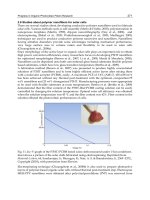
connected to an underwater seal apparatus (Fig. 8.7).
Paradoxic motion of the chest wall from moving rib segments (flail chest)
may require intubation. An open wound of the chest wall (open pneumotho-
rax) requires a sterile occlusive dressing taped on three sides, providing a
flutter-type valve effect, followed by insertion of a chest tube.
Respiratory Failure
Respiratory failure is seen in asthma, congestive heart failure, COPD,
trauma (i.e., pulmonary contusion, pneumo-hemothorax) and occasionally
Fig. 8.7. Chest Tube Placement.
137Resuscitation
8
pneumonia. Signs of hypoxia are dyspnea, tachypnea, tachycardia, restless-
ness, gasping respirations and use of accessory ventilatory muscles. Lethargy
and confusion are seen with hypercapnia (see Chapter 6, Oxygen). ABGs
show a PO
2
<50 mmHg and/or a PCO
2
>50 mmHg, implying impending
respiratory failure, although patients with COPD may normally carry a PCO
2
of 60-70 mmHg. A rectal temperature is taken, since the person is mouth-
breathing. Treatment: endotracheal intubation is usually required, although
some cases may respond to continuous positive airway pressure (CPAP).
Initial settings on a volume-cycled respirator are: oxygen 100%, tidal vol-
ume 15 cc/kg, respiratory rate 16.
3. CIRCULATION:
Hemorrhage is controlled by pressure. Blood loss is treated with 2 large
bore IVs, 2 liters of normal saline and type-specific or O-neg packed red
blood cells (RBCs). Treatments for hypovolemic, cardiogenic (myocardial
infarction, aortic aneurysms, cardiac tamponade), septic and anaphylactic
shock, as well as hypertensive emergencies requiring resuscitation, are
discussed in Chapter 5.
Pulseless Rhythms
Pulseless rhythms are ventricular fibrillation, pulseless ventricular ta-
chycardia, pulseless electrical activity and asystole, the latter being often
a terminal event. Other rhythms (bradycardias, tachycardias, etc) are dis-
cussed in Chapter 3. One must be careful to examine the patient and not
the monitor. The monitor may show a normal sinus rhythm but the patient
may be apneic or pulseless. Conversely, the monitor may show a chaotic rhythm
or straight-line, but if the patient is alert and conversant, a lead is off.
In ventricular fibrillation (VF), the electrical activity of the heart is
chaotic and no heart-beat is present. Pulseless ventricular tachycardia (pVT)
shows VT but without a pulse and is treated as VF (one must be careful not
to confuse this with ventricular tachycardia with a pulse—see Chapter 3).
Treatment: CPR is begun and the patient is defibrillated as soon as possible
3 times in succession (200, 300, 360 J). If unsuccessful the patient is intu-
bated and CPR is continued. Epinephrine is given 1 mg q 5 min. Vaso-
pressin 40 units may be given as one dose (vasopressin at this dosage is a
vasoconstrictor, and is frequently used in Europe). It has been shown that
antiarrhythmic agents possess minimal efficacy in VF/pVT. The usual
protocol is to give the drug, followed by defibrillation. However, it is
acceptable to give the agent, followed by three shocks. Agents used, in
order of preference, are:
1. Amiodarone 300 mg IV push. A second dose of 150 mg may be
given,
138 Vital Signs and Resuscitation
8
2. Lidocaine 1 mg/kg IV push, and repeat in 5 minutes to a total of
3 mg/kg. Defibrillation may be after each agent, or after each minute
of CPR.
In pulseless electrical activity (PEA), the monitor shows a rhythm, but
the patient has no heart beat—the electrical activity is inadequate to stimu-
late contraction of the heart muscle, or the contraction is so weak as to be
negligible. It is seen in several circumstances, the more common being
hypovolemia and massive acute myocardial infarction. Treatment: this is a
situation in which the patient may be mistakenly assumed to have a pulse.
Always check for a pulse. Unfortunately, the reason for this lethal condition
is often not known. CPR is begun, intubation is performed, IV access is
obtained and epinephrine 1 mg IV push is given every 5 minutes. If electri-
cal bradycardia is present, atropine 1mg IV is given every 5 minutes to a
total of 0.04mg/kg. Because this condition is reversible in some circum-
stances, as a last resort bicarb 1 meq/kg and a 200 cc bolus x 2 of normal
saline may be tried.
Asystole, or a straight line on the monitor, is treated with CPR, transcu-
taneous pacing if the rhythm occurred suddenly, epinephrine 1 mg IV every
5 min and atropine 1 mg IV q 5 min (total 0.04 mg/kg). This is often a
terminal nonrhythm, indicating death.
Some bradycardias and tachycardias may require resuscitative measures
(see Chapter 3).
Fig. 8.8. Ventricular Fibrillation. Reprinted with permission from: Merck, Sharp &
Dohm, Division of Merck & Co., Inc.
Fig. 8.9. Pulseless Ventricular Tachycardia. Reprinted with permission from: Merck,
Sharp & Dohm, Division of Merck & Co., Inc.
139Resuscitation
8
Fig. 8.10. Ventricular Fibrillaton/Pulseless Ventricular Tachycardia Algorithm. Re-
printed with permission from: Guidelines for 2000 for Cardiopulmonary Resuscita-
tion and Emergency Cardiovascular Care, American Heart Association.
140 Vital Signs and Resuscitation
8
4. DEFIBRILLATION and DISABILITY
(Level of Consciousness)
In the hospital and emergency medical services (EMS) settings, “D” for
Defibrillation is added to the ABCs. When a monitor or “quick look” paddles
show ventricular fibrillation or pulseless ventricular tachycardia, the patient
is defibrillated immediately as per the above protocol. In the trauma and
other settings, “D” also represents “Disability”, or level of consciousness.
Assessment and management for level of consciousness and therapy for
increased intracranial pressure is described in Chapter 6 (see Figs. 6.4, 6.5).
Fig. 8.12. Asystole. Reprinted with permission from: Guidelines for 2000 for
Cardiopulmonary Resuscitation and Emergency Cardiovascular Care, American
Heart Association.
Fig. 8.11. Pulseless Electrical Activity (PEA). Reprinted with permission from: Merck,
Sharp & Dohm, Division of Merck & Co., Inc.
141Resuscitation
8
Fig. 8.13. Pulseless Electrical Activity Algorithm. Reprinted with permission from:
Guidelines for 2000 for Cardiopulmonary Resuscitation and Emergency Cardiovas-
cular Care, American Heart Association.
142 Vital Signs and Resuscitation
8
Fig. 8.14. Asystole Algorithm. Reprinted with permission from: Guidelines for 2000
for Cardiopulmonary Resuscitation and Emergency Cardiovascular Care, American
Heart Association.
143Resuscitation
8
Fig. 8.15. Resuscitation Protocol.
144 Vital Signs and Resuscitation
8
Pediatric Resuscitation
Pediatric Basic Life Support
1. Establish unresponsiveness. Tap the child and speak loudly.
2. Call for appropriate help.
3. AIRWAY. Open airway using jaw thrust or chin lift (if trauma sus-
pected, stabilize neck, use jaw thrust).
4. BREATHING. Look, listen and feel for breathing. If no breath-
ing, 2 slow breaths mouth to mouth (nose closed). In infants (1
year and less) rescue mouth over nose and mouth. Rescue breath-
ing at 20 breaths per minute.
5. CIRCULATION. If no breathing, begin chest compressions alter-
nating with ventilations, 2 or 3 fingers lower sternum (heel of one
hand with larger children), 1/2 to 1 inch deep, 100 per minute (for
healthcare providers: pulse check, if pulse <60 begin chest compres-
sions). Compression/ventilation ratio 5:1 1-2 rescuers.
Fig. 8.16. Peds Jaw Thrust.
Pediatric Advanced Life Support
1. AIRWAY
As in the adult, the head is tilted and either the jaw thrust or chin lift
used to open the airway. In the child requiring intubation, the patient is first
ventilated by bag-valve-mask with 100% oxygen. If the child is slightly breath-
ing, gentle positive-pressure should be carefully timed with voluntary respira-
tions. Unlike the adult where two assistants are required to adequately bag the
patient, one assistant is often sufficient. In the infant, the jaw is supported with
145Resuscitation
8
the base of the middle and 4th fingers. In older children, the fingertips of
the 3rd, 4th and 5th fingers are placed on the ramus of the mandible to hold
the jaw forward and extend the head. Endotracheal intubation is always via the
orotracheal route (nasotracheal intubation is not performed in children). Rapid
sequence intubation (RSI) is accomplished as in the adult (see earlier section).
Trauma
In major trauma, the c-spine is immobilized and the jaw thrust is per-
formed. The oral cavity is inspected for foreign bodies, vomitus, broken
teeth and suctioned using a hard-tipped suction catheter of appropriate size.
Not only must the C-spine be cleared but the child must be cleared neuro-
logically. If the history and physical exam indicate a possible spinal cord
injury (spinal cord injury without radiographic abnormality—SCIWORA)
the C-collar is left on and the patient is cleared by the neurosurgeon.
Airway Obstruction
Diagnosing a foreign body in the airway may pose a difficult problem
unless complete obstruction occurs. Offenders are nuts, toy parts, round
candies and aluminum “pop-tops”. Complete obstruction in an infant is
treated by a variation of the Heimlich maneuver: the infant is held prone in
the left hand and forearm and 5 back blows are delivered between the shoul-
der blades with the heel of the right hand. Then the infant is turned over,
with the head lower than the body, and 5 quick chest thrusts are delivered
on the lower third of the sternum. The mouth is opened and, if visualized,
the foreign body is removed. The finger sweep and rescue breathing are per-
formed. Nearly all larger foreign bodies are captured at this point. Smaller
foreign bodies will be moved into a mainstem bronchus. In the child over 1-
2 years, the Heimlich maneuver is similar to the adult.
If the patient can not be adequately bagged or intubated, a needle
cricothyrotomy is performed by inserting a 14 or 16g angiocath through the
cricothyroid membrane. The needle is removed, the cannula is secured and
attached to oxygen tubing using a “Y” connector, at 20 breaths per minute:
1 second inhalation, 2 seconds exhalation. A surgical cricothyrotomy is not
performed in children less than 9 years old (Figs. 8.17, 8.18).
2. BREATHING
As in the adult, the lungs are auscultated for equal breath sounds. Breath-
ing children receive 100% oxygen by nonrebreather mask. Comatose patients
are intubated to protect the airway. Nonbreathing children are bagged with
a bag-valve-mask and 100% oxygen and are intubated.
Trauma
In trauma, if signs of tension pneumothorax are present (respiratory dis-
tress, distended neck veins, tracheal deviation), needle decompression is
146 Vital Signs and Resuscitation
8
accomplished through the 2nd intercostal space above the 3rd rib at the
midclavicular line, followed by chest tube placement at the 5th interspace
anterior to the mid-axillary line. An open pneumothorax is treated with an
occlusive dressing and a chest tube as described for the adult.
Fig. 8.17. Infant Foreign-Body Airway Obstruction.
147Resuscitation
8
Respiratory Failure
In respiratory failure, oxygen by mask should be administered to all seri-
ously ill or injured children. A nasopharyngeal airway is placed in the con-
scious patient. Respiratory failure should be anticipated when the following
signs are present:
1. Increased respiratory effort. Tachypnea is the first sign of respira-
tory distress in infants. Other signs are restlessness, use of accessory
muscles with nasal flaring, inspiratory/intercostal/sternal retractions
and tachycardia. Stridor is an inspiratory high-pitched sound of
upper airway obstruction. Wheezing may be present. Grunting is
caused by premature closure of the glottis during early expiration
in an attempt to increase airway pressure.
2. Diminished level of consciousness or response to pain.
3. Poor skeletal muscle tone.
4. Cyanosis is a late sign.
Treatment of Respiratory Failure:
1. Open airway.
2. 100% oxygen by mask.
3. If the patient is not moving air, begin bag-mask ventilations with
small volumes and prepare for endotracheal intubation.
Fig. 8.18. Peds Needle Cricothyrotomy.
148 Vital Signs and Resuscitation
8
3. CIRCULATION
In a child with no pulse, chest compressions are begun and an intrave-
nous line is secured. If intravenous access cannot be obtained within 1-2
minutes, intraosseous access should be performed at the proximal medial
tibia. The next step depends on the reason for the arrest. In adults it is often
cardiac, but in children it is usually respiratory or traumatic. In the trauma
Fig. 8.19. Peds Ventricular Fibrillation/Asystole/PEA Algorithm. Reprinted with per-
mission from: Guidelines for 2000 for Cardiopulmonary Resuscitation and Emer-
gency Cardiovascular Care, American Heart Association.
149Resuscitation
8
setting, hemorrhage is identified and controlled. Hemorrhagic shock is treated
with normal saline boluses (20 cc/kg x 3) and type-specific or O-neg packed
red cells (10 cc/kg) as indicated.
Ventricular fibrillation/pulseless ventricular tachycardia, asystole and
pulseless electrical activity are rare occurrences in pediatrics. In the hospi-
tal, a call is made for quick-look paddles/monitor, with appropriate therapy
depending on the arrhythmia (Fig. 8.19, see also Figs. 8.8, 8.9, 8.11, 8.12).
4. DISABILITY (Level of Consciousness)
“D” for Disability is similar to the adult and represents Level of Con-
sciousness. As mentioned in Chapter 7, the Glasgow Coma Scale has been
modified for infants ages 0-23 months and children ages 2-5 years (Pediatric
Glasgow Coma Scale—PGCS). Since the total scores are the same as for the
adult, intubation is still required for a score of 8 or less. As in the adult,
AVPU is sometimes substituted for the PGCS in the field (see Chapter 6).
The PGCS is now an integral part of the Revised Trauma Score (RTS) (see
Figs. 7.8 and 7.9). A separate Pediatric Trauma Score (PTS) has also been
developed that does not use the GCS. Children with an RTS of less than 12
or a PTS of less than 8 are at increased risk for morbidity and should be
evaluated at a trauma center. Management of coma and therapy for increased
intracranial pressure is described in Chapter 6 (Figs. 6.4, 6.5) (Fig. 8.20).
Neonatal Resuscitation
During delivery, as soon as the head is delivered, the mouth and the nose
are suctioned. If meconium is present, intubation is performed and suction
is applied through the endotracheal tube. The newborn is dried, warmed,
positioned supine, suctioned and tactile stimulation is applied. For low
Apgars, oxygen is administrated (see Chapter 7). If the heart rate is less than
Fig. 8.20. Pediatric Trauma Score (PTS).
150 Vital Signs and Resuscitation
8
100 beats per minute, the newborn is ventilated with a bag-valve-mask at 40
breaths per minute. If bag-mask ventilation cannot raise the heart-rate above
100, intubation is required. Chest compressions (90 per minute) and venti-
lations (30 per min) are performed if the heart rate is less than 60. If the
heart-rate remains below 60 after intubation, epinephrine 0.01 mg/kg
(1:10,000) is administered by way of the umbilical vein. Neonatal resuscita-
tion is sometimes necessary because of drug ingestion, particularly crack/
cocaine, by the mother. If the history indicates drug abuse by the mother,
naloxone (0.1 mg/kg) IV is given (Fig. 8.21).
Fig. 8.21. Neonatal Resuscitation Algorithm. Reprinted with permission from: Guide-
lines for 2000 for Cardiopulmonary Resuscitation and Emergency Cardiovascular
Care, American Heart Association.
151Resuscitation
8
Special Resuscitation Cases
A multitude of drugs, toxins and circumstances may cause near-arrest or
arrest. Space does not permit a discussion of them. Hypothermia and hyper-
thermia are described in Chapter 2. Some toxins, including carbon monox-
ide, are discussed in Chapter 6.
Electrical and Lightning Injuries
Arrest may occur from high voltage direct current (> 1000 volts). Do not
attempt removal until current is shut off. With low voltage exposure, such as
120 volts of alternating current (AC) from a hair dryer or radio, for example,
removal can be accomplished with cloth, rubber or wood. High voltage (DC)
contact usually causes asystole; low voltage (AC) usually causes ventricular
fibrillation. A lightning strike may carry up to a billion volts of DC energy
and may strike directly, obliquely or “splash” from contact as it strikes the
ground or at an object and spreads out to involve the victim. Treatment: the
ABCs are assessed and CPR is begun, followed by intubation and IV access.
Defibrillation or treatment for asystole is performed as required.
Hyperkalemia
Although hypokalemia and hyperkalemia may both cause arrest (in hy-
pokalemia the heart stops in systole; in hyperkalemia it stops in diastole),
hyperkalemia is the more potentially fatal condition, sometimes seen in the
dialysis patient (i.e., K+ level above 8 mEq/L). Less common causes are crush
injuries, burns, acidosis and some drugs (i.e., digoxin, ACE-inhibitors). It is
often impossible to learn the reason for the arrest other than a fragmentary
history (i.e., dialysis). Treatment: the ABC’s are assessed and CPR is begun,
followed by intubation and IV access. Defibrillation or treatment for asys-
tole is instituted as needed. If the cause is known, 10 cc of a 10% solution of
calcium chloride is administered. Calcium stabilizes the cardiac cell mem-
brane, preventing the continued adverse effect of the increased K+. If the
resuscitation is successful, glucose 2 amps + regular insulin 10 units IV push
plus bicarb 1 amp will lower the K+ by moving it into the cells, thus decreas-
ing the level in the bloodstream.
Pregnancy
The main causes of cardiac arrest in pregnancy are pulmonary embolism,
trauma (high homicide), hemorrhage and pre-existing cardiac disease. Pul-
monary embolism is discussed in Chapter 4. Hemorrhagic shock is discussed
in Chapter 5. Treatment: the cause is often not known (other than trauma).
Usual therapies such as heparin for pulmonary embolism will probably not
be effective. The patient in late pregnancy is turned to the left and a roll or
Cardiff wedge is placed under the right flank and hip for CPR. Appropriate
therapy for cardiac arrhythmias and blood loss (IV NS and O-neg packed
152 Vital Signs and Resuscitation
8
cells) is instituted. If pulmonary embolism seems likely, thrombolytics may
be tried. Emergency cesarean section is performed if 5 minutes have elapsed,
the fetus is viable (>20 weeks) and therapy has not been successful. The
results are poor if the C-section is delayed for up to 15 minutes.
Submersion (Near-Drowning)
NOTE: the 2000 International Liaison Committee on Resuscitation
(ILCOR) recommends using the word “submersion” and discarding “near-
drowning” (“drowning” is considered submersion with death at the scene).
Teenagers and toddlers are frequently affected. Drugs, alcohol (60% of teen-
age drownings), cervical spine injury, seizure disorders, mental retardation,
child abuse and suicide are contributory. Death/arrest is from laryngospasm
and asphyxia. Hypothermia is frequently present. Treatment: CPR is begun
after removal from the water, with C-spine protection if a head-injury is
suspected. On EMS arrival, the ABC’s are assessed (including C-spine im-
mobilization) and CPR continued as needed. Supplemental oxygen by mask
is administered to all patients, with fast transport. In the emergency depart-
ment IV access is secured, and intubation is performed as needed. Appropri-
ate therapy for cardiac arrhythmias is instituted. If hypothermia is present,
rewarming measures are begun and resuscitative measures are continued until
the temperature is 90˚F (see Chapter 2).
References
1. American College of Surgeons. Advanced trauma life support. Chicago, 1997.
2. American Heart Association and the International Liaison Committee on Resuscita-
tion (ILCOR). Guidelines 2000 for cardiopulmonary resuscitation and emergency
cardiovascular care. Baltimore: Lippincott, Williams & Wilkins, 2000.
3. American Academy of Pediatrics/American Heart Association. Pediatric advanced life
support. Dallas, 1997.
4. Barkin R, Rosen P. Emergency pediatrics: A guide to ambulatory care. St. Louis: CV
Mosby, 1999.
5. Berg R et al. Bystander cardiopulmonary resuscitation: Is ventilation necessary? Cir-
culation 1993; 88:1907.
6. Bessen H. Hypothermia. In: Tintinalli J et al. Emergency Medicine: A Comprehen-
sive Study Guide. New York: McGraw-Hill, 2000.
7. Cobb L et al. Influence of cardiopulmonary resuscitation prior to defibrillation in
patients with out-of-hospital ventricular fibrillation. JAMA 1999; 281:1182.
8. Colquhoun M et al. ABC of Resuscitation. London: BMJ Books, 1999.
9. Cummins R, Hazinski M. Cardiopulmonary resuscitation techniques and instruc-
tion: When does evidence justify revision? Ann Em Med 1999; 34:780.
10. Datner E, Promes S. Resuscitation issues in pregnancy. In: Tintinalli J et al. Emer-
gency Medicine: A Comprehensive Study Guide. New York: McGraw-Hill, 2000.
11. Dayton L. Secrets of a bolt from the blue. New Scientist 1993; 140:16.
12. De Maio V et al. Cardiac arrest witnessed by emergency medical services personnel:
Descriptive epidemiology, prodromal symptoms, and predictors of survival. Ann Em
Med 2000; 35:138.
13. de Vos R et al. In-hospital cardiopulmonary resuscitation: Prearrest morbidity and
outcome. Arch Int Med 1999; 159:845.
153Resuscitation
8
14. Ewy G. Cardiopulmonary resuscitation—Strengthening the links in the chain of sur-
vival. New Engl J Med 2000; 342:1599.
15. Finer N et al. Cardiopulmonary resuscitation in the very low birth weight infant: The
Vermont Oxford Network experience. Pediatrics 1999; 104:428.
16. Goodlin S et al. Factors associated with use of cardiopulmonary resuscitation in seri-
ously ill hospitalized adults. JAMA 1999; 282:2333.
17. Guohua L et al. Cardiopulmonary resuscitation in pediatric trauma patients: Survival
and functional outcome. J Trauma 1999; 47:1.
18. Hallstrom A et al. Cardiopulmonary resuscitation by chest compression alone or with
mouth-to-mouth ventilation. N Engl J Med 2000; 342:1546.
19. Hals G, Crump T. The pregnant patient: Guidelines for management of common
life-threatening medical disorders in the emergency department. Em Med Reports
2000; 21:53.
20. Jiva T. Critical care of pregnant women, Part 1: Pulmonary edema, ARDS, throm-
boembolism. J Crit Illness 2000; 15:316.
21. Kirkegaard-Nielsen H et al. Rapid tracheal intubation with rocuronium. Anesthesiol-
ogy 1999; 91:131.
22. Krumholz H et al. Resuscitation preferences for heart-failure patients likely to change.
Circulation 1998; 98:648.
23. Kudenchuk P. Amiodarone for resuscitation after out-of-hospital cardiac arrest due to
ventricular fibrillation. N Engl J Med 1999; 341:871.
24. Landwirth J. Ethical issues in pediatric and neonatal resuscitation. Ann Emerg Med
1993; 22:502.
25. Losek J. Hypoglycemia and the ABC’s (sugar) of pediatric resuscitation. Ann Emerg
Med 2000; 35:43.
26. Markovchick V, Pons P. Emergency Medicine Secrets. Philadelphia: Hanley & Belfus, 1999.
27. Noe M et al. Mechanical ventilation may not be essential for initial cardiopulmonary
resuscitation. Chest 1995; 108:821.
28. Patterson M. Resuscitation update for the pediatrician. Ped Clin N Am 1999; 46:1285.
29. Reed W. Near-drowning: Life-saving steps. Phys and SpoRTSmed 1998; 26:31.
30. Ryan J. Immersion deaths and swim failure: Implications for resuscitation and pre-
vention. Lancet 1999; 354:613.
31. Ryan T et al. 1999 update: ACC/AHA guidelines for the management of patients
with acute myocardial infarction: Executive summary and recommendations. Circu-
lation 1999; 100:1016.
32. Singer A et al. Emergency Medicine PEARLS. Philadelpia: FA Davis, 1996.
33. Soll R. Consensus and controversy over resuscitation of the newborn infant. Lancet
1999; 354:4.
34. Strange G et al. Pediatric Emergency Medicine. New York: McGraw-Hill, 1996.
35. Tapson V. Management of the critically ill patient with pulmonary embolism. J Crit
Illness (supplement) July 2000.
36. Tyson J et al. Viability, morbidity and resource use among newborns of 501 to 800 g
birth weight: National Institute of Child Health and Human Development Neonatal
Research Network. JAMA 1996; 276:1645.
37. Van Hoeyweghen R et al. Quality and efficiency of bystander CPR. Resuscitation
1993; 26:47.
38. Weil M, Tang W. CPR—Resuscitation of the Arrested Heart. Philadelphia: WB
Saunders, 1999.


