ALZHEIMER''''S DISEASE: ITS DIAGNOSIS AND PATHOGENESIS - PART 3 pptx
Bạn đang xem bản rút gọn của tài liệu. Xem và tải ngay bản đầy đủ của tài liệu tại đây (1.64 MB, 10 trang )
ALZHEIMER'S DISEASE 187
Unresolved Issues
CAA
Insoluble A~
plaque
Soluble AI3 4 X~
Microvascular
damage
~d flow
Disturbances
Cell death
,S
x,
NFT Inflammation '
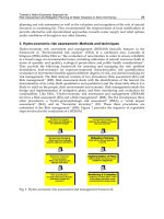
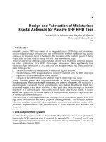
FIG. 4. Many unresolved issues continue to plague our understanding of the pathogene-
sis of AD. Although it is known that neuronal death can occur due to NFT formation and
cerebrovascular disease, such as cerebral amyloid angiopathy (CAA), altered perfusion, or mi-
crovascular pathology (pale arrows), the exact role of AB and inflammation are unknown (dark
arrows). A/3 deposition and inflammation are both universal findings in the brain of AD, and
the former is necessary for a pathological diagnosis of AD. However, whether either results in
neuronal death, and if so by what mechanism, is yet to be determined.
major mechanism of neuronal degeneration occurs in AD. Unfortunately,
the lack of a readily identifiable marker for this neuronal loss has made the
identification of its cause extremely difficult.
A study has shown that the prolyl isomerase, Pinl, is sequestered into
the NFT and depleted in the brains of AD patients (Lu
et al.,
1999).
188
JILLIAN J. KRIL AND GLENDA M. HALLIDAY
Depletion of Pinl may induce neuron death via mitotic arrest and apopto-
sis prior to the development of NFTs (Lu
et al.,
1996). The neuron-specific
activator for cell proteins involved in the mitotic cycle is p35, which is pro-
teolytically cleaved to produce p25, a fragment found to accumulate in the
brains of patients with AD (Patrick
et al.,
1999). Application of Aft 1-42 in-
duces the conversion of p35 to p25 (Lee
et al.,
2000). p25 links with cell
cycle-dependent kinase 5 to hyperphosphorylate tau and promote apopto-
sis (Lee
et al.,
2000). Degenerating neurons in APP V717F Aft-producing
transgenic mice show chromatin segmentation and condensation, as well as
increased TUNEL staining, suggestive of apoptosis (Nijhawan
et aL,
2000).
This supports a link between Aft deposition and apoptosis (Fig. 4). Increased
TUNEL staining (Druganow
et al.,
1995; Lassmann
et al.,
1995; Smale
et al.,
1995; Bancher
et al.,
1997), as well as cleaved caspase 3 (Selznick
et al.,
1999;
Stadelmann
et al.,
1999), an enzymatic marker of apoptosis, are found in
vulnerable brain regions in AD. APP has been identified as a specific sub-
strate for caspase 3 with the resultant peptides (including Aft), inducing
apoptosis (Gervais
et al.,
1999). Other apoptotic-specific caspases can also
cleave APP (Pellegrini
et al.,
1999), and the resultant C-terminal fragment
from such cleavage has been called C31 (Lu
et aL,
2000). C31 is also a potent
inducer of apoptosis and was found in the brain of patients with AD (Lu
et al.,
2000), whereas caspase deficient mice are resistant to this form of cell
death (Nakagawa
et al.,
2000).
Despite these studies that suggest apoptosis occurs in AD, apoptotic bod-
ies and blebbing are not features of AD neuronal degeneration. In addition,
the time sequence of such events remains to be determined. The chronic
nature of the neurodegeneration in AD does not fit well with the more rapid
time course of apoptosis, which is believed to take only weeks or months at
most (Stadelmann
et al.,
1999). Other mechanisms of neuronal death, such
as necrosis, were also demonstrated in AD (Wolozin and Behl, 2000b). In-
deed, the same triggers may cause either apoptosis or necrosis, including Aft
toxicity, oxidative stress, excitotoxicity, ischemia, and removal of trophic fac-
tors. The distinction between apoptotic and necrotic mechanisms, however,
may be somewhat false given that neurons may begin with necrosis and then
convert to apoptosis or alternatively begin with apoptosis and then undergo
necrosis (Wolozin and Behl, 2000b).
Although Aft plaques are necessary for a diagnosis of AD, like NFTs, they
are poorly related to the degree of neuronal loss. However, studies suggest
the intracellular accumulation of Aft may be neurotoxic (Fig. 4). There is an
additional site of APP cleavage within the endoplasmic reticulum that gives
rise to intracellular Aft 1-42/43, which over time reaches the concentration
necessary for fibril formation (Hartmann, 1999; Wilson
et al.,
1999). Cell
rupture would release this intracellular Aft into the surrounding extracellu-
lar milieu, which could stimulate further amyloid deposition. Although most
ALZHEIMER'S DISEASE 189
cell types express APE neurons produce the highest amount and preferen-
tially use the intracellular pathways for Aft production (Hartmann, 1999). It
is difficult to know how to prove or refute this model of AD neuronal vulnera-
bility, although it is of interest that Aft is not deposited within the vulnerable
hippocampal formation or entorhinal cortex (Arnold
et al.,
1991) and no
neuronal loss occurs in elderly APP-transgenic mice who show considerable
A/3 deposits (Irizarry
et al.,
1997a, 1997b). Interestingly, a study identified
nonpyramidal neurons containing Aft 1-42 around amyloid plaques in AD
patients (Mochizuki
et al.,
2000), suggesting preserved neurons may con-
centrate these peptides intracellularly.
In contrast, a number of studies suggest that soluble A/3, and particularly
Aft 1-40, is synaptotoxic without causing plaque formation or overt cell death
(Mucke
et al.,
2000). Reductions in soluble A/31 40 concentrations correlate
with synaptic loss in patients with AD (Lue
et al.,
1999). Interestingly, in the
same patients, soluble A/31-40 levels correlate with cerebrovascular amyloid
angiopathy and ApoEe4 allele frequency (Lue
et al.,
1999), suggesting a
greater influence on vascular changes than neuronal degeneration.
E SUMMARY
Taken together, these studies suggest a multifactorial origin of neuronal
loss in AD where a number of primary and secondary factors may cause
neuronal death (Fig. 4). More work is needed to link all the potential cellular
events that underlie the clinical symptoms of AD. At present, we do not have a
good understanding of the association between A/3 deposition (required for
a diagnosis of AD) and the degenerative process. The link between soluble
A/3 and brain atrophy needs to be clarified, and mechanisms of cell death
other than NFT formation (and possibly apoptosis) need to be elucidated.
It will be important to determine the time sequence of these events to target
appropriate therapeutic measures.
IV. Genetic Influences
As many as 50% of patients with AD have at least one first-degree relative
with dementia (Writing Committee Lancet Conference 1996, 1996), and
numerous studies have investigated family history as a risk factor for AD.
Nine of the 14 case control studies reviewed byJorm (1990) showed a sig-
nificantly increased risk of AD in subjects with a positive family history. The
odds ratios ranged from 2.1 to 9.9 and reflect data obtained from prevalence
and incidence studies.
190 JILLIAN J. KRIL AND GLENDA M. HALLIDA¥
A.
DOMINANT INHERITANCE
It is estimated that between 5% and 10% of AD cases have a demon-
strable pattern of inheritance. These cases, although rare, provide valuable
insights into the pathogenesis of AD. To date, three genes have been iden-
tified. These are APP mutations on chromosome 21, presenilin-1 (PS-1)
mutations on chromosome 14, and presenilin-2 (PS-2) mutations on chro-
mosome 1. Each of these genes have an autosomal dominant pattern of
inheritance, although PS-2 does not appear to have complete penetrance
(St. George-Hyslop, 2000). These three identified genes do not fully account
for all autosomal dominant cases of AD, suggesting other genes are yet to
be identified.
The APP gene encodes a transmembrane protein of 770 amino acids
from which Aft is derived (see section III.C above). The normal function
of APP is not known, although it is highly conserved and expressed ubiqui-
tously. In addition to AD, mutations in APP can also result in hereditary cere-
bral haemorrhage with amyloidosis-Dutch type (HCHWA-D). Mutations in
the APP gene are mostly located in or around the amyloidogenic portion of
the molecule, especially near the three secretase sites.
Mutations in the PS-1 gene are the most common of the early-onset
familial AD mutations, accounting for 30-50% of all autosomal dominant
cases. PS-1 is a transmembrane protein that is also expressed ubiquitously
and has six or eight transmembrane domains (Checler, 1999). There is an
increasing body of evidence that suggests the presenilins function as the
y-secretase, or in close association with y-secretase, in the production of A/3
(Checler, 1999; Ray
et al.,
1999; Wolfe
et al.,
1999) and thus increase the pro-
duction Afll-42/43. More than 50 mutations in PS-1 have been identified.
The majority of these are missense mutations and are scattered throughout
the molecule. In addition, a number of splice acceptor mutations that cause
the deletion of the sequence encoded by exon 9 were also described (Kwok
et al.,
1997; Crook
et al.,
1998; Smith
et al.,
2001). A proportion of PS-1
mutations with a deletion of exon 9 have AD with spastic paraparesis (SP;
Crook
et al.,
1998; Verkkoniemi
et al.,
2000). In AD+SP, there is progressive
weakness and wasting of the lower extremities and a later age of onset of
dementia has been described in some of these families (Smith
et al.,
2001).
The pathology of exon 9 mutations is also interesting in that very large,
noncored, and faintly neuritic plaques are described (Crook
et al.,
1998;
Smith
et al.,
2001). These have been termed "cotton-wool" plaques because
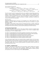
of their size and uniform appearance (Fig. 5).
The PS-2 gene encodes a transmembrane protein that is 67% homol-
ogous to PS-1 (Checler, 1999). Unlike APP and PS-1, PS-2 is expressed
more strongly in peripheral tissues (pancreas, cardiac, and skeletal muscle)
ALZHEIMER'S DISEASE 191
FIG. 5. Photomicrographs of the temporal neocortex of a patient with a presenilin-1
(PS-1) mutation. In the upper panel, both neuritic (arrows) and diffuse plaques can be seen.
The diffuse plaques (inset) in these patients are unusual because they are large, only faintly neu-
ritic, and lack cores. They have been termed "cotton wool" plaques and are tound exclusively
in patients with PS-I mutations.
than in the brain (St. George-Hyslop, 2000). A small number of families with
missense mutations in PS-2 have been identified, indicating they are much
rarer than PS-1 mutations. The exact mechanism by which PS-2 mutations
cause AD is unclear, although because of its sequence homology with PS-1,
it is believe to have a similar function.
192 JILLIAN J. KRIL AND GLENDA M. HALLIDAY
The mechanism common to the known mutations is an increased pro-
duction of Aft 1-42/43 and an increased rate of aggregation of Aft plaques
(see Wolozin and Behl, 2000a, for commentary). However, it appears that
the PS mutations may also be involved in other aspects of the pathology
of AD by participating in cell death due to apoptosis and in the phos-
phorylation of tau (see Checler, 1999; Czech
et al.,
2000). Our knowledge
of AD has advanced substantially since the identification of the mutations
responsible for familial forms of AD and of the presenilins in particular. This
rapidly moving field of research provides valuable insights into the disease
processes and has the potential for the development of strategies for ther-
apeutic intervention. However, it is still unknown whether the knowledge
gained from studying these cases is generally applicable to the majority of
AD patients. In addition, the knowledge gained has still not elucidated the
cause(s) of sporadic AD.
B. GENETIC RISK FACTORS
Apart from dominant inheritance, the clustering of dementia within
families must be viewed as evidence for the role of an individual's genotype in
determining their risk of AD. The apolipoprotein E (ApoE) gene, found on
chromosome 19, encodes three isoforms of e2, e3, and e4, and the presence
of the e4 allele has been found to increase the risk ofAD (Katzman, 1994;
Strittmatter and Roses, 1995). ApoE is involved in lipid transport and is
present in the serum (Uterman, 1994). An association between ApoE e4
and AD was first described in 1993 in both sporadic (Saunders et
al.,
1993)
and familial (Corder
et al.,
1993) AD. It has subsequently been confirmed
in many other studies of early- and late-onset AD and a variety of other
neurological diseases, including other dementias (e.g., Roses, 1996; Stevens
et al.,
1997; Horsburgh
et al.,
2000). In addition, an allelic dose dependence
has been shown where subjects who are homozygous for e4 have a greater
risk of AD at an earlier age than those who are heterozygous (Corder
et al.,
1993). In this study of families with late-onset AD, subjects with no e4 had a
mean age of onset of 84.3 years compared with 75.5 years in those with one
s4 allele and 68.4 years with two alleles.
In addition to its effect on age of onset, ApoE genotype has also been
shown to influence, albeit variably, the response to drug treatment. A poorer
response to the cholinesterase inhibitor tacrine has been shown in patients
with AD who possess the ApoE e4 allele than those who do not (Poirier
et al.,
1995), although this effect has not been found in all studies (MacGowan
et al.,
1998). In addition, only patients with e4 showed improvement in
ALZHEIMER'S DISEASE
193
cognitive performance when treated with a drug that facilitiates noradrener-
gic and vasopressinergic activity in the brain (Richard et
al.,
1997). Some de-
bate also exists over whether ApoE genotype modifies the type or amount of
AD pathology in individuals carrying the e4 allele. Several studies (Schmechel
et al.,
1993; Nagy et
al.,
1995; Overmyer
et al.,
1999), but not all (Morris
et al.,
1995; Landen et
al.,
1996), have found an increase in the density of neu-
rofibrillary tangles and senile plaques in AD. Moreover, the correlation with
brain pathology is further complicated by the finding that normal subjects
in their forties and older who possess an e4 allele have smaller right hip-
pocampi than those without e4 (Tohgi et
al.,
1997). It is unclear whether
this finding represents a lifelong trait or is an indicator of "preclinical" AD.
Longitudinal studies on such groups of subjects are necessary to clarify this
issue. In patients with AD, greater brain atrophy (Lehtovirta
et al.,
1995;
Juottonen
et al.,
1998b) and an increased rate of atrophy has been found in
individuals with e4 (Wahlund
et al.,
1999). However, this association has not
been found in all studies (Barber
et al.,
1999).
The mechanism of action of ApoE is not fully elucidated. ApoE is in-
volved in the regulation of the transport of cholesterol and phospholipid
and has an important role in the distribution of these molecules during peri-
ods of membrane remodeling, such as synaptic plasticity and membrane re-
pair. In addition, ApoE-lipid complexes are believed to assist in the removal
of Aft via the low-density lipoprotein-related receptor (Wolozin and Behl,
2000a). Isoform differences in the behavior of ApoE have been identified
(e.g., Strittmatter
et al.,
1993; Nathan
et al.,
1994), and these are believed to
underlie the susceptibility to AD in individuals with the e4 allele (Horsburgh
et al.,
2000; Wolozin and Behl, 2000a).
C. SUMMARY
In addition to these genetic factors, other modifying influences have
been identified (e.g., HLA, butyrylcholinesterase K, ~ 1 antichymotrypsin) ;
however, the exact nature of the relationship between genotype and disease
susceptibility remains obscure. Although there is strong evidence for an as-
sociation between ApoE e4 and AD, the presence of e4 is not causative
or is it necessary to develop AD. For these reasons, it is recommended
that ApoE not be used for predictive testing (American College of Medical
Genetics/American Society of Human Genetics Working Group on ApoE
and Alzhemer's Disease, 1995). Similar results are likely for other genetic
risk factors. Nevertheless, such genotypes are important variables to be
considered in research studies examining aspects of the pathogenesis
194 JILLIAN J. KRIL AND GLENDA M. HALLIDAY
and progression of AD, especially as reports of monozygotic twins that are
discordant for AD (Creasey
et al.,
1989) suggest that inheritability is not
solely responsible for one's risk of AD. Few studies are integrating the multi-
ple genotype analyses required to understand genetic versus environmental
influences.
V. Inflammation and Anti-inflammatory Drugs
Numerous lines of evidence suggest a link between brain inflammation
and AD (see Gahtan and Overmier, 1999; Halliday
et al.,
2000a). Initial ev-
idence from clinical studies for a role of anti-inflammatory drugs in the
prevention of AD came from case control studies that examined arthritis
as a risk factor and found a reduced risk of dementia in patients who con-
sumed anti-inflammatory drugs (Broe
et al.,
1990; Breitner, 1996). However,
a number of similar studies were unable to identify a significant reduction in
risk (e.g., Heyman
et al.,
1984). This inconsistency may reflect the relatively
small samples examined in each study individually because a meta-analysis
of 17 studies showed a reduced risk of AD dementia in patients taking both
steroidal and nonsteroidal anti-inflammatory drugs (NSAIDs; McGeer
et al.,
1996). It should be noted, however, that the majority of these studies were
of cross-sectional design where significant biases exist in selection of cases
for study and the reporting of drug use (Stewart
et al.,
1997).
Antigens of the major histocompatibility complex are intimately associ-
ated with inflammation and polymorphisms of the genes encoding these
proteins have been associated with an increased risk of disease. In partic-
ular, CNS and peripheral diseases with an inflammatory basis occur more
commonly in subjects who have a particular HLA genotype; notable among
these is the association between rheumatoid arthritis and HLA-DR4 (Khan
et al.,
1983; Stastny
et al.,
1988). A number of different associations were de-
scribed between AD and HLA alleles. In late-onset patients who do not have
ApoE e4 alleles, an increased risk of AD was found in patients with HLA-
DR1, 2, or 3, and a reduced risk was found in patients with HLA-DR4 or 6
(Curran
et al.,
1997). However, these findings were not replicated by others
(Middleton
et al.,
1999b), or only partly replicated (Neill
et al.,
1999), and
the converse relationship (HLA-DR3 is protective) was found in a study of
autopsy-confirmed cases ofAD (Culpin
et al.,
1999). In addition, an earlier
age of onset by 3 years has been reported in subjects with HLA-A2 com-
pared with other alleles (Payami
et al.,
1997; Combarros
et al.,
1998), and
when the patient's ApoE status was examined, the effect of HLA-A2 and
ALZHEIMER'S DISEASE 195
ApoE e4 appeared to be additive (Payami
et al.,
1997). A similar additive
effect of HLA-A2 and ApoE e4 has been found in early-onset familial AD
(Ballerini
et al.,
1999). Other associations with HLA alleles were reported
(Small
et al.,
1991; Middleton
et al.,
1999a), but these studies are yet to
be replicated. It is therefore unclear whether the initial studies implicat-
ing anti-inflammatory medications as protective for AD are due to a direct
effect on brain inflammation or are associated with genotype and disease
susceptibility.
To date, there have been only three longitudinal studies analyzing the
question of drug protection in AD. Two of these studies (Stewart
et al.,
1997; Prince
et aL,
1998) found a beneficial effect of NSMDs. The Baltimore
Longitudinal Study of Aging found a reduced risk of AD among users of
NSMDs and aspirin, which was increased the longer the drugs were used
(Stewart
et al.,
1997). Prince and colleagues (1998) showed less decline in
some tests of cognitive function in NSMD users, although the benefit was
reduced in older subjects. In contrast, a study of Australians ages 70 years or
older (mean age of 80) found that NSMDs or aspirin provided no protection
against cognitive decline or incidence of dementia over a 3- to 4-year period
(Henderson
et al.,
1997). Taken together, these studies suggest that some
protection is conferred at ages when susceptibility is relatively low. It may be
that sufficient protection occurs only with long-term drug usage.
It is therefore not surprising that clinical trials aimed at assessing the role
of NSMDs in preventing AD produced conflicting results. Rogers and col-
leagues (1993) performed a study ofindomethacin in 28 patients and found
a small but significant slowing of cognitive decline in the treated patients.
Conversely, Scharf and colleagues (1999) used an NSMD in combination
with a gastroprotective agent and found no difference between groups in
measures of cognitive performance. Drop-out rates in both studies were
considerable (up to 50%) and follow-up times short (around 6 months), so
neither study can be considered conclusive. Nevertheless, on cross-sectional
analysis cognitive performance is improved in AD patients taking NSMDs
and aspirin (Broe
et al.,
2000) compared with their nontreated counter-
parts. Interestingly, this effect was present at low doses of aspirin, which are
not considered to be anti-inflammatory suggesting the effect of these drug
is not through reducing inflammation but through some other, possibly
peripheral mechanism (Broe
et al.,
2000).
Neuropathological studies demonstrated a close relationship between
Aft plaques and both reactive astrocytes and microglia (Rozemuller
et al.,
1992; McGeer and McGeer, 1995; Halliday
et al.,
2000b). Mthough a glial
response might be expected to occur secondary to the degeneration in AD,
evidence suggests the inflammatory response itself may contribute to the
196 JILLIAN J. KRIL AND GLENDA M. HALLIDAY
pathology of AD. Many of the proteins of the complement pathway, together
with acute phase proteins, are found in Aft plaques (see Walker, 1998) and
are believed to be synthesized by microglia. In addition, activated microglia
synthesize and excrete a number of inflammation-related substances that
have been shown to be neurotoxic in rats (Weldon
et al.,
1998), and it has
been suggested that microglia might facilitate Aft deposition (see Gahtan
and Overmier, 1999). Overall, the data show that patients with AD have an
active immune response in the brain.
An age-related increase in inflammatory microglia has also been found
(Mattiace
et al.,
1990; Mackenzie and Munoz, 1998) which may reflect the
brain's reaction to the increased AD-type pathology in aging or, alternatively,
indicate changes to the immune status of the elderly brain. Interestingly, this
age-associated increase in activated microglia is ameliorated by NSAID use
(Mackenzie and Munoz, 1998), unlike AD patients where NSAID use does
not decrease inflammation (Halliday
et al.,
2000b). This suggests the disease
process itself stimulates an immune response. Whether inflammation is a
primary cause for the neurodegeneration in AD or a secondary event to
aid in its clearance is still unclear because the sequence of these events is
still poorly understood (Fig. 4). Although some epidemiological and clin-
ical evidence suggests a beneficial effect of treatment with NSAIDs, other
research suggests any such benefit is mediated through a noninflammatory
mechanism (Broe
et al.,
2000; Halliday
et al.,
2000b). A clearer picture of
the sequence of the early and subsequent cellular events in patients with AD
would help clarify any direct role of inflammation in the disease process.
The enhanced immune response in AD patients is now being used for
a new type of treatment, Aft peptide immunization (Schenk
et al.,
2000).
Immunization trials are about to commence following the dramatic find-
ings that transgenic mice that overproduce APP and deposit Aft can recover
following immunization (Schenk
et al.,
2000). Specifically, when the mice
were immunized at a young age, they developed little if any Aft depositions
with advancing age. Moreover, the progression of both neuritic dystrophy
and astrogliosis were significantly reduced in the treated animals, suggest-
ing the immunization had benefits beyond simply reducing Aft deposition.
When immunization was begun at later ages when the mice exhibit Aft de-
position, further Aft deposition was blocked and somewhat reversed, as was
the neuritic dystrophy and astrogliosis. In addition, remaining Aft deposits
were often actively metabolized by microglia cells, questioning the premise
that reduction of the activity of these cells by anti-inflammatory medica-
tions would be of benefit in AD. These studies support the concept that the
immune system may be harnessed into an appropriately targeted therapy
for AD. If the trials of Aft immunization are effective in AD, it will provide
compelling evidence for its causative role in AD.