Case Files Neurology - part 2 pptx
Bạn đang xem bản rút gọn của tài liệu. Xem và tải ngay bản đầy đủ của tài liệu tại đây (326.33 KB, 50 trang )
APPROACH TO DYSTONIA
Definitions
Dystonia—Sustained muscle contractions cause twisting and repetitive
movements or abnormal postures.
Myoclonus—Sudden, involuntary jerking of a muscle or group of muscles.
Opisthotonos—Great rigid spasm of the body with the back fully arched
and the heels and head bent back.
Clinical Approach
Dystonia is classified according to etiology, as idiopathic or symptomatic.
Primary dystonia is defined as a condition with no etiology that can be iden-
tified, and dystonia is the sole or major symptom. Primary dystonias are further
subdivided by criteria such as age of onset, distribution of affected body parts,
presence of diurnal variation of symptoms, responsiveness to drugs, and genetic
markers. Secondary dystonia refers to dystonia in the context of a neurologic
disease in which dystonia is only one of several symptoms or in which dysto-
nia is the result of an environmental insult. There are at least 15 genetic causes
of dystonia. Generalized dystonia tends to have its onset in childhood. A three–-
base-pair guanine–adenine–guanine (GAG) deletion in exon 5 of DYT1
(TOR1A) is the most frequent cause of early onset, generalized dystonia starting
in a limb and is known as DYT-1 dystonia. However, there is a large phenotypic
variability even within families with an identical mutation. Primary generalized
torsion dystonia is a progressive, disabling disorder that usually begins in child-
hood and is linked to several genetic loci. Many cases are inherited as autosomal
CLINICAL CASES 33
Table 3–1
DIFFERENTIAL DIAGNOSIS OF DYSTONIA
Secondary dystonias
Drug-induced tardive dystonias
Antipsychotic drugs: dopamine receptor–blocking older typical and newer atypical
drugs
Anxiolytic drug: buspirone
Antidepressant agents: selective serotonin-reuptake inhibitors
Dopaminergic drugs: levodopa and dopamine agonists
Antiemetic drugs: metoclopramide
Antiseizure drugs: phenytoin, carbamazepine, gabapentin
Cerebral palsy
Wilson disease
Mitochondrial encephalopathies
Neuroacanthocytosis
Pantothenate kinase–associated neurodegeneration (Hallervorden–Spatz disease)
Fahr disease
dominant traits caused by a deletion in the torsin A gene (DYT1 locus), result-
ing in the deletion of glutamate in torsin A, a brain protein of unknown func-
tion with highest concentrations in the substantia nigra.
Penetrance is 30–40%, and clinical expression varies from generalized dysto-
nia to occasional adult-onset focal dystonias. It begins as a focal action dystonia
before the middle of the third decade of life with most cases beginning in child-
hood. Because of its rarity and unfamiliar features, it is sometimes misdiagnosed
a psychogenic disorder. Approximately 65% of cases progress to a generalized or
multifocal distribution, 10% become segmental, and 25% remain focal.
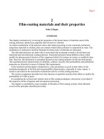
Childhood-onset cases commonly evolve to generalized dystonia, which pro-
duces severe disability owing to serious gait and posture abnormalities (Fig. 3–1).
This can result in a life-threatening condition called status dystonicus. The
diagnosis of DYT-1 can be made by commercially available testing.
Most primary dystonias have normal routine neuroimaging studies. [18F]-
fluorodeoxyglucose and positron emission tomography (PET) has been used
with a novel regional network analytical approach to identify a reproducible
pattern of abnormal regional glucose metabolism in primary torsion dystonia.
This pattern is not specific for the DYT1 genotype, can be present in other pri-
mary dystonia genotypes, and is not routinely available.
34 CASE FILES: NEUROLOGY
Figure 3–1. Incapacitating postural deformity in a young man with dystonia.
(With permission from Ropper AH, Brown RH. Adams and Victor’s principles
of neurology, 8th ed. New York: McGraw-Hill; 2005: Fig. 4–5c.)
In any given case, the first consideration is whether this represents a sec-
ondary dystonia, particularly one which is amenable to effective treatment,
including discontinuation of offending agents. Some clues that dystonia is sec-
ondary include:
• History of trauma or exposure to drugs, infections, cerebral anoxia
• Dystonia at rest, rather than with action, at its onset
• Atypical site for age of onset—for example, leg onset in an adult, cranial
onset in a child
• Early onset of speech abnormality
• Hemidystonia
• Presence of abnormalities other than dystonia on neurologic examination
or general medical examination
• Nonphysiologic findings suggesting a psychogenic basis
• Abnormal brain imaging
• Abnormal laboratory tests
Table 3–2 summarizes common etiologies of secondary dystonia. The current
functional model of basal ganglia suggests that dystonia results from abnormally
low or generally abnormal pattern of activity of basal ganglia output structures:
the internal segment of globus pallidus (GPi) and substantia nigra pars reticulata.
CLINICAL CASES 35
Table 3–2
CAUSES OF SECONDARY DYSTONIA
Hereditary disorders associated with
Neurodegeneration (Huntington disease, juvenile Parkinson disease (parkin),
Wilson disease, lysosomal storage disorders, Rett syndrome)
Dystonia-plus syndromes (dopa-responsive dystonia, myoclonus-dystonia, rapid-
onset dystonia-parkinsonism)
Acquired/exogenous causes (Medication: dopamine receptor-blocking agents,
Antiepileptic agents, levodopa, dopamine agonists, calcium-channel blockers;
Toxins: manganese, carbon monoxide, carbon disulphide, methanol, wasp sting;
Perinatal cerebral injuries: cerebral palsy, kernicterus; Vascular lesions: stroke,
arteriovenous malformation, antiphospholipid syndrome ; Infection: encephalitis,
subacute sclerosing panencephalitis, HIV/AIDS, abscess; Brain tumors; paraneo-
plastic syndromes; demyelination: multiple sclerosis, pontine myelinolysis;
Trauma: head trauma, cervical cord injury; Structural: atlanto-axial subluxation,
Klippel-Feil syndrome, Arnold-Chiari malformation)
Parkinson disease and other parkinsonian disorders (progressive supranuclear
palsy, corticobasal degeneration, multiple system atrophy)
Other movement disorders (tic disorders, familial paroxysmal kinesigenic dyski-
nesias, familial paroxysmal non-kinesigenic dyskinesias, episodic ataxia syndromes)
This low activity consequently disinhibits the motor thalamus and cortex, giv-
ing rise to abnormal movements. In addition, drugs that inhibit the action of
dopamine (through type 2 dopamine [D2] receptors) can cause acute or chronic
dystonia. This seems to be mediated by disinhibition of cholinergic neurons.
Symptomatic treatment of dystonia in the past has employed primarily phar-
macologic agents. These include systemic agents such as levodopa, blockers of
central muscarinic cholinergic receptors, benzodiazepines, and baclofen.
Anatomically targeted administration of agents is also feasible including botu-
linum toxin and intrathecal administration of baclofen. There is mounting evi-
dence that the most effective treatment for generalized dystonia is high-frequency
stimulation of the GPi, through the surgical placement of a deep brain stimulator.
Comprehension Questions
[3.1] The drug most likely to help dystonic symptoms in a patient with DYT-1
dystonia is:
A. Haloperidol
B. Trihexyphenidyl (Artane)
C. Phenytoin
D. Chlorpromazine
[3.2] A 12-year-old boy has the acute onset of sustained contractions of the
left leg and right arm as well as loss of sensation above the neck. The
severity of the symptoms is highly variable. The most likely diagnosis is:
A. DYT-1 dystonia
B. Acute dystonia from a medication
C. Bilateral ischemic infarction of the globus pallidi
D. Psychogenic disorder
E. A right spinal cord hemisection syndrome
[3.3] A 32-year-old woman is seen in the emergency department. She has no
medical problems nor allergies to medications. She receives a medica-
tion intravenously and has an acute dystonic reaction with muscle
spasm of the neck. Which of the following drugs is most likely respon-
sible for this reaction?
A. Haloperidol
B. Trihexyphenidyl (Artane)
C. Phenytoin
D. Levodopa
36
CASE FILES: NEUROLOGY
CLINICAL CASES 37
Answers
[3.1] B. Trihexyphenidyl (Artane) is an antimuscarinics anticholinergic.
[3.2] D. This is likely psychogenic because there is a physiologically incon-
gruent examination.
[3.3] A. Haloperidol is a potent blocker of dopamine D2 receptors and is a
common agent responsible for dystonic reactions in otherwise healthy
individuals.
CLINICAL PEARLS
❖ DYT-1 dystonia is an autosomal dominant disease, which can be
confirmed with genetic testing.
❖ DYT-1 and other primary dystonias usually have the abnormal
movements in association with action early in the course of the
disease.
❖ In mild cases of DYT-1 and other primary generalized dystonias,
systemic drugs, such as anticholinergics, benzodiazepines, and
baclofen may control symptoms, in severe cases, deep brain stim-
ulation of the globus pallidi may be required.
REFERENCES
Albanese A. The clinical expression of primary dystonia. J Neurol 2003;250:1145–1151.
Albanese A, Barnes MP, Bhatia KP, et al. A systematic review on the diagnosis and
treatment of primary (idiopathic) dystonia and dystonia plus syndromes: report
of an EFNS/MDS-ES Task Force. Eur J Neurol 2006;13(5):433–444.
Geyer HL, Bressman SB. The diagnosis of dystonia. Lancet Neurol 2006;5:780–790.
Krauss JK, Yianni J, Loher TJ, et al. Deep brain stimulation for dystonia. J Clin
Neurophysiol 2004;21(1):18–30.
Manji H, Howard RS, Miller DH, et al. Status dystonicus: the syndrome and its
management. Brain 1998;121:243–252.
Tarsy D, Simon DK. Dystonia. N Engl J Med 2006;355:818–829.
This page intentionally left blank
❖
CASE 4
The patient is a 55-year-old man in good health until about 6 months ago. At
that time he noticed development of a tremor. He has no other complaints. On
examination, there is a tremor in the right arm at rest and while he walks, he
has a sustained tremor in both arms, and to some degree during finger-nose-
finger maneuver (fairly fine and without an obvious rhythm). He has a poker
face and a slow, deliberate gait. Tone is increased in the right arm and leg. The
physical examination is otherwise unremarkable. He and his wife deny his use
of alcohol or any other medications.
◆
What is the most likely diagnosis?
◆
What is the next diagnostic step?
◆
What is the next step in therapy?
ANSWERS TO CASE 4: Parkinson Disease
Summary: This is a middle-aged man with asymmetric onset of tremor. In
addition he has mild poverty of movement (otherwise known as akinesia of the
face and body), tremor at rest, as well as increased tone.
◆
Most likely diagnosis: Parkinson disease.
◆
Next diagnostic step: Do an MRI of the brain to evaluate other
disorders in the differential diagnosis.
◆
Next step in therapy: If the current symptoms are causing the patient
disability, initiate therapy with either dopamine agonist or monoamine
oxidase type B (MAO-B) inhibitor.
Analysis
Objectives
1. Understand the differential diagnosis of parkinsonism.
2. Know the clinical characteristics of Parkinson disease.
3. Describe the usefulness of different imaging modalities for evaluating
spinal cord injury and the importance of patient age.
4. Be aware of the different treatment options for Parkinson disease and
their role and liabilities.
Considerations
The patient described in the case above has tremor at rest, rigidity, and hypoki-
nesia, which are the three cardinal features of Parkinson disease–and consti-
tute the syndrome of parkinsonism. The fourth of the cardinal features is
postural instability, which in idiopathic Parkinson disease typically has onset
several years later. The most common cause of parkinsonism is idiopathic
Parkinson disease. A careful search for secondary causes of parkinsonism should
be undertaken such as a history of medication use (antipsychotic agents),
metabolic or structural diseases of the brain (hydrocephalus), and infectious
etiologies. MRI of the brain is typically performed. Levodopa is a standard
agent used to treat the symptoms of Parkinson disease; unfortunately, no agent
has been shown to slow the progress of the disease.
APPROACH TO SUSPECTED PARKINSON DISEASE
Definitions
Substantia nigra—(Latin for “black substance”) or locus niger is a heteroge-
neous portion of the midbrain, and a major element of the basal ganglia sys-
tem. It consists of the pars compacta, pars reticulata, and the pars lateralis.
40
CASE FILES: NEUROLOGY
Lewy body—an eosinophilic, round inclusion found in the cell cytoplasm
of substantia nigra, the nucleus basalis of Meynert, locus ceruleus, dor-
sal raphe, and the dorsal motor nucleus of cranial nerve X. They contain
alpha-synuclein, a presynaptic protein, the function of which is
unknown. Neurofilament proteins and ubiquitin are other important con-
stituents of Lewy bodies.
Clinical Approach
Parkinson disease is a disorder that gets its name from the Essay on the
Shaking Palsy by James Parkinson. Features of Parkinson disease can be
expressed in other ways including: difficulty arising from a chair, difficulty
turning in bed, micrographia, masked face, stooped, shuffling gait with
decreased arm swing; and sialorrhea. Although Parkinson disease is thought of
as a motor disorder, sensory systems are also affected. Loss of sense of smell
is almost universal. Pain is very common. Other system involvement can result
in autonomic disturbance, depression, a variety of speech disturbances includ-
ing dysarthria, palilalia, and stuttering. In Parkinson’s monograph, he specifi-
cally stated “the senses and intellect are preserved.” Research has shown that
isolated cognitive deficits are extremely common in Parkinson disease, espe-
cially executive dysfunction. In addition approximately 50% of patients
develop dementia.
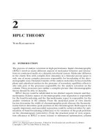
The most obvious pathologic feature of Parkinson disease is loss of pig-
ment in the substantia nigra caused by loss of neurons in this region. The
remaining neurons may show an intra-cytoplasmic eosinophilic inclusion
called a Lewy body (Fig. 4–1). These neurons project rostrally in the brain to
innervate the striatum as well as the cerebral cortex. Parkinson disease is asso-
ciated with marked striatal dopamine (DA) depletion and is considered by
many to be a striatal dopamine deficiency syndrome. At death, DA loss is
greater than 90%, and approximately 70% DA loss results in symptom expres-
sion. Severity of DA loss best correlates with bradykinesia in Parkinson
disease—the correlation with tremor is very poor. In recent years, we have
seen a much more comprehensive picture of the pathologic destruction by
Parkinson disease, which helps us to understand the wide variety of signs and
symptoms besides bradykinesia. Other morphologic and chemical deficits
have also been demonstrated in the brains of patients with Parkinson disease
in the cholinergic pedunculopontine nucleus, noradrenergic locus coeruleus,
serotonergic raphe nuclei, and glutamatergic centromedian/parafascicularis
complex of the thalamus. Still, there are many signs and symptoms that are
atypical for Parkinson disease and should raise our level of vigilance that
another disorder is present. These include:
• Early onset of, or rapidly progressing, dementia
• Rapidly progressive course
CLINICAL CASES 41
• Supranuclear gaze palsy
• Upper motor neuron signs
• Cerebellar signs—dysmetria, ataxia
• Urinary incontinence
• Early symptomatic postural hypotension
• Early falls
The majority of cases of Parkinson disease are unknown. Familial Parkinson
disease, while rare, does occur, and is most commonly associated with a muta-
tion of the parkin gene, which is inherited in an autosomal recessive pattern.
This mutation is the most common cause of early onset Parkinson disease,
without Lewy bodies. Routine neuroimaging is usually normal in Parkinson
disease. Functional imaging designed to visualize the dopamine innervation of
the striatum, especially in combination with other imaging techniques may
provide a way to positively identify the disease, however these techniques are
still under investigation and are not available under routine clinical circum-
stances. Imaging is useful, however, to identify some of the other entities in
the differential diagnosis.
The differential diagnosis of parkinsonism includes the following
categories:
42
CASE FILES: NEUROLOGY
Figure 4–1. Lewy body on microscopy. (With permission from Ropper AH,
Brown RH. Adams and Victor’s principles of neurology, 8th ed. New York:
McGraw-Hill; 2005: Fig. 39–5.)
• Drug-induced (antipsychotics, metoclopramide)
• Toxin-induced
• Metabolic
• Structural lesions (vascular parkinsonism, etc.)
• Hydrocephalus (normal-pressure hydrocephalus [NPH])
• Infections
Differential Diagnosis
Parkinson disease is most often mimicked by other neurodegenerative disor-
ders, most commonly by multiple system atrophy (MSA). This comes in two
major clinical forms: MSA–P, resembles Parkinson disease except that tremor
is less prominent, and the disorder tends to be quite symmetric. MSA–C, also
called olivopontocerebellar atrophy, presents as a cerebellar syndrome. Both
forms may have prominent autonomic insufficiency—including orthostatic
hypotension and impotence. Conventional MRI usually show abnormalities.
Dementia with Lewy bodies is a disorder with prominent cognitive dys-
function as well as parkinsonism. The typical clinical hallmarks include early
onset dementia, delusions and hallucinations, fluctuations in consciousness
and myoclonus. Although listed as a separate entity, there is much controversy
about whether this is Parkinson disease or represents parts of the clinical spec-
trum of the same pathologic entity. Although parkinsonism can be seen in
Alzheimer disease, it is a rare finding, and dementia is usually the primary
clinical syndrome.
Corticobasalganglionic degeneration typically has unilateral, coarse
tremor, rigidity, increased reflexes as well as limb apraxia/limb dystonia/alien
limb phenomenon. This disorder is the only one that typically has the asym-
metric appearance of Parkinson disease. Progressive supranuclear palsy is
characterized by supranuclear downgaze palsy (inability to voluntarily look
down) and square wave jerks on extraocular motion testing. These patients
typically have an upright rather than flexed posture. Also frequent falls can be
an early finding. Tremor is not common, and there is a pseudobulbar emotion-
ality. As mentioned, several drugs, especially dopamine antagonists (typical
neuroleptics, anti-nauseants) can cause drug-induced parkinsonism.
Treatment Options
Treatment is initiated when the patient’s quality of life is affected and usually
consists of either levodopa or a dopamine agonist. Because no treatment cur-
rently arrests the degenerative process, symptomatic treatment is the mainstay
of therapy. This includes pharmacologic and surgical interventions. Physical
measures such as physical therapy, speech therapy, and exercise are important
and have a major impact on the lives of patients with Parkinson disease.
CLINICAL CASES 43
Pharmacologic therapy:
• Dopaminergic agents are the mainstay of treatment for the cardinal
features of Parkinson disease.
• Levodopa crosses the blood–brain barrier, whereas dopamine does
not; levodopa is converted to dopamine in the brain. Peripheral break-
down in the gut is inhibited by the addition of inhibitors of aromatic
amino acid decarboxylase (dichloroisoprenaline [DCI]), carbidopa.
Thus, a Carbidopa/Levodopa formulation is popularly prescribed.
Levodopa can also be broken down peripherally by the enzyme
catechol-O-methyltransferase (COMT) so COMT inhibitors such as
entacapone and tolcapone are often employed. A therapeutic trial of
levodopa can confirm Parkinson disease because more than 90% of
patients with pathologically proven Parkinson disease have a good to
excellent response to adequate doses of levodopa (at least 600 mg/d
levodopa with DCI).
• Dopamine agonists cross the blood–brain barrier and act directly as
primarily D2-type receptors without requiring conversion. These
agents include pramipexole, ropinirole, and bromocriptine.
• MAO-B inhibitors such as selegiline and rasagiline can improve symp-
toms in both patients with mild disease (as monotherapy) and patients
already on levodopa therapy. Anticholinergics such as trihexyphenidyl or
diphenhydramine (Benadryl) are used primarily to combat tremor, but
have many side effects especially in older individuals.
• Amantadine is felt to act primarily by blocking glutamate N-methyl-D-
aspartate (NMDA) receptors and has a mild attenuation of the cardinal
symptoms of resting tremor and dystonia. Recently, amantadine has been
shown to help alleviate levodopa induced dyskinesias.
Although no treatment slows the degeneration of Parkinson disease, disease
mortality been reduced by levodopa therapy. Over time, the response to lev-
odopa becomes unstable, resulting in motor fluctuations, which are exag-
gerated clinical manifestations; also, patients can develop troublesome
abnormal involuntary choreiform and dystonic movements called dyskinesias.
There is good evidence that starting treatment with a dopamine agonist rather
than levodopa delays the onset of dyskinesias. Thus, those patients at high risk
for developing dyskinesia probably should be treated initially with dopamine
agonists.
Younger patients are more at risk for dyskinesia and are likely to be treated
for long periods of time (the average age of onset of Parkinson disease is approx-
imately 59 years). Although levodopa is the most efficacious agent for the treat-
ment of Parkinson disease, for mild Parkinson disease, dopamine agonists have
comparable benefit. In patients that still have an excellent response to levodopa
except for motor fluctuations and dyskinesias, surgical treatment that inhibits the
subthalamic nucleus with high-frequency stimulation can provide excellent
44
CASE FILES: NEUROLOGY
CLINICAL CASES 45
relief of the cardinal symptoms of disease. However, placement of a deep
brain stimulation (DBS) appears to be the preferable surgical therapy. It is
less invasive, more reversible, and can be adjusted to the individual patient, and
remarkable results can be seen. In addition, inhibition of the ventrolateral thala-
mus can be very effective for treatment of tremor.
Comprehension Questions
[4.1] Which of the following signs is most suggestive of Parkinson disease
rather than the other neurodegenerative diseases?
A. Unilateral resting tremor
B. Supranuclear downed gaze palsy
C. Orthostatic hypotension early in the course of the disease
D. Early falls
E. Abnormal cerebral MRI
[4.2] Which of the following medications is most likely to be able to help
both relieve cardinal features of Parkinson disease as well as reduce
drug-induced dyskinesias?
A. Levodopa
B. Dopamine agonists
C. Amantadine
D. Anticholinergics
E. Haloperidol
[4.3] Which of the following medications would be most likely to cause
drug-induced parkinsonism?
A. Trihexyphenidyl
B. Metoclopramide
C. Diazepam
D. Carbidopa
E. Levodopa
Answers
[4.1] A. Resting tremor is an early manifestation of Parkinson disease.
[4.2] C. Amantadine can decrease the incidence of levodopa induced
dyskinesia.
[4.3] B. Antiemetic agents such as prochlorperazine (Compazine) and meto-
clopramide can cause a drug-induced parkinsonism.
46 CASE FILES: NEUROLOGY
CLINICAL PEARLS
❖ The cardinal features of Parkinson disease are resting tremor, rigid-
ity, bradykinesia, and postural instability.
❖ Parkinson disease is usually an asymmetric disorder.
❖ Postural instability leading to falls occurs relatively late in the clin-
ical course of Parkinson disease.
❖ Failure to respond clinically to even large doses of levodopa is rel-
atively strong evidence that the patient does not have idiopathic
Parkinson disease.
❖ The mainstay of therapy for Parkinson disease is levodopa, which
can lead to dyskinesia.
REFERENCES
Hardy J, Cai H, Cookson MR, et al. Genetics of Parkinson’s disease and parkin-
sonism. Ann Neurol 2006;60:389–398.
Horstink M, Tolosa E, Bonuccelli U, et al. European Federation of Neurological
Societies; Movement Disorder Society—European Section. Review of the thera-
peutic management of Parkinson’s disease. Report of a joint task force of the
European Federation of Neurological Societies and the Movement Disorder
Society-European Section. Part I: early (uncomplicated) Parkinson’s disease.
Eur J Neurol 2006;13:1170–1185.
de Lau LM, Breteler MM. Epidemiology of Parkinson’s disease. Lancet Neurol
2006;5:525–535.
Pahwa R, Factor SA, Lyons KE, et al; Quality Standards Subcommittee of the
American Academy of Neurology. Practice parameter: treatment of Parkinson
disease with motor fluctuations and dyskinesia (an evidence-based review):
report of the Quality Standards Subcommittee of the American Academy of
Neurology. Neurology 2006;66:983–995.
Tolosa E, Wenning G, Poewe W. The diagnosis of Parkinson’s disease. Lancet
Neurol 2006;5:75–86.
❖
CASE 5
This 57-year-old man of Portuguese descent noticed that he had difficulty
marching in line as a soldier. From age 20 until the age of 40 he had a slow
progression of symptoms. Since then he experienced a rapidly progressing gait
disturbance, diplopia, dyssynergia, and paraesthesia in the limbs. At age 45 he
was confined to a wheelchair. On examination, he was intellectually normal
but had severe dysarthria and constant drooling. He had bulging eyes, slow
saccades, and impaired voluntary up- and down-gaze but no nystagmus. He
had fasciculations and dyscoordination of the tongue but no facial fascicula-
tions. A general moderate muscle weakness and atrophy were revealed, but
muscle tone was normal. Tendon reflexes were absent, but there were bilateral
Babinski signs. Deep senses were impaired, and coordination was impaired by
severe ataxia, dysmetria, and dysdiadochokinesia. A constant static tremor was
seen in the hands. His mother and paternal grandfather as well as his sister and
her son also had problems with gait, which were progressive and began during
adulthood. MRI of the brain revealed cerebellar folial atrophy.
◆
What is the most likely diagnosis?
◆
What is the next diagnostic step?
◆
What is the next step in therapy?
ANSWERS TO CASE 5: Ataxia, Spinocerebellar
Summary: This is a case of an essentially healthy man who had the insidious
onset and gradual progression of the syndrome heralded by gait difficulties,
which were later characterized as ataxia.
◆
Most likely diagnosis: Autosomal dominant cerebellar degeneration
with additional neurologic features with normal cognition–most likely
spinocerebellar ataxia type 3 (SCA-3).
◆
Next diagnostic step: DNA confirmation of diagnosis.
◆
Next step in management: Supportive care, genetic counseling,
rehabilitation.
Analysis
Objectives
1. Describe the movement disorder of ataxia.
2. List the differential diagnosis of ataxia including genetic and non-
genetic etiologies.
Considerations
As stated, this essentially healthy man had an insidious onset and gradual pro-
gression of a syndrome heralded by gait difficulties, which were later charac-
terized as ataxia. It later caused dysarthria, abnormal saccades, probable lower
motor neuron findings, neuropathy, and upper motor neuron deficits. This clin-
ical picture suggests a multiple system degeneration with the most prominent
feature being ataxia, and poor coordination on voluntary movements. These are
typically caused by problems either with a motor control as a result of pathology
of the cerebellum or its connections or pathologic proprioception because of
pathology in sensory pathways. Ataxias can either be isolated or seen as part of
the syndrome in conjunction with other neurologic abnormalities or abnormal-
ities in other body systems. This patient has other neurologic abnormalities but
no evidence at least at this time of other body system involvement. In addition,
there is strong familial involvement; specifically, there are four successive gen-
erations affected in his family, and both sexes are affected. Although familial
disorders are not necessarily genetic, this extensive involvement actually sug-
gests an autosomal dominant disorder. This is reinforced by the fact that auto-
somal recessive ataxias tend to have other body systems involved, whereas this
is not the case with adult-onset autosomal dominant disease.
It is worth considering some nongenetic causes of ataxia as they would
suggest other management issues, although most present over a much shorter
time course. Recognizable causes including trauma, toxic and metabolic
factors, neoplasms, and autoimmune mechanisms. Paraneoplastic cerebellar
48 CASE FILES: NEUROLOGY
degenerations (PCD) associated with specific tumor type antineuronal anti-
bodies are a relatively frequent cause of late-onset ataxia and are characterized
by a subacute progressive course and would prompt discovery and treatment
of the underlying neoplasm. More rarely (and controversially), subacute spin-
ocerebellar degeneration is associated with nonparaneoplastic immune dis-
eases such as gluten intolerance. In addition, hormonal abnormalities, such as
thyroid hormone deficiency can cause ataxia.
APPROACH TO AUTOSOMAL DOMINANT
CEREBELLAR ATAXIA
Definitions
Ataxia—an unsteady and clumsy motion of the limbs or torso caused by a
failure of the gross coordination of muscle movements.
Trinucleotide repeat expansion disease—caused by stretches of DNA in
a gene that contain the same trinucleotide sequence repeated many
times. These repeats are a subset of unstable microsatellite repeats that
occur throughout all genomic sequences. If the repeat is present in a
gene, an expansion of the repeat results in a defective gene product and
often disease.
Clinical Approach
Harding (1983) proposed a useful clinical classification for late onset autoso-
mal dominant cerebellar ataxias. In addition, sporadic cerebellar syndromes
include idiopathic forms of obscure etiology characterized by progressive
ataxia, autonomic failure, and extrapyramidal features, such as multiple sys-
tem atrophy (MSA). In Harding’s system autosomal dominant cerebellar
ataxia I (ADCA I), cerebellar ataxia is associated with additional features
related to the optic nerve (extra) pyramidal system, cerebral cortex, and periph-
eral nerves. ADCA II is associated with pigmentary macular dystrophy, and
ADCA III is a pure late onset cerebellar syndrome.
Since 1993 autosomal dominant cerebellar ataxias have been increasingly
characterized in terms of their genetic locus and are referred to as spinocere-
bellar ataxia. At this point there are more than 25 such disorders, and the num-
ber is increasing. The most common types are listed in Table 5–1. Many of
these can be definitively diagnosed by DNA testing. Clinical characterization
however is helpful in limiting the number of tests required.
There are several gene mutations on different chromosomes that cause spin-
ocerebellar ataxia, and the gene frequency within different populations varies
considerably. In general, the incidence is thought to be approximately 1.5 per
100,000 people, with equal gender distribution. Most of the ADCAs are caused
by a genetic defect that involves an expansion in the DNA sequence, and most
of these are trinucleotide repeat expansions (SCA types 1–3, 6–10, 12, and 17).
CLINICAL CASES 49
Table 5–1
SELECT LIST OF AUTOSOMAL DOMINANT SPINOCEREBELLAR ATAXIAS
INCIDENCE OCULOMOTOR
DISEASE (%) LOCUS PROTEIN MUTATION CLINICAL FEATURES ABNORMALITIES
SCA-1 6 6p23 Ataxin-1 CAG repeats Ataxia, dysarthria, Nystagmus, hypermetric
(38–83) pyramidal signs, saccades, slow saccades,
peripheral neuropathy, ophthalmoparesis
hyperreflexia,
cognitive impairment
SCA-2 14 12q24 Ataxin-2 CAG repeats Ataxia, dysarthria, Slow saccades,
(35–64) peripheral neuropathy, ophthalmoplegia
hyporeflexia, dementia,
myoclonus
SCA-3 21 14q32 CAG repeats Ataxia, dysarthria, Lid retraction, nystagmus,
Ataxin-3 (61–84) spasticity, parkinsonism, saccade dysmetria,
amyotrophy ophthalmoparesis,
square-wave jerks
SCA-6 15 19p13 CAG repeats Ataxia, dysarthria, Nystagmus (60% down-
CACNA1A (20–33) sometimes episodic beating), saccadic pursuit
ataxia, very slow
progression, lack of
family history
SCA-7 5 3p14 CAG repeats Ataxia, dysarthria, Saccadic smooth
Ataxin-7 (37- > 300) retinopathy, peripheral pursuit, slow saccades
neuropathy, pyramidal
signs, infantile phenotypes
SCA-8 2–5 13q21 CTG (3’UTR) Ataxia, dysarthria, Nystagmus, saccadic
(100–250) mild sensory neuropathy pursuit
Source: C Mariotti, R Fancellu, S Di Donato. An overview of the patient with ataxia. J Neurol 2005;252:511–518.
Other types of repeat expansions that cause SCA have been discovered. For
example, SCA-10 involves an ATTCT repeat expansion of the SCA10 gene,
and SCA-8 involves an expansion in the SCA8 gene with the nucleotides CTG
repeated. Finally, SCA-4 involves a mutation in a gene that does not involve a
trinucleotide repeat expansion.
The average age of onset for all of these types is from 20 to 30 years of age
except for SCA-6, which usually occurs between the ages of 40 and 50. People
with SCA-8 usually develop symptoms in their late 30s. SCA-2 patients usu-
ally develop dementia and slow eye movements. SCA-8 patients, who have nor-
mal life spans, and SCA-1 patients generally both have very active reflexes.
SCA-7 patients develop visual loss. In SCA types 1–3 and 7, there can be an
earlier age of onset with increased severity (called anticipation) from one gen-
eration to the next. The size of the repeat expansion zone in the affected genes
roughly correlates with the severity and age of onset. Penetrance is quite high;
however, there are rare cases in which people do not develop symptoms. The
reason for the lack of complete penetrance is currently unknown.
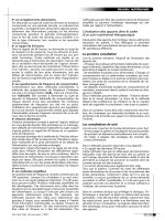
The diagnosis of spinocerebellar ataxia is initially suspected by the adult-
onset of symptoms. An MRI or CT of the brain can detect atrophy (wasting) of
the cerebellum, and a variety of other subcortical structures (Fig. 5–1). A molec-
ular genetic test to determine the gene that has the trinucleotide repeat expansion
52
CASE FILES: NEUROLOGY
Figure 5–1. Sagittal MRI of the brain in spinocerebellar ataxia. (With permis-
sion from Kasper DL, Braunwal E, Fauci A, et al. Harrison’s principles of
internal medicine, 16th ed. New York: McGraw-Hill; 2004: Fig. 352–1.)
can be helpful in quickly identifying other carriers in the family. Many of these
disorders can now be confirmed by DNA testing. Rather than just ordering all
available DNA tests (which can be quite expensive) there are algorithms that can
focus the testing by use of clinical signs; especially retinal degeneration, promi-
nent involvement of noncerebellar symptoms, age of onset, eye-movement dis-
orders, reduced stochastic velocity, and pyramidal signs. The clinical features of
these disorders are listed in the accompanying table (see Table 5–1).
Once the genetic defect is characterized, family members can also be tested.
Unfortunately, genetic testing is not always 100% informative. There are rare
cases of spinocerebellar ataxia diagnosed clinically that cannot be explained by
any of the known genetic defects. It is estimated that in approximately 50–60%
of white persons with a dominant familial form of cerebellar ataxia, DNA test-
ing can provide a definitive diagnosis.
SCA-3 or Machado-Joseph disease (MJD) is the most common SCA sub-
type in most populations. The phenotype is one of the most variable among
SCAs. The presenting syndromes for SCA-3 include pure cerebellar ataxia,
familial parkinsonism, hereditary spastic paraplegia, hereditary neuropathy,
and restless legs syndrome (RLS). A rarely recognized but common and rather
specific sign of SCA-3 is impaired temperature discrimination in all limbs and
even trunk and face. Pseudoexophthalmos (bulging eyes caused by lid retrac-
tion), faciolingual myokymia, and dystonia have been thought to be character-
istic, but not specific, signs of SCA-3.
SCA-3 MJD is an autosomal dominantly inherited disorder with variable
expression first described by Nakano and coworkers (1972) in an American
family of Portuguese-Azorean descent. Since then more families with MJD
have been reported worldwide. Three different clinical subtypes are described:
Type I with an early onset (20–30 years of age), pyramidal and extrapyrami-
dal signs, progressive external ophthalmoplegia (PEO), and minor cerebellar
deficits; type II with an intermediate age of onset. At neuropathological exam-
ination, degeneration of the cerebellum and the thoracic cord is always pres-
ent in SCA-3, but degeneration of the striatum, substantia nigra, basis pons,
oculomotor nuclei, and peripheral nerves is variable.
Treatment
There is no cure for ADCA and no treatment to slow the progression of the
disease. Nevertheless, supportive treatment is important. Drugs that help con-
trol tremors are not effective for treating cerebellar tremors, but can be effec-
tive for parkinsonism, dystonia, RLS (restless leg syndrome) and a variety of
other neurologic symptoms. Physical therapy does not likely slow the progres-
sion of loss of coordination or muscle wasting, but affected patients should be
encouraged to be active. Occupational therapy can be helpful in developing
ways to accommodate the patient in performing daily activities. Walkers and
other devices can assist the patient to have mobility. Other modifications such
as ramps for a wheelchair, heavy eating utensils, and raised toilet seats can
CLINICAL CASES 53
make patients more independent. Speech therapy and computer-based com-
munication aids often help as the person loses his or her ability to speak.
Although the nature of the specific mutations can help determine the prog-
nosis, the exact age of onset and the specific symptoms are difficult to determine,
especially for carriers with no symptoms. Ultimately, as with all progressive
degenerative disorders, the disease is fatal. Persons with SCA usually die one to
two decades after symptoms develop. The prognosis for SCA-11 and SCA-6 is
typically less severe, with a very slow worsening of symptoms, and persons
with SCA-8 and SCA-11 have a normal lifespan.
Comprehension Questions
[5.1] A patient with SCA-3, besides having ataxia is very slow with rigidity
and rest tremor. Which of the following drugs is most likely to be help-
ful for these latter symptoms?
A. Carbidopa/Levodopa
B. Haloperidol
C. Diazepam
D. Phenytoin
[5.2] What radiological feature is most characteristic of SCAs:
A. Cerebellar atrophy
B. High T2 signal in the cerebellar cortex
C. High signal lateral to the striatum
D. A high signal “hot cross bun” sign in the brainstem
[5.3] Which familial occurrence pattern would be most suspicious of not
being an ADCA.
A. 4/4 siblings (2 male, 2 female ages 4–12) and father in the same
household with onset within 1 week of each other, but no other first
or second-degree relatives in a large kindred.
B. Male prospectus (affected), father, 1/2 brothers, 0/2 sister, paternal
grandfather and uncle.
C. Male prospectus, neither parent, 1/2 brothers, 1/2 sister, paternal
great-grandfather. [poor penetrance]
D. Male prospectus, neither parent, 0/2 brothers, 0/2 sister, paternal
grandfather and uncle. [poor penetrance]
Answers
[5.1] A–the parkinsonism of SCA is often responsive to levodopa
[5.2] B is characteristic of neoplastic cerebellar degeneration. C and D are
seen with MSA.
[5.3] A is suggestive of a toxic or infectious exposure–B is typical; C and D
might be seen with poor penetrance.
54
CASE FILES: NEUROLOGY
CLINICAL CASES 55
CLINICAL PEARLS
❖ Spinocerebellar ataxias present in adulthood, generally as cerebel-
lar ataxias, often with other neurologic signs but rarely with non-
neurologic system involvement.
❖ DNA testing can be diagnostic, but clinical correlation is helpful in
focused ordering of tests
❖ Pharmacologic therapy does not alter the natural course of cerebel-
lar ataxia but can help to relieve neurological symptoms.
REFERENCES
Bataller L, Dalmau J. Paraneoplastic neurologic syndromes: approaches to diagno-
sis and treatment. Semin Neurol 2003 Jun;23(2):215–224.
Duen AM, Goold R, Giunti P. Molecular pathogenesis of spinocerebellar ataxias.
Brain 2006;129:1357–1370.
Hadjivassiliou M, Grunewald R, Sharrack B, et al. Gluten ataxia in perspective:
epidemiology, genetic susceptibility and clinical characteristics. Brain 2003;
126:685–691.
Harding AE. Hereditary spastic paraplegias. Seminar Neurol 1993;13:333–336.
Løkkegaard T, Nielsen JE, Hasholt L, et al. Machado–Joseph disease in three
Scandinavian families. J Neurol Sci 1998;156:152–157.
Mariotti C, Fancellu R, Di Donato S. An overview of the patient with ataxia. J Neurol
2005;252:511–518.
Schelhaasa HJ, Ippel PF, Beemerb FA, et al. Similarities and differences in the phe-
notype, genotype and pathogenesis of different spinocerebellar ataxias. Eur J
Neurol 2000;7:309–314.
Schöls L, Bauer P, Schmidt T, et al. Autosomal dominant cerebellar ataxias: clinical
features, genetics, and pathogenesis. Lancet Neurol 2004;3:291–304.
This page intentionally left blank
❖
CASE 6
A 65-year-old woman was referred for problems with abnormal involuntary
movements of the mouth and face. She has had good health until 3 years ago
when she developed problems with nausea and constipation. She was placed on
metoclopramide with some relief of symptoms. A complete gastrointestinal
(GI) workup was negative, although it was hypothesized she had decreased
gastric motility. These abnormal movements began approximately 1 year ago.
They have been getting progressively worse. The movements do not interfere
with speech but do interfere with eating. She also occasionally has arching
spasms of the back and neck. Her examination is remarkable for stereotypical
repetitive movements of the tongue and jaw and the sustained arching.
◆
What is the most likely diagnosis?
◆
What is the next step in therapy?