Báo cáo khoa hoc:"A ’G’ chromosome banding study of three cupped oyster species: Crassostrea gigas, Crassostrea angulata and Crassostrea virginica" ppsx
Bạn đang xem bản rút gọn của tài liệu. Xem và tải ngay bản đầy đủ của tài liệu tại đây (863.53 KB, 9 trang )
Original
article
A
’G’
chromosome
banding
study
of
three
cupped
oyster
species:
Crassostrea
gigas,
Crassostrea
angulata
and
Crassostrea
virginica
(Mollusca:
Bivalvia)
Alexandra
Leitão
a
Catherine
Thiriot-Quiévreux
a
Pierre
Boudr
b
Isabel
Malheiro
c
a
Observatoire
océanologique,
Université
P.
et
M.
Curie,
CNRS-INSU,
BP
28,
06230
Villefranche-sur-Mer,
France
b
Laboratoire
de
génétique
et
pathologie,
Station
Ifremer,
BP
133,
17390
La
’Iremblade,
France
c
Instituto
de
ciências
biomédicas
de
Abel
Salazar,
Universidade
do
Porto,
Lg.
Prof.
Abel
Salazar
2,
4000
Porto,
Portugal
(Received
26
May
1999;
accepted
12
August
1999)
Abstract -
The
G-banding
technique
was
performed
on
chromosomes
from
gill
tissue
of
three
cupped
oyster
species:
Crassostrea
gigas,
Crassostrea
angulata
and
Crassostrea
virginica.
Identification
of
the
ten
individual
chromosome
pairs
was
obtained.
Comparative
analysis
of
G-banded
karyotypes
of
the
three
species
showed
that
their
banding
patterns
generally
resembled
each
other,
with
chromosome
pair
3
being
similar
in
all
three
species.
However,
differences
from
one
species
to another
were
also
observed.
The
G-banding
pattern
highlighted
greater
similarities
between
C.
gigas
and
C.
angulata
than
between
these
two
species
and
C.
virginica,
thus
providing
an
additional
argument
for
genetic
divergence
between
these
two
evolutionary
lineages.
C.
gigas
and
C.
angulata
showed
a
different
G-banding
patterns
on
the
two
arms
of
chromosome
pair
7,
which
agrees
with
their
taxonomic
separation.
The
application
of
this
banding
technique
offers
a
new
approach
to
specific
problems
in
oyster
taxonomy
and
genetics.
©
Inra/Elsevier,
Paris
chromosome
/ G-banding
/
Crassostrea
gigas
/
Crassostrea
angulata
/
Crassostrea
virginica
*
Correspondence
and
reprints
E-mail:
Résumé -
Étude
du
marquage
chromosomique
en
bandes
G
chez
trois
espèces
d’huîtres
creuses :
Crassostrea
gigas,
Crassostrea
angulata
et
Crassostrea
virginica.
Le
marquage
chromosomique
en
bandes
G
a
été
réalisé
sur
des
chromosomes
obtenus
à
partir
de
tissu
branchial
de
trois
espèces
d’huîtres
creuses,
Crassostrea
gigas,
Crassostrea
angulata
et
Crassostrea
virginica,
et
a
permis
l’identification
des
dix
paires
de
chromosomes
de
ces
espèces.
L’analyse
comparée
des
caryotypes
marqués
en
bandes
G
a
montré
que
les
principales
bandes
G
présentaient
un
modèle
proche
chez
les
trois
espèces,
la
paire
de
chromosome
3
étant
identique.
Cependant
quelques
différences
ont
pu
être
observées.
Les
caryotypes
de
C.
gigas
et
C.
angulata
ont
révélé
plus
de
similitudes
entre
eux
qu’avec
celui
de
C.
virginica.
Ceci
apporte
un
argument
supplémentaire
à
la
divergence
génétique
entre
ces
deux
lignées
évolutives.
C.
gigas
et
C.
angulata
montrent
un
marquage
en
bandes
G
différent
sur
les
deux
bras
de
la
paire
du
chromosome
7,
ce
qui
corrobore
leur
séparation
taxinomique.
L’application
de
cette
technique
apporte
une
nouvelle
approche
pour
la
taxinomie
et
la
génétique
des
huîtres.
©
Inra/Elsevier,
Paris
chromosome
/
bandes
G
/
Crassostrea
gigas
/
Crassostrea
angulata
/
Crassostrea
virginica
1.
INTRODUCTION
Cytogenetic
investigations
in
oysters
were
first
mainly
concerned
with
data
on
chromosome
number
and
gross
morphology
(e.g.
[1,
22]).
Later,
morphome-
tric
analyses
of
karyotypes
provided
the
characterisation
of
chromosome
mor-
phology
based
on
centromeric
position
(e.g.
[13,
17,
31,
37]).
These
studies
showed
that
oyster
karyotypes
were
symmetrical
and
interspecific
differences
consisted
in
the
occurrence
and
differing
proportions
of
metacentric
and
sub-
metacentric
chromosomes
[18,
21].
The
application
of
differential
staining
techniques,
such
as
Ag-NORs
for
nucleolar
organiser
regions
and
C-banding
for
constitutive
heterochromatin
allowed
the
identification
of
some
specific
chromosome
pairs
in
the
karyotypes
of
oyster
species
[14-16,
18,
19,
39].
More
recent
techniques,
such
as
fluorescent
in
situ
hybridization,
have
been
tested
in
Crassostrea
gigas
[5,
9],
and
others,
such
as
fluochrome
staining
and
restriction
endonuclease
treatment,
have
been
carried
out
in
other
bivalve
species
[25,
26].
But
although
the
data
obtained
using
these
differential
staining
techniques
provide
a
better
knowledge
of
the
karyotypes
of
bivalve
species,
they
do
not
allow
the
identification
of
all
individual
chromosomes.
The
G-banding
technique,
defined
as
a
system
of
alternating
dark
and
light
bands
throughout
the
length
of
the
euchromatic
parts
of
chromosomes
[35],
allows
the
identification
of
each
individual
chromosome
which
enables
one
to
prepare
precise
and
detailed
karyotypes.
This
technique
has
been
routinely
used
in
vertebrate
cytogenetics,
especially
in
mammals
(e.g.
[11,
12,
34,
41]).
Only
a
few
studies
have
focused
on
lower
vertebrates,
such
as
fishes
(e.g.
[2,
6,
7,
24])
and
on
invertebrates
such
as
insects
(e.g.
[4,
23,
33]).
Among
bivalves,
G-banding
patterns
have
been
attempted
in
Mytilus
[25,
27]
and
in
the
oyster
Crassostrea
virginica
[32].
In
this
study,
G-banding
patterns
are
described
in
three
cupped
oys-
ters:
Crassostrea
gigas
(Thunberg),
the
Pacific
oyster,
Crassostrea
angulata
(Lamark),
the
Portuguese
oyster
and
Crassostrea
virginica
(Gmelin),
the
East-
ern
American
oyster.
2.
MATERIALS
AND
METHODS
2.1.
Biological
material
Specimens
of
each
taxon
were
reared
at
the
Ifremer
hatchery
in
La Tremblade
(Charente-Maritime,
France).
Specimens
of
Crassostrea
gigas
were
collected
from
the
Seudre
estuary,
where
this
species
was
introduced
from
Japan
[8]
and
is
currently
farmed
on
a
large
scale.
Specimens
of
Crassostrea
angulata
were
collected
in
Setubal
bay
(Portugal),
then
acclimated
at
the
Ifremer
hatchery.
Their
taxonomic
status
was
assessed
using
mitochondrial
DNA
markers
as
described
in
Boudry
et
al.
(3!.
Specimens
of
Crassostrea
virginica
were
imported
from
a
wild
population
located
in
Shippagan,
New
Brunswick
(Canada)
and
acclimated
at
the
Ifremer
hatchery.
These
oysters
were
maintained
in
common
quarantine
facilities
until
reproduction,
and
their
progenies
were
sampled
for
chromosome
analysis.
2.2.
Chromosome
preparation
Whole
juvenile
animals
were
incubated
for
7-9
h
in
a
0.005
%
solution
of
colchicine
in
seawater.
Because
cell
cultures
are
not
yet
available
for
molluscs,
we
used
growing
somatic
tissues
such
as
gills
as
a
source
of
mitoses.
After
dissection,
gills
were
treated
for
30
min
in
0.9
%
sodium
citrate.
The
material
was
fixed
in
a
freshly
prepared
mixture
of
absolute
alcohol
and
acetic
acid
(3:1)
with
three
changes
of
20
min
each.
Slide
preparations
were
made
from
pieces
of
gill
tissue
from
each
individual,
using
an
air
drying
technique
(38!.
2.3.
G-banding
G-banding
was
performed
by
the
ASG
method
(acetic/saline/Giemsa)
after
Sumner
et
al.
[36].
Chromosome
preparations
were
treated
for
1
h
at
60 °C
in
2
x
SSC
(0.3
M
sodium
chloride:
0.03
M
trisodium
citrate).
After
rinsing
in
distilled
water,
the
slides
were
stained
with
2
%
Giemsa-stain
in
phosphate
buffer,
pH
6.8
for
90
min.
Best
results
were
obtained
by
banding
within
5-
10
days
of
chromosome
preparation.
Photographs
of
G-banded
metaphases
were
taken
with
a
Zeiss
III
photomi-
croscope.
Karyotypes
were
made
on
the
basis
of
length,
centromeric
position
and
banding
pattern.
Because
we
were
working
with
somatic
tissues,
we
had
to
use
many
animals
to
obtain
a
sufficient
number
of
mitoses.
Moreover,
the
lat-
ter
showed
different
levels
of
condensation
making
the
number
of
cells
we
could
work
on
even
smaller.
Thus,
in
total,
18
G-banded
karyotypes
were
examined
in
C.
gigas,
20
in
C.
angulata
and
21
in
C.
virginica.
3. RESULTS
Establishing
a
repetitive
G-banding
pattern
requires
a
similar
degree
of
condensation
of
chromosomes
to
be
compared.
Although
a
large
number
of
metaphases
was
observed,
we
selected
only
those
with
similar
degrees
of
condensation
for
interpretation.
Figure
1 presents
an
example
of
a
G-banded
metaphase
of
one
of
the
three
species
studied,
C.
gigas.
In
figure
2,
haploid
G-banded
karyotypes
are
shown
to
facilitate
comparison
of
homologous
chromosome
pairs
between
the
three
species
studied.
The
karyotype
of
C.
gigas
consists
of
ten
metacentric
chromosomes,
that
of
C.
angulata
has
nine
metacentric
and
one
submetacentric
(no.
8)
and
the
karyotype
of
C.
virginica
includes
eight
metacentric
and
two
submetacentric
(nos
4
and
8)
chromosomes
!18).
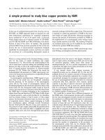
Figure
3
gives
a
schematic
representation
of the
G-banding
patterns
obtained
for
the
three
species.
Owing
to
differences
in
condensation
of
chromosomes
only
major
G-bands
were
used
to
compare
the
three
species,
with
an
emphasis
on
their
number
rather
than
their
position.
Chromosome
1:
on
the
short
arm,
both
C.
gigas
and
C.
angulata
show
two
major
bands,
while
in
C.
virginica
four
major
bands
are
present.
All
three
species
present
four
bands
on
the
long
arm.
Chromosome
2:
on
the
short
arm,
C.
gigas
shows
three
bands
while
in
C.
angulata
and
C.
virginica
two
major
bands
are
present.
On
the
long
arm,
the
three
species
are
characterised
by
two
major
bands.
Chromosome
3:
the
pattern
is
very
similar
across
the
three
species,
being
characterised
by
two
major
bands
at
the
extremities
of
the
short
arm
and
one
large
band
at
the
centre
of
the
long
arm.
Chromosome
4:
on
the
short
arm,
in
C.
gigas
and
in
C.
angulata,
the
bands
are
observed
in
subtelomeric
and
near
centromeric
positions
while
C.
virginica
is
characterised
by
one
centrally
located
major
band.
Four
bands
are
located
on
the
long
arm
of
the
three
species
differing
slightly
in
their
position,
which
was
probably
caused
by
the
different
degrees
of
condensation
of
the
chromosomes.
Chromosome
5:
the
three
species
present
three
bands
on
the
short
arm.
On
the
long
arm,
the
G-banding
pattern
is
different
between
the
three
species.
Chromosome
6:
on
the
short
arm,
three
bands
are
observed
in
the
three
species.
On
the
long
arm,
C.
gigas
and
C.
angulata
show
four
successive
bands,
while
in
C.
virginica
the
banding
pattern
is
characterised
by
the
presence
of
only
one
major
median
band.
Chromosome
7:
C.
gigas
shows
two
bands
on
the
short
arm
and
two
on
the
long
arm.
In
C.
virginica
and
C.
angulata,
three
bands
are
seen
on
both
the
short
arm
and
the
long
arm.
These
differ
slightly
in
intensity
and
position.
Chromosome
8:
the
short
arm
in
all
three
species
is
characterised
by
the
presence
of
one
major
band.
There
are
three
major
bands
on
the
long
arm
of
C.
gigas
and
C.
angulata
and
four
bands
in
C.
virginica.
Chromosome
9:
on
the
short
arm,
two
bands
are
seen
in
the
three
species.
On
the
long
arm,
C.
gigas
presents
three
near
equivalent
bands
differing
from
C.
angulata
and
C.
virginica
which
are
characterised
by
the
presence
of
one
major
band.
Chromosome
10:
the
three
species
are
characterised
by
two
bands
at
the
extremities
of
the
short
arm.
Two
major
bands
are
seen
on
the
long
arm
of
C.
gigas
and
C.
angulata
while
three
are
present
in
C.
virginica.
4.
DISCUSSION
The
application
of
G-banding
to
three
species
of
oysters:
C.
gigas,
C.
angulata
and
C.
virginica,
allowed
individual
identification
of
the
chromosomes
which
makes
it
possible
to
prepare
accurate
karyotypes
and
describe
the
respective
idiograms.
Comparison
with
previous
G-banding
analysis
of
C.
virginica
[32]
showed
that
the
number
of
G-bands
identified
as
black
bands
(i.e.
the
most
distinct)
was
similar
in
chromosome
pairs
5
and
9
but
quite
different
in
the
remaining
chromosome
pairs.
This
can
be
explained
by
i)
different
karyotypes
of
the
oys-
ter
populations
studied
(6
m-4
sm
in
Tabasco,
Mexico;
8
m-2
sm
in
our
popula-
tion),
ii)
different
techniques
used,
iii)
different
condensation
of
chromosomes.
The
major
G-bands
of
the
three
Crassostrea
species
studied
here
show
a
sim-
ilar
pattern
on
the
whole
chromosome
3,
on
the
short
arms
of
chromosomes
5,
6,
8,
9
and
10
and
on
the
long
arms
of
chromosomes
1,
2
and
4.
C.
gigas
and
C.
angulata
present
additional
similarities
on
the
short
arms
of
chromosomes
1
and
4,
and
on
the
long
arms
of
chromosomes
6,
8
and
10.
These
two
taxa,
often
considered
as
the
same
species
!28!,
have
been
differentiated
by
mitochondrial
DNA
analysis
[3,
30]
and
karyotype
analysis
[18].
G-banding
highlights
sim-
ilarities
between
these
two
taxa,
except
for
chromosome
7
where
both
arms
are
shown
to
be
different.
This
difference
corroborates
their
taxonomic
sepa-
ration.
C.
angulata
and
C.
virginica
also
display
additional
similarities
in
the
number
of
major
G-bands
on
the
whole
chromosome
7,
the
short
arm
of
chro-
mosome
2
and
the
long
arm
of
chromosome
9,
but
they
differ
on
the
short
arms
of
chromosomes
1
and
4,
and
on
the
long
arms
of
chromosomes
5,
6,
8
and
10.
Karyological
differences
between
these
two
species
have
been
previ-
ously
observed
!18!.
C.
virginica
contrasts
with
C.
gigas
on
the
short
arms
of
chromosomes
1,
2,
4
and
7,
and
on
the
long
arms
of
chromosomes
5,
6,
7,
8,
9
and
10.
Genetic
divergence
between
C.
gigas
and
C.
virginica
has
been
demon-
strated
by
molecular
phylogenies
[20,
29]
and
by
karyotype
analysis
!18!.
The
differences
in
G-banding
pattern
between
C.
gigas
and
C.
virginica
substantiate
their
genetic
difference.
Therefore,
from
the
analysis
of
the
banding
karyotypes
of
the
three
species,
we
can
conclude
that
they
generally
resemble each
other
with
chromosome
pair
3
being
similar
in
all
the
three
species.
However,
differences
were
observed
from
one
species
to
another,
showing
that
there
is
a
higher
resemblance
between
the
banded
karyotypes
of
C.
gigas
and
C.
angulata
than
between
these
two
species
and
C.
virginica.
Because
of
the
economic
and
ecological
importance
of
oysters,
genetic
inves-
tigations
are
of
special
interest.
In
this
respect,
the
identification
of
structural
chromosomal
features
could
be
very
useful
in
gene
mapping,
hybrid
breeding
or
stock
conservation
programmes.
The
individual
identification
of
the
chro-
mosomes
by
G-banding
will
provide
a
better
knowledge
of
the
aneuploidy
phe-
nomenon
reported
in
oysters
(e.g.
[40])
by
identifying
missing
chromosomes.
Similarly,
G-banding
could
also
provide
a
very
valuable
technique
for
chromo-
some
segregation
studies
on
triploid
and
tetraploid
oysters
[10].
The
applica-
tions
of
G
chromosome
banding
are
therefore
numerous
and
represent
a
useful
new
tool
in
oyster
genetics.
ACKNOWLEDGEMENT
This
work
was
supported
by
a
French-Portuguese
co-operation
(no.
158
C
1),
by
a
research
training
project
(contract
no.
FAIR
GT
97-3599)
and
part
of
Genephys
program
(Contract
no.
FAIR
95-421).
We
thank
S.
Sabini
and
S.
Heurtebise
for
excellent
technical
assistance
and
H.
McCombie
for
advice
on
the
English.
REFERENCES
[1]
Ahmed
M.,
Cytogenetics
of oysters,
Cytologia
38
(1973)
337-346.
[2]
Blaxhall
P.C.,
Chromosome
karyotyping
of
fish
using
conventional
and
G-
banding
methods,
J.
Fish
Biol.
22
(1983)
417-424.
[3]
Boudry
P.,
Heurtebise
S.,
Collet
B.,
Cornette
F.,
Gérard
A.,
Differentiation
between
populations
of
the
Portuguese
oyster,
Crassostrea
angulata
(Lamarck)
and
the
Pacific
oyster,
Crassostrea
gigas
(Thunberg)
revealed
by
mtDNA
RFLP
analysis,
J.
Exp.
Mar.
Biol.
Ecol.
226
(1998)
279-291.
[4]
Bregman
A.A.,
Q-,
C-,
and
G-banding
patterns
in
the
germ-line
and
somatic
chromosomes
of
Miastor
sp.
(Diptera:
Cecidomydae),
Chromosoma
53
(1975)
119-
130.
[5]
Clabby
C.,
Goswami
U.,
Flavin
F.,
Wilkins
N.P.,
Houghton
J.A.,
Powell
R.,
Cloning,
characterisation
and
chromosomal
location
of
a
satellite
DNA
from
the
Pacific
oyster,
Crassostrea
gigas,
Gene
168
(1996)
205-209.
[6]
Gold
J.R.,
Li
Y.C.,
Trypsin
G-banding
of
North
American
cyprinid
chromo-
somes:
Phylogenetic
considerations,
implications
for
fish
chromosome
structure,
and
chromosomal
polymorphism,
Cytologia
56
(1991)
199-208.
[7]
Gold
J.R.,
Li
Y.C.,
Chromosomal
NOR
karyotypes
and
genome
size
variation
among
squawfishes
of the
genus
Ptychocheilus
(Teleostei:
Cyprinidae),
Copeia
1
(1994)
60-65.
[8]
Grizel
H.,
H6ral
M.,
Introduction
into
France
of
the
Japanese
oyster
(Cras-
sostrea
gigas),
J.
Cons.
Int.
Explor.
Mer
47
(1991)
399-403.
[9]
Guo
X.,
Allen
S.K.,
Fluorescence
in
situ
hybridization
of
vertebrate
telomere
sequence
to
chromosome
ends
of
the
Pacific
oyster,
Crassostrea
gigas
Thunberg,
J.
Shellfish
Res.
16
(1997)
87-89.
[10]
Guo
X.M.,
Allen
S.K.,
Sex
and
meiosis
in
autotetraploid
Pacific
oyster,
Crassostrea
gigas
(Thunberg),
Genome
40
(1997)
397-405.
!11)
lannuzi
L., Di
Meo
G.P., Perucatti
A.,
An
improved
characterization
of
goat
chromosomes
by
means
of
G-
and
R-band
comparison,
Hereditas
120
(1994)
245-251.
[12]
lannuzzi
L.,
Di
Meo
G.P.,
Perucatti
A.,
G-
and
R-banded
prometaphase
karyotypes
in
goat
(Capra
hircus
L.),
Caryologia
49
(1996)
267-277.
[13]
leyama
H.,
Chromosomes
of
the
oysters,
Hyatissa
i!bricata
and
Dendostrea
folium
(Bivalvia:
Pteriomorpha),
Venus,
Jpn.
J.
Malac.
49
(1990)
63-68.
[14]
Insua
A.,
Thiriot-(auievreux
C.,
The
characterization
of
Ostrea
denselamel-
losa
(Mollusca,
Bivalvia)
chromosomes:
karyotype,
constitutive
heterochromatin
and
nucleolus
organizer
regions,
Aquaculture
97
(1991)
317-325.
[15]
Insua
A.,
Thiriot-(!7uievreux
C.,
Karyotype
and
nucleolus
organizer
regions
in
Ostrea
puelchana
(Bivalvia:
Ostreidae),
Veliger
36
(1992)
215-219.
[16]
Ladron
de
Guevara
B.,
Winkler
F.,
Palma
C.,
Karyotype
description
and
the
position
of
the
nucleolar
organizer
region
(NOR)
in
the
Chilean
oyster
Tiostrea
chilensis
(Philippi)
Chanley
and
Dinamani,
in:
Beaumont
A.R.
(Ed.),
Genetics
and
Evolution
of
Aquatic
Organisms,
University
of
Wales,
Bangor,
UK,
1992,
pp.
399-405.
[17]
Ladron
de
Guevara
B.,
Winkler
F.,
Rodriguez-Romero
F.,
Palma-Rojas
C.,
Comparative
karyology
of
four
american
oyster
species,
Veliger
39
(1996)
260-266.
[18]
Leitao
A.,
Boudry
P.,
Labat
J.P.,
Thiriot-(auievreux
C.,
Comparative
kary-
ological
study
of
cupped
oyster
species,
Malacologia
41
(1999)
175-186.
[19]
Li
X.X.,
Havenhand
J.N.,
Karyotype,
nucleolus
organizer
regions
and
consti-
tutive
heterochromatin
in
Ostrea
angasi
(Mollusca:
Bivalvia):
evidence
of
taxonomic
relationship
within
Ostreidae,
Mar.
Biol.
127
(1997)
443-448.
[20]
Littlewood
D.J.T.,
Molecular
phylogenies
of
cupped
oysters
based
on
partial
28S
rRNA
gene
sequences,
Mol.
Phylog.
Evol.
3
(1994)
221-229.
[21]
Longwell
A.C.,
Stiles
S.S.,
Chromosomes,
biology
and
breeding,
in:
Kennedy
V.S.,
Newell
R.T.E.,
Eble
F.
(Eds.),
The
Eastern
Oyster:
Crassostrea
virginica,
College
Park
Maryland,
Maryland
Sea
Grant
College
XVI,
1996,
pp.
443-465.
[22]
Longwell
A.C.,
Stiles
S.S.,
Smith
D.G.,
Chromosome
complement
of
the
American
oyster
Crassostrea
virginica
as
seen
in
meiotic
and
cleaving
eggs,
Can.
J.
Genet.
Cytol.
9
(1967)
845-856.
[23]
Lorite
P.,
Chica
E.,
Palomeque
T.,
G-banding
and
chromosome
condensa-
tion
in
the
ant,
Tapino!rca
nigerrimum,
Chromosome
Res.
4
(1996)
77-79.
[24]
Luo
C.,
Multiple
chromosomal
banding
in
grass
carp,
Ctenopharyngodon
idellus,
Heredity
81
(1998)
481-485.
[25]
Martinez-Lage
A.,
Gonzalez-Tizon
A.,
Méndez
J.,
Characterization
of
differ-
ent
chromatin
types
in
Mytilus
galloprovincialis
L.
after
C-banding,
fluorochrome
and
restriction
endonuclease
treatments,
Heredity
72
(1994)
242-249.
[26]
Martinez-Lage
A.,
Gonzalez-Tizon
A.,
Méndez
J.,
Chromosomal
markers
in
three
species
of
the
genus
Mytilus
(Mollusca:
Bivalvia),
Heredity
74
(1995)
369-375.
[27]
Méndez
J., Pasantes
J.J., Martinez
Exposito
M.J.,
Banding
pattern
of
mus-
sel
(Mytilus
galloprovincialis)
chromosomes
induced
by
2xSSC/Giemsa-stain
treat-
ment,
Mar.
Biol.
106
(1990)
375-377.
[28]
Menzel
R.W.,
Portuguese
and
Japanese
oysters
are
the
same
species,
J.
Fish
Res.
Board
Can.
31
(1974)
453-456.
[29]
O’Foighil
D.,
Gaffney
P.M.,
Hilbish
T.J.,
Differences
in
mitochondrial
16
S
ribosomal
gene
sequences
allow
discrimination
among
American
(Crassostrea
virginica
(Gmelin)]
and
Asian
[C.
gigas
(Thunberg)
C.
ariakensis
Wakiya]
oyster
species,
J.
Exp.
Mar.
Biol.
Ecol.
192
(1995)
211-220.
[30]
O’Foighil
D.,
Gaffney
P.M.,
Wilbur
A.E.,
Hilbish
T.J.,
Mitochondrial
cy-
tochrome
oxidase
I
gene
sequences
support
an
Asian
origin
for
the
Portuguese
oyster
Crassostrea
angulata,
Mar.
Biol.
131
(1998)
497-503.
[31]
Rodriguez-Romero
F., Uribe-Alcocer
M.,
Laguarda-Figueras
A.,
Cytogenetic
study
of
an
oyster
population
of
the
species
Crassostrea
virginica
Gmelin
from
the
coast
of
Tabasco,
Mexico,
Venus,
Jpn.
J.
Malac.
37
(1978)
83-86.
[32]
Rodriguez-Romero
F.,
Laguarda-Figueras
A.,
Uribe-Alcocer
M.,
Rojas-Lara
M.L.,
Distribution
of
&dquo;G&dquo;
bands
in
the
karyotype
of
Crassostrea
virginica,
Venus,
Jpn.
J.
Malac.
38
(1979)
180-184.
[33]
Staiber
W.,
Preferential
pairing
in
the
germ
line
limited
chromosomes
of
Acricotopus
lucidus
(Diptera,
Chironomidae),
Heredity
66
(1991)
191-201.
[34]
Stanyon
R.,
Ardito
G.,
Lamberti
L.,
Bigatti
P.,
The
banded
karyotypes
of
Macaca
fuscata
compared
with
Cercocebus
aterrimus,
Folia
Primatol.
41
(1983)
137-
146.
[35]
Sumner
A.T.,
Chromosome
Banding,
Unwin
Hyman
Ltd,
1990.
[36]
Sumner
A.T.,
Evans
H.J.,
Buckland
R.A.,
New
technique
for
distinguishing
between
human
chromosomes,
Nature
232
(1971)
31-32.
[37]
Thiriot-(auievreux
C.,
Analyse
comparée
des
caryotypes
d’Ostreidae
(Bi-
valvia),
Cah.
Biol.
Mar.
25
(1984)
407-418.
[38]
Thiriot-(auievreux
C.,
Ayraud
N.,
Les
caryotypes
de
quelques
espèces
de
Bivalves et
de
Gastéropodes
marins,
Mar.
Biol.
70
(1982)
165-172.
[39]
Thiriot-(auievreux
C.,
Insua
A.,
Nucleolar
organiser
region
variation
in
the
chromosomes
of
three
oyster
species,
J.
Exp.
Mar.
Biol.
Ecol.
157
(1992)
33-40.
[40]
Thiriot-(auievreux
C., Pogson
G.H., Zouros
E.,
Genetics
of
growth
rate
vari-
ation
in
bivalves:
aneuploidy
and
heterozygosity
effects
in
a
Crassostrea
family,
Genome
35
(1992)
39-45.
[41]
Vassart
M.,
Séguéla
A.,
Hayes
H.,
Chromosomal
evolution
in
gazelles,
J.
Hered.
86
(1995)
216-227.