Digital histology an interactive cd atlas with review text phần 9 ppsx
Bạn đang xem bản rút gọn của tài liệu. Xem và tải ngay bản đầy đủ của tài liệu tại đây (315.03 KB, 22 trang )
᭜
Numerous lateral processes surround and invest the maturing
germ cells. The most basal of these processes forms a series
of tight junctions with similar processes of adjacent Sertoli
cells.
᭜
Blood-testis barrier is formed by occluding junctions that unite
the basal processes of adjacent Sertoli cells forming a:
Basal compartment with access to blood-borne materials
and which contains spermatogonia and earliest primary
spermatocytes
Adluminal compartment
– Provides a unique microenvironment for developing
germ cells that protects these cells from immunologic
attack and concentrates hormones needed for sperm
production
– Contains later primary spermatocytes, secondary sperma-
tocytes, and spermatids
᭜
Functions
Mediate exchange of nutrients to germ cells
Form blood-testis barrier to protect developing germ cells
from immunologic attack
Break down excess spermatid cytoplasm
Produce testicular fluid
Secrete androgen-binding protein that binds to and concen-
trates testosterone in the seminiferous epithelium
Produce inhibin, which inhibits the secretion of follicle stim-
ulating hormone (FSH) from the adenohypophysis
Orchestrate movement of germ cells through germinal
epithelium and facilitate cytodifferentiation and subsequent
release of spermatozoa into the lumen of the seminiferous
tubule
᭹
Germ cells (spermatogenic cells)
᭜
Form a stratified germinal (seminiferous) epithelium
᭜
Cell types
Spermatogonia
– Are diploid cells resting on the basement membrane
– Are of two varieties. Type A spermatogonia divide mitoti-
cally to perpetuate self and to form type B cells. Type B
spermatogonia divide mitotically to form primary sperma-
tocytes.
18.
Male Reproductive System 185
– Undergo incomplete cytokinesis so resulting cells remain
attached to each other during spermatogenesis
– Divide mitotically to produce primary spermatocytes
Primary spermatocytes
– Are the largest germ cells; each nucleus is 1.5 times larger
than that of a spermatogonium.
– Form in the basal compartment, then probably migrate
through the tight junctions between Sertoli cell processes to
the adluminal compartment
– Remain in prophase about one-third of the spermatogenic
cycle, so many are seen. Nuclei contain highly condensed
chromosomes.
– Are diploid cells that complete meiosis I (reductional divi-
sion) to form secondary spermatocytes
Secondary spermatocytes
– Are haploid cells whose pale staining nuclei are similar in
size to those of the spermatogonia nuclei
– Are present for only eight hours of the entire 64-day sper-
matogenic cycle; therefore, very few are seen.
– Divide by meiosis II (equational division) to form
spermatids
Spermatids
– Are haploid cells whose nuclei are initially about two-
thirds the size of spermatogonia nuclei
– Are located near the lumen of the seminiferous tubules
– Do not divide but undergo cytodifferentiation to form
spermatozoa
• Intercellular bridges break down.
• Nucleus condenses and elongates.
• Acrosome forms. An acrosome is a modified lysosome
containing enzymes to aid the sperm in penetrating the
zona pellucida surrounding the secondary oocyte.
• Flagellum forms.
• Excess cytoplasm is shed.
Spermatozoa
– Are haploid cells
– Are anatomically mature, but incapable of fertilization at
this time
186
Digital Histology
– Are released from Sertoli cells into the lumen of the semi-
niferous tubules
Spermatogenesis
➢ Spermatogenesis is defined as the process by which diploid somatic
cells (spermatogonia) in the basal compartment become haploid sper-
matozoa lying free in the lumen of the seminiferous tubules.
➢ Stages
᭹
Spermatocytogenesis. Mitotic divisions of spermatogonia (diploid)
to form primary spermatocytes (diploid); cytokinesis is
incomplete.
᭹
Meiosis. Two cell divisions convert diploid cells to haploid
(i.e., reduction of chromosomes and DNA by half); cytokinesis is
incomplete.
᭜
Meiosis I. Primary spermatocytes (diploid) form secondary
spermatocytes (haploid).
᭜
Meiosis II. Secondary spermatocytes form spermatids (haploid).
᭹
Spermiogenesis. Cytodifferentiation of spermatids (haploid) into
spermatozoa (haploid)
᭹
Spermiation. Release of mature sperm into lumen of seminiferous
tubule
18.
Male Reproductive System 187
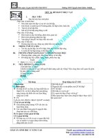
FIGURE 18.4. The stages and cells of spermatogenesis.
➢ Under control of follicle stimulating hormone (FSH) from the ante-
rior pituitary
➢ One cycle lasts about 64 days, with a new cycle beginning in any
given location about every 16 days.
Course of Sperm Within the Testis
➢ Seminiferous tubules, convoluted portion. Germinal epithelium where
sperm production occurs; sperm are released into the lumen of this
tubule from Sertoli cells.
➢ Seminiferous tubules, straight portion (tubuli recti)
᭹
Lined by simple columnar epithelium whose cells resemble Sertoli
cells
᭹
Connects convoluted portion of seminiferous tubules with rete
testis
➢ Rete testis
᭹
Is a meshwork of channels within mediastinum of testis
᭹
Lined by simple cuboidal cells, many of which possess a single
flagellum
᭹
Connects the straight portion of the seminiferous tubules with
efferent ducts in the epididymis
Genital Ducts External to the Testis
Epididymis
➢ The epididymis is a comma-shaped organ lying posterior to the testis
that is divided into head, body, and tail subdivisions.
➢ Head region composition
᭹
Efferent ducts
᭜
Connect rete testis with duct of epididymis
᭜
Consist of about 12 ducts, each of which is coiled into a cone
shape. Each duct connects with the rete testis at the apex of
the cone adjacent to the testis. All ducts anastomose to form
the single duct of the epididymis at the bases of the cones.
᭜
Form coni vasculosi (singular, conus vasculosus) that are com-
posed of one coiled efferent duct plus its surrounding connec-
tive tissue, containing abundant blood vessels.
᭜
Are lined with a simple epithelium composed of alternating
taller, ciliated cells and shorter cuboidal cells with lysosomes.
188
Digital Histology
Therefore, efferent ducts present a characteristic, scalloped
border adjacent to the lumen. A thin muscularis layer surrounds
the epithelium.
᭜
Function. Ciliated cells propel spermatozoa toward duct of
epididymis while cuboidal cells absorb testicular fluid.
᭹
Duct of epididymis. A single duct formed by fusion of efferent ducts
➢ Body and tail regions
᭹
Contains the remainder of the duct of the epididymis
᭜
Highly coiled, single tube (6m long) formed by union of effer-
ent ducts in the head region
᭜
Lined by tall pseudostratified columnar epithelium with stere-
ocilia, which decreases in height from head to tail regions;
creates a smooth lumen when compared with efferent ducts
᭜
Smooth muscle layer surrounds epithelium and increases in
thickness and number of layers from head to tail
᭹
Function
᭜
Storage and maturation site for sperm
᭜
Absorption of excess testicular fluid
᭜
Movement of sperm toward ductus deferens
Ductus (Vas) Deferens
➢ The ductus deferens is a thick muscular tube carrying sperm from duct
of epididymis to the ejaculatory duct.
18.
Male Reproductive System 189
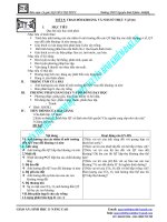
FIGURE 18.5. Components of the epididymis.
➢ Structure
᭹
Mucosa
᭜
Pseudostratified columnar epithelium with stereocilia sur-
rounds a narrow lumen.
᭜
Thin lamina propria
᭜
Longitudinal folds produce an irregular lumen.
᭹
Thick muscularis. Inner and outer longitudinal, middle circular
layers of smooth muscle
➢ Course
᭹
Located in spermatic cord in the inguinal canal with spermatic
artery, pampiniform venous plexus, and a nerve plexus
᭹
Enters abdominal cavity, crosses above entrance of ureter into
bladder, and enlarges to form the ampulla, which lies posterior to
urinary bladder
᭹
Is joined by duct of the seminal vesicle just before it enters the
prostate
➢ Function. Transports sperm from epididymis to ejaculatory duct in
prostate
Ejaculatory Duct
➢ The ejaculatory duct is formed by the union of the ductus deferens
with the duct of the seminal vesicle.
➢ No muscle layer is retained from the ductus deferens.
➢ The ejaculatory duct traverses the prostate gland to join the prosta-
tic urethra.
Urethra
➢ Prostatic urethra. Within prostate; lined with transitional epithelium
➢ Membranous urethra. Pierces skeletal muscle of the urogenital
diaphragm; lined with stratified or pseudostratified columnar
epithelium
➢ Penile urethra (discussed with penis)
Genital Glands
Seminal Vesicle
➢ Seminal vesicles are paired glands lying posterior to the urinary
bladder.
➢ Each is composed of a single, highly tortuous tube.
190
Digital Histology
➢ Function. Add sperm-activating substances, such as fructose, citrate,
proteins, and prostaglandins to seminal fluid; provides bulk of
seminal fluid
➢ Structure
᭹
Pseudostratified columnar epithelium with many secretory gran-
ules overlies a thin layer of connective tissue. These tissues are
thrown into an intricate system of primary, secondary, and tertiary
folds that produce a pattern of arcades, dividing the central lumen
into fragments.
᭹
A thin layer of smooth muscle surrounds the tube.
Prostate Gland
➢ The prostate, a single gland, is the largest of the genital glands and
surrounds the prostatic urethra.
➢ 30–50 tubuloalveolar glands, opening onto the prostatic urethra, can
be divided into groups depending on their location.
➢ Capsule. Dense connective tissue with abundant smooth muscle;
septa from the capsule also possesses smooth muscle fibers and
partition the gland into indistinct lobes.
➢ Usually lined by a pseudostratified columnar epithelium whose
height will vary with its activity
➢ Prostatic concretions. Lamellated, spherical bodies that are the con-
densation of secretory products. The number of concretions increases
with age.
➢ Function. Contributes a thin, milky fluid to semen that is rich in citric
acid and acid phosphatase
18.
Male Reproductive System 191
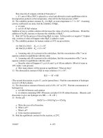
FIGURE 18.6. Major genital glands and their associated passageways.
Penis
Composition
➢ Three cylindrical masses of erectile tissue
᭹
Corpora cavernosa. Paired dorsal cylinders
᭹
Corpus spongiosum (corpus cavernosum urethrae)
᭜
Single, ventral cylinder that houses the penile urethra
᭜
Expands to terminate in glans penis that caps the two corpora
cavernosa
➢ Structure
᭹
Outer covering of skin (epidermis and dermis)
᭹
Tunica albuginea
᭜
Capsule of dense, nonelastic connective tissue surrounding the
three cylinders
᭜
Thicker around corpora cavernosa than around corpus
spongiosum
᭜
Forms an incomplete septum between the corpora cavernosa
᭹
Structure of erectile tissue
᭜
Sponge-like cavernous spaces (venous spaces) separated by con-
nective tissue trabecula with smooth muscle fibers
᭜
Deep artery in each corpus cavernosum supplies blood to
Nutritive arteries that supply trabecula
Helicine arteries that supply cavernous spaces
Process of Erection
➢ Flaccid state is effected by a minimal blood flow to the penis. This
blood flow is regulated by the continuous input of the sympathetic
division of the autonomic nervous system on the tone of the smooth
muscle in the penile vasculature.
➢ Erection
᭹
Parasympathetic division of autonomic nervous system effects
relaxation of smooth muscle (vasodilation) of the deep and helicine
arteries.
᭹
The subsequent filling of the cavernous spaces expands these
vessels against the tunica albuginea, causing the penis to become
erect and turgid.
᭹
Corpus spongiosum does not become as erect as the other
cavernous bodies because the tunica albuginea is thinner here.
Therefore, sperm can be transported during ejaculation.
192
Digital Histology
18.
Male Reproductive System 193
Testis
Intratesticular ducts
Testis
Tunica albuginea
Tunica vaginalis
Processes vaginalis
Tunica vaginalis, parietal layer
Mesothelium
Mediastinum
Rete testis
Seminiferous tubules, convoluted
portion
Seminiferous tubules, straight
portion
Epididymis
Body and tail
Coni vasculosi
Coni vasculosi, vasculature
Duct of epididymis
Efferent ducts
Efferent ducts, cilia
Efferent ducts, epithelium
Efferent ducts, lysosomes
Head
Pseudostratified columnar
epithelium with stereocilia
Rete testis
Smooth muscle
Spermatozoa
Spermatic cord
Ductus deferens
Ductus deferens, epithelium
Ductus deferens, lamina propria
Ductus deferens, smooth muscle
Nerves
Pampinoform plexus of veins
Testicular artery
Ejaculatory duct
Penis
Cavernous spaces
Corpora cavernosa
Corpus spongiosum
➢ Return to flaccid state occurs with decline of parasympathetic
activity.
Penile Urethra
➢ The penile urethra is located within corpus spongiosum (corpus cav-
ernosum urethrae).
➢ Microscopic anatomy
᭹
Lined by pseudostratified columnar epithelium that becomes
stratified squamous moist in fossa navicularis, the terminal
enlargement in the glans penis
᭹
Glands of Littre
᭜
Mucus-secreting glands
᭜
Originate in mucus-secreting recesses of the urethra and extend
obliquely toward the base of the penis
᭜
Secrete a mucous fluid that is the initial ejaculate; provides
lubrication
Structures Identified in This Section
194
Digital Histology
Deep artery
Glands of Littre
Helicine artery
Trabeculae
Tunica albuginea
Urethra
Seminal vesicles
Arches
Epithelium
Lamina propria
Lumen
Smooth muscle
Prostate
Epithelium
Glands
Prostatic concretions
Smooth muscle
General Concepts
➢ The eyes are complex photoreceptive organs located in the bony
orbits of the skull. Movement of the eye is accomplished by a set of
extrinsic ocular muscles, which insert on the outer surface of the
globe.
➢ Each eye consists of image-forming structures, a photoreceptive
retina, and a fibrous globe to provide support.
➢ The eye is protected by an eyelid, a moveable fold of skin that covers
the anterior surface of the globe.
Eyelid
➢ Protective covering of the eye.
➢ Components
᭹
Covered on its outer surface by thin skin; possesses hair follicles,
eyelashes, sebaceous glands, and sweat glands
᭹
Tarsal plate. Region of dense fibrous and elastic connective tissues
within the eyelid that provide support
᭹
Meibomian glands. Specialized sebaceous glands on the inner
surface of the eyelid whose secretions add to the tear film to reduce
evaporation
CHAPTER
19
Eye
195
Digital Histology: An Interactive CD Atlas with Review Text, by Alice S. Pakurar and
John W. Bigbee
ISBN 0-471-64982-1 Copyright © 2004 John Wiley & Sons, Inc.
᭹
Contains the obicularis oculi muscle
᭹
Conjunctiva. Lines the inner surface, consisting of a stratified
columnar epithelium with goblet cells; the conjunctiva is reflected
onto the globe as the bulbar conjunctiva, which is continuous with
the corneal epithelium.
Eyeball (Globe)
➢ Composed of three layers or tunics
᭹
Fibrous tunic consisting of the sclera and cornea
᭹
Vascular tunic or uveal tract consisting of the iris, ciliary body, and
choroid
᭹
Neural tunic consisting of the retina
➢ Contains three chambers
᭹
Anterior chamber is the space between the cornea and the iris, filled
with aqueous humor fluid.
᭹
Posterior chamber lies between the iris anteriorly and the lens, ciliary
body, and zonule fibers posteriorly; filled with aqueous humor
᭹
Vitreous chamber is located behind the lens and is filled with a
gelatinous substance called the vitreous body.
Fibrous Tunic of the Eye—Outer Tunic
➢ Sclera
᭹
Opaque layer composed of dense, irregular connective tissue;
forms the outer layer of the posterior four-fifths of the globe
196
Digital Histology
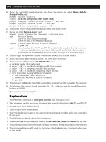
FIGURE 19.1. Midsagittal section of the eyeball.
᭹
Gives shape and support for the globe
᭹
Provides insertion points for extraocular muscles
➢ Cornea
᭹
Anterior continuation of the sclera, covering the anterior one-fifth
of the eye
᭹
Transparent and avascular; transparency results from the ordered
arrangement of its collagen fibers and low state of tissue
hydration.
᭹
Convex curvature aids in focusing light (refraction).
᭹
Layers (anterior to posterior)
᭜
Corneal epithelium. Covers the anterior surface of the cornea;
composed of a moist, stratified squamous epithelium that is con-
tinuous with the bulbar conjunctiva at the limbus
᭜
Bowman’s membrane. Acellular collagenous layer beneath the
corneal epithelium
᭜
Stroma. Multiple layers of parallel collagen fibers constitute the
majority of the cornea. The collagen fibers in each layer are
arranged at about right angles to adjacent layers. The highly
ordered arrangement of these fibers contributes to the trans-
parency of the cornea.
᭜
Descemet’s membrane. Thickened basal lamina of the corneal
endothelium
᭜
Corneal endothelium. Simple squamous epithelium covering the
posterior surface of the cornea; regulates the hydration state of
the stroma
➢ Corneo-scleral junction (limbus)
᭹
Transition zone between the cornea and the sclera
᭹
Bowman’s membrane ends and the corneal epithelium thickens at
this junction.
᭹
Trabecular meshwork. Irregular channels in the stroma that are lined
by endothelium. Drains the aqueous humor from the anterior
chamber to maintain proper intraocular pressure. The channels of
the trabecular meshwork merge to form the canal of Schlemm, a
ring-like sinus that encircles the limbus and drains into the venous
system.
Vascular Tunic (Uveal Tract) of the Eye—Middle Tunic
➢ Choroid
᭹
Highly vascular, cellular layer lying beneath the sclera; this layer
is richly pigmented due to the large numbers of melanocytes. Its
19.
Eye 197
inner portion is the choriocapillary layer, which contains large
numbers of small vessels and capillaries and serves a nutritive
function for the retina.
᭹
Bruch’s membrane. A thin layer separating the retina from the chori-
ocapillary layer; represents the combined basal laminae of the cap-
illary endothelium and the pigment epithelium of the retina and
an intervening network of elastic and collagen fibers
➢ Ciliary body
᭹
Anterior expansion of the choroid forming a ring that encircles the
lens; appears triangular in cross-section
᭹
Composed of a core of connective tissue and muscle; lined on its
vitreal surface by two layers of columnar cells, an inner pigmented
epithelium and an outer layer of nonpigmented cells. This layer,
the nonsensory retina, represents the attenuated anterior part of
the sensory layer of the retina.
᭹
Ciliary processes
᭜
Ridge-like extensions from the ciliary body
᭜
Zonule fibers. Emerge from between the processes and attach to
the lens capsule
᭜
The aqueous humor is produced by the epithelium of the ciliary
processes.
᭹
Ciliary muscles. Smooth muscle fibers that insert on the sclera and
ciliary body; contraction of circularly arranged fibers releases
tension on the zonule fibers, allowing the lens to assume a more
spherical shape, thus providing for focusing on near objects
(accommodation). Contraction of radially oriented smooth muscle
fibers results in flattening of the lens, thus providing for focusing
on far objects.
➢ Iris
᭹
Disc-shaped structure that arises from the anterior margin of the
ciliary body; separates anterior and posterior chambers and par-
tially covers the lens
᭹
Composed of loose connective tissue that is covered on its anterior
surface by an incomplete layer of pigment cells and fibroblasts. Its
posterior surface is covered by a double layer of pigmented epithe-
lial cells.
᭹
Pupil. Central opening in the iris, the diameter is regulated by con-
traction of two sets of intrinsic smooth muscle in the iris.
᭜
Dilator pupillae muscle. Derived from the more anterior, pig-
mented epithelial layer; consists of radially oriented cells whose
contraction widens the aperture of the pupil
198
Digital Histology
᭜
Constrictor pupillae muscle. Consists of circularly oriented smooth
muscle fibers surrounding the pupil; contraction of these fibers
decreases the diameter of the pupil.
Retina—Inner Tunic
➢ Inner-most of the three layers, forming a cup-shaped structure.
The posterior portion is photosensitive and extends forward to the
ciliary body, terminating as the ora serrata. The nonphotosensitive
anterior portion is reduced in thickness and number of layers and
forms the posterior lining of the ciliary body and the posterior lining
of the iris.
➢ The photosensitive portion contains the photoreceptors, which trans-
duce light into nervous impulses, and neurons, which perform the
initial integration of the visual signals.
➢ Overview of retinal cytoarchitecture
᭹
Basic plan of the retina consists of a three-cell pathway
᭜
Rods and cones. Photoreceptors that transduce light energy into
neural activity and form the photoreceptor layer; their nuclei are
located in the outer nuclear layer.
᭜
Bipolar cells. Synapse with rods and cones; nuclei are located in
the inner nuclear layer.
᭜
Ganglion cells. Synapse with bipolar cells; cell bodies are located
in the ganglion cell layer; axons from these cells form the optic
nerve fiber layer as they pass toward the optic disc, head of the
optic nerve.
᭹
Regions of synaptic integration
᭜
Outer plexiform layer. Location of synapses of rods and cones
with bipolar cells
᭜
Inner plexiform layer. Location of synapses of bipolar cells and
ganglion cells
➢ Layers of the retina–from outer to inner
᭹
Composed of 10 layers. The naming of the layers is based on their
position relative to the path of the neural conduction, not the path
of light.
᭹
Pigment epithelium
᭜
Cytoplasm contains numerous melanin granules to absorb light
and reduce reflection
᭜
Columnar epithelial cells with apical microvilli whose bases are
adherent to Bruch’s membrane in the choroid
19.
Eye 199
᭜
Cells posses a cylindrical sheath that surrounds the apical tips
of the photoreceptors; these sheaths aid in phagocytosis and
digestion of membranous discs shed by the photoreceptors.
᭹
Photoreceptor layer
᭜
Composed of rods and cones
᭜
Rods are sensitive to low light intensity, outnumber cones and
are located throughout the retina
᭜
Cones are less numerous than rods, sensitive to high intensity
light and respond to color. Cones provide greater visual acuity
and are concentrated in the fovea centralis. (See below.)
᭜
Outer segment. Contains flattened, membranous discs that
contain the visual pigments rhodopsin (rods) and iodopsins
(cones).
᭜
Inner segment. Separated from the outer segment by a constric-
tion, contains the major synthetic and energy-producing
organelles.
᭹
External limiting membrane. Not a true membrane; formed by ad-
herent junctions of Mueller cells, modified astrocytes, with the
photoreceptors
᭹
Outer nuclear layer. Location of the nuclei of rods and cones
᭹
Outer plexiform layer. Region of synaptic contacts between pho-
toreceptor axons and bipolar cell dendrites
᭹
Inner nuclear layer. Location of cell bodies of bipolar cells. Also
present are additional neurons, amacrine and horizontal cells.
᭹
Inner plexiform layer. Location of synaptic contacts between bipolar
cell axons and ganglion cell dendrites.
᭹
Ganglion cell layer. Location of cell bodies of ganglion cells
᭹
Optic nerve fiber layer. Collections of unmyelinated ganglion cell
axons that pass toward the optic disc, the head of the optic nerve,
where they exit to form the optic nerve (cranial nerve II).
᭹
Internal limiting membrane. Formed by the basal portions of Mueller
cells
➢ Fovea centralis. Region of the retina providing greatest visual acuity,
consists entirely of cones; other retinal layers are displaced centri-
fugally to allow for an unimpeded path for the light to reach the
photoreceptors.
➢ Optic disc (“blind spot”). Region composed only of axons from
retinal ganglion cells as they pass through the sclera to form the optic
nerve
200
Digital Histology
19.
Eye 201
Chambers
Anterior chamber
Posterior chamber
Vitreous chamber
Eyelid
Conjunctiva
Eyelash
Hair follicles
Meibomian glands
Skin
Tarsus
Fibrous tunic
Bowman’s membrane
Canal of Schlemm
Cornea
Corneal endothelium
Corneal epithelium
Corneo-scleral junction (limbus)
Descemet’s membrane
Sclera
Stroma
Trabecular meshwork
Lens
Lens capsule
Lens fibers
Subcapsular epithelium
Neural tunic
External limiting membrane
Fovea centralis
Ganglion cell layer
Inner nuclear layer
Inner plexiform layer
Internal limiting membrane
Optic disc
Optic nerve
Optic nerve fiber layer
Ora serrata
Outer nuclear layer
Outer plexiform layer
Photoreceptor layer containing
rods and cones
Lens
➢ Biconcave, transparent, and elastic
➢ Suspended by radially oriented zonule fibers that extend from the
ciliay body to insert into the lens capsule
➢ Structure of the lens
᭹
Lens capsule. A thickened basal lamina, produced by the subcap-
sular epithelium, surrounds the entire lens.
᭹
Subcapsular epithelium. Simple cuboidal epithelium, present only on
the anterior surface of the lens; apical surfaces of the cells are
directed toward the center of the lens.
᭹
Lens fibers. Derived from cells of the subcapsular epithelium pri-
marily in the equatorial region of the lens; lens fibers are highly
differentiated cells that lose their organelles and become filled with
crystallin proteins.
➢ Contraction of the ciliary muscle releases tension on the zonule fibers,
allowing the lens to assume a more spherical shape which provides
for focusing on near objects (accommodation).
Structures Identified in This Section
202
Digital Histology
Pigmented epithelium
Vascular tunic (uveal tract)
Bruch’s membrane
Chorio-capillary layer
Choroid
Ciliary body
Ciliary muscle
Ciliary processes
Constrictor pupillae muscle
Dilatory pupillae muscle
Iris
Melanocytes
Pupil
Components
➢ External ear. Receives sound waves, transmitting them to the tym-
panic membrane
➢ Middle ear. Transmits movement of the tympanic membrane by three
ear ossicles to fluid in the inner ear
➢ Inner ear. Contains a receptor that responds to these fluid vibrations
for the perception of sound. Additional receptors in the inner ear
respond to the effects of gravity and motion of the head to maintain
equilibrium.
External Ear
➢ Auricle or pinna. Shallow appendage on the lateral surfaces of the
head that is formed by thin skin covering a framework of elastic
cartilage
➢ External auditory meatus. Short tube leading to the tympanic
membrane
᭹
The thin skin, lining the meatus, possesses ceruminous glands.
Their secretions combine with those of adjacent sebaceous glands
to form cerumen, a thick, waxy product.
CHAPTER
20
Ear
203
Digital Histology: An Interactive CD Atlas with Review Text, by Alice S. Pakurar and
John W. Bigbee
ISBN 0-471-64982-1 Copyright © 2004 John Wiley & Sons, Inc.
᭹
Support provided by
᭜
Elastic cartilage in the outer portion
᭜
Temporal bone in the inner portion
➢ Tympanic membrane (ear drum) separates external from the middle
ear.
᭹
Composition (from exterior to interior). Thin skin, two layers of
collagen and elastic fibers with radial then circular arrangements,
and a mucous membrane that is continuous with that lining the
middle ear
᭹
Attachment of the malleus, an ear ossicle, to the inner surface pulls
the tympanic membrane into a flattened, cone shape.
Middle Ear (Tympanic Cavity)
➢ The middle ear or tympanic cavity is a cavity within the temporal bone
that is bounded by the tympanic membrane laterally and the bony
wall of the inner ear medially. It communicates with the mastoid air
cells posteriorly, and with the nasopharynx, via the Eustachian tube,
anteriorly.
➢ Structure
᭹
Lined by a mucous membrane whose epithelium is predominately
simple squamous
204
Digital Histology
FIGURE 20.1. Schematic illustration of the three subdivisions of the ear.
᭹
Ear ossicles, small bones, transmit vibrations from the tympanic
membrane to the inner ear.
᭜
Components
Malleus. Attached to the tympanic membrane
Incus. Interconnects malleus with stapes
Stapes. Footplate of the stapes fits into the oval window of the
inner ear
᭜
Ossicles are connected to each other by ligaments and are
covered with mucosa.
᭜
Small muscles attached to malleus (tensor tympani) and stapes
(stapedius) modulate vibrations of these ossicles.
᭹
Eustachian tube (auditory tube)
᭜
Connects middle ear with the nasopharynx
᭜
Is lined by a mucous membrane whose epithelium becomes
pseudostratified near the nasopharynx. Cilia associated with
this epithelium beat toward the pharynx.
᭜
Is supported first by bone and then by cartilage and fibrous
tissue as it nears the nasopharynx
᭜
Is usually collapsed but opens during swallowing to equilibrate
air pressure
20. E
ar 205
FIGURE 20.2. Coronal section through the skull showing the three subdivisions of
the ear in the temporal bone.
᭹
Oval window and round window
᭜
Openings in the petrous portion of the temporal bone that form
the medial wall of the middle ear
᭜
The oval window is occupied by the footplate of the stapes.
᭜
The round window is covered by a membrane that bulges to
relieve pressure in the cochlea that originates from motion of the
stapes at the oval window.
᭹
Mastoid air spaces, located in the mastoid process of the temporal
bone, communicate posteriorly with the middle ear.
Inner Ear
➢ The inner ear is located in the petrous portion of the temporal bone.
➢ Components
᭹
Osseous labyrinth. Series of interconnected tubular and cavernous
spaces in the petrous portion of the temporal bone that are lined
with periosteum and filled with perilymph fluid
᭜
Vestibule. Centrally located chamber; communicates with
middle ear via the oval window
᭜
Semicircular canals
Are three tubular spaces that communicate with and lie pos-
terolaterally to the vestibule
Are oriented in three mutually perpendicular planes
An enlargement at one end of each canal, adjacent to the
vestibule, houses the ampulla of the semicircular ducts.
᭜
Cochlea. An osseous tube that connects with and lies anterome-
dially to the vestibule
Tube is coiled into a spiral shape with 2.5 turns, resembling a
snail shell.
The tube’s spiraling in the temporal bone results in the
formation of a central, bony axis for the cochlea called
the modiolus, which resembles a screw. The threads of the
screw project into the cochlea and are called the osseous spiral
lamina.
The modiolus houses the cochlear division of cranial nerve VIII
and its sensory ganglion, the spiral ganglion.
᭹
Membranous labyrinth. Series of interconnected ducts and chambers
that are suspended within the osseous labyrinth. Contain the fluid,
endolymph. These ducts and chambers contain receptors for
hearing and for static and kinetic senses.
206
Digital Histology