Báo cáo y học: "Genome-wide deletion mutant analysis reveals genes required for respiratory growth, mitochondrial genome maintenance and mitochondrial protein synthesis in Saccharomyces cerevisia" pot
Bạn đang xem bản rút gọn của tài liệu. Xem và tải ngay bản đầy đủ của tài liệu tại đây (1.83 MB, 18 trang )
Genome Biology 2009, 10:R95
Open Access
2009Merz and WestermannVolume 10, Issue 9, Article R95
Research
Genome-wide deletion mutant analysis reveals genes required for
respiratory growth, mitochondrial genome maintenance and
mitochondrial protein synthesis in Saccharomyces cerevisiae
Sandra Merz
*
and Benedikt Westermann
*†
Addresses:
*
Institut für Zellbiologie, Universität Bayreuth, Universitätsstraße 30, 95440 Bayreuth, Germany.
†
Bayreuther Zentrum für
Molekulare Biowissenschaften (BZMB), Universität Bayreuth, Universitätsstraße 30, 95440 Bayreuth, Germany.
Correspondence: Benedikt Westermann. Email:
© 2009 Merz and Westermann; licensee BioMed Central Ltd.
This is an open access article distributed under the terms of the Creative Commons Attribution License ( which
permits unrestricted use, distribution, and reproduction in any medium, provided the original work is properly cited.
Yeast respiratory genes<p>A genome-wide deletion mutant analysis in budding yeast reveals genes required for respiratory growth, mitochondrial genome main-tenance and mitochondrial protein synthesis.</p>
Abstract
Background: The mitochondrial respiratory chain produces metabolic energy by oxidative
phosphorylation. Biogenesis of the respiratory chain requires the coordinated expression of two
genomes: the nuclear genome encoding the vast majority of mitochondrial proteins, and the
mitochondrial genome encoding a handful of mitochondrial proteins. The understanding of the
molecular processes contributing to respiratory chain assembly and maintenance requires the
systematic identification and functional analysis of the genes involved.
Results: We pursued a systematic, genome-wide approach to define the sets of genes required for
respiratory activity and maintenance and expression of the mitochondrial genome in yeast. By
comparative gene deletion analysis we found an unexpected phenotypic plasticity among
respiratory-deficient mutants, and we identified ten previously uncharacterized genes essential for
respiratory growth (RRG1 through RRG10). Systematic functional analysis of 319 respiratory-
deficient mutants revealed 16 genes essential for maintenance of the mitochondrial genome, 88
genes required for mitochondrial protein translation, and 10 genes required for expression of
specific mitochondrial gene products. A group of mutants acquiring irreversible damage
compromising respiratory capacity includes strains defective in assembly of the cytochrome c
oxidase that were found to be particularly sensitive to aging.
Conclusions: These data advance the understanding of the molecular processes contributing to
maintenance of the mitochondrial genome, mitochondrial protein translation, and assembly of the
respiratory chain. They revealed a number of previously uncharacterized components, and provide
a comprehensive picture of the molecular processes required for respiratory activity in a simple
eukaryotic cell.
Published: 14 September 2009
Genome Biology 2009, 10:R95 (doi:10.1186/gb-2009-10-9-r95)
Received: 27 July 2009
Accepted: 14 September 2009
The electronic version of this article is the complete one and can be
found online at /> Genome Biology 2009, Volume 10, Issue 9, Article R95 Merz and Westermann R95.2
Genome Biology 2009, 10:R95
Background
Mitochondria are the major sites of metabolic energy produc-
tion in animals and most other eukaryotic organisms. Elec-
trons generated by the oxidation of nutrients are passed along
the respiratory chain and finally transferred to molecular oxy-
gen in a process called oxidative phosphorylation. Energy
released by the passage of electrons is stored as a proton gra-
dient across the mitochondrial inner membrane and har-
vested by the ATP synthase to produce ATP from ADP and
phosphate [1]. In an average human individual, ATP is syn-
thesized at an astonishing rate of 9 × 10
20
molecules per sec-
ond, totaling an amount of 65 kg per day [2]. In most
eukaryotic organisms, the respiratory chain consists of five
multi-subunit complexes: complex I, NADH dehydrogenase;
complex II, succinate dehydrogenase; complex III, cyto-
chrome bc
1
complex; complex IV, cytochrome c oxidase; and
complex V, ATP synthase [1]. In some organisms, including
baker's yeast, Saccharomyces cerevisiae, complex I is
replaced by an alternative NADH dehydrogenase that con-
sists of a single amino acid chain [3,4].
Biogenesis of the respiratory chain depends on coordinated
expression of gene products encoded by the nuclear and mito-
chondrial genomes. The vast majority of the approximately
1,000 proteins that make up the mitochondrial proteome is
encoded by nuclear genes, while a small number of protein-
coding genes have been retained in the mitochondrial
genome during the evolution of eukaryotic cells - thirteen in
humans, eight in Saccharomyces cerevisiae, and as little as
three in the protist Plasmodium falciparum [5]. Proteins
encoded by the mitochondrial genome are generally
restricted to a few respiratory chain complex subunits and - in
some organisms - components required for synthesis and
assembly of mitochondria-encoded proteins [5]. In order to
express this handful of mitochondrial genes, the cell synthe-
sizes about 200 nuclear-encoded proteins that are devoted to
mitochondrial genome maintenance and gene expression
[6,7].
S. cerevisiae is a powerful model organism to genetically dis-
sect the pathways required for maintenance of respiratory
activity because it is capable of satisfying its energy require-
ments with ATP generated by fermentation. Thus, oxidative
phosphorylation and the presence of the mitochondrial
genome are dispensable as long as fermentable carbon
sources, such as glucose or fructose, are present in the growth
medium. Even when oxygen is available, yeast cells generate
ATP primarily by glycolysis with ethanol as an end product of
fermentation. Most respiratory functions are repressed under
these conditions by catabolite repression [8]. Only when fer-
mentable carbon sources become limiting, genes required for
respiration are induced, and ATP is generated by metaboliz-
ing non-fermentable carbon sources, such as ethanol, glycerol
or lactate [9,10]. Yeast mutants defective in oxidative phos-
phorylation are unable to grow on media containing non-fer-
mentable carbon sources. On media containing limiting
amounts of fermentable carbon sources, these mutants form
smaller colonies than wild-type strains. The term petite has
been coined to describe this characteristic phenotype [11].
The originally isolated petite mutants that were described in
the 1940s were later found to have long deletions in the mito-
chondrial genome (termed [rho
-
]) or completely lack mito-
chondrial DNA (termed [rho
0
]). Mutants with lesions in the
mitochondrial genome are referred to as cytoplasmic petite,
whereas respiratory-deficient strains carrying mutations in
the nuclear genome are referred to as nuclear petite or pet
mutants [12]. Nuclear pet genes include, but are not limited
to, genes encoding respiratory chain components, factors
required for folding and assembly of respiratory chain subu-
nits, proteins required for mitochondrial DNA (mtDNA)
inheritance, mitochondrial RNA and protein synthesis, and
components of the machinery determining mitochondrial
morphology [12-14].
By the end of the last century, more than 200 complementa-
tion groups and more than 100 pet genes had been identified
by classic yeast genetic methods [12,13,15]. The availability of
the yeast gene deletion library nowadays allows systematic
and comprehensive screens to assign functions to almost all
of the approximately 4,800 non-essential yeast genes [16].
Here, we aimed at a large-scale functional analysis of respira-
tory-deficient yeast mutants to define the complement of
genes a yeast cell requires for mitochondrial gene expression
and respiratory activity. Comparative gene deletion analysis
revealed a surprising phenotypic plasticity of respiratory-
deficient mutants and allowed us to identify ten novel genes
that are essential for respiratory activity in yeast. By system-
atic functional tests of respiratory-deficient mutants we
obtained a comprehensive picture of the molecular processes
required for respiratory activity and maintenance and expres-
sion of the mitochondrial genome in yeast.
Results and discussion
Genes required for respiratory growth
Two independent screens of the yeast deletion library have
previously revealed two partially overlapping sets of pet
genes. By plating the homozygous diploid yeast deletion
library on media containing glycerol as a carbon source, Dim-
mer et al. [14] identified 341 deletion mutants that were una-
ble to grow. In a very similar approach, Luban et al. [17]
identified a set of 355 respiratory-deficient clones by screen-
ing the MATa yeast deletion library. While about two-thirds
of the mutants in each screen were found to be respiratory-
deficient also in the other screen, a surprisingly large number
of mutants were isolated only once [17]. It seems unlikely that
this is due to differences in the genetic background, because
both screens have been conducted in largely isogenic strains,
BY4743 and BY4741 [18]. Here, we screened the MATα dele-
tion library (BY4742 background) to obtain a third set of res-
piratory-deficient mutants. This was then compared with the
Genome Biology 2009, Volume 10, Issue 9, Article R95 Merz and Westermann R95.3
Genome Biology 2009, 10:R95
data obtained by Dimmer et al. [14] and Luban et al. [17]. The
MATα deletion library contained 319 mutants that were una-
ble to grow on glycerol-containing medium (Additional data
file 1). Of these, 176 are common to all three sets of pet genes
(Figure 1a). In the following we will refer to these genes as
highly penetrant pet genes. 125 genes have been identified in
two of three screens, and 237 genes have been identified only
once (pet genes unique to this study are listed in Additional
data file 2). Nineteen additional pet genes (not included in the
set of 176 highly penetrant pet genes) were only covered by
one or two libraries. Based on data from the Saccharomyces
Genome Database [19] and manual annotation, we grouped
all genes according to their frequency of occurrence in pet
screens and the intracellular location and function of their
gene products (Additional data file 3).
Strikingly, 129 out of the 176 pet genes found in all three
screens encode proteins known to be located in mitochondria,
corresponding to 73.3% (Figure 1b; Additional data file 3).
The fraction of genes encoding mitochondrial proteins was
reduced to 52.1% for pet genes found in two of three screens,
and as low as 14.7% for pet genes that were found only once
(Figure 1b; Additional data file 3). This demonstrates a clear
correlation of the penetrance of pet phenotypes with mito-
chondrial functions of the affected gene products. The major-
ity of the 176 pet genes found in all libraries encode proteins
devoted to maintenance and expression of the mitochondrial
genome and assembly of the respiratory chain (Figure 1c;
Additional data file 3). Thirteen open reading frames (ORFs)
are unlikely to encode proteins, because they overlap with
other known genes (Additional data file 3), reducing the
number of protein-coding genes to 163.
Differences of growth behavior of strains taken from different
versions of the deletion libraries could either reflect inherent
properties of the mutant strains, they could be due to techni-
cal differences between the various screens, or they could
mean that a given deletion in one collection is wrong (as it has
occasionally been observed by us and others; for example,
strains not correct in the MATα library include Δrpo41 lack-
ing the mitochondrial RNA polymerase). We reasoned that
incorrect mutants will be enriched among strains that showed
respiratory competence in one screen but were respiratory-
deficient in the two other screens because it is more likely that
a specific phenotype is obscured rather than generated by
chance. To test this, we checked the genotypes of 29 mutants
taken from the MATα library by PCR. Nineteen randomly
chosen mutants were tested that were respiratory-competent
in the MAT
α library, but respiratory-deficient in the MATa
and homozygous diploid library. Of these, six mutants
(Δyal012w, Δybl038w, Δydl202w, Δydr268w, Δyor205c,
and Δypl029w) contained exclusively the wild-type allele,
seven mutants (Δydr231c, Δydr332w, Δyil036w, Δyjr090c,
Δymr066w, Δypr047w, and Δypr124w) contained a mixture
of deletion and wild-type alleles, and six mutants were found
to have the correct genotype (Δyal047c, Δybr163w,
Δydr323c, Δykl148c, Δyml081c-a, and Δyml129c). In addi-
tion, we tested ten mutants that showed a pet phenotype only
in the MATα library, but not in the MATa and homozygous
diploid library, and ten mutants, that showed a pet phenotype
in all three screens. All mutants of the two latter groups were
found to have the correct genotype. This means whenever a
wrong deletion was detected, a pet phenotype was obscured
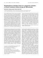
Nuclear pet genes of S. cerevisiaeFigure 1
Nuclear pet genes of S. cerevisiae. (a) The numbers of pet mutants
identified in three screens of the yeast deletion library are indicated.
References: Dimmer et al. [14], Luban et al. [17]. (b) The intracellular
location of proteins encoded by pet genes has been grouped according to
their frequency of occurrence in screens of the deletion library. The graph
is a summary of data contained in Additional data file 3. (c) Cellular
functions of proteins encoded by highly penetrant pet genes. Functions
have been assigned according to data from the Saccharomyces Genome
Database [19] and manual annotation. Red indicates mitochondrial
proteins, green known extra-mitochondrial proteins, grey unknown
proteins, and white dubious ORFs overlapping with known protein-coding
genes.
(a)
(b)
(c)
Genome Biology 2009, Volume 10, Issue 9, Article R95 Merz and Westermann R95.4
Genome Biology 2009, 10:R95
by the presence of the wild-type allele, whereas all respira-
tory-deficient mutants tested were found to have the correct
genotype. We conclude that several discrepancies of growth
phenotypes can be ascribed to wrong genotypes that are
present in the deletion library. However, the fact that a rela-
tively large number of mutants with confirmed correct geno-
types show differences in their growth behavior points to a
pronounced phenotypic plasticity of pet mutants. Further-
more, the correlation of the penetrance of pet phenotypes
with mitochondrial localization of gene products (Figure 1b)
is a clear indication that the phenotypic variability is not only
due to wrong deletions present in the mutant libraries, but
also reflects biological processes.
Eight highly penetrant pet genes (YDR065w, YGR150c,
YJL046w, YLL033w, YLR091w, YMR293c, YOR305w, and
YPR116w) encode previously uncharacterized proteins, and
earlier studies have revealed a respiratory-deficient pheno-
type for two additional ORFs of unknown function that were
not covered by all three yeast deletion libraries, YNL213c [20]
and YJL062w-a [21]. We confirmed the identity of these
mutant strains by PCR and named the genes RRG1 through
RRG10 (for 'Required for respiratory growth') as the func-
tional analysis described below proves that their products are
novel factors required for respiratory growth.
Comparative growth analysis on different non-
fermentable carbon sources
We asked whether the respiratory-deficient phenotype
observed for the 319 pet mutants isolated from the MATα
deletion library is specific to glycerol metabolism or reflects a
general lack of respiration competence. To test this, we plated
the mutants also on complete media containing lactate or eth-
anol as sole carbon sources. The vast majority (305 strains,
corresponding to 95.6% of the pet mutants) failed to grow on
all non-fermentable carbon sources that were tested. Of the
remainder, seven mutants showed a growth defect only on
glycerol-containing medium, seven on glycerol or ethanol-
containing media, and one mutant on glycerol or lactate con-
taining media (Additional data file 4). As pet phenotypes are
highly reproducible even on different carbon sources we con-
clude that our screen gives a largely accurate estimate of res-
piratory deficiencies in the MATα deletion library.
Restoration of respiratory activity by mating with
Δmip1 and by cytoduction of [rho
+
] mitochondria
In order to define the genetic basis of respiratory deficiency,
we subjected the complete set of 319 pet mutants isolated
from the MATα deletion library to various functional tests
(Figure 2). As a petite phenotype is often associated with the
complete or partial loss of the mitochondrial genome [13], we
first asked whether the pet mutants contain functional
mtDNA. To test this, pet mutants were mated with a strain
lacking the mtDNA polymerase Mip1. As the Δmip1 strain is
[rho
0
] [22], resulting heterozygous diploid strains are able to
grow on glycerol-containing medium only if functional
mtDNA is provided by the pet mutant mating partner. We
observed restoration of respiratory activity in 157 hetero-
zygous diploid strains demonstrating that the parental pet
strains possessed an intact mitochondrial genome. In con-
trast, 162 strains failed to grow on glycerol-containing
medium after mating, suggesting that the parental pet
mutants were [rho
-
] or [rho
0
].
The complementation test with Δmip1 does not discern
whether the protein encoded by the pet gene is obligatorily
required for maintenance of mtDNA, or whether a functional
mitochondrial genome had been spontaneously lost in the pet
mutant during many generations of growth. To discriminate
between these possibilities, we replenished cells with mito-
chondria containing a wild-type [rho
+
] mitochondrial
genome by cytoduction. In brief, pet mutants were crossed
with a [rho
+
] donor strain that carries a kar1 mutation to pre-
vent karyogamy in the zygote. After counterselection against
nuclear chromosomes of the donor strain, growth of the hap-
loid progeny was assessed on glycerol-containing media. Res-
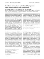
Summary of the systematic functional analysis of 319 pet mutants isolated from the MATα yeast deletion libraryFigure 2
Summary of the systematic functional analysis of 319 pet mutants isolated
from the MATα yeast deletion library. Grey boxes indicate groups of
mutants that were further analyzed, black boxes indicate the final level of
resolution of functional analysis. See text for details.
Genome Biology 2009, Volume 10, Issue 9, Article R95 Merz and Westermann R95.5
Genome Biology 2009, 10:R95
toration of respiratory activity after cytoduction was observed
in 67 pet mutants, whereas 252 strains failed to grow on non-
fermentable carbon sources.
Combining the results from the Δmip1 mating test and the
cytoduction experiment allowed us to define four classes of
pet mutants (Figure 3; Additional data file 5). Class I mutants
were not rescued either by mating with Δmip1 or by cytoduc-
tion; class II mutants were rescued by mating with Δmip1 as
well as by cytoduction; class III mutants were rescued only by
mating with Δmip1, but not by cytoduction; and class IV
mutants were rescued only by cytoduction, but not by mating
with Δmip1. The basic properties of these classes are summa-
rized in Table 1. In the following, the various classes of pet
mutants are further examined (Figure 2).
Genes required for maintenance of mtDNA
The 118 class I mutants were [rho
-
] or [rho
0
] and remained
respiratory-deficient after introduction of functional mito-
chondria. This group of mutants is expected to include all
components that are essential for maintenance of a [rho
+
]
genome. In addition, we expected it to contain components
deletion of which leads to a gradual loss of mtDNA and, at the
same time, induces respiratory deficiency due to lack of func-
tions not directly related to mtDNA maintenance. To discern
between these possibilities, we subjected all class I mutants to
various functional tests. First, we tested for the presence of
mtDNA by DAPI (4',6-diamidino-2-phenylindole) staining
immediately after cytoduction. Second, we tested growth on
YPG medium after adaptation to the medium by pre-culture
on YPG containing low amounts of glucose. And third, we
tested mitochondrial protein translation activity by SDS-
PAGE and autoradiography after labeling cycloheximide-
treated cells with
35
S methionine.
Genes essential for maintenance of mtDNA were defined by
the following criteria: At least 95% of the cells observed by
DAPI staining after cytoduction were devoid of mtDNA and
the remainder contained less than five mtDNA nucleoids per
cell. This phenotype was observed after cytoduction in the
Δmip1 mutant lacking the mtDNA polymerase and, therefore,
is indicative of instantaneous loss of mtDNA. In addition,
cells lacking genes essential for maintenance of mtDNA were
expected to be unable to grow on YPG after adaptation to the
carbon source, and they were unable to produce even trace
amounts of mitochondria-encoded proteins. Sixteen mutants
were identified that matched these criteria (Table 2). We pro-
pose that the gene products lacking in these mutants are par-
ticularly important for maintenance of mtDNA. As expected,
this group includes several components known to be involved
in mtDNA metabolism: the mtDNA polymerase Mip1 [22];
mtDNA helicases Hmi1 [23] and Pif1 [24]; Apn1, a DNA
repair protein active in the nucleus and mitochondria [25];
and aconitase, Aco1, an enzyme of the citric acid cycle that has
an additional role in mtDNA maintenance [26].
It has been observed that a block of mitochondrial protein
synthesis leads to a rapid and quantitative loss of mtDNA
[27]. However, the reasons for this phenomenon are still
unknown. Here, we observed instantaneous loss of mtDNA in
cells lacking Mrpl37, Mtf1, Mtg2, Rsm24, and Slm5, which
are all required for mitochondrial transcription or transla-
tion, and in a deletion mutant lacking the dubious ORF
YKL091w, which overlaps with the MRPL38 gene encoding a
mitochondrial ribosomal protein. Loss of mtDNA at a rela-
tively high rate was also observed in several other class I
mutants lacking components of the mitochondrial protein
synthesis machinery. These findings underscore the impor-
tance of mitochondrial protein synthesis for maintenance of
mtDNA. Moreover, rapid loss of mtDNA was observed in the
Δatp4 mutant lacking ATPase subunit b. This is consistent
with earlier observations [28]; however, the molecular rea-
sons are not understood [29]. Also, Δpet100 mutants lacking
a factor required for cytochrome c oxidase assembly showed
rapid loss of mtDNA. As loss of mtDNA in Δatp4, Δmrpl37,
Δmtf1, Δmtg2, Δpet100, Δrsm24, and Δslm5 occurs instanta-
neously (as rapid as in Δmip1) we consider it likely that repli-
cation and/or inheritance of mtDNA is actively suppressed in
these strains. These results point to an active role of Atp4,
Mrpl37, Mtf1, Mtg2, Pet100, Rsm24, and Slm5 in regulating
mtDNA abundance in yeast mitochondria.
Table 1
Classes of pet mutants
Class Respiration after mating with Δmip1 Respiration after cytoduction Associated gene functions
I - - Genes essential for maintenance of mtDNA (16 mutants);
or genes essential for respiration with gradual loss of
mtDNA (102 mutants)
II + + Additional effects of extra-genomic factors and/or acquired
mitochondrial damage (23 mutants)
III + - Genes essential for respiration but not for maintenance of
mtDNA (134 mutants)
IV - + Genes dispensable for respiration, gradual loss of mtDNA
(44 mutants)
Genome Biology 2009, Volume 10, Issue 9, Article R95 Merz and Westermann R95.6
Genome Biology 2009, 10:R95
Other factors required for mtDNA inheritance are Mgm1,
Doc1 and the newly identified protein Rrg5. Mgm1 is a
dynamin-related protein required for mitochondrial genome
maintenance by mediating mitochondrial fusion [30-32].
Doc1 is involved in cyclin proteolysis as a processivity factor
required for the ubiquitination activity of the anaphase pro-
moting complex (APC) [33]. Intriguingly, Doc1 has been
found in the mitochondrial proteome [6,34]. Thus, it is
tempting to speculate that it links mtDNA replication and/or
inheritance to the cell cycle. The RRG5 gene (YLR091w)
encodes a protein of unknown function. Its sequence does not
show similarities to any characterized protein. As the Rrg5
protein has been localized to mitochondria [6,34,35], we pro-
pose that it is a novel factor essential for maintenance of
mtDNA.
In addition to class I mutants, 44 pet mutants were identified
that were not complemented by mating with Δmip1 but could
be rescued by cytoduction. These strains are able to maintain
newly re-introduced mtDNA when they are grown on non-
fermentable carbon sources (class IV; Additional data file 5).
It is conceivable that these mutants have a tendency to spon-
taneously lose their mitochondrial genome when they are
grown on fermentable carbon sources for longer times. This
has been observed previously for Δmdm31 and Δmdm32
mutants that showed a pet phenotype in the screen performed
by Dimmer et al. [14], but not in the screens performed by
Luban et al. [17] and in the screen reported here. Freshly
made Δmdm31 and Δmdm32 deletion mutants have been
found to be able to maintain [rho
+
] mtDNA [36]. However,
mtDNA is not stably inherited and is gradually lost after sev-
eral generations of growth in glucose-containing medium
[36]. To test this systematically for all class IV mutants, we
replenished mtDNA by cytoduction and then passaged the
strains in liquid YPD medium for 10 days to allow for loss of
mtDNA. Presence or absence of mtDNA was assayed by DAPI
staining immediately after cytoduction and after 10 days of
replicative growth. In all strains, at least 90% of the cells con-
tained mtDNA directly after cytoduction. Continued growth
in glucose-containing medium led to increased loss of mtDNA
in many mutants (Additional data file 6), suggesting that
gradual loss of mtDNA accounts for the pet phenotype in
many class IV mutants. Only few mutants maintained
mtDNA as stably as the wild type (Additional data file 6). We
consider it possible that these mutants require more genera-
tion times or special growth conditions to induce loss of
mtDNA, or that these mutants rapidly accumulate mtDNA
point mutations or deletions rendering the mitochondrial
genome non-functional over time. Interestingly, 77% of the
class IV mutants have not been found in the screens by Dim-
mer et al. [14] and Luban et al. [17], suggesting that many of
the affected genes are only indirectly related to maintenance
of respiratory activity.
Genes required for protein translation in mitochondria
Next, we asked which genes are required for mitochondrial
protein synthesis. Mutants defective in this process are
expected to be found in either class I or class III. Class I con-
tains mutants that have lost their mtDNA as a consequence of
blocked mitochondrial translation activity, whereas class III
contains mutants that are defective in translation but main-
tain an intact mitochondrial genome. In order to be able to
test for mitochondrial protein synthesis activity, we replen-
ished wild-type mtDNA in class I mutants by cytoduction.
After this treatment, mtDNA could be visualized by DAPI
staining in 102 mutants, whereas 16 mutants lacking genes
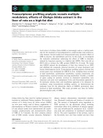
Classes of pet mutantsFigure 3
Classes of pet mutants. The left column indicates genotypes of haploid pet
mutant strains taken from the deletion library carrying a deletion in the
nuclear genome (Δyfg1, 'your favourite gene 1') and either no mtDNA
([rho
0
]; alternatively these strains might be [rho
-
]) or a wild type-like
mitochondrial genome ([rho
+
]; labeled in red). The middle column
indicates genotypes of heterozygous diploid strains after mating with
Δmip1. The right column indicates genotypes of haploid deletion mutants
after having received [rho
+
] mitochondria from a donor strain by
cytoduction. Respiratory-competent mitochondria are labeled in red, and
respiratory-competent yeast cells are depicted on a red background. Class
I mutants contain either [rho
+
] or [rho
0
] or [rho
-
] mitochondria after
cytoduction. See text for details.
Genome Biology 2009, Volume 10, Issue 9, Article R95 Merz and Westermann R95.7
Genome Biology 2009, 10:R95
essential for maintenance of mtDNA immediately became
[rho
0
] (see above; Table 2). For class III mutants, we rea-
soned that some strains might be unable to grow on medium
containing glycerol as the sole carbon source because of syn-
ergistic effects of compromised mitochondrial function in
combination with catabolite repression, which reduces the
expression of genes required for respiration [8]. Therefore,
we first relieved catabolite repression in all class III mutants
by growth on glycerol-containing medium supplemented
with limiting amounts of fermentable carbon source (3% glyc-
erol/0.1% glucose) before replicating the strains on glycerol-
containing medium. After this treatment, 77 strains were able
to grow on plates containing glycerol as the sole carbon
source (Additional data file 7). We conclude that the gene
products lacking in these mutants are dispensable for respira-
tion.
Then, we tested mitochondrial translation in a total number
of 159 deletion mutants (102 class I mutants with replenished
mtDNA and 57 class III mutants unable to grow on glycerol-
containing medium after adaptation to the carbon source).
Strains were grown to logarithmic growth phase in medium
containing fermentable carbon sources, before cytosolic
translation was stopped by the addition of cycloheximide.
Newly synthesized mitochondrial proteins were labeled with
35
S methionine, and cell extracts were analyzed by SDS-PAGE
and autoradiography.
Mitochondrial translation products could not be detected in
88 mutants (Table 3). We conclude that these genes are
required for mitochondrial protein synthesis. Encoded pro-
teins include 39 subunits of the mitochondrial ribosome and
several additional components required for mitochondrial
transcription, translation or assembly of the respiratory chain
[37]. In addition, mitochondrial translation activity was
absent in several mutants lacking proteins known to be
required for mtDNA inheritance, such as Fzo1 [38,39], Mhr1
[40], Msh1 [41], or Mgm101 [42]. Supposedly, in these strains
Table 2
Genes essential for maintenance of mtDNA
Genes encoding components involved in mtDNA metabolism
*YKL114c APN1 Involved in repair of DNA damage; located in nucleus and
mitochondria
YLR304c ACO1 Aconitase; also independently required for mtDNA
maintenance
YML061c PIF1 DNA helicase; active in nucleus and mitochondria
YOL095c HMI1 Mitochondrial inner membrane localized DNA helicase
YOR330c MIP1 Catalytic subunit of the mitochondrial DNA polymerase
Genes encoding components involved in mitochondrial
transcription and translation
*YBR268w MRPL37 Mitochondrial ribosomal protein
*YCR024c SLM5 Mitochondrial asparaginyl-tRNA synthetase
*YDR175c RSM24 Mitochondrial ribosomal protein of the small subunit
YHR168w MTG2 Associates with mitochondrial ribosome; possible role in
ribosome assembly
*YKL169c Dubious ORF; partially overlaps with MRPL38
YMR228w MTF1 Mitochondrial RNA polymerase specificity factor
Genes encoding components involved in oxidative
phosphorylation
*YDR079w PET100 Specifically facilitates the assembly of cytochrome c oxidase
YPL078c ATP4 Subunit b of the stator stalk of mitochondrial F
1
F
0
ATP
synthase
Other genes
*YGL240w DOC1 Required for the ubiquitination activity of the anaphase
promoting complex
*YLR091w RRG5 Unknown function
YOR211c MGM1 Mitochondrial GTPase involved in fusion
The list indicates systematic and standard names of genes essential for maintenance of newly re-introduced mtDNA in class I pet mutants. The
cellular roles of the proteins are indicated according to the Saccharomyces Genome Database [19] or manually annotated. Genes that were
previously not known to be essential for maintenance of mtDNA are indicated with an asterisk.
Genome Biology 2009, Volume 10, Issue 9, Article R95 Merz and Westermann R95.8
Genome Biology 2009, 10:R95
Table 3
Genes essential for mitochondrial translation
Genes encoding mitochondrial ribosomal proteins YBL090W/MRP21; YBR146W/MRPS9; YBR251W/MRPS5; YBR282W/MRPL27;
YCR003W/MRPL32; YCR071C/IMG2; YDL045W-A/MRP10; YDR115W;
YDR337W/MRPS28; YDR347W/MRP1; YEL050C/RML2; YER050C/RSM18;
YGL129C/RSM23; YGR076C/MRPL25; YGR215W/RSM27; YGR220C/MRPL9;
YHR147C/MRPL6; YJL063C/MRPL8; YJL096W/MRPL49; YKL003C/MRP17;
YKL138C/MRPL31; YKL155C/RSM22; YKL170W/MRPL38; YKR006C/MRPL13;
YKR085C/MRPL20; YLR312W-A/MRPL15; YLR439W/MRPL4; YMR158W/
MRPS8; YMR188C/MRPS17; YMR193W/MRPL24; YMR286W/MRPL33;
YNL081C/SWS2; YNL177C/MRPL22; YNL185C/MRPL19; YNL252C/MRPL17;
YNR037C/RSM19; YOR150W/MRPL23; YOR158W/PET123; YPL173W/
MRPL40
Other genes encoding known proteins
*YBL019w APN2 Class II abasic (AP) endonuclease involved in repair of DNA damage
*YBR179c FZO1 Transmembrane GTPase required for mitochondrial fusion
YDL044c MTF2 Mitochondrial protein involved in mRNA splicing and protein synthesis
*YDR194c MSS116 Mitochondrial RNA helicase, required for splicing of group II introns
*YDR296w MHR1 Involved in repair, recombination and maintenance of mitochondrial DNA
*YER145c FTR1 Iron permease that mediates high-affinity iron uptake
*YER154w OXA1 Component of the mitochondrial protein export machinery
*YGL071w RCS1 Transcription factor regulates genes involved in iron uptake and cell size
YGL143c MRF1 Mitochondrial peptide chain release factor
YGR171c MSM1 Met-tRNA synthetase, mitochondrial
YHL038c CBP2 Mitochondrial splicing factor
YHR011w DIA4 tRNA synthetase, may be involved in mitochondrial function
YHR038w RRF1 Mitochondrial ribosome recycling factor
*YHR051w COX6 Cytochrome c oxidase subunit VI
YHR091c MSR1 Arginyl-tRNA synthetase of mitochondria
*YHR120w MSH1 Involved in mitochondrial DNA repair
YJL102w MEF2 Mitochondrial translation elongation factor
*YJL209w CBP1 Required for COB mRNA stability or 5' processing
*YJR144w MGM101 Mitochondrial genome maintenance protein
*YKL016c ATP7 ATP synthase subunit d
*YKL134c OCT1 Mitochondrial intermediate peptidase
YKL194c MST1 Mitochondrial threonyl tRNA synthase
YLR067c PET309 Specific translational activator for the COX1 mRNA
YLR069c MEF1 Mitochondrial translation elongation factor G
*YLR070c XYL2 Xylitol dehydrogenase
YLR139c SLS1 Protein involved in mitochondrial metabolism
*YLR295c ATP14 ATP synthase subunit h
YMR064w AEP1 Required for accumulation of transcript of ATP9/OLI1
*YMR089c YTA12 Involved in proteolytic and chaperone activities in the inner membrane
YMR097c MTG1 Likely functions in assembly of the large ribosomal subunit
*YMR098c ATP25 Required for the stability of ATP9 mRNA
*YMR267w PPA2 Inorganic pyrophosphatase, mitochondrial
*YMR287c DSS1 RNase, associates with the ribosome, turnover of aberrant RNAs
YNL073w MSK1 Lysyl-tRNA synthetase, mitochondrial
YOL033w MSE1 Glutamyl-tRNA synthetase, mitochondrial
*YOR065w CYT1 Cytochrome c
1
YOR187w TUF1 Translation elongation factor Tu, mitochondrial
YPL097w MSY1 Tyrosyl-tRNA synthetase, mitochondrial
YPL104w MSD1 Aspartyl-tRNA synthetase, mitochondrial
*YPL148c PPT2 Activates mitochondrial acyl carrier protein
Genome Biology 2009, Volume 10, Issue 9, Article R95 Merz and Westermann R95.9
Genome Biology 2009, 10:R95
- and likely also in other class I mutants - the mitochondrial
genome had been largely lost or damaged during growth of
the strains in the time between cytoduction and the labeling
reaction. It should be noted that strain-dependent effects
might also play a role, because, for example, Δpet309 was
observed to be completely translation-inactive here, whereas
mitochondrial translation products could be observed when
this mutant was constructed in the W303 genetic background
[43]. Five genes (RRG1, YGR102c, RRG2, RRG6, and RRG8)
encode uncharacterized proteins, and two dubious ORFs
(YDR114c and YNL184c) overlap with genes encoding mito-
chondrial ribosomal proteins. A possible role of Rrg1, Rrg2,
Rrg6, and Rrg8 as novel components required for mitochon-
drial protein synthesis is discussed below.
Specific alterations of the pattern of newly translated mito-
chondrial proteins were observed in ten mutants (Figure 4
and Table 4). A role in the expression of specific mitochon-
dria-synthesized proteins has already been described for
Aep2 [44], Cbs2 [45], Mrs1 [46], Mss51 [47], Pet54 [48,49],
and Pet494 [50]. We observed that the pattern of mitochon-
drial translation products was also altered in Δcoq3, Δcyc3,
Δrrg10, and Δvma8 mutants. Coq3 is required for the biosyn-
thesis of ubiquinone (coenzyme Q) in mitochondria [51]. We
observed that mutant cells show a strong reduction of Cox1
(Figure 4, lane 11). Cyc3 is the mitochondrial cytochrome c
heme lyase that attaches the heme cofactor to apo-cyto-
chrome c in the intermembrane space [52]. Strikingly,
mutant mitochondria show a strong reduction of Cox1 and
cytochrome b and generate an additional protein band above
Cox3 (Figure 4, lane 2), pointing to a role of Cyc3 also in the
biogenesis of other mitochondrial proteins. Rrg10 is an
uncharacterized mitochondrial protein that might play a spe-
cific role in the expression of the mitochondrial COX1 gene
(Figure 4, lane 7), as discussed below. Cox1 and Atp6 are also
reduced in the Δvma8 mutant lacking a subunit of the vacu-
*YPL254w HFI1 Component of the ADA complex
*YPL271w ATP15 Epsilon subunit of F
1
-ATP synthase
ORFs encoding unknown proteins
*YDR065w RRG1 Unknown function, protein is detected in highly purified mitochondria
*YDR114c Dubious ORF, overlaps with YDR115w
*YGR102c Unknown function, protein is detected in highly purified mitochondria
*YGR150c RRG2 Unknown function, protein is detected in highly purified mitochondria
*YMR293c RRG6 Unknown function, protein is detected in highly purified mitochondria
*YNL184c Dubious ORF unlikely to encode a protein
*YPR116w RRG8 Unknown function, GFP-tagged protein in mitochondria
The list indicates systematic and standard names of genes required for protein translation activity in class I and III pet mutants. The cellular roles of
the proteins are indicated according to the Saccharomyces Genome Database (SGD) [19] or manually annotated. The list of genes has been matched
to entries in SGD (biological process term: translation and cellular component term: mitochondrion). Genes that were previously not known to be
required for mitochondrial translation are indicated with an asterisk.
Table 3 (Continued)
Genes essential for mitochondrial translation
Table 4
Genes required for expression of specific mitochondrial translation products
*YAL039c CYC3 Cytochrome c heme lyase
YDR197c CBS2 Mitochondrial translational activator of the COB mRNA
*YEL051w VMA8 Subunit D of the vacuolar H
+
-ATPase (V-ATPase)
YGR222w PET54 Binds to the 5' untranslated region of the COX3 mRNA to activate its translation; also binds to the COX1
group I intron AI5 beta to facilitate splicing
YIR021w MRS1 Required for the splicing of two mitochondrial group I introns
*YJL062w-a RRG10 Protein of unknown function
YLR203c MSS51 Required for translation of COX1 mRNA
YMR282c AEP2 Likely involved in translation of the mitochondrial ATP9 mRNA
YNR045w PET494 Mitochondrial translational activator specific for the COX3 mRNA
*YOL096c COQ3 Component of a mitochondrial ubiquinone-synthesizing complex
The list indicates systematic and standard names of genes required for synthesis of only a subset of mitochondria-encoded proteins. The cellular
roles of the proteins are indicated according to the Saccharomyces Genome Database [19] or manually annotated. Genes that were previously not
known to be required for expression of specific mitochondrial translation products are highlighted with an asterisk.
Genome Biology 2009, Volume 10, Issue 9, Article R95 Merz and Westermann R95.10
Genome Biology 2009, 10:R95
olar H
+
ATPase [53], suggesting that expression of these pro-
teins is particularly sensitive to changes in cell metabolism
(Figure 4, lane 4).
Other genes important for respiration
In sum, 61 respiratory-deficient mutants showed a wild-type-
like mitochondrial translation pattern (Additional data file
8). We conclude that these genes are not essential for mito-
chondrial genome maintenance or mitochondrial protein
synthesis. This group contains 32 genes encoding known
mitochondrial proteins, many of which are required for
assembly of the respiratory chain. Eighteen genes encode
known extra-mitochondrial proteins, and 11 ORFs are
uncharacterized. Five of the uncharacterized ORFs are
unlikely to encode proteins because they overlap with known
protein-coding genes, whereas six ORFs (YDL129w,
YDL133w, YDL033w/RRG4, YNL213c/RRG9, YOL071w,
and YOL083w) might encode novel proteins involved in
maintenance of respiratory activity. Possible roles of Rrg4
and Rrg9 in this process are discussed below.
Half of the pet genes encoding extra-mitochondrial proteins
are associated with vacuolar functions (Additional data file
8). Moreover, a surprisingly large number of genes encoding
V-ATPase subunits are highly penetrant pet genes (Figure 1c;
Additional data file 3). What might be the function of the vac-
uole in maintenance of respiratory activity in yeast? We sug-
gest three possibilities. First, vacuolar functions in metabolite
storage or in cytosolic ion and pH homeostasis [54,55] might
interfere with mitochondrial metabolism. Second, loss of V-
ATPase activity has been reported to render cells hypersensi-
tive to oxidative stress [56-58], which might have an impact
on mitochondrial functions as well. And third, the vacuole is
the terminal compartment receiving cellular components
destined for degradation by autophagic pathways. As also
mitochondria are degraded by autophagy in yeast [59], it is
possible that the vacuole plays an important role in mitochon-
drial quality control and turn-over. The high number of pet
mutants lacking V-ATPase subunits clearly demonstrates that
there is an important - as yet not fully understood - functional
relationship between the vacuole and mitochondria.
Contribution of acquired defects to maintenance of
respiratory activity
The respiratory-deficient phenotype of 23 pet mutants was
rescued by mating with Δmip1 as well as by cytoduction (class
II; Additional data file 9). These mutants contained a [rho
+
]
mitochondrial genome, as indicated by the mating experi-
ment. In addition, three independently performed cytoduc-
tion experiments suggest that replenishment of cytoplasmic
material reproducibly restores and maintains respiratory
growth, at least for a few generations. These observations
point to the possibility that respiratory competence may
involve acquired properties that are not strictly linked to the
nuclear or mitochondrial genotype. In order to corroborate
this assumption, we tested whether cytoduction with a [rho
0
]
donor strain would also restore respiratory growth. Rescue
was observed in 11 strains (Additional data file 9), suggesting
that, at least in some cases, cytoplasmic components other
than mtDNA are able to improve respiratory functions. We
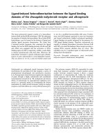
Mitochondrial protein synthesis in pet mutants showing an altered translation patternFigure 4
Mitochondrial protein synthesis in pet mutants showing an altered translation pattern. Yeast strains were grown in raffinose-containing minimal medium to
logarithmic growth phase, cytosolic translation was stopped by the addition of cycloheximide, and newly synthesized mitochondrial proteins were labeled
by the addition of
35
S methionine. After an incubation of 30 minutes at 30°C, labeling of mitochondrial proteins was stopped by the addition of cold
methionine and chloramphenicol, and cell extracts were analyzed by SDS-PAGE, transfer of proteins to nitrocellulose and autoradiography. All mutants
have been analyzed in at least three independent experiments. The samples shown here have all been analyzed on the same gel (one lane has been spliced
out as indicated by the thin line between lanes 3 and 4). For each strain the same amount of total cellular protein has been loaded per lane. Mutants that
were previously not known to be affected in the synthesis of specific mitochondria-encoded proteins are in bold letters. Alterations of the translation
pattern mentioned in the text are marked with asterisks. Black asterisks mark bands that are absent, and the white asterisk marks an additional band
present in Δcyc3. WT, wild type.
Genome Biology 2009, Volume 10, Issue 9, Article R95 Merz and Westermann R95.11
Genome Biology 2009, 10:R95
hypothesize that respiratory deficiency may be an acquired
phenotype that does not exclusively depend on the genotype.
Among ten class II mutants lacking known mitochondrial
proteins (Δcoq5, Δcoq10, Δcox10, Δcox16, Δcox19, Δmct1,
Δmss2, Δnfu1, Δslm3, and Δsom1) are four mutants that are
specifically defective in the assembly of the cytochrome c oxi-
dase (COX complex). Cox10 is required for the synthesis of
the heme A cofactor [60,61], Cox19 is a metallochaperone that
delivers copper to the COX complex [62], Mss2 is required for
the membrane translocation of the carboxyl terminus of the
mitochondria-encoded Cox2 protein [63], and Cox16 contrib-
utes to assembly of the COX complex by an as yet unknown
mechanism [64]. Intriguingly, all four of these proteins are
required for assembly of COX subunits at a post-translational
stage. While respiratory-deficiency in Δcox10, Δcox16,
Δcox19, and Δmss2 mutants has been documented before
[60,62-64], we asked whether acquired properties might con-
tribute to the loss of respiratory activity in these mutants. To
exclude effects due to differences in mtDNA copy number, we
first quantified the abundance of the mitochondrial COX3
gene by RT-PCR. We found that mtDNA is stably maintained
in Δcox10, Δcox16, Δcox19, and Δmss2 mutants at a level very
similar to wild-type cells (Figure 5a).
Next, we tried to rescue the deletion mutants with plasmids
encoding wild-type copies of the respective genes under con-
trol of their endogenous promoters. Remarkably, after
growth on selective medium a substantial number of trans-
formants remained respiratory-defective after complementa-
tion with the respective wild-type gene (Figure 5b). The
occurrence of respiratory-deficient clones was not induced by
the transformation procedure per se because transformation
of wild-type cells with the same plasmids yielded 100% respi-
ration-competent clones (not shown). In order to test
whether Δcox10,
Δcox16, Δcox19, and Δmss2 clones lose prop-
erties required for respiratory competence over time, we sub-
jected the deletion mutants to chronological aging [65], that
is, continued incubation of stationary phase cultures. Mutant
cells were incubated on glucose-containing medium for sev-
eral days at room temperature before transformation with the
complementing plasmids. Under these conditions, the frac-
tion of clones that could not be rescued increased to 60 to 81%
for mutant cells, whereas only 6% of aged wild-type clones
were observed to be respiratory-deficient after transforma-
tion (Figure 5b). This suggests that mitochondria in Δcox10,
Δcox16, Δcox19, and Δmss2 cells become irreversibly dam-
aged over time, producing a respiratory-deficient phenotype
that cannot be rescued any more. Apparently, this damage is
already induced during vegetative growth and is markedly
enhanced during aging.
As mitochondrial metabolism and aging are linked to the gen-
eration of potentially harmful reactive oxygen species (ROS)
[66] we asked whether ROS accumulate in COX assembly
mutants. High levels of ROS generated in yeast cells convert
the non-fluorescent compound dihydrorhodamine 123
(DHR) to the oxidized fluorescent chromophore rhodamine
123 [67]. Upon incubation of young wild-type Δcox10, Δcox16,
Δcox19, and Δmss2 cultures with DHR (8 h in liquid YPD
medium) only very few cells showed significant staining (Fig-
ure 5c). After continued incubation (32 h), about 60% of wild-
type cells and 90 to 98% of mutant cells showed significant
rhodamine staining (Figure 5c). Very similar results were
obtained when aging was allowed for up to 80 h (not shown).
Furthermore, we noticed that rhodamine staining in wild-
type cells was relatively faint and often restricted to tubular
structures (presumably representing the mitochondrial net-
work), whereas the signal was much stronger and dispersed
throughout the cytosol in mutant cells (Figure 5c). We con-
clude that Δcox10, Δcox16, Δcox19, and Δmss2 cells produce
elevated ROS levels during chronological aging. Presumably,
ROS induce irreversible damage to mitochondrial proteins,
lipids and/or mtDNA, thereby preventing rescue of the
mutant phenotype by transformation with complementing
plasmids. On the other hand, replenishment of fresh mito-
chondria by cytoduction might improve respiratory perform-
ance, at least for a limited time. It remains to be shown
whether accumulation of ROS-induced damage is a general
feature of class II mutants.
Novel components essential for respiratory growth
All previously uncharacterized RRG genes analyzed herein
can be clearly related to mitochondrial functions. Proteins
Rrg1, Rrg2, and Rrg5 through Rrg10 have been localized to
mitochondria by high-throughput green fluorescent protein
(GFP) fusion protein localization [35] and/or mitochondrial
proteome analysis [6,34]. The Rrg3 protein carries a putative
mitochondrial presequence, whereas the intracellular loca-
tion of Rrg4 remains unknown. Functional properties of RRG
genes are summarized in Table 5.
Δrrg1, Δrrg2, Δrrg4, Δrrg5, Δrrg6, Δrrg8, and Δrrg9 are
class I pet mutants lacking a functional mitochondrial
genome. DAPI staining revealed defects in the organization of
mtDNA that emerged early after introduction of wild-type
mitochondrial genomes by cytoduction in Δrrg1, Δrrg2,
Δrrg6, Δrrg8, and Δrrg9 mutants. Nucleoids appeared larger
compared to the wild type, the number of nucleoids per cell
was reduced, and several cells were completely devoid of
mtDNA (not shown). These observations suggest that Rrg1,
Rrg2, Rrg6, Rrg8, and Rrg9 play an important role in mainte-
nance of mtDNA. Immediate and complete loss of mtDNA
after cytoduction in the Δrrg5 mutant indicates an essential
role of Rrg5 for maintenance of mtDNA (see above).
Interestingly, Rrg2 contains a pentatricopeptide (PPR) motif.
PPR protein-encoding genes can be found in virtually all
sequenced eukaryotic genomes, but are particularly abundant
in plants. PPR proteins are localized in plastids and mito-
chondria where they are involved in the control of various
stages of gene expression [68]. Lack of mitochondrial transla-
Genome Biology 2009, Volume 10, Issue 9, Article R95 Merz and Westermann R95.12
Genome Biology 2009, 10:R95
Figure 5 (see legend on next page)
(a) (b)
(c)
Genome Biology 2009, Volume 10, Issue 9, Article R95 Merz and Westermann R95.13
Genome Biology 2009, 10:R95
tion activity and early loss of mtDNA observed here are con-
sistent with a role of Rrg2 in control of mitochondrial gene
expression.
Δrrg3 is a class III pet mutant able to maintain a [rho
+
]
genome and wild-type-like mitochondrial protein translation
activity. Although a mitochondrial location of Rrg3 has not
been shown experimentally, the Mitoprot program [69] pre-
dicts the presence of a mitochondrial presequence with a high
probability (0.9484). Mutants lacking Rrg3 (alternative name
Aim22) show an increased petite frequency [70]. The protein
has high homology to lipoate-protein ligase A family mem-
bers [71]. Thus, it is conceivable that Rrg3 mediates the
attachment of the lipoic acid cofactor to mitochondrial mul-
tienzyme complexes, such as pyruvate dehydrogenase, α-
ketoglutarate dehydrogenase, glycine decarboxylase or oth-
ers. Intriguingly, it has recently been reported that lipoate-
protein ligase activity is important for maturation of RNase P,
an enzyme that processes mitochondrial precursor tRNAs
[72]. It will be interesting to determine whether Rrg3 plays a
specific role in this process.
The Δrrg4 mutant has recently been identified as one of 86
gene deletion mutants that show an increased assembly of
Rad52, a central protein of the homologous recombination
machinery, in subnuclear foci reflecting DNA repair centers.
Therefore, the gene has been named IRC19 (for 'increased
recombination centers') [73]. Interestingly, several other
genes related to mitochondrial function were also isolated in
this screen, including CBT1, COX16, MRP17, MRPL1,
MRPS16, and YMR31. It has been suggested that an increase
of oxidative damage due to impaired respiratory chain func-
tions might stimulate spontaneous DNA lesions in the
nucleus and, therefore, constitutes a functional link between
mitochondrial respiration and DNA repair processes in the
nucleus [73]. As a Rrg4-GFP fusion protein can not be visual-
ized in cells [35], the intracellular location of Rrg4 remains
unknown.
The RRG6 gene has recently been found in a screen for com-
ponents involved in remodelling of the endoplasmic reticu-
lum (ER). It has been named HER2 (Hmg2-induced ER
remodelling); however, its molecular role in shaping the ER
membrane remained unknown [74]. As the Rrg6 protein has
been localized to mitochondria by both GFP tagging and pro-
Acquired phenotypes of COX assembly mutantsFigure 5 (see previous page)
Acquired phenotypes of COX assembly mutants. (a) Quantification of mtDNA. Yeast strains were grown overnight in liquid glucose-containing medium.
Total DNA was isolated and the copy number of the mitochondrial COX3 gene was related to that of the nuclear GAL4 gene by RT-PCR and calculation of
the 2
-ΔΔC
T
value. Error bars indicate standard deviations of triplicate PCR reactions. (b) Complementation test. Δcox10, Δcox16, Δcox19, and Δmss2 strains
taken from the MATα yeast deletion library have been transformed with single copy plasmids carrying the respective complementing wild-type alleles
under control of their endogenous promoters. Wild-type cells (WT) were transformed with an empty vector. Young cells were grown on complete
medium at 30°C overnight before transformation (light bars). Aged cells were incubated on complete medium at room temperature for 14 to 28 days
before they were transferred to fresh plates, grown at 30°C overnight, and transformed with complementing plasmids (dark bars). Three days after
transformation, colonies were replicated on plates containing fermentable or non-fermentable carbon sources, and the percentage of respiratory-deficient
transformants was determined. Error bars indicate standard deviations of three independent experiments. (c) ROS accumulation. Yeast strains were
grown for the indicated time periods in liquid glucose-containing medium (YPD), stained by the addition of DHR and analyzed by differential interference
microscopy (left panels) and fluorescence microscopy (right panels). All fluorescent micrographs were taken with identical camera settings.
Table 5
Functional properties of newly described RRG genes
Systematic name Mitochondrial
localization
Δmip1 mating Cytoduction Nucleoids after
cytoduction
Translation activity
after cytoduction
RRG1 YDR065w [34] - - Altered Absent
RRG2 YGR150c [34,35] - - Altered Absent
RRG3 (AIM22) YJL046w [69] + - WT WT
RRG4 (IRC19) YLL033w Unknown - - WT WT
RRG5 (GEP5) YLR091w [6,34,35] - - Absent Absent
RRG6 (HER2) YMR293c [6,34,35] - - Altered Absent
RRG7 YOR305w [35] + + ND ND
RRG8 YPR116w [35] - - Altered Absent
RRG9 YNL213c [34] - - Altered WT
RRG10 YJL062w-a [34,35] + - WT Altered
References refer to published evidence of mitochondrial localization of Rrg proteins; + indicates rescue by mating with Δmip1 or cytoduction,
respectively. WT, wild type-like; ND, not determined. See text for details.
Genome Biology 2009, Volume 10, Issue 9, Article R95 Merz and Westermann R95.14
Genome Biology 2009, 10:R95
teome analysis [6,34,35], we propose that its primary func-
tion is related to maintenance of respiratory activity. The
protein is highly homologous to bacterial glutamyl-tRNA
amidotransferases, and a role in mitochondrial protein syn-
thesis is consistent with our observation that mitochondrial
translation is blocked in the Δrrg6 mutant (Table 3).
Recently, RRG5 (alternative name GEP5) and RRG6 (alterna-
tive names GEP6 or HER2) have been shown to genetically
interact with genes encoding prohibitin ring complexes in the
mitochondrial inner membrane [75]; however, the functional
significance of this interaction is not yet understood.
Δrrg7 is a class II pet mutant presumably acquiring respira-
tory deficiency independent of its genotype. The RRG7 gene
encodes a mitochondrial protein [35] that has homologs in
fungi and other lower eukaryotes. Its function in mitochon-
drial biogenesis is currently unknown; however, the deletion
mutant has been reported to exhibit increased sensitivity to
the synthetic tripeptide arsenical 4-(N-(S-glutathiony-
lacetyl)amino) phenylarsenoxide that targets mitochondria
by inactivating the adenine nucleotide translocator. This drug
inhibits proliferation of actively dividing endothelial cells and
is an inhibitor of angiogenesis during tumor formation [76].
Δrrg10 is a class III pet mutant able to maintain a [rho
+
]
genome. The RRG10 gene encodes a small mitochondrial pro-
tein [34,35] of only 85 amino acid residues. Analysis of the
mitochondrial translation pattern revealed a reduction of
Cox1, suggesting that Rrg10 plays a specific role in transcrip-
tion or maturation of mitochondrial mRNAs and/or transla-
tion or assembly of mitochondrial gene products.
Conclusions
Surprisingly, only a limited number of mutants reproducibly
show a pet phenotype when different versions of the yeast
deletion library are screened for growth on non-fermentable
carbon sources. While some differences can be ascribed to
wrong deletions present in the library, most of the variations
are likely due to intrinsic properties of the mutant strains. We
present four lines of evidence suggesting that the plasticity of
pet phenotypes is much greater than previously anticipated.
First, several deletions produce different phenotypes in dif-
ferent versions of the deletion library (Figure 1a, b; Additional
data file 3); second, a number of mutants lose mtDNA at a
high rate upon continued incubation in glucose-containing
medium (Additional data file 6); third, respiratory deficiency
can be reversed by relief of catabolite repression in a relatively
large number of mutants (Additional data file 7); and fourth,
several [rho
+
] pet mutants accumulate irreversible damage
resulting in an improvement of respiratory performance after
cytoduction (Figure 5; Additional data file 9). It is a challenge
for the future to examine further contributions of environ-
mental factors, nutrient supply, and possible epigenetic
mechanisms to phenotypic plasticity.
Comparative gene deletion analysis enabled us to define by
stringent criteria a set of 163 protein-coding genes (13 dubi-
ous ORFs subtracted from the 176 mutants found in all pet
screens of the library) that are obligatorily required for respi-
ratory metabolism in yeast. These include ten largely unchar-
acterized genes, RRG1 through RRG10. Remarkably, almost
all of these highly penetrant mutants (95%) have been
reported to show decreased fitness on non-fermentable car-
bon sources when the whole-genome pool of yeast deletion
mutants was analyzed [77,78]. While the approach pursued
by Steinmetz et al. [77] resulted in a relatively large set of
genes potentially required for respiratory growth (466 genes,
43.1% of which encode mitochondrial proteins), the compar-
ative gene deletion approach pursued here apparently is more
selective (176 genes, 73.3% of which encode mitochondrial
proteins). A high resolution of our comparative gene deletion
analysis is also apparent from a comparison with the results
we obtained after our first screen of the deletion library
reported in the Dimmer et al. study [14], which yielded 341
pet genes (only 54.8% of which encode mitochondrial pro-
teins). Our present work suggests that 165 of these originally
identified mutants do not reproducibly give rise to a pet phe-
notype and should be considered as important but not essen-
tial for respiratory growth of yeast. Thus, Figure 1c gives a
significantly improved representation of cellular functions of
genes essential for respiration. A recent study by Hess et al.
[70] reports a computational prediction of 193 candidate
genes and subsequent analysis of their possible roles in mito-
chondrial biogenesis. They found that Δrrg2 and Δrrg6
mutants are respiratory deficient, and that the Δrrg3 mutant
shows an increased petite frequency [70]. However, the
remaining seven RRG genes were only found by the compar-
ative gene deletion analysis described here, demonstrating
the value of our approach.
The systematic functional analysis of pet mutants reported
here uncovered roles of 8 novel components in mtDNA main-
tenance, 30 novel components in mitochondrial protein syn-
thesis, and 4 novel components in expression of specific
mitochondrial translation products. We suggest that these
data may serve as positive lists for genes important for respi-
ratory growth, mtDNA maintenance and mitochondrial pro-
tein synthesis. It should be pointed out that components
might have been missed that are encoded by redundant genes
or that are not correct in the deletion library. Furthermore,
some genes might be specifically required only under certain
growth conditions or in certain genetic backgrounds. While a
mechanistic understanding of the molecular processes con-
tributing to respiratory activity will require further rigorous
experimentation, the systematic large-scale functional analy-
sis of pet mutants reported here is a first step towards a defi-
nition of the complements of genes required for maintenance
of the mitochondrial genome and mitochondrial protein
translation. Together with integrated analyses of different
genomic and proteomic approaches [78], combination of
computational approaches with quantitative experimentation
Genome Biology 2009, Volume 10, Issue 9, Article R95 Merz and Westermann R95.15
Genome Biology 2009, 10:R95
[70], and the construction of protein interaction networks
[79] it will contribute to an understanding of the systems
properties of mitochondria with steadily increasing resolu-
tion.
Materials and methods
Yeast strains and plasmids
Yeast strains used in this study were isogenic to BY4741,
BY4742 and BY4743 [18], with the exception of strain J1361
[80], which was used for cytoduction. The [rho
0
] cytoduction
donor strain was generated by growth of J1361 overnight in
YPD medium supplemented with 50 μg/ml ethidium bro-
mide. Complete loss of mtDNA was controlled by DAPI stain-
ing. The MATα gene deletion library [16] and its supplement
covering newly assigned small ORFs [21] was obtained from
BioCat (Heidelberg, Germany), and MATa single mutant
Δmip1 was obtained from EUROSCARF (Frankfurt, Ger-
many). Plasmid pRS416/MSS2 was constructed by PCR
amplification of the MSS2 gene using primers 5' AAA GGA
TCC GAT TTT ATG TGT GGA ATG CTA ACG ATG AAC and 5'
AAA CTC GAG CTC TAA CAG TAT TTC CTA ATT ATT TCA
TAG GTA AC and subcloning into the BamHI and XhoI sites
of vector pRS416 [81]. Plasmid pRS416/COX16 was con-
structed by PCR amplification of the COX16 gene using prim-
ers 5' AAA GGA TCC AAT ATT ACC GTG AAT ATC GCG AGC
TAC and 5' AAA CTC GAG AGG TAT TTA CAA TCA TTT CCT
AGA CAT TCT and subcloning into the BamHI and XhoI sites
of vector pRS416. For complementation tests, yeast strains
were transformed with plasmids pG12/T4 [60] expressing
COX10, pRS416/COX16, pG188/T1 [62] expressing COX19,
or pRS416/MSS2.
PCR analyses to confirm the identity of deletion mutants were
performed in a way that one primer was homologous to a
sequence within the coding region or within the deletion
marker cassette, respectively, whereas the other primer was
homologous to a sequence outside the deleted part of the
gene. Thus, a PCR product can be generated only if the correct
allele corresponding to the primer combination is present.
Primers used to confirm the identity of yeast deletion mutants
are listed in Additional data file 10.
Yeast genetic methods
S. cerevisiae was cultivated and manipulated according to
standard procedures [82]. For screening for respiratory-defi-
cient mutants, yeast deletion strains were manually trans-
ferred with a sterile pinning tool from 96-well plates to rich
media plates with either 2% glucose as fermentable (YPD) or
3% glycerol as non-fermentable (YPG) carbon source. The
screening of the entire library on YPG was performed once.
The screening procedure was as similar as possible to the
screen performed earlier by us in the Dimmer et al. study
[14]. Respiratory-deficient mutants were screened in addition
on media containing 3% ethanol or 3% lactate (pH adjusted to
7.0 with NaOH) as non-fermentable carbon sources. The
growth behavior was evaluated by visual inspection after 3
days (YPD) or 6 days (YPG and other non-fermentable media)
of incubation at 30°C.
For high-throughput complementation tests with Δmip1,
yeast deletion strains were transferred with a sterile pinning
tool from 96-well plates to a lawn of MATa Δmip1 cells on
YPD plates. After incubation overnight to allow for mating,
cells were replica-plated two times on plates containing min-
imal SD medium selective for diploid cells. Then, growth on
YPG plates was determined as above. Cytoduction was per-
formed as described [80]. Cytoduction experiments were
repeated at least three times. To adapt yeast deletion strains
to non-fermentable carbon sources, cells were transferred
from YPD plates to YPG plates containing 0.1% glucose, rep-
lica-plated once on YPG/0.1% glucose and then replica-plated
on YPG.
Analysis of mitochondrial translation products
Labeling of mitochondrial translation products in vivo was
performed essentially as described [83] with the following
minor modifications: cytosolic translation was stopped with
0.3 mg/ml cycloheximide, the labeling reaction was per-
formed for 30 minutes, and the chase reaction was performed
for 15 minutes. Mitochondrial translation products were ana-
lyzed by SDS-PAGE, transfer to nitrocellulose and autoradi-
ography.
Assay of accumulation of irreversible damage during
chronological aging
Using sterile toothpicks, cells were taken from glycerol stocks
and spread on YPD plates as patches of about 2 cm
2
size.
Plates were incubated for 14 to 28 days at room temperature
to allow chronological aging. Then, a small amount of aged
cells (or young cells taken directly from glycerol stocks as a
control) was spread on a fresh YPD plate, incubated overnight
at 30°C, and transformed according to the rapid transforma-
tion protocol described by Truong and Gietz [84]. Using ster-
ile toothpicks, at least 100 transformants were transferred as
short streaks (approximately 1 cm) to fresh SD plates selective
for the marker of the transformed plasmids. Plates were incu-
bated for 1 day at 30°C and then replica-plated on YPD and
YPG using sterile velvet. Carbon source-dependent growth of
transformants was visually scored after 2 to 3 days at 30°C.
Staining of mtDNA, DHR staining and microscopy
Staining of mtDNA with DAPI in methanol-fixed cells was as
described [30]. For the analysis of ROS production, 1 μl DHR
(2.5 mg/ml in DMSO) was added to 500 μl cell suspension
and incubated for 2 h at 30°C. Cells were harvested by centrif-
ugation, washed in phosphate-buffered saline, resuspended
in phosphate-buffered saline and analyzed by microscopy.
Epifluorescence microscopy was performed using a Zeiss Axi-
oplan 2 microscope equipped with a HBO 100 mercury lamp,
Zeiss filter sets 01 and 09 and a Plan-Neofluar 100× 1.30 NA
Ph3 oil objective (Carl Zeiss Lichtmikroskopie, Göttingen,
Genome Biology 2009, Volume 10, Issue 9, Article R95 Merz and Westermann R95.16
Genome Biology 2009, 10:R95
Germany). Images were recorded with an Evolution VF Mono
Cooled monochrome camera (Intas, Göttingen, Germany)
and processed with Image Pro Plus 5.0 and Scope Pro 4.5
software (MediaCybernetics, Silver Springs, MD, USA).
RT-PCR
Yeast strains were grown overnight in liquid YPD medium
and DNA was extracted using the YeaStar™ Genomic DNA
Kit (Zymo Research, Orange, USA) according to the manufac-
turer's instructions. PCR reactions were performed in 20 μl
volume in 96-well plates using Maxima™ SYBR Green qPCR
Master Mix (2×) (Fermentas, St Leon-Rot, Germany) accord-
ing to the manufacturer's instructions in an ABI PRISM 7000
Sequence Detection System (Applied Biosystems, Foster City,
CA, USA). The following primers were used: GAL4 forward, 5'
TTT CTC CTG GCT CAG TAG GGC; GAL4 reverse, 5' AGT
TAC GAG AGG GTG GAC GGT; COX3 forward, 5' ATT GAA
GCT GTA CAA CCT ACC GAA TT; COX3 reverse, 5' CCT GCG
ATT AAG GCA TGA TGA. Data were analyzed with Sequence
Detection Software Version 1.2.3 7000 System SDS software
Core Application (Applied Biosystems) and calculated
according to the 2
-ΔΔC
T
-method [85].
Abbreviations
COX: cytochrome c oxidase; DAPI: 4',6-diamidino-2-phe-
nylindole; DHR: dihydrorhodamine 123; ER: endoplasmic
reticulum; GFP: green fluorescent protein; mtDNA: mito-
chondrial DNA; ORF: open reading frame; PPR: pentatr-
icopeptide; ROS: reactive oxygen species.
Authors' contributions
SM performed the experiments, SM and BW conceived the
study, analyzed the data and wrote the manuscript.
Additional data files
The following additional data are available with the online
version of this paper: a table listing pet genes isolated from
the MATa deletion library (Additional data file 1); a table list-
ing pet genes unique to this study (Additional data file 2); a
table listing pet genes grouped according to their occurrence
in pet screens and localization and function of the encoded
gene products (Additional data file 3); a table listing pet genes
producing growth defects only on specific carbon sources
(Additional data file 4); a table listing mutants belonging to
four classes of pet genes (Additional data file 5); a table show-
ing quantification of loss of mtDNA in class IV pet mutants
(Additional data file 6); a table listing pet genes dispensable
for respiration (Additional data file 7); a table listing genes
required for respiratory activity in class I and III pet mutants
that show a wild-type pattern of mitochondrial translation
products (Additional data file 8); a table listing genes possibly
affecting mitochondrial function in combination with
acquired defects (Additional data file 9); a table listing prim-
ers used to confirm the identity of yeast deletion mutants
(Additional data file 10).
Additional data file 1pet genes isolated from the MATα deletion librarypet genes isolated from the MATα deletion library.Click here for fileAdditional data file 2pet genes unique to this studypet genes unique to this study.Click here for fileAdditional data file 3pet genes grouped according to their occurrence in pet screens and localization and function of the encoded gene productspet genes grouped according to their occurrence in pet screens and localization and function of the encoded gene products.Click here for fileAdditional data file 4pet genes producing growth defects only on specific carbon sourcespet genes producing growth defects only on specific carbon sources.Click here for fileAdditional data file 5Mutants belonging to four classes of pet genesMutants belonging to four classes of pet genes.Click here for fileAdditional data file 6Quantification of loss of mtDNA in class IV pet mutantsQuantification of loss of mtDNA in class IV pet mutants.Click here for fileAdditional data file 7pet genes dispensable for respirationpet genes dispensable for respiration.Click here for fileAdditional data file 8Genes required for respiratory activity in class I and III pet mutants that show a wild-type pattern of mitochondrial translation prod-uctsGenes required for respiratory activity in class I and III pet mutants that show a wild-type pattern of mitochondrial translation prod-ucts.Click here for fileAdditional data file 9Genes possibly affecting mitochondrial function in combination with acquired defectsGenes possibly affecting mitochondrial function in combination with acquired defects.Click here for fileAdditional data file 10Primers used to confirm the identity of yeast deletion mutantsPrimers used to confirm the identity of yeast deletion mutants.Click here for file
Acknowledgements
This work was supported by the Deutsche Forschungsgemeinschaft. We
thank Robert JD Reid for making yeast strain J1361 available to us, Alexan-
der Tzagoloff for plasmids pG12/T4 and pG188/T1, Alexander Kastaniotis
and Johannes Herrmann for helpful discussions, and Melanie Krist for her
contributions to some experiments.
References
1. Saraste M: Oxidative phosphorylation at the fin de siècle. Sci-
ence 1999, 283:1488-1493.
2. Lane N: Power, Sex, Suicide. Mitochondria and the Meaning of Life
Oxford, UK: Oxford University Pressa; 2005.
3. de Vries S, Van Witzenburg R, Grivell LA, Marres CA: Primary
structure and import pathway of the rotenone-insensitive
NADH-ubiquinone oxidoreductase of mitochondria from
Saccharomyces cerevisiae. Eur J Biochem 1992, 203:587-592.
4. Marres CA, de Vries S, Grivell LA: Isolation and inactivation of
the nuclear gene encoding the rotenone-insensitive internal
NADH: ubiquinone oxidoreductase of mitochondria from
Saccharomyces cerevisiae. Eur J Biochem 1991, 195:857-862.
5. Gray MW, Burger G, Lang BF: Mitochondrial evolution. Science
1999, 283:1476-1481.
6. Sickmann A, Reinders J, Wagner Y, Joppich C, Zahedi R, Meyer HE,
Schönfisch B, Perschil I, Chacinska A, Guiard B, Rehling P, Pfanner N,
Meisinger C: The proteome of Saccharomyces cerevisiae mito-
chondria. Proc Natl Acad Sci USA 2003, 100:13207-13212.
7. Grivell LA, Artal-Sanz M, Hakkaart G, de Jong L, Nijtmans LG, van
Oosterum K, Siep M, Spek H van der: Mitochondrial assembly in
yeast. FEBS Lett 1999, 452:57-60.
8. Gancedo JM: Yeast carbon catabolite repression. Microbiol Mol
Biol Rev 1998, 62:334-361.
9. Piskur J, Rozpedowska E, Polakova S, Merico A, Compagno C: How
did Saccharomyces evolve to become a good brewer? Trends
Genet 2006, 22:183-186.
10. Johnston M: Feasting, fasting and fermenting. Glucose sensing
in yeast and other cells. Trends Genet 1999, 15:29-33.
11. Ephrussi B, Hottinguer H, Tavlitzki J: Action de l'acriflavine sur les
levures II. Étude génétique du mutant "petite colonie". Ann
Inst Pasteur 1949,
76:419-442.
12. Tzagoloff A, Dieckmann CL: PET genes of Saccharomyces cerevi-
siae. Microbiol Rev 1990, 54:211-225.
13. Contamine V, Picard M: Maintenance and integrity of the mito-
chondrial genome: a plethora of nuclear genes in the bud-
ding yeast. Microbiol Mol Biol Rev 2000, 64:281-315.
14. Dimmer KS, Fritz S, Fuchs F, Messerschmitt M, Weinbach N, Neupert
W, Westermann B: Genetic basis of mitochondrial function
and morphology in Saccharomyces cerevisiae. Mol Biol Cell 2002,
13:847-853.
15. Bolotin-Fukuhara M, Grivell LA: Genetic approaches to the study
of mitochondrial biogenesis in yeast. Antonie Van Leeuwenhoek
1992, 62:131-153.
16. Giaever G, Chu AM, Ni L, Connelly C, Riles L, Veronneau S, Dow S,
Lucau-Danila A, Anderson K, Andre B, Arkin AP, Astromoff A, Bakk-
oury ME, Bangham R, Benito R, Brachat S, Campanaro S, Curtiss M,
Davis K, Deutschbauer A, Entian KD, Flaherty P, Foury F, Garfinkel
DJ, Gerstein M, Gotte D, Güldener U, Hegemann JH, Hempel S, Her-
man Z, et al.: Functional profiling of the Saccharomyces cerevi-
siae genome. Nature 2002, 418:387-391.
17. Luban C, Beutel M, Stahl U, Schmidt U: Systematic screening of
nuclear encoded proteins involved in the splicing metabo-
lism of group II introns in yeast mitochondria. Gene 2005,
354:72-79.
18. Brachmann CB, Davies A, Cost GJ, Caputo E, Li J, Hieter P, Boeke JD:
Designer deletion strains derived from Saccharomyces cerevi-
siae S288C: a useful set of strains and plasmids for PCR-
mediated gene disruption and other applications. Yeast 1998,
14:115-132.
19. Issel-Tarver L, Christie KR, Dolinski K, Andrada R, Balakrishnan R,
Ball CA, Binkley G, Dong S, Dwight SS, Fisk DG, Harris M, Schroeder
M, Sethuraman A, Tse K, Weng S, Botstein D, Cherry JM: Saccharo-
Genome Biology 2009, Volume 10, Issue 9, Article R95 Merz and Westermann R95.17
Genome Biology 2009, 10:R95
myces Genome Database. Methods Enzymol 2002, 350:329-346.
20. Wysocki R, Roganti T, Van Dyck E, de Kerchove D'Exaerde A, Foury
F: Disruption and basic phenotypic analysis of 18 novel genes
from the yeast Saccharomyces cerevisiae. Yeast 1999,
15:165-171.
21. Kastenmayer JP, Ni L, Chu A, Kitchen LE, Au WC, Yang H, Carter
CD, Wheeler D, Davis RW, Boeke JD, Snyder MA, Basrai MA: Func-
tional genomics of genes with small open reading frames
(sORFs) in S. cerevisiae. Genome Res 2006, 16:365-373.
22. Foury F: Cloning and sequencing of the nuclear gene MIP1
encoding the catalytic subunit of the yeast mitochondrial
DNA polymerase. J Biol Chem 1989, 264:20552-20560.
23. Sedman T, Kuusk S, Kivi S, Sedman J: A DNA helicase required for
maintenance of the functional mitochondrial genome in Sac-
charomyces cerevisiae. Mol Cell Biol 2000, 20:1816-1824.
24. Lahaye A, Stahl H, Thines-Sempoux D, Foury F: PIF1: a DNA heli-
case in yeast mitochondria. EMBO J 1991, 10:997-1007.
25. Vongsamphanh R, Fortier PK, Ramotar D: Pir1p mediates translo-
cation of the yeast Apn1p endonuclease into the mitochon-
dria to maintain genomic stability. Mol Cell Biol 2001,
21:1647-1655.
26. Chen XJ, Wang X, Kaufman BA, Butow RA: Aconitase couples
metabolic regulation to mitochondrial DNA maintenance.
Science 2005, 307:714-717.
27. Myers AM, Pape LK, Tzagoloff A: Mitochondrial protein synthesis
is required for maintenance of intact mitochondrial
genomes in Saccharomyces cerevisiae.
EMBO J 1985,
4:2087-2092.
28. Paul MF, Velours J, Arselin de Chateaubodeau G, Aigle M, Guerin B:
The role of subunit 4, a nuclear-encoded protein of the F
0
sector of yeast mitochondrial ATP synthase, in the assembly
of the whole complex. Eur J Biochem 1989, 185:163-171.
29. Duvezin-Caubet S, Rak M, Lefebvre-Legendre L, Tetaud E, Bonnefoy
N, di Rago JP: A "petite obligate" mutant of Saccharomyces cer-
evisiae : functional mtDNA is lethal in cells lacking the delta
subunit of mitochondrial F
1
-ATPase. J Biol Chem 2006,
281:16305-16313.
30. Jones BA, Fangman WL: Mitochondrial DNA maintenance in
yeast requires a protein containing a region related to the
GTP-binding domain of dynamin. Genes Dev 1992, 6:380-389.
31. Merz S, Hammermeister M, Altmann K, Dürr M, Westermann B:
Molecular machinery of mitochondrial dynamics in yeast.
Biol Chem 2007, 388:917-926.
32. Wong ED, Wagner JA, Gorsich SW, McCaffery JM, Shaw JM, Nunnari
J: The dynamin-related GTPase, Mgm1p, is an intermem-
brane space protein required for maintenance of fusion com-
petent mitochondria. J Cell Biol 2000, 151:341-352.
33. Carroll CW, Morgan DO: The Doc1 subunit is a processivity
factor for the anaphase-promoting complex. Nat Cell Biol 2002,
4:880-887.
34. Reinders J, Zahedi RP, Pfanner N, Meisinger C, Sickmann A: Toward
the complete yeast mitochondrial proteome: multidimen-
sional separation techniques for mitochondrial proteomics.
J Proteome Res 2006, 5:1543-1554.
35. Huh WK, Falvo JV, Gerke LC, Carroll AS, Howson RW, Weissman
JS, O'Shea EK: Global analysis of protein localization in bud-
ding yeast. Nature 2003, 425:686-691.
36. Dimmer KS, Jakobs S, Vogel F, Altmann K, Westermann B: Mdm31
and Mdm32 are inner membrane proteins required for
maintenance of mitochondrial shape and stability of mito-
chondrial DNA nucleoids in yeast. J Cell Biol 2005, 168:103-115.
37. Towpik J: Regulation of mitochondrial translation in yeast.
Cell Mol Biol Lett 2005, 10:571-594.
38. Rapaport D, Brunner M, Neupert W, Westermann B: Fzo1p is a
mitochondrial outer membrane protein essential for the
biogenesis of functional mitochondria in Saccharomyces cere-
visiae. J Biol Chem 1998, 273:20150-20155.
39. Hermann GJ, Thatcher JW, Mills JP, Hales KG, Fuller MT, Nunnari J,
Shaw JM: Mitochondrial fusion in yeast requires the trans-
membrane GTPase Fzo1p. J Cell Biol
1998, 143:359-373.
40. Ling F, Shibata T: Recombination-dependent mtDNA parti-
tioning: in vivo role of Mhr1p to promote pairing of homolo-
gous DNA. EMBO J 2002, 21:4730-4740.
41. Reenan RA, Kolodner RD: Characterization of insertion muta-
tions in the Saccharomyces cerevisiae MSH1 and MSH2 genes:
evidence for separate mitochondrial and nuclear functions.
Genetics 1992, 132:975-985.
42. Meeusen S, Tieu Q, Wong E, Weiss E, Schieltz D, Yates JR, Nunnari
J: Mgm101p is a novel component of the mitochondrial
nucleoid that binds DNA and is required for the repair of oxi-
datively damaged mitochondrial DNA. J Cell Biol 1999,
145:291-304.
43. Zambrano A, Fontanesi F, Solans A, de Oliveira RL, Fox TD, Tzagoloff
A, Barrientos A: Aberrant translation of cytochrome c oxidase
subunit 1 mRNA species in the absence of Mss51p in the
yeast Saccharomyces cerevisiae. Mol Biol Cell 2007, 18:523-535.
44. Ackerman SH, Gatti DL, Gellefors P, Douglas MG, Tzagoloff A:
ATP13, a nuclear gene of Saccharomyces cerevisiae essential
for the expression of subunit 9 of the mitochondrial ATPase.
FEBS Lett 1991, 278:234-238.
45. Rödel G: Two yeast nuclear genes, CBS1 and CBS2, are
required for translation of mitochondrial transcripts bearing
the 5'-untranslated COB leader. Curr Genet 1986, 11:41-45.
46. Kreike J, Schulze M, Pillar T, Korte A, Rödel G: Cloning of a nuclear
gene MRS1 involved in the excision of a single group I intron
(bI3) from the mitochondrial COB transcript in S. cerevisiae.
Curr Genet 1986, 11:185-191.
47. Perez-Martinez X, Broadley SA, Fox TD: Mss51p promotes mito-
chondrial Cox1p synthesis and interacts with newly synthe-
sized Cox1p.
EMBO J 2003, 22:5951-5961.
48. Costanzo MC, Seaver EC, Fox TD: The PET54 gene of Saccharo-
myces cerevisiae: characterization of a nuclear gene encoding
a mitochondrial translational activator and subcellular local-
ization of its product. Genetics 1989, 122:297-305.
49. Valencik ML, Kloeckener-Gruissem B, Poyton RO, McEwen JE: Dis-
ruption of the yeast nuclear PET54 gene blocks excision of
mitochondrial intron aI5 beta from pre-mRNA for cyto-
chrome c oxidase subunit I. EMBO J 1989, 8:3899-3904.
50. Costanzo MC, Fox TD: Product of Saccharomyces cerevisiae
nuclear gene PET494 activates translation of a specific mito-
chondrial mRNA. Mol Cell Biol 1986, 6:3694-3703.
51. Marbois B, Gin P, Faull KF, Poon WW, Lee PT, Strahan J, Shepherd
JN, Clarke CF: Coq3 and Coq4 define a polypeptide complex
in yeast mitochondria for the biosynthesis of coenzyme Q. J
Biol Chem 2005, 280:20231-20238.
52. Dumont ME, Ernst JF, Hampsey DM, Sherman F: Identification and
sequence of the gene encoding cytochrome c heme lyase in
the yeast Saccharomyces cerevisiae. EMBO J 1987, 6:235-241.
53. Graham LA, Hill KJ, Stevens TH: VMA8 encodes a 32-kDa V1 sub-
unit of the Saccharomyces cerevisiae vacuolar H(+)-ATPase
required for function and assembly of the enzyme complex.
J Biol Chem 1995, 270:15037-15044.
54. Klionsky DJ, Herman PK, Emr SD: The fungal vacuole: composi-
tion, function, and biogenesis. Microbiol Rev 1990, 54:266-292.
55. Kane PM: The where, when, and how of organelle acidification
by the yeast vacuolar H+-ATPase. Microbiol Mol Biol Rev 2006,
70:177-191.
56. Kane PM: The long physiological reach of the yeast vacuolar
H
+
-ATPase. J Bioenerg Biomembr 2007, 39:415-421.
57. Thorpe GW, Fong CS, Alic N, Higgins VJ, Dawes IW: Cells have dis-
tinct mechanisms to maintain protection against different
reactive oxygen species: oxidative-stress-response genes.
Proc Natl Acad Sci USA 2004, 101:6564-6569.
58. Milgrom E, Diab H, Middleton F, Kane PM: Loss of vacuolar pro-
ton-translocating ATPase activity in yeast results in chronic
oxidative stress. J Biol Chem 2007, 282:7125-7136.
59. Kissova I, Salin B, Schaeffer J, Bhatia S, Manon S, Camougrand N:
Selective and non-selective autophagic degradation of mito-
chondria in yeast. Autophagy 2007, 3:329-336.
60. Nobrega MP, Nobrega FG, Tzagoloff A: COX10 codes for a pro-
tein homologous to the ORF1 product of Paracoccus denitri-
ficans and is required for the synthesis of yeast cytochrome
oxidase. J Biol Chem 1990, 265:14220-14226.
61. Tzagoloff A, Nobrega M, Gorman N, Sinclair P: On the functions of
the yeast COX10 and COX11 gene products. Biochem Mol Biol
Int 1993, 31:593-598.
62. Nobrega MP, Bandeira SC, Beers J, Tzagoloff A: Characterization
of COX19, a widely distributed gene required for expression
of mitochondrial cytochrome oxidase. J Biol Chem 2002,
277:40206-40211.
63. Broadley SA, Demlow CM, Fox TD: Peripheral mitochondrial
inner membrane protein, Mss2p, required for export of the
mitochondrially coded Cox2p C tail in Saccharomyces cerevi-
siae. Mol Cell Biol 2001, 21:7663-7672.
64. Carlson CG, Barrientos A, Tzagoloff A, Glerum DM: COX16
encodes a novel protein required for the assembly of cyto-
Genome Biology 2009, Volume 10, Issue 9, Article R95 Merz and Westermann R95.18
Genome Biology 2009, 10:R95
chrome oxidase in Saccharomyces cerevisiae. J Biol Chem 2003,
278:3770-3775.
65. Kaeberlein M, Burtner CR, Kennedy BK: Recent developments in
yeast aging. PLoS Genet 2007, 3:e84.
66. Balaban RS, Nemoto S, Finkel T: Mitochondria, oxidants, and
aging. Cell 2005, 120:483-495.
67. Madeo F, Fröhlich E, Ligr M, Grey M, Sigrist SJ, Wolf DH, Fröhlich KU:
Oxygen stress: a regulator of apoptosis in yeast. J Cell Biol
1999, 145:757-767.
68. Andrés C, Lurin C, Small ID: The multifarious roles of PPR pro-
teins in plant mitochondrial gene expression. Physiol Plant
2007, 129:14-22.
69. Claros MG, Vincens P: Computational method to predict mito-
chondrially imported proteins and their targeting
sequences. Eur J Biochem 1996, 241:779-786.
70. Hess DC, Myers CL, Huttenhower C, Hibbs MA, Hayes AP, Paw J,
Clore JJ, Mendoza RM, Luis BS, Nislow C, Giaever G, Costanzo M,
Troyanskaya OG, Caudy AA: Computationally driven, quantita-
tive experiments discover genes required for mitochondrial
biogenesis. PLoS Genet 2009, 5:e1000407.
71. Morris TW, Reed KE, Cronan JEJ: Identification of the gene
encoding lipoate-protein ligase A of Escherichia coli. J Biol
Chem 1994, 269:16091-16100.
72. Schonauer MS, Kastaniotis AJ, Hiltunen JK, Dieckmann CL: Intersec-
tion of RNA processing and the type II fatty acid synthesis
pathway in yeast mitochondria. Mol Cell Biol 2008,
28:6646-6657.
73. Alvaro D, Lisby M, Rothstein R: Genome-wide analysis of Rad52
foci reveals diverse mechanisms impacting recombination.
PLoS Genet 2007, 3:e228.
74. Federovitch CM, Jones YZ, Tong AH, Boone C, Prinz WA, Hampton
RY:
Genetic and structural analysis of Hmg2p-induced ER
remodeling in S. cerevisiae. Mol Biol Cell 2008, 19:4506-4520.
75. Osman C, Haag M, Potting C, Rodenfels J, Dip PV, Wieland FT, Brüg-
ger B, Westermann B, Langer T: The genetic interactome of pro-
hibitins links their function to cardiolipin and
phosphatidylethanolamine in mitochondria. J Cell Biol 2009,
184:583-596.
76. Dilda PJ, Don AS, Tanabe KM, Higgins VJ, Allen JD, Dawes IW, Hogg
PJ: Mechanism of selectivity of an angiogenesis inhibitor from
screening a genome-wide set of Saccharomyces cerevisiae
deletion strains. J Natl Cancer Inst 2005, 97:1539-1547.
77. Steinmetz LM, Scharfe C, Deutschbauer AM, Mokranjac D, Herman
ZS, Jones T, Chu AM, Giaever G, Prokisch H, Oefner PJ, Davis RW:
Systematic screen for human disease genes in yeast. Nat
Genet 2002, 31:400-404.
78. Prokisch H, Scharfe C, Camp DG 2nd, Xiao W, David L, Andreoli C,
Monroe ME, Moore RJ, Gritsenko MA, Kozany C, Hixson KK, Mottaz
HM, Zischka H, Ueffing M, Herman ZS, Davis RW, Meitinger T, Oef-
ner PJ, Smith RD, Steinmetz LM: Integrative analysis of the mito-
chondrial proteome in yeast. PLoS Biol 2004, 2:e160.
79. Perocchi F, Jensen LJ, Gagneur J, Ahting U, von Mering C, Bork P,
Prokisch H, Steinmetz LM: Assessing systems properties of
yeast mitochondria through an interaction map of the
organelle. PLoS Genet 2006, 2:e170.
80. Lettier G, Feng Q, de Mayolo AA, Erdeniz N, Reid RJ, Lisby M,
Mortensen UH, Rothstein R: The role of DNA double-strand
breaks in spontaneous homologous recombination in S. cer-
evisiae. PLoS Genet 2006, 2:e194.
81. Sikorski RS, Hieter P: A system of shuttle vectors and host
strains designed for efficient manipulation of DNA in Saccha-
romyces cerevisiae. Genetics 1989, 122:19-27.
82. Sherman F: Getting started with yeast. Methods Enzymol 1991,
194:3-21.
83. Westermann B, Herrmann JM, Neupert W: Analysis of mitochon-
drial translation products in vivo
and in organello in yeast.
Methods Cell Biol 2001, 65:429-438.
84. Truong D, Gietz R: Yeast transformation. Methods Microbiol 2007,
36:45-54.
85. Livak KJ, Schmittgen TD: Analysis of relative gene expression
data using real-time quantitative PCR and the 2(-Delta Delta
C(T)) Method. Methods 2001, 25:402-408.