Báo cáo y học: " Immunity and other defenses in pea aphids, Acyrthosiphon pisum" ppt
Bạn đang xem bản rút gọn của tài liệu. Xem và tải ngay bản đầy đủ của tài liệu tại đây (1.54 MB, 17 trang )
RESEA R C H Open Access
Immunity and other defenses in pea aphids,
Acyrthosiphon pisum
Nicole M Gerardo
1*
, Boran Altincicek
2
, Caroline Anselme
3,4
, Hagop Atamian
5
, Seth M Barribeau
1
, Martin de Vos
6
,
Elizabeth J Duncan
7
, Jay D Evans
8
, Toni Gabaldón
9
, Murad Ghanim
10
, Adelaziz Heddi
3
, Isgouhi Kaloshian
5
,
Amparo Latorre
11,12
, Andres Moya
11,12
, Atsushi Nakabachi
13
, Benjamin J Parker
1
, Vincente Pérez-Brocal
3,11,12
,
Miguel Pignatelli
11,12
, Yvan Rahbé
3
, John S Ramsey
6
, Chelsea J Spragg
1
, Javier Tamames
11,12
, Daniel Tamarit
11,12
,
Cecilia Tamborindeguy
14,15
, Caroline Vincent-Monegat
3
, Andreas Vilcinskas
2
Abstract
Background: Recent genomic analyses of arthropod defense mecha nisms suggest conse rvation of key elements
underlying responses to pathogens, parasites and stresses. At the center of pathogen-induced immune responses
are signaling pathways triggered by the recognition of fungal, bacterial and viral signatures. These pathways result
in the production of response molecules, such as antimicrobial peptides and lysozymes, which degrade or destroy
invaders. Using the recently sequenced genome of the pea aphid (Acyrthosiphon pisum), we conducted the first
extensive annotation of the immune and stress gene repertoire of a hemipterous insect, which is phylogenetically
distantly related to previously characterized insects models.
Results: Strikingly, pea aphids appear to be missing genes present in insect genomes characterized to date and
thought critical for recognition, signaling and killing of microbes. In line with results of gene annotation,
experimental analyses designed to characterize immune response through the isolation of RNA transcripts and
proteins from immune-challenged pea aphids uncovered few immune-related products. Gene expression studies,
however, indicated some expression of immune and stress-related genes.
Conclusions: The absence of genes suspected to be essential for the insect immune response suggests that the
traditional view of insect immunity may not be as broadly applicable as once thought. The limitations of the aphid
immune system may be representative of a broad range of insects, or may be aphid specific. We suggest that
several aspects of the aphid life style, such as their association with microbial symbionts, could facilitate survival
without strong immune protection.
Background
Aphids face numerous environmental challenges, includ-
ing infection by diverse pathogens and parasites. These
pressures include parasitoid wasps, which consume their
hosts as they develop inside, and a variety of viral, bac-
terial and f ungal pathogens. Both parasitoid wasp and
fungal pathogens cause significant decline of natural
aphid populations [1,2], and have been suggested as
potential agents for b iocontrol of these agriculturally
destructive pests. While facing such challenges, aphids
also cope with predators and abiotic stresses, such as
extreme temp erature fluctuations. Thus, like most
insect s, aphids must attempt to survive in a harsh, com-
plex environment.
Insects have a number of defense mechanisms. First,
many insects, including aphids, behaviorally avoid preda-
tors, pathogens, and environmental stressors [3-6].
When stressors cannot be avoided, insects have a p ro-
tective cuticle and gut pH inhospitable to many foreign
organisms. If these barriers fail, immunological defense
mechanisms recognize the invader, triggering a signaling
cascade and response. While insects do not have adap-
tive, antigen-based responses typical of vertebrates,
insects do have innate immune responses, which include
clotting, phagocytosis, encapsulation, and production of
* Correspondence:
1
Department of Biology, Emory University, O Wayne Rollins Research Center,
1510 E. Clifton Road NE, Atlanta, GA, 30322, USA
Gerardo et al. Genome Biology 2010, 11:R21
/>© 2010 Gerardo et al.; licensee BioMed Central Ltd. This is an open a ccess article distributed under th e terms of the Creative Commons
Attribu tion License ( 2.0), which permits unrestricted use, distribution, and reprodu ction in
any medium, provided the original work is properly cited.
antimicrobial s ubstances [7,8]. Phagocytosis and encap-
sulation are referred to as cellular resp onses as they are
mediated by blood cells [9] . Reponses vary de pending
on the invader, with antimicrobial peptides being central
to combating microbes and encapsulation being central
to combating larger invaders, such as parasitoids. Until
recently, it was presumed that insects were limited to
these non-specific innate immune responses and had no
specific immunity (for exam ple, the antigen-based
immune response of humans). There is, however,
increasing evidence for the ability of insects to mount
specific immune responses [10].
Here we focus on the iden tification of aphid genes
thatareknowntoplayaroleintherecognitionand
degradation of microbial pathogens in other insects, as
these are the invertebrate defense processes that are
best understood. In the fruit fly Drosophila melanoga-
ster, recognition of an invasive microbe leads to signal
production via f our pathways (Toll, immunodeficiency
(IMD), c-Jun N-terminal kinase (JNK), and Janus
kinase/Si gnal transducers and activators of transcription
(JAK/STAT)) [11]. Each pathway is activated in response
to particular pathogens [12]. Signaling triggers the pro-
duction of a multitude of effectors, including, most
notably, antimicrobial peptides (AMPs). Insect AMPs
may be 1,000-fold induced in microbe-challenged insects
compared to basal levels. In insect genomes annotated
to date, these pathways appear well conserved, with
most of the key components found across flies (Droso-
phila sp p.) , mosquitoe s (Aedes aegypti, Anopheles gam-
biae), bees (Apis mellifera)andbeetles(Tribolium
castaneum) [13-17].
Because aphids and other insects face diverse chal-
lenges, we propose models for several genes critical to
other elements of insect stress responses. These include
genes encoding heat shock proteins (HSPs), which are
synthesized in almost all living organisms when exposed
to high temperatures or stress [18]. We also suggest
models for genes involved in the synthesis of the alarm
pheromone (E)- b farnesene, which aphids release in the
presence of predators [19]. While there are undoubtedly
many other genes involved in stress and i mmunological
responses, our selection of genes for exploration pro-
vides a broad survey of the known insect immune and
stress repertoire and w ill serve as a basis for future
exploration of more specific responses.
The pea aphid genome provides novel insights into
arthropod immunity for two reasons. First, most of our
understanding of i nsect immune and stress responses
comes from holometabolous insects, the group of
insects with complete metamorphisis, such as flies, but-
terflies, beetles and bees. The genome of the hemimeta-
bolous pea aphid, Acyrthosiphon pisum, may thus
provide novel insight into immunity and defense i n
more basal, non-holometabolous insects, which have
incomplete metamorphisis.Second,aphidsareunique
amongst the arthropods sequenced to date in that they
are intimately dependent on both obligate and faculta-
tive bacterial symbionts for their survival. The aphid
symbiont community includes Buchnera ap hidicola,
obligate and intracellular Gram-negative bacteria that
have the ability to synthesize required amino acids not
readily available in the aphid diet. Beyond this obligate
symbiosis, aphids frequently host one or more additional
Gram-negative bacterial symbionts, including most nota-
bly Hamiltonella defensa, Serratia symbiot ica and
Regiella insecticola [20,21]. Unlike Buchnera,whichis
present in all aphids and is thus considered a primary
symbiont, these bacteria are considered to be facultative,
secondary symbionts, because their presence varies
within an aphid species [22]. Secondary symbiotic bac-
teriahavebeenshowntoinfluenceseveralaspectsof
aphid ecology, including heat tolerance and resistance to
parasites and pathogens [23-26]. Specifically, both H.
defensa and S. symbiotica confer protection against
parasitoid wasp development [27,28], and R. insecticola
decreases A. pisum m ortality after exposure to the fun-
gal pathogen Pandora neoaphidis [29]. These are some
of the best-studied examples of symbiont-conferred pro-
tection [30].
Aphids thus provide an excellent opportunity to study
the immune system of an organism that is dependent
on microbial symbionts but is hampered by parasites
and pathogens. Despite this, little work has been done
to characterize the aphid immune response. Altincicek
et al. [31] found that compared to other insects, stab-
bing a pea aphid with bacteria elicits reduced lysozyme-
like (muramidase) activity, and no detectable activity
against live bacteria in hemolymph assays. Furthermore,
suppression subtraction hybridization (SSH) of bacterial-
challenged aphids uncovered no antimicrobial peptides
and few genes of known immune function [31]. These
results a re surprising given that similar studies in other
insects demonstrate that antimicrobial peptide produc-
tion and upregulation of immune-related genes is a
common feature of the insectimmuneresponsethat
can be captured in functional assays such as SSH
[32-35]. This suggests that aphids have a significantly
reduced or altered immune repertoire.
Using the recently sequenced genome of the pea aphid
clone LSR1, in this study, we take two approaches to
study immunity and stress in pea aphids. First, we assay
presence/absence of a subset of known immune and
stress-related genes. Second, we combine functional
assays targeting the production of RNA and proteins to
gain insight into how pea aphids respond to various
challenges. Overall, our results suggest that pea aphids
are missing many genes central to immune function in
Gerardo et al. Genome Biology 2010, 11:R21
/>Page 2 of 17
other insects, and that, although pea aphids do mount
some response to challenges, the overall immune-
response of pea aphids is more limited than that of
other insects studied to date.
Results and discussion
Overview of annotation
We focused our manual annotation efforts on a subset
of genes involved in the innate, humoral immune
response contributing to recognition, signaling and
response to bacteria and fungi in arthropods. We also
manually annotated some genes involved in more gen-
eral stress responses (for example, HSPs). All annota-
tions are based on the recently completed sequencing
of pea aphid clone LSR1 [36]. All genes manually
annotated, as well as those genes that we found to be
missing in the pea aphid genome, are listed in Table
S1 in Additional file 1. Also in this table, BLAST-
based searches revealed that another aphid, Myzus per-
sicae (green peach aphid), has putative homologs for
many immune and stress related genes identified in
the pea ap hid.
Annotation of microbial recognition genes
Peptidoglycan receptor proteins
Upon microbial invasion, Drosophila utilize several
pathogen recognition receptors (PRRs) to detect patho-
gen-specific molecular patterns (for example, cell-sur-
face motifs) [37]. PRRs include peptidoglycan receptor
proteins (PGRPs), which recognize peptidoglycans pre-
sent in cell walls of Gram-positive and Gram-negative
bacteria. PGRP-based recognition activates both the Toll
and IMD/JNK pathways. PGR Ps are highly conserved,
with mammals and insect PGRPs sharing a 160 amino
acid domain [38,39]. Thus, it is surprising that pea
aphids, in contrast to all other sequenced insects, appear
to have no PGRPs. One other sequenced arthropod, the
crustacean Daphia pulex, is also missing PGRPs [40].
Gram-negative binding proteins
GNBPs (Gram-negative binding proteins, a historical
misnomer) are thought to detec t Gram-positive bacteria
[41]. GNBPs and PGRPs are suspected to form a com-
plex. GNBPs then hydrolyze Gram-positive peptidogly-
cans into small fragments, which are detected by PGRPs
[41,42]. Aphids have t wo GNBP paralogs, GNBP1 and
GNBP2 (see Figure S1a in Additional file 1). Because
GNBPs are thought to form a complex with PGRPs, the
presence of GNBPs without PGRPs in aphids, as well as
in the crustacean D. pulex [40], calls into question
whether GNBPs play a role in bacterial detection in
these o rganisms. Some GNBPs and similar proteins are
knowntofunctioninfungalrecognition[42],which
may be the primary f unction of these molecules in
aphids.
Lectins
Lectins are a diverse group of sugar binding proteins.
Many lectins function in insect immune recognition by
binding to polysaccharide chains on the surface of
pathogens [43]. Drosophila c-type lectins also appear to
facilitate encapsulation of parasitoid invaders, by mark-
ing surfaces for hemocyte recruitment [44]. Aphids have
five c-type lectin paralogs.
Galectins are another widely-distributed group of lec-
tins [45]. In mosquitoes, galectins are upregulated in
response to both bacterial and ma laria parasite infection
[46,47]. Insect galectins are thought to be involved in
either pathogen recognition, via recognition of b-galac-
toside, or in phagocytosis [45]. Aphids have two galectin
paralogs.
Class C scavenger receptors
In Drosophila , Scavenger receptors exhibit broad affinity
towards both Gram-positive and Gram-negative bacteria,
but not yeast [48]. Pathogen recognition by class C sca-
venger receptors in Drosophila facilitates phagocytosis,
and natural genetic variation of Drosophila scavenger
receptors is correlated with variation in the ability to
suppress bacterial infec tion [49]. While D. melanogaster
has f our class C scavenger receptor homologs, A. gam-
biae and A. mellifera have only one. Pea aphids appear
to have no class C scavenger receptors.
The Nimrod superfamily and Dscam
Several members of the Nimrod superfamily appear to
function as receptors in phagocytosis and bacterial bind-
ing [50,51]. Such insect genes include eater and nimrod.
Many of these genes are characterized by a specific EGF
(epidermal growth factor) repeat, and are duplicated in
the genomes of D. melanogaster, T. cast aneum and A.
mellifera [52]. We were unable to identif y any EGF
motif genes in the pea aphid genome.
Complex alternative splicing of Dscam (Down syn-
drome cell adhesion molecule ) generates diverse surface
receptors sometimes employed in arthropod innate
immune d efenses [53-55]. Tho ugh we did not manually
annotate this complex gene as a part of this initial aphid
immune gene project, we did ide ntify multiple predicted
protein sequences coded by t he aphid genome with
strong similarity to Dscam in other insects [GenBank:
XP_001951010, XP_001949262, XP_001945921,
XP_001951684, XP_001942542]. Further investigations
will be ne cessary to determi ne the acti vity and hyper-
variability of these genes and their transcripts in aphids.
Annotation of signaling pathways
The Toll signaling pathway
The Toll pathway is a signaling cascade involved in both
development and innate immunity. In Drosophila, dele-
tion of many of the component genes leads to increased
susceptibility to many Gram-positive bacteria and fungal
Gerardo et al. Genome Biology 2010, 11:R21
/>Page 3 of 17
pathogens [11], and some Gram-negative bacteria and
viruses [12]. In addition, upregulation of many compo-
nents of the Toll pathway is observed following parasi-
toid wasp invasion [56]. The Toll pathway appears to be
intact in pea aphids. We found convincing matches for
genes encoding the extracellular cytokine spätzle, the
transmembrane receptor Toll, the tube and MyD88
adaptors, the kinase pelle, the inhibitor molecule cactus
(a homolog of IkB), cactin, Pellino, Traf, and the trans-
activator dorsal (Figure 1). The latter two genes are
duplicated.
As in other insects, there are several gene families
associated with the Toll pathway that are represented in
aphids. First, aphids seem to h ave multi ple spätzles that
segregate with Drosophila spätzles 1, 2, 3, 4 a nd 6 in
phylogenetic analyses (Figure S1b in Additional file 1).
Second, aphids also have a s uite of serine proteases and
serine protease inhibitors (serpins). Though we did not
manually annotate serine proteases and serpins as a part
of this init ial aphid immune gene project, we did iden-
tify multiple predicted protein sequences in the aphid
genome with strong similarity to serine proteases and
serpins in other insects. In insects, these molecules
function in digestion, embryonic development and
defense responses towards both microbial and parasitoid
wasp invaders [57-59]. In the absence of microbial
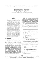
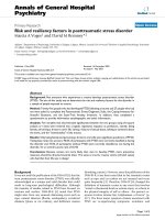
Figure 1 Some key insect recognition, signaling and response genes are missing in the pea aphid. Previously sequenced genomes of
other insects (flies, mosquitoes, bees, beetles) have indicated that immune signaling pathways, seen here, are conserved across insects. In
aphids, missing IMD pathway members (dashed lines) include those involved in recognition (PGRPs) and signaling (IMD, dFADD, Dredd, REL).
Genes encoding antimicrobial peptides common in other insects, including defensins and cecropins, are also missing. In contrast, we found
putative homologs for most genes central to the Toll, JNK and JAK/STAT signaling pathways.
Gerardo et al. Genome Biology 2010, 11:R21
/>Page 4 of 17
challenge, the serpin necrotic prevents activation of the
Toll pathway, but upon immunological challenge, the
Toll pathway is triggered by a cascade of serine pro-
teases, including persephone, which is thought to be
specific to fungal challenge [41]. Though it is not clear
which of the many aphid serine proteases is homologous
to persephone, it is likely that pea aphids have serine
proteases cap able of triggering the Toll pathway. Finally,
aphids also have multiple genes encoding Toll receptors,
which function as transmembrane receptors in both
mammals and insects. While nine single-copy Toll
genes ha ve been identified in D. melanogaster (Toll1 to
Toll9), it seems t hat pea aphids, like other insects, lack
some of these genes, but have multiple copies of others
(Figure S1c in Additional file 1). In other organisms,
some, but not all, Tolls serve a role in immune function,
while others function in developmental processes
[60-62]. For aphids, i t is n ot yet clear what role each
Toll serves.
The JAK/STAT signaling pathway
Like the Toll pathway, in Drosophila,theJAK/STAT
pathway is involved in both development and immunity.
The JAK/STAT pathway is the least understood of the
core insect immune pathways. JAK/STAT pathway
induction appears to lead to overproliferation of hemo-
cytes, upregulation of thiolester-containing proteins
(TEPs), and an antiviral response [63]. Changes in gene
expression following parasitoid wasp invasion of Droso-
phila larvae suggest a role for the JAK/STAT pathway
in parasitoid response [56]. Pea aphids have homologs
of all core JAK/STAT genes, including genes encoding
the cytokine receptor domeless, JAK tyrosine kinase
(aka Hopscotch), and the STAT92E transcription factor
(Figure 1). STAT92E appears to be duplicated. No
homologs were found for upd (unpaired), considered a
key ligand in Drosophila JAK/STAT induction. This
ligand is also missing in other insects (for example, A.
mellifera) [14].
IMD and JNK signaling pathways
Surprisingly, pea aphid s appear to be missing many cru-
cial components of the IMD signaling pathway. This
pathway is critical for fighting Gram-negative bacteria in
Drosophila [11,64], and IMD pathway member knock-
outs influence susceptibility to some Gram-positive bac-
teria and fungi as well [12]. IMD-associated genes
missing in pea aphids include PGRPs(seeabove),IMD,
dFADD, Dredd and Relish (Rel) (Figure 1). In contrast,
conserved one-to-one orthologs of these same ge nes are
found across Drosophila, Apis, Aedes, Anopheles and
Tribolium [13]. Cursory BLAST-based searches for these
genes in other arthropods suggest that some may be
missing (Figure 2). Pea aphids do have homologs for a
few pathway members ( TAB, TAK, kenny, Iap2 and
IRD5; Figure 1).
While missing IMD-associated genes, pea aphids have
plausible orthologs for most components of the JNK
pathway (Figure 1). In Drosophila, the JNK pathway reg-
ulates many developmental processes, as well as wound
healing [65], and has been proposed to play a role in
antimicrobial peptide gene expression and cellular
immune responses [11,66]. Genes present include hep,
basket,andJRA . Searches for homologs to the Droso-
phila kayak (kay) gene found an apparently similar tran-
scription factor encoding gene in the A. pisum genome
[GenBank: X P_001949014], but this match was largely
restricted to the leucine zipper region, and failed tests of
reciprocity.
The absence of IMD but presence of JNK in pea
aphids is surprising as, in Drosophila, t he IMD signaling
pathway leads to activation of components of the JNK
signaling pathway [11]. Specifically, when TAK, a pro-
tein kinase of the IMD pathway, is activated, it triggers
the JNK pathway. Whether TAK can be activated with-
out the res t of the IMD pathway is unknown. An alter-
native IMD-independent activation of JNK, via the
inducer Eiger [67], has been proposed in Drosophila
[66]. As Eiger is present in the pea aphid, this mode of
activation may serve a critical role in any aphid JNK-
based immune response.
Annotation of recognition genes
Antimicrobial peptides
Introduction of microbes into most insects leads to the
production of AMPs by the fat body, an insect immune-
response tissue, and occasionally by hemocytes and
other tissues [68-71]. These peptides are secreted into
the hemolymph, where they exhibit a broad range of
activities against fungi and bacteria. The mechanisms of
AMP action are poorly understood, but at least in som e
cases (for example, drosomycin in Drosophila), AMPs
destroy invading microbes by disrupting microbial cell
membranes, leading to cell lysis [71].
Antimicrobial peptides are diverse and ubiquitous.
They tend to be small molecules (<30 kDa) specialized
at attacking particular microbial classes (that is, Gram-
positive bacteria, fungi, and so on) [68,69]. While some
antimicrobial peptides are found in only a single insect
group (for example, metchnikowin is found only in
Drosophila), others are widely dispersed across eukar-
yotes (for example, defensins are present in fungi,
plants and animals). Genomics, coupled with proteo-
mics, has revealed that all sequenced insects, and
many other insects, have multiple types of antimicro-
bial peptides (Figure 2). Pea aphids, surprisingly, are
missing many of the antimicrobial peptides common
to other insects. For example, while all insect genomes
annotated t hus far have genes encoding defensins [13],
homology-based searches, phylogenetic-based analyses,
Gerardo et al. Genome Biology 2010, 11:R21
/>Page 5 of 17
transcriptomics (see below), and proteomics (see
below) failed to find any signatures of defensins i n the
pea aphid genome. The presence of defensins in the
human louse Pedicu lus humanus (Figure 2), and in the
ancient apterygote insect, the fire brat Thermobia
domestica [34], suggests that defensins have been lost
during aphid evolution.
Extensive searches for genes encoding insect cecro-
pins, drosocin (and other proline-rich arthropod AMPs),
diptericin (and other glycine-rich AMPs), drosomycin,
metchnikowin, formicin, moricin, spingerin, gomesin,
tachyplesin, polyphemusin, andropin, gamb icin, and vir-
escein also revealed no hits. Weak hits were found for
genes that encode for two antimicrobial peptides in
other invertebrates: megourin [UniProtKB: P83417], ori-
ginally isolated from another aphid species, the vetch
aphid Megoura viciae (P Bulet et al., unpublished data)
and penaeidin [UniProtKB: P81058], originally isola ted
from the shrimp Penaeus vannamei. The putative pea
aphid Megourin (scaffold EQ11086, positions 45,752 to
45,892), however, is highly diverged from that of M.
viciae (31% identity) and, compared to its M. viciae
counterparts, seems to have a shorter carboxy-terminal
region containing a stop- codon (Figure S2 in Additional
file 1). Using three different primer pairs, we were
unable to amplify products of this putative Meg ourin
from cDNA generated for expression analyses (see
below). The highly divergent Penaeidin [GenBank:
ACYPI37769] (Figure S2 in Additional file 1) also did
not amplify from cDNA.
We found six Thaumatin homologs in the A. pisum
genome that show overall sequence and predicted struc-
ture similarities to plant thaumatins (Figure 3a, b).
Thaumatin-like proteins are disulfide-bridged polypep-
tides of about 200 residues. Some thaumatins possess
antifungal activity in plant tissues after infection [72].
Recently, a thaumatin found in the beetle T. castaneum
was shown to inhibit spore germination of the filamen-
tous fungi Beauveria bassiana and Fusarium culmorum
[32]. Phylogenetic analyses revealed that A. pisum thau-
matins form a monophyletic group closely related to
beetle thaumatins (Figure 3c). Since thaumatin-like
genes are conspicuously absent f rom the genomes of
Drosophila, Apis, Anopheles, Pediculus and Ixodes (Fig-
ure 2), our findings indicate that thaumatins may repre-
sent ancient d efense molecules t hat have b een lost in
several insect species, or have been independently
acquired in aphids and beetles. The monophyly of aphid
and beetle thaumatins provides no indication of an ori-
gin of novel acquisition (Figure 3c).
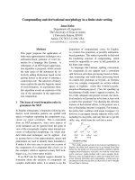
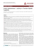
Figure 2 Gene families impl icated in arthropod immun ity suggest unique featur es of the pea aphid immune system. Black indicates
present (copy number is indicated, when known), white indicates absent, and gray indicates equivocal or unknown. Values for D. melanogaster,
A. gambiae, T. castanateum, A. mellifera, and some D. pulex genes are based on published analyses [13,14,16,17,40]. For previously unannotated D.
pulex genes, as well as for I. scapularis and P. humanus genes, we determined presence via cursory BLAST searches against available genome
databases [127,128] (wfleabase.org, vectorbase.org) using both D. melanogaster and A. pisum protein sequences as queries. Gene presence for
Ixodes was confirmed based on previous studies [129]. Future comprehensive annotation of the Pedicularis and Ixodes immune gene sets may
reveal the presence of additional genes and lack of functionality of others. PPO, prophenoloxidase.
Gerardo et al. Genome Biology 2010, 11:R21
/>Page 6 of 17
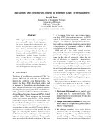
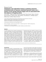
Figure 3 Evolutionarily conserved thaumatins are present in pea aphids and plants. (a) The three-dimensional structure of the pea aphid
thaumatin ACYPI009605 (top) was calculated using the published crystallographic structure of a sweet cherry (plant) thaumatin 2AHN_A
(bottom) [130] and Swissmodel [131], revealing that both thaumatins are similar in structure. However, one exposed loop, encircled by a dotted
line, shows a significant difference in structure, suggesting possible adaptation to different targets. (b) Similarities are also revealed in the
alignment of the pea aphid thaumatin with the plant thaumatin. A predicted signal sequence of the pea aphid thaumatin is underlined.
Identical amino acids are highlighted in red. (c) Maximum likelihood phylogeny of thaumatins, indicating branches leading to nematode, plant,
insect and bacteria-specific clades. Red highlights the sweet cherry thaumatin. Blue highlights the pea aphid thaumatins. Asterisks indicate
approximate likelihood ratio test support >80. Abbreviations: Api, A. pisum; Cac, Catenulispora acidiphila; Cel, Caenorhabditis elegans; Mtr,
Medicago truncatula; Pav, Prunus avium; Tca, Tribolium castaneum; Tpr, Trifolium pretense.
Gerardo et al. Genome Biology 2010, 11:R21
/>Page 7 of 17
Lysozymes
Lysozymes represent a family of enzymes that degra de
bacterial cell walls by hydrolyzing the 1,4-beta-linkages
between N-acetyl-D-glucosamine and N-acetylmuramic
acid in peptidoglycan heteropolymers [73]. They are ubi-
quitously distributed among living organisms and are
believed to be essential for defense against bacterial
infection. Lysozymes are classified into several types
(that is, c (chicken), g (goose), i (invertebrate), plant,
bacteria and phage types). C-type lysozymes are the
most common for metazoa, being found in all verte-
brates examined thus far and many invertebrates,
including all the previously sequenced insects. For
example, D. melanogaster and A. gambiae have at least
seven and nine loci for c-type lysozymes, respectively
[74,75]. Insects also have i-type homologs, but their bac-
teriolytic activities are unclear [76].
Unlike other insects sequenced thus far, similarity
searches demonstrated that A. pisum lacks genes for c-
type lysozymes. The a nalysis further verified that the
genome also lacks genes for g-type, plant-type, and
phage-type lysozymes. Only three genes for i-type
homologs were detected in the genome (Figure S1d in
Additional file 1). One of them, Lys1, is highly expressed
in the bacteriocyte [77]. Two others, Lys2 and Lys3,are
located adjacent to Lys1.
Notably, two genes that appear to have been trans-
ferred from bacterial genomes to the A. pisum genome
encode bacteriolytic enzymes [36] . One is for a chimeric
protein that consists of a eukaryotic carboxypeptidase
and a bacterial lysozyme. The other (AmiD)encodesN-
acetylmuramoyl-L-alanine amidase, which is not a true
lysozyme (1,4-beta-N-acetylmuramidase) but similarly
degrades bacterial cell walls. While some of these bac-
teriolytic-related genes are highly expressed in the bac-
teriocyte, and lysozymes appear to be upregulated in
response to som e challenges (see gene expression study,
below), assays of bacterioltyic activity of hemolymph
from immune-challenged aphids suggest that aphid
hemolymph has weak to no lysozyme-like activity [31].
Further studies will determine the role of these gene
products.
Chitinases
Chitinases are enzymes that degra de chitin (a long-chain
polymer of N-acetyl -D-glucosamine), hydrolyzing 1,4-
beta-linkages between N-acetyl-D-glucosamines. Chiti-
nases and lysozymes represent a superfamily of hydro-
lases, and their catalytic activities are similar. Indeed,
some chitinases show lysozyme activity and vice versa
[73]. In insects, chitinases are used to degrade the chitin
in the exoskeleton and peritrophic membrane during
molting, and some are suspected to have antifungal
activity, as fungal cell walls also consist of chitin [78].
Similarity searches followed by phylogenetic analyses
demonstrated that the genome of A. pisum encodes
seven genes for putative chitinase-like proteins [79].
Further studies are required to determine the biochem-
ical properties and substrate specificity of these chiti-
nase-like proteins.
TEPs and Tots
Some TEPs can covalently attach to pathogens and
parasites in order to ‘mark’ them for phagocytosis [80].
Like other insects, aphids have multiple Tep paralogs.
Both are homologous to TepIII (Figure S1e in Addi-
tional file 1). Homologs of TepI, TepII and TepIV were
not found. In contrast, no Turandot (Tot) genes, which
encode small peptides induced by severe stress and sep-
ticinjuryinDrosophila [81-83], have been found in
aphids or in other insects other than Drosophila spp.
Both TEPs and Tots are thought to be regulated by the
JAK/STAT pathway.
Prophenoloxidase
Phenoloxidase-mediated melanin formation characteris-
tically accompanies wound clotting, phagocytosis and
encapsulation of pathogens and parasites [84]. In insects,
the inactive enzyme prophenoloxidase ( ProPO) is acti-
vated by serine proteases to yield phenoloxidase [85].
Aphids appear to have two prophenoloxidase homologs
(ProPO1, ProPO2; Figure S1f in Additional file 1), which
are homologous t o D. melanogaster Diphenol oxidase
A3 [Flybase: CG2952].
Nitric oxide synthase
Production of nitric oxide is mediated by the enzyme
nitric oxide synthase. Nitric oxide is a highly unstable
free radical gas that has been shown to be toxic to both
parasites and pathogens. In insects, No s is upregulated
after both parasite and Gram-negative bacterial infection
[86,87]. Like other insects, pea aphids have one Nos
homolog.
Heat shock proteins
Though called HSPs, these proteins are produced in
response to a range of stresses in both eukaryotic and
prokaryotic organisms [18]. They serve as chaperones,
facilitating protein folding and stabilization, and as pro-
teases, mediating the degradation of damaged proteins.
HSPs may also serve as signaling proteins during
immune responses [18,88]. In many insects, including
aphids, HSPs h ave been shown to be upregulated after
septic injury and microbial infection [31,89-92]. We
identified 15 HSPs of varying molecular weight in pea
aphids (Figure S1g in Additional file 1).
Gluthione-S-tranferases
Gluthione-S-tranferases comprise a diverse class of
enzymes that detoxify stress-causing agents, including
toxic oxygen free radical species. They are upregulated
in some arthropods upon oxidative stress [93] and
microbial challenge [89,94]. Pea aphids have at least 18
genes encoding gluthione-S-tranferases and many other
Gerardo et al. Genome Biology 2010, 11:R21
/>Page 8 of 17
detoxification enzymes that likely play a role in stress
responses [95]. Ramsey et al. [95] identified many of the
genes encoding detoxification enzymes in A. pisum and
in Myzus persicae.
Alarm pheromone production
In response to predators, aphids release an alarm phero-
mone that causes neighboring aphids to become more
mobile and to produce more winged than unwinged off-
spring [19,96]. These winged offspring have the ability
to disperse to enemy-free space. While many insects
produce a suite of chemicals that constitute an alarm
signal, the aphid alarm pheromone is dominated by a
single compound, (E)-b farnesene [97]. While the genes
underlying alarm pheromone production have not been
fully characterized, we have identified a Farnesyl dipho-
sphate synthase (FPPS)andanIsoprenyl diphosphate
synthase (IPPS), w hich may underlie alarm pheromone
production [98].
Functional assays
Gene expression
We utilized real-time quantitative PCR to conduct a
preliminary investigation of the expression of 23 recog-
nition, signaling and response genes in aphids subjected
to a number of infection and stress treatments (see Sup-
plementary materials and Table S2 in Additional file 1).
While future studies with more biological replicates will
be necessary to fully survey gene regulation in the face
of stress and infection, this initial survey indicates that
aphids do express these genes under both control and
infection/stress conditions (Tables S4 and S5 in Add i-
tionalfile1).Thissuggeststhatthesegenesarefunc-
tional even in the absence of many other missing
immune-related genes.
Oneexpressionpatternseeninthisinitialsurveyis
of particular note. Unlike other insect immune expres-
sion studies, we found no strong upregulation of anti-
microbial peptides, which frequently exhibit ten-fold or
greater upregulation in the face of infection. For exam-
ple, while Altincicek et al. [32] observed 20-fold upre-
gulation of Thaumatins in tribolium beetles after
stabbing with lipopolysaccaride endotoxin derived from
Escherichia coli, we saw modest upregulatio n (approxi-
mately 2-fold) of only one Thaumatin (Thm2)after
stabbing aphids (Table S5 in Additional file 1).
Furthermore, despite the fact that they are known to
suppress fungal germination in beetles, the Thaumatin
homologs were not upregulated after fungal infection
at the time point included in this study, and were only
approximately two-fold upregulated at two additional
time points and in a follo w-up fungal infection experi-
ment (data not shown) [32]. The role of thaumatins in
fighting microbial infections, however, should not be
discounted, as they may function in the absence of
significant upregulation (tha t is, they may be constitu-
tively expressed) .
Exploration of ESTs from infected and uninfected aphids
In the first of two EST-based experiments, we compared
a cDNA library synthesized from the guts of A. pisum
that had been fed a Gram-negative pathogen, Dickeya
dadantii[99], to a cDNA library synthesized from unin-
fected guts. Strikingly, no standard immune-related
gen es, such as antimicrobial peptides, were identified in
the infected sample. The main functional classes differ-
entially expressed were the ‘biopolymer metabolism ’
class, many members of which were down-regulated in
infected guts, and ‘transport’ or ‘establishment of locali-
zation’ classes, whose genes were upregulated in infected
guts (Table S6 in Additional file 1). The ‘ immune
response’ class, in contrast, was only represented by five
genes. Four of these five genes were in the uninfected
library, while only one, encoding a leucyl-aminopepti-
dase, was identified from the infected library; the
immune function of leucyl-aminopeptidases is not well
understood. Moreover, the ‘ response to stress/external
stimulus/biotic stimulus’ classes were not overrepre-
sented in the infected gut library.
In a separate experiment, to further identify aphid
immune-relevant genes, we utilized SSH to compare
cDNA from E. coli-infected aphids and cDNA from
unchallenged aphids. To obtain genes expressed at dif-
ferent phases of the i mmune respons e, three RNA sam-
ples were extracted 3, 6 a nd 12 hours after E. coli
infection and mixed prior to cDNA synthesis.
Among the 480 ESTs t hat were sequenced from the
subtracted library [GenBank: GD185911 to GD186390],
we found s ome genes with similarity to proteases and
protease inhibitors but few other immune-related pro-
teins. Interestingly, SSH-based EST analysis failed to
identify any PRRs, such as PGRPs or GNBPs, or any
ant imicrobial pepti des (Table S7 in Additional file 1). It
is noteworthy th at this aphid experiment was conducted
in parallel to a similar Sitophilus weevil experiment,
where many immune-related genes (more than 18% of
ESTs) were identified, including antibacterial peptides
and PRRs [35]. This suggests that the paucity of
immune genes identified in A. pisum is not a technical
issuebutmaybeaspecificfeatureofaphids[31].In
addition, dot blot analysis demonstrated that only a few
genes (less than 5%) were differentially expressed
between E. coli-stabbed and unstabbed aphids. These
findings indicate that, in contrast to other insects, either
aphids respond only w eakly to challenge with E. coli or
aphid genes and pathways directed against these bacteria
are expressed only constitutively.
High performance liquid chromatography
HPLC peptide analyses targeting production of small
peptides (for example, antimicrobial peptides) were run
Gerardo et al. Genome Biology 2010, 11:R21
/>Page 9 of 17
on hemolymph samples from pea aphids challenged by
three microorganisms: E. coli (Gram-negative bacteria),
Micrococcus luteus (Gram-positive bacteria) and Asper-
gillus fumigatus (fungi). Profiles were compared between
control, infected and sterile-st abbed aphids at 6, 12 and
18 hours after challenge. When identified, the produc-
tion of small peptides was maximal at 18 hours. In E.
coli-t reated samples, no upregula tion could be identified
(Figure 4a), in M. luteus-treated samples, there was
modest upregulation (data no t shown), and in A. fumi-
gatus-treated samples, there was a significant response,
though few peaks (Figure 4b ). In contrast, a respo nse
profile to E. coli from another obligate symbiotic insect
(the weevil, Sitophilus oryzae) exhibited at least five
well-distinguishable upregulated peaks (Figure 4c).
Response being restricted to Gram-positive bacteria and
fungi is consistent with previous identification of
megourin, an antimicrobial peptide in the aphid
Megoura viciae, which appears to have activity against
Gram-positive bacteria and fungi, but not against Gram-
negative bacteria (P Bulet, unpublished) . Because so few
distinguishable peaks were present in the aphid samples,
we did not choose to identify the associated products,
but overall the presence of few inducible peptides sug-
gests a peculiar scarcity of antimicrobial peptides in
aphids.
Conclusions
Aphids are one of only a few genomic models for hemi-
metabolous insects, yet until recently, virtually nothing
was known about aphid immune and stress response
systems. Here, by coupling gene anno tation with
functional assays, we see evidence that aphids have
some defense systems common to other arthrop ods (for
example, the Toll and JAK/STAT signaling pathways,
HSPs, ProPO). Surprisingly, however, several of the
genes thought central to arthropod innate immunity are
missing in aphids (for example, PGRPs, the IMD signal-
ing pathway, defensins, c-type lysozymes). This calls into
question the generality of the current model of insect
immunity, and it remains to be determined h ow aphids
protect themselves from the diverse pathogens and para-
sites that they face.
The fact that we cannot find aphid homologs to many
insect immune genes could be a consequence of the
large evolutionary distance between aphids and the taxa
(in most c ases, flies, mosquitoes and bees) from which
these genes are known (that is, the split between the
ancestors of aphids and these taxa occurred approxi-
mately 350 million years ago [100]), making it challen-
ging to find divergent genes via homology-based
searches, even when using highly sensitive methods as
done here. Though we cannot preclude this possibility
in all cases, in some cases, similar homology-based
methods are able to recover homologs in even more dis-
tantly related taxa. For example, querying genome data-
bases w ith Drosophila genes via BLAST recovers
putative homologs of PGRPs and defensins in P. huma-
nus (human body louse) and in Ixodes scapul aris (deer
tick) (Figure 2). The divergence time between Droso-
phila and these taxa is equal to or greater than that
between Drosophila and aphids. Moreover, for some
cases, we could identify genomic regions similar to func-
tional genes in other species, but these regions contain
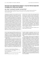
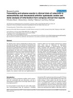
Figure 4 HPLC traces of inducible hemolymph peptides in the pea aphid compared to the rice weevil. Representative traces (solid, red
lines) are from insects 18 hours after microbial challenge; traces generated from 18 hour control insects are overlaid (dashed, black lines).
Phenylthiourea (PTU) served as an internal standard. Arrows indicate peaks that are significantly upregulated (solid, red arrows) or downregulated
(dashed, black arrows). (a) Profile from pea aphids challenged with E. coli, showing no upregulated response. (b) Profile from pea aphids
challenged with the fungus A. fumigatus, showing some differential peaks. (c) For comparison, profile from rice weevils (Sitophilus oryzae)
challenged with E. coli, showing several differentials peaks at multiple retention times.
Gerardo et al. Genome Biology 2010, 11:R21
/>Page 10 of 17
large insertions or stop codons (for example, the puta-
tive antimicrobial peptide Megourin), indicating they are
the result of pseudogenization.
One potential explanation for the lack of known
immune-related genes in pea aphids is that aphids
mount an alter native, but equal, immune response. Our
functional analyses, as well as those of Altincicek et al.
[31], found little evidence for an alternative response. In
EST and HPLC analyses , few novel EST s or pe ptide sig-
nals were recovered from immune-challenge aphids rela-
tive to their unchallenged controls. It should be noted,
however, that these challenges were primarily limited to
exposure to E. coli bacteria. When testing for expression
of a few immune genes in r esponse to a wider array of
challenges, we do see some evidence of an aphid
immune and stress response. Future expression studies,
including large-scale transcriptional and proteomic stu-
dies, will extend this work and allow for m ore compre-
hensive characterization of the full complementation of
aphid immune responses.
While we have focused mainly on the humoral com-
ponent of the innate immune response, it is interesting
to note that there is some evidence that the cellular
comp onent of pea aphids’ innate immune response may
also be different to that seen in other insects. While
many insects encapsulate parasitoid wasp larvae,
smothering them to death with hemocyt es (insect
immune cells), aphids appear not to have this layer of
protection [101,102]. Aphids, however, appear to recruit
some hemocytes to parasitoid eggs, suggesting that cel-
lular immuni ty may play an alterna tive, though possibly
more limited, role [101]. Better insights into the capacity
of the aphid immune system will require further investi-
gation of both the humoral and cellular components of
aphid immunity.
The lack of genomic and molecular data regarding
immune systems of aphid relati ves makes it difficult to
establish whether the pea aphid immune system is
unique. There are, however, a number of aspects of
aphid ecology that could facilitate ecological success
without a strong immune defense. Altincicek et al. [31]
proposed three hypotheses to explain the apparent lack
of antimicrobial defenses. First, they suggested that con-
trary to Drosophila, whose natural environment consists
of decaying fruit tha t is c olonized by many microbes,
aphids exploit phloem sap, which only occasionally con-
tains bacteria and rarely contains entomopathogens.
Thus, the risk of encountering pathogens while feeding
is more limited. This assumption, however, is only partly
true. While probing plants, aphids are capable of acquir-
ing pathogenic bacteria from the surface of their host
plants’ leaves [103], and aphids become host to a diverse
assemblage of bacteria and fungi under stressful condi-
tions [104], some of which are pathogenic (NM
Gerardo, unpublished data). Furthermore, Sitophilus
weevils, which when challenged with E. coli significantly
upregulate immune genes [35], spend their entire larval
and nymph stages within sterile cereal grains, indicating
that a sterile diet is not likely to explain the absence of
antibacterial defenses in aphids.
Altincicek et al. [31] also suggest that aphids may
invest in terminal reproduction in response to an
immune challenge, rather than in a costly immune
response. In their study, stabbed aphids produced signif-
icantly more offspring than untreated aphids within 24
hours of injury . Such an increase in reproduction upon
challenge is not uncommon for inver tebrates. Biompha-
laria snails [105,106], Acheta crickets [107], Daphnia
waterfleas [108], and Drosophila flies [109] have all been
shown to increase their invest ment in reproduction in
response to infection. Yet, Drosophila still mount a
complex immune response. Furthermore, aphids do not
increase their reproductiv e effor t in the face of all
immune challenges: fungal infection reduces the number
of offspring A. pisum produce within 24 hours of inocu-
lation [110], and response to stabbing with bacteria
seems to b e specific to t he aphid genotype and to the
location of the stab (Barribeau, unpublished data).
Therefore, though aphids have the capacity to reproduce
many offspring prior to succumbing to some pathogens,
it seems that immune competence would still provide
increased fitness.
Even without increased reproduction following infec-
tion, the prolific reproductive capacity of aphids suggests
these insects, in general, may invest most resources
towards rapid, early onset reproduction rather than
towards fewer, though better-protected offspring (aka, in
terms of classical ecological theory, aphids may be r-
select ed rather k-selection organisms [ 111]). Recent the-
ory of the evolution of immunity suggests that suc h
organisms may specifically invest less in costly immune
responses [112,113]. Many characteristics of aphids,
including their rapid generation time, short life span
and small body size all fit a model of r-selection [114].
Drosophila spp., however, also exhibit many of these
characteri stics and still inve st in a strong defense
repertoire.
The third hypothesis proposed by Altincicek et al. [31]
concerning the evolution and maintenance of aphid
defense relies on the presence of secondary symbionts
that can be found extracellularly in aphids [115]. A.
pisum is protected against fungal pathogens by one of
these secondary symbionts, Regiella insecticola [29], and
also against the parasitoid wasp Aphidius ervi by
another secondary symbiont, Hamiltonella defensa [27].
Such symbiont-mediated host protection may explain
why aphids have a reduc ed (or specialized) antimicrobial
defense. This hypothesis seems plausible with regard to
Gerardo et al. Genome Biology 2010, 11:R21
/>Page 11 of 17
thecostofimmunegeneexpressionversusthebenefit
of protection by the secondary endosymbionts. However,
it does not explain how the secondary endosymbionts
(as Gram-negative bacteria), often present in aphid
hemolymph, are themselves perceived and controlled by
the aphid immune system. Thus, it is challenging to say
whether the presence of secondary symbionts is a cause
or a consequence of reduced antimicrobial activity.
Potentially, all of these forces could shape the evolu-
tion of aphid stress and immune responses. In order to
test these hypotheses (for example, reproductive invest-
ment, symbiont-mediated host protection), we need
more studies characterizi ng the global aphid response
under more conditions, and in more aphid species.
Potential insight from aphid relatives with different life-
styles (for example, those not associated with secondary
symbionts, or those that live in soil or other microbe-
rich habitats) may be particularly helpful. More broadly,
as the pea aphid is the first published genome of a
hemimetabolous insect, future analyse s of the immune
and stress related genes of more insects in this group
will facilitate the reconstruction of the evolutionary his-
tory of innate immunity and other defenses.
Materials and methods
Bioinformatic screening of the pea aphid genome
Immune and stress gene candidates from other insects
(for example, D. melanogaster, A. aegypti, A. gambiae,
A. mellifera) were used to query the pea aphid genom e.
Most searches utilized the blastp search function to
search for hits against the predicted A. pisum proteome
[116]. For some gene families and putative paralogs,
protein sequences were aligned to sequ ences from other
insects and outgroups using ClustalW [117]. These
alignments, as well as available EST and full length
cDNA sequences, served to refine aphid gene models
(exon/intron boundaries, and so on), and to facilitate
phylogenetic analyses. In addition, a comprehensive
database of all available EST sequences from the green
peach aphid, Myzus persicae,wasscreenedusingtblastn
to search for potential homologs to all immune and
stress genes annotated in the pea aphid.
For genes that could not be found in the proteome,
we also conducted a tblastn search against all contigs
and unassembled reads. Then, a final, more sensitive
profile-based search was performed for those immune
defense proteins that produced no hits with BLAST
searches. For this analysis, insect and other species pro-
tein sequences belonging to the family of interest were
retrieved from NCBI and aligned with MUSCLE [118].
A hidden Markov model for the alignment was built
and calibrated using HMMER [119]. This was used to
perform a profile-based search (hmmsearch) against the
six-frame translated sequences of the assembled pea
aphid genome and the unassembled reads. Additionally,
a similar search with PFAM profiles [120] was also per-
formed for those families encoding PFAM domains in
their sequences. Whenever a significant hit was found,
the genomic region was analyzed to discard the possibi-
lity that it encoded a pseudogene (presence of stop
codons, absence of relevant domains, and so on).
Phylogenetic analyses of selected protein families were
performed using their corresponding maximum likeli-
hood phylogenetic trees from the pea aphid phylome
[36], deposited in PhylomeDB [121]. When necessary,
additional sequences were added to the original Phylo-
meDB alignment, realigned with MUSCLE and used to
reconstruct a maximum likelihood phylogenetic tree,
using the JTT (Jones-Taylor-Thornton) model as imple-
mented in PhyML v2.4.4 [1 22], assuming a discrete
gamma-distribut ion model with four rate categories and
invariant sites, and estimating the gamma shape para-
meter and the fraction of invariant sites. Cladograms
were edited using Dendrogram [123].
Exploration of ESTs from infected and uninfected aphids
In the first experiment, two EST libraries (one control,
one infected) were generated by standard procedures
using a SMART cDNA kit (Clontech, Mountain View,
California, USA), starting from approximately 1,000 dis-
sected A. pisum midguts for each library. The aphids
were clonal, young, reproducing asexuals, which were
either fed on control diet or infected by feeding on arti -
ficial diet with the G ram-negative aphid pathogen Dick-
eya d adan tii at 10
6
bacteria per milliliter [99]. Twenty-
four hours after infection, control and treated aphids
were dissected, and complete guts were transferred
immediately into RNeasy solution (Qiagen Valencia,
California, USA). ESTs were sequenced according to
procedures in Sabater-Munoz et al. [124].
In another EST-based experiment utilizing SSH and
dot-blot technology, we treated aphids (clone LL01)
with rifampicin as described in Rahbé et al. [125] to
reduce symbiont load. We challenged wingless fourth-
instar aposymbiotic aphids by stabbing them with nee-
dles previously dipped into a pellet of overnight cultures
of E. coli (TOP10, Invitrogen Carlsbad, California, USA),
and then maintained them on fava plants. At 3, 6, and
12 hours post-treatment, we stored surviving aphid s at
-80°C. To identify genes that are differe ntially expressed
in response to septic injury, we performed SSH using
RNAs from immune challenged (3, 6 and 12 hours post-
treatment) and untreated aposymbiotic aphids, using the
SMART PCR cDNA Synthesis Kit and the PCR-Select
cDNA Subtraction Kit (Clontech) according to the man-
ufacturer’s instructions and as described in Anselme et
al. [35]. After transformation by electroporation, we
recovered approximately 1,500 colonies from LB agar
Gerardo et al. Genome Biology 2010, 11:R21
/>Page 12 of 17
plates. We plasmid extracted and sequenced 500 ran-
domly picked colonies (NucleoSpin® Plasmid Kit,
Macherey-Nagel, Düren, Germany) utilizing the sequen-
cing center at the University of Valencia (Spain). We
compared all sequences against UniProt using blastx.
Immune -related gene sequences (Table S7 in Additional
file 1) were then compared to the aphid genome using
blastn.
To analyze the differential expression status of each
EST, we conducted a dot-blot experiment. Briefly, we
amplified 344 ESTs from the SSH library by colony PCR
with nested PCR primers 1 and 2R from the PCR-Select
cDNA Subtraction Kit. We then spotted 10 μlfrom
each PCR product onto two different membranes
(Amersham Hybond™-N, GE Healthcare Life Sciences,
Piscataway, New Jersey, U SA) using a Bio-Dot Microfil-
tration System (Biorad, Hercules, California, USA). We
hybridized membranes with radiolabeled cDNA probes
generated by reverse-transc ription from RNA extracted
from either aposymbiotic aphids stabbed with E. coli or
unstabbed aposymbiotic aphids. We synthesized these
probes using the Super Script™ First Strand Synthesis
system (Invitrogen) for RT-PCR and [a-
32
P]dCTP, and
purified them using Quick Spin Column (Roche Molecu-
lar Biochemical s, Indianapolis, Indiana, USA). A fter
exposing blots for up to 24 hours to a Storm PhosphorI-
mager imaging plate (GE Healthcare Life Sciences), we
analyzed differential expression by comparison of band
intensities between the two membranes. We did not,
however, normalize the data, as we failed to see any sig-
nal from the Gapdh gene, though the same amount of
each PCR product was loaded on both membranes.
HPLC
Aphids were challenged by abdominal puncture with tri-
ple-0 needles dipped in a solution of Gram-negative
bacteria (E. coli strain Top10), Gram-positive bacteria
(M. luteus)orfungalspores(A. fumigatus). For each
microbial treatment, five hemolymph samples from 50
aphids each were collected at four times points (t = 0, 6,
12 and 18 hours).
Hemolymph was flash-extrac ted by centrifuging (1
minute, 10,000 g, 4°C) live aphids through a 1 ml pip-
ette tip and directly into 40 μl 0.1% trifluoractetic acid
contaning 10 μl of saturated phenylthiourea (PTU) for
phenolo xidase inhibition. Resulting samples wer e highly
similar to pure hemolymph samples obtained by leg
bleeding (>95% band identity by silver-stained SDS-
PAGE).
After initial collection, tips were removed and the
samples were centrifuged for 5 minutes at 15,000 g. Fol-
lowing addition of 70 μl trifluoractetic acid 0.1%, the
supernatant sat for 1 ho ur at 4°C to allow for protein
precipitation prior to a final 10-minute centrifugation at
15,000 g to recover peptides. Samples were evaporated
and stored at -20°C until use in HPLC. Chromatography
was performed on standard peptide C18-3 00Å reverse
phase columns using water acetonitrile gradients [126].
For retention time standardization, PTU served as an
internal standard, and samples were analyzed by area-
normalization to unchallenged sample peaks (retention
time = 14 minutes, preceding PTU).
Additional file 1: Supplementary methods for the gene expressio n
study and supplementary tables and figures. Table S1: pea aphid
immune and stress gene list. Table S2: samples for quantitative PCR
expression study. Table S3: primers for quantitative PCR expression study.
Table S4: relative expression of recognition and signaling genes. Table S5:
relative expression of response genes. Table S6: gut EST library statistics.
Table S7: list of selected ESTs from the subtracted library. Figure S1:
maximum likelihood phylogenies of selected immune and stress gene
families. Figure S2: alignments of putative antimicrobial peptides
megourin and penaeidin. Figure S3: survival curves for experimental
infections associated with quantitative PCR study.
Abbreviations
AMP: antimicrobial peptide; EST: expressed sequence tag; GNBP: Gram-
negative binding protein; HPLC: high performance liquid chromatography;
HSP: heat shock protein; IMD: immunodeficiency; JAK/STAT: Janus kinase/
Signal transducers and activators of transcription; JNK: c-Jun N-terminal
kinase; PGRP: petidoglycan receptor protein; ProPO: prophenoloxidase; PTU:
phenylthiourea; PRR: pathogen recognition receptor; SSH: suppression
subtractive hybridization; TEP: thiolester-containing protein.
Acknowledgements
We thank Angela Douglas, Nancy Moran, Tom Little and members of the
International Aphid Genomics Consortium for insightful discussion, and
Charles Godfray and two anonymous reviewers for comments that
enhanced this manuscript. Cultures of Z. occidentalis were provided by the
USDA ARS Collection of Entomopathogenic Fungal Cultures. Samples of
ALPV and virus-infection protocols were provided kindly by Bryony Bonning
and Liljana Georgievska. Comparison with M. persicae was supported by
USDA grant 2005-35604-15446 to Georg Jander.
Author details
1
Department of Biology, Emory University, O Wayne Rollins Research Center,
1510 E. Clifton Road NE, Atlanta, GA, 30322, USA.
2
Interdisciplinary Research
Center, Institute of Phytopathology and Applied Zoology, Justus-Liebig-
University of Giessen, Heinrich-Buff-Ring 26-32, D-35392 Giessen, Germany.
3
Université de Lyon, INRA, INSA-Lyon, IFR41 BioEnvironnement et Santé,
UMR203 BF2I, Biologie Fonctionnelle Insectes et Interactions, Bat. Louis-
Pasteur 20 ave Albert-Einstein, F-69621 Villeurbanne, France.
4
UMR
Interactions Biotiques et Santé Végétale, INRA 1301-CNRS 6243-Université de
Nice-Sophia Antipolis, 400 routes des Chappe, F-06903 Sophia-Antipolis
cedex, France.
5
Department of Nematology, Graduate Program in Genetics,
Genomics and Bioinformatics, University of California, 900 University Ave,
Riverside, CA 92521, USA.
6
Boyce Thompson Institute for Plant Research,
Ithaca, NY 14853, USA.
7
Genetics Otago and The Laboratory for Evolution
and Development, Department of Biochemistry, University of Otago, Box 56,
Dunedin 9054, New Zealand.
8
USDA-ARS Bee Research Lab, BARC-East Bldg
476, Beltsville, MD 20705, USA.
9
Bioinformatics and Genomics Programme,
Centre for Genomic Regulation (CRG), Doctor Aiguader 88, 08003 Barcelona,
Spain.
10
Department of Entomology, The Volcani Center, Bet Dagan 50250,
Israel.
11
Instituto Cavanilles de Biodiversidad y Biología Evolutiva, Universitat
de València, Avenida Blasco Ibañez 13, 46071 València, Spain.
12
CIBER en
Epidemiología y Salud Pública (CIBEResp) and Centro Superior de
Investigación en Salud Pública (CSISP), Conselleria de Sanidad (Generalitat
Valenciana), Avenida de Cataluña 21, 46020 València, Spain.
13
Advanced
Science Institute, RIKEN, 2-1 Hirosawa, Wako, Saitama 351-0198, Japan.
14
Plant Pathology and Plant-Microbe Biology Department, Cornell University,
Gerardo et al. Genome Biology 2010, 11:R21
/>Page 13 of 17
Tower Road, Ithaca, NY 14853, USA.
15
Department of Entomology, Texas
A&M, College Station, TX 77843-2475, USA.
Authors’ contributions
NMG, SMB, and MG were group leaders for the project. NMG, BA, HA, SMB,
MDV, EJD, JDE, AM, MG, IK, AN, BJP, MP, JSR, JT, DT, and CT designed and
performed manual gene annotation. TG and SMB conducted phylogenetic
analyses. BA and AV conceived of and conducted analyses of Thaumatin.
SMB, NMG, CS and BJP performed experiments and analyses for the gene
expression study. CA, AH, VPB, AM, and AL conceived of and conducted the
SSH study, and CVM constructed the aphid gut libraries. YR conducted the
HPLC study. The manuscript was prepared by NMG, SMB, CA, TG and YR
with input from MDV, BA, AN, AV and AH. All authors have read and
approved the final version of the manuscript.
Received: 22 August 2009 Revised: 7 October 2009
Accepted: 23 February 2010 Published: 23 February 2010
References
1. Snyder WE, Ives AR: Interactions between specialist and generalist natural
enemies: Parasitoids, predators, and pea aphid biocontrol. Ecology 2003,
84:91-107.
2. Hufbauer RA: Aphid population dynamics: does resistance to parasitism
influence population size? Ecol Entomol 2002, 27:25-32.
3. Hatano E, Kunert G, Bartram S, Boland W, Gershenzon J, Weisser WW: Do
aphid colonies amplify their emission of alarm pheromone? J Chem Ecol
2008, 34:1149-1152.
4. Tarpy DR: Genetic diversity within honeybee colonies prevents severe
infections and promotes colony growth. Proc Biol Sci 2003, 270:99-103.
5. Ha EM, Oh CT, Ryu JH, Bae YS, Kang SW, Jang IH, Brey PT, Lee WJ: An
antioxidant system required for host protection against gut infection in/
Drosophila. Dev Cell 2005, 8:125-132.
6. Francke DL, Harmon JP, Harvey CT, Ives AR: Pea aphid dropping behavior
diminishes foraging efficiency of a predatory ladybeetle. Entomologia
Experimentalis Et Applicata 2008, 127:118-124.
7. Gagneux S, DeRiemer K, Van T, Kato-Maeda M, de Jong BC, Narayanan S,
Nicol M, Niemann S, Kremer K, Gutierrez MC, Hilty M, Hopewell PC,
Small PM: Variable host-pathogen compatibility in Mycobacterium
tuberculosis. Proc Natl Acad Sci USA 2006, 103:2869-2873.
8. Govind S: Innate immunity in Drosophila: pathogens and pathways. Insect
Sci 2008, 15:29-43.
9. Strand MR: The insect cellular immune response. Insect Sci 2008, 15:1-14.
10. Schulenburg H, Boehnisch C, Michiels NK: How do invertebrates generate
a highly specific innate immune response? Mol Immunol 2007,
44:3338-3344.
11. Boutros M, Agaisse H, Perrimon N: Sequential activation of signaling
pathways during innate immune responses in Drosophila. Dev Cell 2002,
3:711-722.
12. Dionne MS, Schneider DS: Models of infectious diseases in the fruit fly
Drosophila melanogaster. Dis Model Mech 2008, 1:43-49.
13. Zou Z, Evans JD, Lu ZQ, Zhao PC, Williams M, Sumathipala N, Hetru C,
Hultmark D, Jiang HB: Comparative genomic analysis of the Tribolium
immune system. Genome Biol 2007, 8
:R177.
14. Evans JD, Aronstein K, Chen YP, Hetru C, Imler JL, Jiang H, Kanost M,
Thompson GJ, Zou Z, Hultmark D: Immune pathways and defence
mechanisms in honey bees Apis mellifera. Insect Mol Biol 2006, 15:645-656.
15. Waterhouse RM, Kriventseva EV, Meister S, Xi ZY, Alvarez KS,
Bartholomay LC, Barillas-Mury C, Bian GW, Blandin S, Christensen BM,
Dong YM, Jiang HB, Kanost MR, Koutsos AC, Levashina EA, Li JY,
Ligoxygakis P, MacCallum RM, Mayhew GF, Mendes A, Michel K, Osta MA,
Paskewitz S, Shin SW, Vlachou D, Wang LH, Wei WQ, Zheng LB, Zou Z,
Severson DW, et al: Evolutionary dynamics of immune-related genes and
pathways in disease-vector mosquitoes. Science 2007, 316:1738-1743.
16. Christophides GK, Zdobnov E, Barillas-Mury C, Birney E, Blandin S, Blass C,
Brey PT, Collins FH, Danielli A, Dimopoulos G, Hetru C, Hoa N, Hoffmann JA,
Kanzok SM, Letunic I, Levashina EA, Loukeris TG, Lycett G, Meister S,
Michel K, Muller HM, Osta MA, Paskewitz SM, Reichhart JM, Rzhetsky A,
Troxler L, Vernick KD, Vlachou D, Volz J, von Mering C, et al: Immunity-
related genes and gene families in Anopheles gambiae. Science 2002,
298:159-165.
17. Sackton TB, Lazzaro BP, Schlenke TA, Evans JD, Hultmark D, Clark AG:
Dynamic evolution of the innate immune system in Drosophila. Nat
Genet 2007, 39:1461-1468.
18. Pockley AG: Heat shock proteins as regulators of the immune response.
Lancet 2003, 362:469-476.
19. Kunert G, Otto S, Rose USR, Gershenzon J, Weisser WW: Alarm pheromone
mediates production of winged dispersal morphs in aphids. Ecol Lett
2005, 8:596-603.
20. Moran NA, Russell JA, Koga R, Fukatsu T: Evolutionary relationships of
three new species of Enterobacteriaceae living as symbionts of aphids
and other insects. Appl Environ Microbiol 2005, 71:3302-3310.
21. Moran NA, Telang A: Bacteriocyte-associated symbionts of insects - a
variety of insect groups harbor ancient prokaryotic endosymbionts.
Bioscience 1998, 48:295-304.
22. Sandstrom JP, Russell JA, White JP, Moran NA: Independent origins and
horizontal transfer of bacterial symbionts of aphids. Mol Ecol 2001,
10:217-228.
23. Tsuchida T, Koga R, Fukatsu T: Host plant specialization governed by
facultative symbiont. Science 2004, 303:1989-1989.
24. Russell JA, Moran NA: Costs and benefits of symbiont infection in aphids:
variation among symbionts and across temperatures. Proc Biol Sci 2006,
273:603-610.
25. Montllor CB, Maxmen A, Purcell AH: Facultative bacterial endosymbionts
benefit pea aphids Acyrthosiphon pisum under heat stress. Ecol Entomol
2002, 27
:189-195.
26. Chen DQ, Montllor CB, Purcell AH: Fitness effects of two facultative
endosymbiotic bacteria on the pea aphid, Acyrthosiphon pisum, and the
blue alfalfa aphid, A. kondoi. Entomologia Experimentalis Et Applicata 2000,
95:315-323.
27. Oliver KM, Moran NA, Hunter MS: Variation in resistance to parasitism in
aphids is due to symbionts not host genotype. Proc Natl Acad Sci USA
2005, 102:12795-12800.
28. Oliver KM, Russell JA, Moran NA, Hunter MS: Facultative bacterial
symbionts in aphids confer resistance to parasitic wasps. Proc Natl Acad
Sci USA 2003, 100:1803-1807.
29. Scarborough CL, Ferrari J, Godfray HCJ: Aphid protected from pathogen
by endosymbiont. Science 2005, 310:1781-1781.
30. Haine ER: Symbiont-medi ated protection. Proc Biol Sci 2008, 275:353-361.
31. Altincicek B, Gross J, Vilcinskas A: Wounding-mediated gene expression
and accelerated viviparous reproduction of the pea aphid Acyrthosiphon
pisum. Insect Mol Biol 2008, 17:711-716.
32. Altincicek B, Knorr E, Vilcinskas A: Beetle immunity: Identification of
immune-inducible genes from the model insect Tribolium castaneum.
Dev Comp Immunol 2008, 32:585-595.
33. Altincicek B, Vilcinskas A: Analysis of the immune-inducible transcriptome
from microbial stress resistant, rat-tailed maggots of the drone fly
Eristalis tenax. BMC Genomics 2007, 8:326.
34. Altincicek B, Vilcinskas A: Identification of immune-related genes from an
apterygote insect, the firebrat Thermobia domestica. Insect Biochem Mol
Biol 2007, 37:726-731.
35. Anselme C, Perez-Brocal V, Vallier A, Vincent-Monegat C, Charif D, Latorre A,
Moya A, Heddi A: Identification of the weevil immune genes and their
expression in the bacteriome tissue. BMC Biol 2008, 6:43.
36. The International Aphid Genomics Consortium: Genome sequence of the
pea aphid Acyrthosiphon pisum. PloS Biol 2010, 8:e1000313.
37. Kaneko T, Silverman N: Bacterial recognition and signalling by the
Drosophila IMD pathway. Cell Microbiol 2005,
7:461-469.
38. Steiner H: Peptidoglycan recognition proteins: on and off switches for
innate immunity. Immunol Rev 2004, 198:83-96.
39. Werner T, Liu G, Kang D, Ekengren S, Steiner H, Hultmark D: A family of
peptidoglycan recognition proteins in the fruit fly Drosophila
melanogaster. Proc Natl Acad Sci USA 2000, 97:13772-13777.
40. McTaggert SJ, Conlon C, Colbourne JK, Blaxter ML, Little TJ: The
components of the Daphnia pulex immune system as revealed by
complete genome sequencing. BMC Genomics 2009, 10:175.
41. Lemaitre B, Hoffmann J: The host defense of Drosophila melanogaster.
Annu Rev Immunol 2007, 25:697-743.
42. Goftar M, Gobert V, Matskevich AA, Reichhart JM, Wang CS, Buft TM,
BeIvin M, Hoffmann JA, Ferrandon D: Dual detection of fungal infections
in Drosophila via recognition of glucans and sensing of virulence factors.
Cell 2006, 127:1425-1437.
Gerardo et al. Genome Biology 2010, 11:R21
/>Page 14 of 17
43. Tanji T, Ohashi-Kobayashi A, Natori S: Participation of a galactose-specific
C-type lectin in Drosophila immunity. Biochem J 2006, 396:127-138.
44. Ao JQ, Ling EJ, Yu XQ: Drosophila C-type lectins enhance cellular
encapsulation. Mol Immunol 2007, 44:2541-2548.
45. Pace KE, Baum LG: Insect galectins: Roles in immunity and development.
Glycoconj J 2004, 19:607-614.
46. Dimopoulos G, Seeley D, Wolf A, Kafatos FC: Malaria infection of the
mosquito Anopheles gambiae activates immune-responsive genes during
critical transition stages of the parasite life cycle. EMBO J 1998,
17:6115-6123.
47. Dimopoulos G, Richman A, dellaTorre A, Kafatos FC, Louis C: Identification
and characterization of differentially expressed cDNAs of the vector
mosquito, Anopheles gambiae. Proc Natl Acad Sci USA 1996,
93:13066-13071.
48. Ramet M, Pearson A, Manfruelli P, Li XH, Koziel H, Gobel V, Chung E,
Krieger M, Ezekowitz RAB: Drosophila scavenger receptor Cl is a pattern
recognition receptor for bacteria. Immunity 2001, 15:1027-1038.
49. Lazzaro BP: Elevated polymorphism and divergence in the class C
scavenger receptors of Drosophila melanogaster and D. simulans. Genetics
2005, 169:2023-2034.
50. Ju JS, Cho MH, Brade L, Kim JH, P ark JW, Ha NC, Soderhall I, Soderhall K,
Brade H, Lee BL: A novel 40-kDa protein containing six repeats of an
epidermal growth factor-like domain functions as a pa ttern
recognition protein for lipopolysaccharide. J Immunol 20 06,
177:1838-1845.
51. Kurucz E, Markus R, Zsamboki J, Folkl-Medzihradszky K, Darula Z, Vilmos P,
Udvardy A, Krausz I, Lukacsovich T, Gateff E, Zettervall CJ, Hultmark D,
Ando I: Nimrod, a putative phagocytosis receptor with EGF repeats in
Drosophila plasmatocytes. Curr Biol 2007, 17:649-654.
52. Somogyi K, Sipos B, Penzes Z, Kurucz E, Zsamboki J, Hultmark D, Ando I:
Evolution of genes and repeats in the Nimrod superfamily. Mol Biol Evol
2008, 25:2337-2347.
53. Dong YM, Taylor HE, Dimopoulos G: AgDscam, a hypervariable
immunoglobulin domain-containing receptor of the Anopheles gambiae
innate immune system. PLoS Biol 2006, 4:e229.
54. Brites D, McTaggart S, Morris K, Anderson J, Thomas K, Colson I, Fabbro T,
Little TJ, Ebert D, Du Pasquier L: The Dscam homologue of the crustacean
Daphnia is diversified by alternative splicing like in insects. Mol Biol Evol
2008, 25:1429-1439.
55. Watson FL, Puttmann-Holgado R, Thomas F, Lamar DL, Hughes M,
Kondo M, Rebel VI, Schmucker D: Extensive diversity of Ig-superfamily
proteins in the immune system of insects. Science 2005, 309:1874-1878.
56. Schlenke TA, Morales J, Govind S, Clark AG: Contrasting infection
strategies in generalist and specialist wasp parasitoids of Drosophila
melanogaster. PloS Pathogens 2007, 3:1486-1501.
57. Kanost MR: Serine proteinase inhibitors in arthropod immunity. Dev
Comp Immunol 1999, 23:291-301.
58. Krem MM, Di Cera E: Evolution of enzyme cascades from embryonic
development to blood coagulation. Trends Biochem Sci 2002, 27:67-74.
59. Rawlings ND, Barrett AJ: Evolutionary families of peptidases. Biochem J
1993, 290:205-218.
60. Imler JL, Hoffmann JA: Toll receptors in Drosophila: a family of molecules
regulating development and immunity. Toll-Like Receptor Family Members
and Their Ligands Berlin: SpringerBeutler B, Wagner H 2002, 270:63-79,
[Current Topics in Microbiology and Immunology, volume 270].
61. Leulier F, Lemaitre B: Toll-like receptors - taking an evolutionary
approach. Nat Rev Genet 2008, 9:165-178.
62. Tauszig S, Jouanguy E, Hoffmann JA, Imler JL: Toll-related receptors and
the control of antimicrobial peptide expression in Drosophila. Proc Natl
Acad Sci USA 2000, 97:10520-10525.
63. Agaisse H, Perrimon N: The roles of JAK/STAT signaling in Drosophila
immune responses. Immunol Rev 2004, 198:72-82.
64. Lemaitre B, Kromermetzger E, Michaut L, Nicolas E, Meister M, Georgel P,
Reichhart JM, Hoffmann JA: A recessive mutation, immume-deficiency
(IMD), defines 2 distinct control pathways in the Drosophila host-
defense. Proc Natl Acad Sci USA 1995, 92:9465-9469.
65. Delaney JR, Stoven S, Uvell H, Anderson KV, Engstrom Y, Mlodzik M:
Cooperative control of Drosophila immune responses by the JNK and
NF-kappa B signaling pathways. EMBO J 2006, 25:3068-3077.
66. Bidla G, Dushay MS, Theopold U: Crystal cell rupture after injury in
Drosophila requires the JNK pathway, small GTPases and the TNF
homolog Eiger. J Cell Sci 2007, 120:1209-1215.
67. Igaki T, Kanda H, Yamamoto-Goto Y, Kanuka H, Kuranaga E, Aigaki T,
Miura M: Eiger, a TNF superfamily ligand that triggers the Drosophila JNK
pathway. EMBO J 2002, 21:3009-3018.
68. Bulet P, Stocklin R: Insect antimicrobial peptides: Structures, properties
and gene regulation. Protein Pept Lett 2005, 12:3-11.
69. Bulet P, Stocklin R, Menin L: Anti-microbial peptides: from invertebrates
to vertebrates. Immunol Rev 2004, 198:169-184.
70. Lemaitre B, Reichhart JM, Hoffmann JA: Drosophila host defense:
Differential induction of antimicrobial peptide genes after infection by
various classes of microorganisms. Proc Natl Acad Sci USA 1997,
94:14614-14619.
71. Hoffmann JA, Reichhart JM: Drosophila innate immunity: an evolutionary
perspective. Nat Immunol 2002, 3:121-126.
72. Shatters RG, Boykin LM, Lapointe SL, Hunter WB, Weathersbee AA:
Phylogenetic and structural relationships of the PR5 gene family reveal
an ancient multigene family conserved in plants and select animal taxa.
J Mol Evol 2006, 63:12-29.
73. Jolles P: Lysozymes: Model Enzymes in Biochemistry and Biology Basel,
Switzerland: Birkhauser 1996.
74. Regel R, Matioli SR, Terra WR: Molecular adaptation of Drosophila
melanogaster lysozymes to a digestive function. Insect Biochem Mol Biol
1998, 28:309-319.
75. Li B, Calvo E, Marinotti O, James AA, Paskewitz SM: Characterization of the
c-type lysozyme gene family in Anopheles gambiae. Gene 2005,
360:131-139.
76. Paskewitz SM, Li B, Kajla MK: Cloning and molecular characterization of
two invertebrate-type lysozymes from Anopheles gambiae. Insect Mol Biol
2008, 17:217-225.
77. Nakabachi A, Shigenobu S, Sakazume N, Shiraki T, Hayashizaki Y, Carninci P,
Ishikawa H, Kudo T, Fukatsu T: Transcriptome analysis of the aphid
bacteriocyte, the symbiotic host cell that harbors an endocellular
mutualistic bacterium, Buchnera. Proc Natl Acad Sci USA 2005,
102:5477-5482.
78. Daimon T, Hamada K, Mita K, Okano K, Suzuki MG, Kobayashi M, Shimada T:
A Bombyx mori gene, BmChi-h, encodes a protein homologous to
bacterial and baculovirus chitinases. Insect Biochem Mol Biol 2003,
33
:749-759.
79. Nakabachi A, Shigenobu S, Miyagishima S: Chitinase-like proteins encoded
in the genome of the pea aphid, Acyrthosiphon pisum. Insect Mol Biol
2010, 19:175-185.
80. Lagueux M, Perrodou E, Levashina EA, Capovilla M, Hoffmann JA:
Constitutive expression of a complement-like protein in Toll and JAK
gain-of-function mutants of Drosophila. Proc Natl Acad Sci USA 2000,
97:11427-11432.
81. Agaisse H, Petersen UM, Boutros M, Mathey-Prevot B, Perrimon N: Signaling
role of hemocytes in Drosophila JAK/STAT-dependent response to septic
injury. Dev Cell 2003, 5:441-450.
82. Ekengren S, Hultmark D: A family of Turandot-related genes in the
humoral stress response of Drosophila. Biochem Biophys Res Comm 2001,
284:998-1003.
83. Ekengren S, Tryselius Y, Dushay MS, Liu G, Steiner H, Hultmark D: A
humoral stress response in Drosophila. Curr Biol 2001, 11:714-718.
84. Nappi AJ, Christensen BM: Melanogenesis and associated cytotoxic
reactions: applications to insect innate immunity. Insect Biochem Mol Biol
2005, 35:443-459.
85. Soderhall K, Cerenius L: Role of the prophenoloxidase-activating system
in invertebrate immunity. Curr Opin Immunol 1998, 10:23-28.
86. Foley E, O’Farrell PH: Nitric oxide contributes to induction of innate
immune responses to Gram-negative bacteria in Drosophila. Genes Dev
2003, 17:115-125.
87. Rivero A: Nitric oxide: an antiparasitic molecule of invertebrates. Trends
Parasitol 2006, 22:219-225.
88. Henderson B, Allan E, CoateS ARM: Stress wars: the direct role of host and
bacterial molecular chaperones in bacterial infection. Infect Immun 2006,
74:3693-3706.
Gerardo et al. Genome Biology 2010, 11:R21
/>Page 15 of 17
89. Aguilar R, Jedlicka AE, Mintz M, Mahairaki V, Scott AL, Dimopoulos G: Global
gene expression analysis of Anopheles gambiae responses to microbial
challenge. Insect Biochem Mol Biol 2005, 35:709-719.
90. Dimopoulos G, Christophides GK, Meister S, Schultz J, White KP, Barillas-
Mury C, Kafatos FC: Genome expression analysis of Anopheles gambiae:
Responses to injury, bacterial challenge, and malaria infection. Proc Natl
Acad Sci USA 2002, 99:8814-8819.
91. Guedes SD, Vitorino R, Domingues R, Tomer K, Correia AJF, Amado F,
Domingues P: Proteomics of immune-challenged Drosophila
melanogaster larvae hemolymph. Biochem Biophys Res Comm 2005,
328:106-115.
92. Ursic-Bedoya RJ, Lowenberger CA: Rhodnius prolixus: Identification of
immune-related genes up-regulated in response to pathogens and
parasites using suppressive subtractive hybridization. Dev Comp Immunol
2007, 31:109-120.
93. Girardot F, Monnier V, Tricoire H: Genome wide analysis of common and
specific stress responses in adult Drosophila melanogaster. BMC Genomics
2004, 5:74.
94. Rudenko N, Golovchenko M, Edwards MJ, Grubhoffer L: Differential
expression of Ixodes ricinus tick genes induced by blood feeding or
Borrelia burgdorferi infection. J Med Entomol 2005, 42:36-41.
95. Ramsey JS, Rider DS, Walsh T, de Vos M, Gordon K, Ponnala L, Roe BA,
Jander G: Comparative analysis of detoxification enzymes in
Acrythrosiphon pisum and Myzus persicae. Insect Mol Biol 2010, 19:155-164.
96. Kislow CJ, Edwards LJ: Repellent odor in aphids. Nature 1972, 235:108-109.
97. Xiangyu JG, Zhang F, Fang YL, Kan W, Zhang GX, Zhang ZN: Behavioural
response of aphids to the alarm pheromone component (E)-beta-
farnesene in the field. Physiol Entomol 2002, 27:307-311.
98. Lewis MJ, Prosser IM, Mohib A, Field LM: Cloning and characterisation of a
prenyltransferase from the aphid Myzus persicae with potential
involvement in alarm pheromone biosynthesis. Insect Mol Biol 2008,
17:437-443.
99. Grenier AM, Duport G, Pages S, Condemine G, Rahbe Y: The
phytopathogen Dickeya dadantii (Erwinia chrysanthemi 3937) is a
pathogen of the pea aphid. Appl Environ Microbiol 2006,
72:1956-1965.
100. Gaunt MW, Miles MA: An insect molecular clock dates the origin of the
insects and accords with palaeontological and biogeographic landmarks.
Mol Biol Evol 2002, 19:748-761.
101. Bensadia F, Boudreault S, Guay JF, Michaud D, Cloutier C: Aphid clonal
resistance to a parasitoid fails under heat stress. J Insect Physiol 2006,
52:146-157.
102. Carver M, Sullivan DJ: Encapsulative defence reactions of aphids
(Hemiptera: Aphididae) to insect parasitoids (Hymenoptera: Aphidiidae
and Aphelinidae). Ecology and Effectiveness of Aphidophaga The Hague: SPB
Academic PublishingNiemczyk E, Dixon AFG 1988, 299-303.
103. Stavrinides J, McCloskey JK, Ochman H: The pea aphid as both host and
vector for the phytopathogenic bacterium, Pseudomonas syringae. Appl
Environ Microbiol 2009, 75:2230-2235.
104. Nakabachi A, Ishikawa H, Kudo T: Extraordinary proliferation of
microorganisms in aposymbiotic pea aphids, Acyrthosiphon pisum. J
Invertebr Pathol 2003, 82:152-161.
105. Minchella DJ, Loverde PT: A cost of increased early reproductive effort in
the snail Biomphalaria glabarata. Am Nat 1981, 118:876-881.
106. Minchella DJ, Leathers BK, Brown KM, McNair JN: Host and parasite
counteradaptations - an example from a fresh-water snail. Am Nat 1985,
126:843-854.
107. Adamo SA: Evidence for adaptive changes in egg laying in crickets
exposed to bacteria and parasites. Anim Behav 1999, 57:117-124.
108. Chadwick W, Little TJ: A parasite-mediated life-history shift in Daphnia
magna. Proc Biol Sci 2005, 272:505-509.
109. Polak M, Starmer WT: Parasite-induced risk of mortality elevates
reproductive effort in male Drosophila. Proc Biol Sci 1998, 265:2197-2201.
110. Baverstock J, Roy HE, Clark SJ, Alderson PG, Pell JK: Effect of fungal
infection on the reproductive potential of aphids and their progeny. J
Invertebr Pathol 2006, 91:136-139.
111. MacArthur RH, Wilson EO: The Theory of Island Biogeography Princeton, NJ:
Princeton University Press 1967.
112. Zuk M, Stoehr AM: Immune defense and host life history. Am Nat 2002,
160
:S9-S22.
113. Miller MR, White A, Boots M: Host life span and the evolution of
resistance characteristics. Evolution 2007, 61:2-14.
114. Pianka ER: On r- and K-selection. Am Nat 1970, 104:592-597.
115. Fukatsu T, Nikoh N, Kawai R, Koga R: The secondary endosymbiotic
bacterium of the pea aphid Acyrthosiphon pisum (Insecta: Homoptera).
Appl Environ Microbiol 2000, 66:2748-2758.
116. Altschul SF, Gish W, Miller W, Myers EW, Lipman DJ: Basic Local Alignment
Search Tool. J Mol Biol 1990, 215:403-410.
117. Thompson JD, Higgins DG, Gibson TJ: Clustal-W - Improving the
sensitivity of progressive multiple sequence alignment through
sequence weighting, position-specific gap penalties and weight matrix
choice. Nucleic Acids Res 1994, 22:4673-4680.
118. Edgar RC: MUSCLE: multiple sequence alignment with high accuracy and
high throughput. Nucleic Acids Res 2004, 32:1792-1797.
119. Eddy SR: Profile hidden Markov models. Bioinformatics 1998, 14:755-763.
120. Bateman A, Birney E, Cerruti L, Durbin R, Etwiller L, Eddy SR, Griffiths-
Jones S, Howe KL, Marshall M, Sonnhammer ELL: The Pfam Protein
Families Database. Nucleic Acids Res 2002, 30:276-280.
121. Huerta-Cepas J, Bueno A, Dopazo JQ, Gabaldon T: PhylomeDB: a database
for genome-wide collections of gene phylogenies. Nucleic Acids Res 2008,
36:D491-D496.
122. Guindon S, Gascuel O: A simple, fast, and accurate algorithm to estimate
large phylogenies by maximum likelihood. Syst Biol 2003, 52:696-704.
123. Huson DH, Richter DC, Rausch C, Dezulian T, Franz M, Rupp R:
Dendroscope: An interactive viewer for large phylogenetic trees. BMC
Bioinformatics 2007, 8:460.
124. Sabater-Munoz B, Legeai F, Rispe C, Bonhomme J, Dearden P, Dossat C,
Duclert A, Gauthier JP, Ducray DG, Hunter W, Dang P, Kambhampati S,
Martinez-Torres D, Cortes T, Moya A, Nakabachi A, Philippe C, Prunier-
Leterme N, Rahbe Y, Simon JC, Stern DL, Wincker P, Tagu D: Large-scale
gene discovery in the pea aphid Acyrthosiphon pisum (Hemiptera).
Genome Biol 2006, 7:R21.
125. Rahbe Y, Delobel B, Febvay G, Chantegrel B: Aphid-specific triglycerides in
symbiotic and aposymbiotic Acyrthosiphon pisum. Insect Biochem Mol Biol
1994, 24:95-101.
126. Chernysh S, Cociancich S, Briand JP, Hetru C, Bulet P: The inducible
antibacterial peptides of the hemipteran insect Palomena prasina:
Identification of a unique family of proline-rich peptides and of a novel
insect defensin. J Insect Physiol 1996, 42:81-89.
127. wFleaBase. [ />128. VectorBase. [ />129. Sonenshine DE, Hynes WL: Molecular characterization and related aspects
of the innate immune response in ticks. Front Biosci 2008, 13:7046-7063.
130. Dall’Antonia Y, Pavkov T, Fuchs H, Breiteneder H, Keller W: Crystallization
and preliminary structure determination of the plant food allergen Pru
av 2. Acta Crystallogr Sect F Struct Biol Cryst Commun 2005, 61:186-188.
131. Swissmodel. [ />132. Koga R, Tsuchida T, Sakurai M, Fukatsu T: Selective elimination of aphid
endosymbionts: effects of antibiotic dose and host genotype, and
fitness consequences. FEMS Microbiol Ecol 2007, 60:229-239.
133. Harada H, Ishikawa H: Experimental pathogenicity of Erwinia aphidicola to
pea aphid, Acyrthosiphon pisum. J Gen Appl Microbiol 1997, 43:363-367.
134. Rahbe Y, Febvay G: Protein toxicity to aphids - an invitro test on
Acyrthosiphon pisum. Entomologia Experimentalis Et Applicata 1993,
67:149-160.
135. Mietkiewski R, Soper RS, Balazy S: Notes on Zoophthora occidentalis
(Thaxter) Batko (entomophthorales, entomophthoraceae). Mycotaxon
1981, 13:41-49.
136. Williamson C, Rybicki EP, Kasdorf GGF, Vonwechmar MB: Characterization
of a new picorna-like virus isolated from aphids. J Gen Virol 1988,
69:787-795.
137. van Munster M, Dullemans AM, Verbeek M, Heuvel van den J, Clerivet A,
Wilk van der F: Sequence analysis and genomic organization of Aphid
lethal paralysis virus: a new member of the family Dicistroviridae. JGen
Virol 2002, 83:3131-3138.
138. Wilson ACC, Dunbar HE, Davis GK, Hunter WB, Stern DL, Moran NA: A dual-
genome microarray for the pea aphid, Acyrthosiphon pisum, and its
obligate bacterial symbiont, Buchnera aphidicola. BMC Genomics 2006,
7:50.
Gerardo et al. Genome Biology 2010, 11:R21
/>Page 16 of 17
139. Rozen S, Skaletsky HJ: Primer3 on the WWW for general users and for
biologist programmers. Bioinformatics Methods and Protocols: Methods in
Molecular Biology Totowa, NJ: Humana PressKrawetz S, Misener S 2000,
365-386.
140. Pea Aphid Gbrowse. [ />aphidbase/].
141. AphidBase Downloads. [ />142. Acyrthosiphon pisum EST Database. [ />doi:10.1186/gb-2010-11-2-r21
Cite this article as: Gerardo et al.: Immunity and other defenses in pea
aphids, Acyrthosiphon pisum. Genome Biology 2010 11:R21.
Submit your next manuscript to BioMed Central
and take full advantage of:
• Convenient online submission
• Thorough peer review
• No space constraints or color figure charges
• Immediate publication on acceptance
• Inclusion in PubMed, CAS, Scopus and Google Scholar
• Research which is freely available for redistribution
Submit your manuscript at
www.biomedcentral.com/submit
Gerardo et al. Genome Biology 2010, 11:R21
/>Page 17 of 17