Intraocular Drug DelIvery - part 2 ppt
Bạn đang xem bản rút gọn của tài liệu. Xem và tải ngay bản đầy đủ của tài liệu tại đây (298.47 KB, 39 trang )
41. Highes L, Maurice DM. A fresh look at iontophoresis. Arch Ophthalmol 1984;
102:1825–1829.
42. Barza M, Peckman C, Baum J. Transscleral iontophoresis of gentamicin in monkeys.
Invest Ophthalmol Vis Sci 1987; 28:1033–1036.
43. Dessouki AL, Yoshizumi MO, Lee D, Lee G. Multiple applications of ocular ionto-
phoresis of foscarnet. Invest Ophthalmol Vis Sci 1987; 38:S1–S117.
44. Gautier S, Kasner L, Behar-Cohen F. Transscleral coulomb controlled iontophoresis of
ganciclovir in rabbits: Safety and pharmacokinetics. Invest Ophthalmol Vis Sci 1997;
38:S147.
45. Lincoff H, Zweifach P, Brodie S, et al. Intraocular injection of lidocaine. Ophthalmol-
ogy 1985; 92:1587–1591.
46. Levine ND, Aronson SB. Orbital infusion of steroids in the rabbit. Arch Ophthalmol
1970; 83:599–607.
47. Hyndiuk RA. Subconjunctival radioactive depot corticosteroid penetration into mon-
key ocular tissue [abstract]. Invest Ophthalmol 1969; 8:352.
48. Hammeshige S, Potts AM. The penetration of cortisone and hydrocortisone into ocular
structures. Am J Ophthalmol 1955; 40:3211–3215.
49. Davis AD, Sarff LD, Hyndiuk RA. Comparison of therapeutic routes in experimental
Pseudomonas keratitis. Am J Ophthamol 1979; 87:710–716.
50. Leibowitz HM, Ryan WJ, Kupferman. Route of antibiotic administration in bacterial
keratitis. Arch Ophthalmol 1981; 99:1420–1423.
51. Behrens-Baumann W, Martell J. Ciprofloxacin concentrations in the rabbit aqueous
humor and vitreous following intravenous and subconjunctival administration. Infec-
tion 1988; 16:54–57.
52. Wine NA, Gornall AG, Basu PK. The ocular uptake of subconjunctivally injected
C14hydrocortisone. Am J Ophthalmol 1964; 58:362–366.
53. Oakley DA, Weeks RD, Ellis PP. Corneal distribution of subconjunctival antibiotics.
Am J Ophthalmol 1976; 81:307–312.
54. Barza, M, Kane A, Baum J. Excretion of gentamicin in rabbit tears after subconjuncti-
val injection. Am J Ophthalmol 1979; 85:118–120.
55. Kane A, Barza M, Baum J. Intravitreal injection of gentamicon in rabbits: effect of
inflammation and pigmentation on half-life. Invest Ophthalmol Vis Sci 1981; 20:593–597.
56. Hyniuk RA, Reagan MG. Radioactive depot-corticosteroid penetration into monkey
ocular tissue. Arch Ophthalmol 1968; 80:499–503.
57. Barry A, Rousseau A, Babineau LM. The penetration of steroids into the rabbit’s
vitreous, choroid and retinal following retrobulbar injections. Can J Ophthalmol
1969; 4:395–399.
58. Weijtens OVD, Sluijs FA, Schoemaker RC, et al. Peribulbar corticosteroid injection:
vitreal and serum concentrations after dexamethasone disodium phosphate injection.
Am J Ophthalmol 1997; 123:358–363.
59. Beer PM, Bakri SJ, Singh RJ, et al. Intraocular concentration and pharmacokinetics of
triamcinolone acetonide after a single intravitreal injection. Ophthalmology 2003;
110:681–688.
60. Moreira CA, Moreira AT, Armstrong DK, et al. In vitro and in vivo studies with
sodium hyaluronate as a carrier for intraocular gentamicin. Acta Ophthalmol 1991;
69:50–56.
61. Moreira CA, Armstrong DK, Jelliffe RW, et al. Sodium hyaluronate as a carrier for
intravitreal gentamicin. An experimental study. Acta Ophthalmol 1990; 68:133.
62. Barza M, Stuart M, Szoka F. Effect of size and lipid composition on the pharmaco-
kinetics of intravitreal liposomes. Invest Ophthalmol Vis Sci 1987; 28:893–900.
63. Tremblay C, Barza M, Szoka F, et al. Reduced toxicity of liposome-associated ampho-
teracin B injected intravitreally in rabbits. Invest Ophthalmol Vis Sci 1985; 26:711–718.
64. Barza M, Baum J, Tremblay C, et al. Ocular toxicity of intravitreally injected liposomal
amphotericin B in rhesus monkeys. Am J Ophthalmol 1985; 100:259–263.
Retinal Drug Delivery 21
65. Liu K-R, Peyman GA, Koobehi B. Efficacy of liposome bound amphteracin B for the
treatment of experimental fungal endophthalmitis in rabbits. Invest Ophthamol Vis Sci
1989; 30:1527–1533.
66. Masuda I, Matsuo T, Yasuda T, Matsuo N. Gene transfer with liposomes to the intra-
ocular tissues by different routes of administration. Invest Ophthalmol Vis Sci 1996;
37:1914–1920.
67. Urtti A, Polansky J, Lui GM, Szoka FC. Gene delivery and expression in human retinal
pigmented epithelial cells: effects of synthetic carriers, serum, extracellular matrix and
viral promoters. J Drug Target 2000; 7:413–421.
68. Abrahm NG, Da Silva JL, et al. Retinal pigmented epithelial cell based gene therapy
against hemoglobin toxicity. Int J Mol Med 1998; 1:657–663.
69. Kimura H, Ogura Y, Honda Y, et al. Intracellular sustained release with biodegradable
polymer microspheres in cultured retinal pigment epithelial cells. Invest Ophthalmol Vis
Sci 1993; 34:1487.
70. Moritera T, Ogura Y, Yoshimura N, et al. Biodegradable microspheres containing
adriamycin in the treatment of proliferative vitreoretinopathy. Invest Ophthalmol Vis
Sci 1992; 33:3125–3130.
71. Pearson PA, Jaffe G, Ashton P. Letter to editor. Am J Ophthalmol 1993; 115:686–687.
72. Berthe P, Baudouin C, Garraffo R, et al. Toxicologic and pharmacokinetic analysis of
intravitreal injections of foscarnet, either alone or in combination with ganciclovir.
Invest Ophthalmol Vis Sci 1994; 35:1038–1045.
73. Pearson PA, Jaffe GJ, Martin DP, et al. Evaluation of a delivery system providing long
term release of cyclosporine. Arch Ophthalmol 1996; 114:311–317.
74. Cobo LM, Forster RK. The clearance of intravitreal gentamicin. Am J Ophthalmol
1981; 92:59–62.
75. Seto C, Araie M, Takase M. Study of fluorescein glucoronide. Graefes Arch Clin Exp
Ophthalmol 1986; 224:113–117.
76. Leeds JM, Kornburst D, Truong L, Henry S. Metabolism and pharmacokinetic analysis
of a phosphothioate oligonucleotide after intravitreal injection (abstract). Pharm Res
1994; 11(suppl):S353.
77. Lam TT, Edward DP, Zhu X-A, Tso MOM. Transcleral inotophoresis of dexametha-
sone. Arch Ophthalmol 1989; 107:1368–1371.
78. Forbes M, Becker B. The transprot of organic anions by the rabbit eye. In vivo trans-
port of iodopyrocet (diodrast). Am J Ophthalmol 1960; 50:867–873.
79. Weiner IM, Blanchard KC, Mudge GH. Factors influencing the renal excretion of
foreign organic acids. Am J Physiol 1964; 207:953–963.
80. Barza M, Kane A, Baum J. Pharmacokinetics of intravitreal carbenicillin, cefazolin and
gentamicin in rhesus monkeys. Invest Ophthalmol Vis Sci 1983; 24:1602–1606.
81. Barza M, Kane A, Baum J. The effects of infection and probenicid on the transport of
carbenicillin from the rabbit vitreous humor. Invest Ophthalmol Vis Sci 1982; 22:720–726.
82. Lutjen-DrecollE, Lonnerholm G, Eichhorn M. Carbonic anhydrase distribution in the
human and monkey eye by light microscopy. Graefes Arch Clin Exp Ophthalmol 1983;
220:285–291.
83. Marmor MF, Negi A. Pharmacologic modifications of subretinal fluid absorption in the
rabbit eye. Arch Ophthalmol 1986; 104:1674–1677.
84. Marmor MF, Maack T. Enhancement of retinal adhesion and subretinal fluid resorp-
tion by acetazolamide. Invest Ophthalmol Vis Sci 1982; 23:121–124.
85. Cox NS, Hay E, Bird AC. Treatment of chronic macular edema. Arch Ophthalmol
1988; 106:1190–1195.
86. Tsuboi S, Pederson JE. Experimental retinal detachment X. Effect of acetazolamide on
vitreous fluorescein disappearance. Arch Ophthalmol 1985; 103:1557–1558.
87. Moldow B, Sander B, Larsoen M, et al. The effect of acetazolamide on passive and
active transport of fluorescein across the blood–retina barrier in retinitis pigmentosa
complicated by macular edema. Graefes Arch Clin Exp Ophthalmol 1998; 236:881–889.
22 Ashton
88. Mallick KS, Zeimer RC, Fishman GA, et al. Transport of fluorescein in the ocular pos-
terior segment in retinitis pigmentosa. Arch Ophthalmol 1984; 102:691–696.
89. Krupin T, Waltman SR. Fluorophotometry in juvenile onset diabetes: long term follow-
up. Jpn J Ophthalmol 1985; 29:139–145.
90. Krupin T, Waltman SR, Szewczyk P, et al. Fluorometric studies on the blood-retinal
barrier in experimental animals. Arch Ophthalmol 1982; 100:631–634.
91. Miyake, K. Vitreous fluorophotometry in aphakic or pseudophakic eyes with persistent
cystoid macular edema. Jpn J Ophthalmol 1985; 29:146–152.
92. Grignolo A, Orzalesi N, Calabria GA. Studies on the fine structure and the rhodopsin
cycle of the rabbit retina in experimental degeneration induced by sodium iodate. Exp
Eye Res 1966; 5:86–97.
93. Kitano S, Hori S, Nagataki S. Transport of fluorescein in the rabbit eye after treatment
with sodium iodate. Exp Eye Res 1988; 46:863–870.
94. Grimes PA, Laties AM. Early morphological alteration of the pigmented epithelium in
streptozocin-induced diabetes: increased surface area of the basal cell membrane. Exp
Eye Res 1980; 30:631–639.
95. Blair NP, Tso MOM, Dodge JT. Pathologic studies of the blood-retinal barrier in the
spontaneously diabetic BB rat. Invest Ophthalmol Vis Sci 1984; 25:302–311.
96. Tso MOM, Cunha-Vaz J, Shih CY. Clinicopathologic study of blood–retinal barrier in
experimental diabetes mellitus. Arch Ophthalmol 1978; 98:725–728.
97. Wallow IHL, Engerman RL. Permeability and patency of the retinal blood vessel in
experimental diabetes. Invest Ophthalmol Vis Sci 1983; 24:1259–1268.
98. Kirber WM, Nichols CVW, Grimes PA, et al. A permeability defect of the retinal pig-
mented epithelium. Occurrence in early streptozocin diabetes. Arch Ophthalmol 1980;
98:725–728.
99. Enea ME, Hollis TM, Kern JAK, Gardner TW. Histamine HI receptors mediate
increased blood–retinal barrier permeability in experimental diabetes. Arch Ophthalmol
1989; 107:270–274.
100. Krupin T, Waltman SR, Oestrich C, et al. Vitreous fluorophotometry in juvenile-onset
diabetes mellitus. Arch Ophthalmol 1978; 96:812–814.
101. Cunha-Vaz J, Faria de Abreu JR, Canmpos AJ, Figo GM. Early breakdown of the
blood retinal barrier in diabetes. Br J Ophthalmol 1975; 59:649.
102. Shires TK, Faeth JA, Pulido JS. Protein levels in the vitreous of rat with streptozotocin-
induced diabetes mellitus. Brain Res Bull 1993; 30:85–90.
103. Hawkins KN. Contribution of plasma proteins to the vitreous of the rat. Curr Eye Res
1986; 5:655–663.
104. Jarus G, Blumenkranz M, Hernandez E, Sosi N. Clearance of intravitreal fluorouracil.
Normal and aphakic vitrectomized eyes. Ophthalmology 1995; 92:91–96.
105. Pearson PA, Hainsworth DP, Ashton P. Clearance and distribution of ciprofloxacin
after intravitreal injection. Retina 1993; 13:326–330.
106. Wingard LB, Zuravleff JJ, Doft BH, et al. Intraocular distribution of intravitreally
administered amphoteracin B in normal and vitrectomized eyes. Invest Ophthalmol
Vis Sci 1989; 30:2184–2189.
107. Perkins SL, Gallemore RP, Yang CH, et al. Pharmacokinetics of the fluocinolone/
5-fluorouracil codrug ion the gas filled eye. Retina 2000; 20:514–519.
108. Doft BH. The endophthalmitis vitrectomy study. Arch Ophthalmol 1991; 109:188–195.
109. Forster RK, Abbott RL, Gelender H. Management of endophthalmitis. Ophthalmology
1980; 87:313–319.
110. Campochiaro PA, Lim JL. Aminoglycoside toxicity in the treatment of endophthalmitis.
Arch Ophthalmol 1994; 112:48–53.
111. Donahue AP, Kowalski RP, Eller AW, et al. Empiric treatment of endophthalmitis. Are
aminoglycosides necessary? Arch Ophthalmol 1994; 112:45–47.
112. Aaberg TM, Flynn HW, Murray TG. Intraocular ceftazidine as an alternative to ami-
noglycosides in the treatment of endophthalmitis. Arch Ophthalmol 1994; 112:18–19.
Retinal Drug Delivery 23
113. Pauriah M, Ong EL. Retrospective study of CMV retinitis in patients with AIDS. Clin
Microbiol Infect 2000; 6:14–18.
114. Henry K, Cantrill H, Fletcher C, et al. Use of intravitreal ganciclovir (dihydroxyprop-
xymethyl guanine) for cytomegalovirus retinitis. Am J Ophthalmol 1987; 103:17–23.
115. Ussery FM, Gibson SR, Conklin RH, et al. Intravitreal ganciclovir in the treatment of
AIDS-associated cytomegalovirus retinitis. Ophthalmology 1988; 95:640–648.
116. Diaz-llopis M, Chipont E, Sanchez S, et al. Intravitreal foscarnet for cytomegalovirus
retinitis in a patient with acquired immunodeficiency syndrome. Am J Ophthalmol
1992; 14:742–747.
117. Hodge WG, Lalonde RG, Sampalis J, Deschenes J. Once weekly intraocular injections
of ganciclovir for maintenance therapy of cytomegalovirus retinitis: clinical and ocular
outcome. J Infect Dis 1996; 174:393–396.
118. Taskintuna I, Rahhal FM, Arevalo JR, et al. Low-dose intravitreal cidofovir (HPMPC)
therapy of cytomegalovirus retinitis in patients with acquired immune deficiency
syndrome. Ophthalmology 1997; 104:1049–1057.
119. Berthe P, Baudouin C, Garraffo R, et al. Toxicologic and pharmacokinetic analysis of
intravitreal injections of foscarnet, either alone or incombination with ganciclovir.
Invest Ophthalmol Vis Sci 1994; 35:1038–1045.
120. Akula SK, Ma PE, Peyman GA, et al. Treatment of cytomegalovirus retinitis with intra-
vitreal injection of liposome encapsulated ganciclovir in a patient with AIDS. Br J
Ophthalmol 1994; 78:677–688.
121. Smith TJ, Pearson AP, Blandford DL, et al. Intravitreal sustained-release ganciclovir.
Arch Ophthalmol 1992; 110:255–258.
122. Sanborn GE, Anand R, Torti RE, et al. Sustained-release ganciclovir therapy for treat-
ment of cytomegalovirus retinitis. Arch Ophthalmol 1992; 110:188–195.
123. Morley MG, Duker J, Ashton P, Robinson M. Replacing ganciclovir implants.
Ophthalmology 1995; 102:388–394.
124. Duker JS, Ashton P, Davis JL, et al. Long-term successful maintenance of bilateral
cytomegalovirus retinitis using exclusively local therapy. Arch Ophthalmol 1996;
14:881–882.
125. Ryan SJ. The pathophysiology of proliferative vitreoretinopathy in its management.
Am J Ophthalmol 1985; 100:188–193.
126. Ruhmann AG, Berliner DL. Influence of steroids on fibrosis. The fibroblast as an assay
system for topical antiinflammatory potency of corticosteroids. J Invest Dermatol 1967;
49:123.
127. Blumenkranz MS, Claflin A, Hajek AS. Selection of therapeutic agents for intraocular
proliferative disease. Cell culture evaluation. Arch Ophthalmol 1984; 102:598–694.
128. Goodman AG, Rail TW, Nies AS, Taylor P. eds. Goodman and Gilman’s Pharmaco-
logical Basis of Therapeutics. New York: Pergamon Press, 1990:1431–1462.
129. Tano Y, Sugita G, Abrams C, Machemer R. Inhibition of intraocular proliferation with
intravitreal corticosteroids. Am J Ophthalmol 1980; 89:131–136.
130. Tano Y, Chandler DB, McCuen BW, Machemer. Glucocorticosteroid inhibition of
intraocular proliferation after injury. Am J Ophthalmol 1981; 91:184–189.
131. McCullen BW, Bessler M, Tano Y, et al. The lack of toxicity of intravitreally adminis-
tered triamcinolone acetonide. Am J Ophthalmol 1981; 91:785–788.
132. Chandler RB, Hida T, Sheta S, et al. Improved efficacy of corticosteroid therapy in an
animal model of proliferative retinopathy by pretreatment. Graefes Arch Clin Exp
Ophthalmol 1987; 225:259–265.
133. Wiedemann P, Lemmen K, Schmiedl R, Heimann K. Intraocular daunorubicin for the
treatment and prophylaxis of traumatic proliferative vitreoretinopathy. Am J Ophthal-
mol 1987; 104:10–14.
134. Blumenkrantz MS, Ophir A, Claflin AJ, Hajek A. Fluorouracil for treatment of massive
periretinal proliferation. Am J Ophthalmol 1982; 94:458–467.
24 Ashton
135. Stern WH, Lewis GP, Erickson PA, et al. Fluorouracil therapy for proliferative vitreo-
retinopathy after vitrectomy. Am J Ophthalmol 1983; 96:33–42.
136. Chung H, Tolentino FI, Cajita VN, et al. BCNU in silicone oil in proliferative vitreo-
retinopathy. 1. Solubility, stability (in vitro and in vivo), and antiproliferative in vitro
studies. Curr Eye Res 1988; 7:1199–1206.
137. Araiz JJ, Refojo MF, Arroyo HM, et al. Antiproliferative effect of retinoic acid in intra-
vitreous silicone oil in an animal model of proliferative vitreoretinopathy. Invest
Ophthalmol Vis Sci 1993; 34:522–530.
138. Steffansen B, Ashton P, Buur A. Intraocular drug delivery. In vitro release studies of
5-Fluorouracil from N-1 alkoxycarbonyl prodrugs in silicone oil. Int J Pharm 1996; 132:
243–250.
139. Joondeph BC, Peyman GA, Khoobehi B, Yue BY. Liposome-encapsulated 5-fluorouracil
in the treatment of proliferative vitreoretinopathy. Ophthalmic Surg 1988; 19:252–256.
140. Skuta GL, Assil K, Parrish RK, et al. Filtering surgery in owl monkeys with the anti-
metabolite; 5-flourouridine S-monophosphate entrapped in muluvesicular liposomes.
Am J Ophthalmol 1987; 103:714–716.
141. Blumenkranz MS, Hartzer MK, Hajek AS. Selection of therapeutic agents for intraocular
proliferative disease, H: Differing antiproliferative activity of the fluoropyrimidines.
Arch Ophthalmol 1987; 105:396–399.
142. Maignen, F, Tilleul P, Billardon C, et al. Antiproliferative activity of a liposomal deliv-
ery system of mitoxantrone on rabbit subconjunctival fibroblasts in an ex-vivo model.
J Ocul Pharmacol Ther 1996; 12:289–298.
143. Rubsamen PE, Davis PA, Hernandez E, et al. Prevention of experimental proliferative
vitreoretinopathy with a biodegradable intravitreal implant for the sustained release of
fluorouracil. Arch Ophthalmol 1994; 112:407–413.
144. Berger AS, Cheng CK, Pearson PA, et al. Intravitreal sustained release corticosteroid
5-fluorouracil conjugate in the treatment of experimental proliferative vitreoretinopathy.
Invest Ophthalmol Vis Sci 1996; 37:2318–2325.
145. Yang CS, Khawley JA, Hainsworth DP, et al. An intravitreal sustained release triamci-
nolone 5-FU codrug in the treatment of experimental proliferative vitreoretinopathy.
Arch Ophthalmol 1998; 116:69–77.
Retinal Drug Delivery 25
2
Blood–Retinal Barrier
David A. Antonetti and Thomas W. Gardner
Departments of Cellular and Molecular Physiology and Ophthalmology,
Penn State College of Medicine, Hershey, Pennsylvania, U.S.A.
Alistair J. Barber
Department of Ophthalmology, Penn State College of Medicine,
Hershey, Pennsylvania, U.S.A.
INTRODUCTION
The blood–retinal barrier controls the flux of fluid and blood-borne elements into the
neural parenchyma, helping to establish the unique neural environment necessary for
proper neural function. Loss of the blood–retinal barrier characterizes a number of
the leading causes of blindness including diabetic retinopathy and age-r elated macu-
lar degeneration. In this chapter, the structure of the tight junctions that constitute
the blood–retinal barrier will be examined with specific emphasis on the transmem-
brane tight junction proteins occludin and claudin, which form the seal between
adjacent endothelial cells. In addition, alterations that occur to the tight junction
proteins in diseases such as diabetic retinopathy will be addressed. Finally, the use
of glucocorticoids to restore barrier properties and the effect of this hormone on
tight junctions will be discussed.
FUNCTION OF THE BLOOD–RETINAL BARRIER
The blood vessels of the retina, like those of the brain, develop a barrier that
partitions the neural parenchyma from the circulating blood. Together with the ret-
inal pigmented epithelium, the blood vessels of the retina create the blood–retinal
barrier. This unique barrier is composed of the junctional complex that includes
the tight junctions, originally called the zonula occludens (ZO), the adherens junc-
tions, and desmosomes. The unique barrier properties of the blood vessels in neural
tissues are the result of well-developed tight junctions. The initial ultrastructural
characterization of this barrier was achieved by electron microscopy. Most notably,
horseradish peroxidase, used as a tracer in electron microscopy, diffuses only up to
the tight junction in brain cortical capillaries: in other tissues without tight junctions,
this marker diffuses out of the vascular lumen (1). Similar studies in the retina with
27
tracers reveal that tight junctions mediate the blood–retinal barrier, preventing
solute flux into the retinal parenchyma (2,3).
This tight control of blood elements into the retinal parenchym a is necessary
for a number of reasons related to neural function. First, the neural tissue maintains
constant exchange of metabolites between glia and neurons. For example, glucose is
metabolized by glia and provided to the neurons as lactate for oxidation and energy
production. Thus, the neural tissue requires a defined and controlled environment.
Second, the ionic environment must be tightly controlled to allow neurons to estab-
lish and control membrane potentials and depolarization in neuronal signaling.
Third, the blood contains amino acids used as protein building blocks as well as inter-
mediate metabolites. These amino acids are used by the neural tissue as signaling mole-
cules; for example, glutamate and aspartate. The blood typically maintains relatively
high concentrations of these excitatory amino acids. Their entry into the neural
parenchyma must be tightly controlled to maintain proper neural signaling. Thus,
the blood–retinal barrier protects neural tissue by regulating flow of essential metabo-
lites into the tissue to control the composition of the extracellular environment.
FORMATION OF THE BLOOD–NEURAL BARRIER
The formation of the tight junction complex and the blood–neural barrier depends
on the close association of glia with the endothelial cells in the capillaries and arteri-
oles traversing the neural tissue. Evidence for glial induction of endothelial barrier
properties comes from a varie ty of experimental approaches. First, on a morphologic
level, astrocytes make close contact with the endothelial cells of both arterioles and
capillaries in the retina. Figure 1 depicts whole mount immunostaining for a specific
tight junction protein, occludin in panel A and in panel B, the same section of retina
stained for glial fibrillary acid protein is shown. This close association between astro-
cytes and endothelia is also observed in brain blood vessels, suggesting a role for glia
in endothelial barrier induction. In the capillary plexus of the retinal outer plexiform
layer, the Mu
¨
ller cells may provide the glial support supplied by the astrocytes in the
Figure 1 Astrocytes make close contact with endothelial cells within the retina. (A) Immu-
nostaining for the tight junction protein occludin reveals a high degree of well-organized tight
junctions in the arterioles and capillaries of the retina. (B) Glial fibrillary acid protein staining
demonstrates that astrocytes make close contact with the endothelial cells within the retina.
28 Antonetti et al.
capillary plexus of the ganglion cell layer. Further support is obtained by coculture
experiments that demonstrate that close contact of astrocytes or brain slices can
confer increased barrier properties to endothelial cells (4–6). In addition, astrocyte-
conditioned media supplemented with agents that increase cAMP can dramatically
increase barrier properties of endothelial cell culture, suggesting a soluble component
may confer barrier properties (7). Final ly, introduction of astrocytes (8) o r Mu
¨
ller cells
adjacent to normal ly leaky b lood vessels increases barrier properties (9). The ability of
glia to induce endothelial barrier properties suggests that loss of the blood–retinal
barrier in eye disease could be related to changes in glial function or association with
the retinal endothelium.
OCULAR DISEASE AND LOSS OF THE BLOOD–RETINAL BARRIER
While normal retinal function requires the blood–retinal barrier, loss of this barrier
characterizes a wide array of retinal complications and precedes neovascularization.
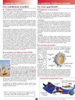
Increased vascular permeability, observed as macular edema, is a common character-
istic of diabetic retinopathy, with a prevalence of 20.1% and 25.4% of type 1 and
type 2 diabetic patients, respectively (10,11). Furthermore, 27% of patients in the
secondary intervention arm of the diabetes control and complications trial developed
macular edema within nine years (12). Indeed, loss of the blood–retinal barrier in
diabetic retinopathy is still one of the earliest detectable events in diabetic retinopa-
thy and macular edema is the clinical feature most closely associated with loss of
vision (13). Loss of the blood–retinal barrier includes increased permeability in both
the blood vessels and retinal pigme nted epithelium but altered vascular permeability
appears to precede changes in the pigmented epithelium in diabetes (14). In addition,
retinal vein occlusion results in blood–retinal barrier breakdown as seen upon vascu-
lar reperfusion, as does uveoretinitis and age-related macular degeneration. Changes
in the pigmented epithelium likely dominat e in the latter. Thus, loss of the normal
blood–retinal barrier is a common feature to many retinal degenerative diseases that
are the leading causes of vision loss in Western society, making development of
therapies to prevent loss of barrier properties or restore barrier properties a high
priority in vision resear ch.
Increased growth factor production from the neural retina and cytokine
production from inflammation both contribute to the loss of the blood–retinal
barrier in diabetic retinopathy. Changes in ocular growth factors and their receptors
include insulin-like growth factor 1 and its binding proteins, platelet-derived growth
factor, fibroblast growth factor, and vascular endothelial growth factor (VEGF) (15–
18). Immunohistochemistry and in situ hybridization studies demonstrate that the
expression of VEGF and its receptors increase by six months of experimentally
induced diabetes within the retinal parenchyma (19–21); in Goto–Kakizaki rats, a
model of type 2 diabetes, the level of hormone is significantly elevated over control
by 28-weeks. In addition, measurements of VEGF content in patients with prolifera-
tive diabetic retinopathy reveal that many, but not all patients, have increased
hormone in the vitreous fluid (22,23) and in epiretinal membranes (24). VEGF
expression in the retina occurs before the onset of proliferative retinopathy, suggest-
ing a role for this growth factor specifically in vascular permeability (25,26).
In addition to neural production of VEGF, inflammation contributes to
vascular permeability as well. Leukostasis increases in the capillaries of the retina
in animals made diabetic by streptozotocin. Inhibition of leukostasis with antibodies
Blood–Retinal Barrier 29
to adhesion molecule intracellular adhesion molecule (ICAM), which block the leu-
kocyte-endothelial interaction, also reduce retinal vascular permeability (27). The
contribution of various cytokines and chemokines to vascular permeability in diabetic
retinopathy are now under intense investigation and a functional role for these cyto-
kines in permeability has already been demonstrated (28). Furthermore, oxygen free-
radicals may cause disruption of the blood–retinal barrier. In vitro studies of the
retinal-pigmented epithelium (29) and endothelial cells (30,31) suggest that hydrogen
peroxide may disrupt barrier properties. Oxygen free-radical production may be due to
an inflammatory response, ischemia reperfusion, or, in the case of diabetes, from dys-
regulation of metabolism. Thus, the contribution of free-radical production on barrier
properties in disease states is an area in need of further study. These studies demon-
strate that multiple insults alter the blood–retinal barrier in diabetic retinopathy.
Understanding how diabetes changes the molecules that constitute this barrier may
provide a means to prevent or reverse the loss of the barrier regardless of the insult.
MOLECULAR ARCHITECTURE OF THE BLOOD–RETINAL BARRIER
Tight junctions confer the barrier properties to the endothelial cells within the retinal
vasculature creating the blood–retinal barrier. The tight junctions are composed of
two transmembrane proteins, occludin and claudin, known to provide barrier prop-
erties. These proteins are linked through adaptor proteins, such as the ZO family
members, to the cell actin cytoskeleton. Occludin and claudin share a common struc-
tural motif; specifically, both proteins span the membrane four times, creating two
extracellular loops that dimerize with proteins in the tight junction of adjacent
endothelial cells, helping to create the paracellular seal. How ever, occludin and
claudin contribute unique functionality to the tight junction. This chapter will focus
on how these transmembrane proteins are involved in barrier formation. Additional
junction-specific proteins may provide important differences to the composition and
function of the junctional complex between endothelial and epithelial cells. For
example, cingulin is an epithelial restricted tight junctio n protein (32,33) and
junction-enriched and associated protei n (JEAP) is an exocrine specific protei n
(34). However, the differences between endothelial cell and retinal pigmented epithe-
lial cell junctional proteins have not yet been characterized.
CLAUDINS
The claudins are made of at least 24 separate gene products whose expression helps
to determine b arrier properties of the tight junctions (35–38). Claudin family members
exhibit distinct tissue expression patterns (39–41). Claudin 5 expression is largely
restricted to the endothelium (42) but in some cases is expressed in retinal vasculature
as well (43). The brain endothelium also expresses claudin 1 (44); however, little has
been do ne to examine additional c laudin expression in the r etinal vasculature. Expres-
sion of claudins in cell lines th at normally lack tight junctions has helped in proposing
important principles. First, claudin expression in cells that do not express additional
junctional components shows that these cells are capable of forming limited strands
that mimic tight junctions in vivo (45). In contrast, occludin forms a punctate staining
pattern with much less extended tigh t junction-like strands (45). However, cotransfec-
tion of occludin with claudins results in occludin integration into the tight junction
30 Antonetti et al.
strands. Expression studies have also demonstrated that claudins can form homo-
meric and heteromeric complexes with specific restrictions. For example, coculture
studies with cells expressing claudin 1, 2, or 3 indicate that claud in 3 inter acts with
claudin 1 and claudin 2 on adjacent cells; however, claudin 1 and claudin 2 do not
interact (46). Finally, gene deletion studies have demonstrated the role of claudins
in barrier formation. Claudin 1-deficient mice die within one day of birth due to
transepidermal water loss (47). Specifically relevant to the blood–neural barrier,
claudin 5-deficient mice demonstrate increased permeability across the blood–retinal
barrier, specifically to molecules of less than 800 Da (48). These studies reveal that
claudins help to create the barriers that comprise the tight junctions.
Specific expression patterns of claudins provide the character of tight junctions,
particularly in relation to electrical resistance. Transfection experiments demonstrate
that expression of claudin isotypes can directly affect ion selectivity and conductance
(49). The effect of charge selectivity was most dramatically shown when three amino
acids in the first extracellular loop of claudin 15 were mutated from a negative charge
to a posit ive charge. This mutation changed the tight junction from allowing Na
þ
flux and prevent ing Cl
À
flux to becoming permissive for Cl
À
flux and inhibitory of
Na
þ
flux. Thus, claudins can form barriers to specific ions and create conductance
channels for other ions. To date, little is known regarding the nature of the tight
junctions in the retina in relation to ionic selectivity.
OCCLUDIN
Occludin is encoded by a single gene but may be alternatively spliced or initiated from
an alternative promoter, yielding novel variants (50,51). The expression of occludin
correlates well with the degree of barrier properties in various tissues. For example,
arterial endothelial cells express 18-fold more occludin protein than venous endothe-
lial cells and form a tighter solute barrier (52). Similarly, occludin is highly expressed
in brain endothelium coincident with the formation of the blood–brain barrier and is
expressed at much lower levels in endothelial cells of non-neuronal tissue, which have
less barrier properties (53). In the retina, the endothelium of the arteries, arterioles,
and capillaries express a relatively high degree of occludin that is well organized at
the cell border. In contrast, the venules and veins express a lower amount of occludin
and localization to the cell border is minimal (43,54).
A number of experiments, performed mostly in epithelial cells, demonstrate that
occludin contributes to the barrier function of tight junctions. Antisense oligonucleo-
tide experiments demonstrate a decrease in barrier properties associ ated with a reduc-
tion of occludin content (52). Expression of chicken occludin in Madin-Darby Canine
Kidney Epithelial (MDCK) cells under the control of an inducible promoter substan-
tially increa sed transcellular electrical resistance (TER) and increased the number of
tight junction strands compared to untreated cells (55). In contrast, synthetic peptides
targeting the second extracellular loop of occludin (OCC2) significantly decreased the
TER and increased the flux of several paracellular tracers in confluent monolayers of
a Xenopus kidney epithelial cell line (56). Furthermore, the OCC2 peptide promotes
the degradation of occludin by competitively inhibiting occludin-mediated cell-cell
adhesion. In a similar study, synthetic peptides homologous to regions of the first
extracellular loop of occludin prevented junction resealing after calcium depletion
and readdition, as measured by TER (57). These studies support a role for occludin
in barrier formation of tight junctions.
Blood–Retinal Barrier 31
Gene-deletion experiments have demonstrated a more complex role for occlu-
din in tight junction barrier formations. Embryonic stem cells from occludin null
mice formed cystic embryoid body structures with an outermost layer of epithelial
cells, simila r to wild-type embryon ic cells (58). Ultrast ructural analysis revealed no
changes in the tight junctions; the tight junction protein Z O-1 exhibited normal locali-
zation at apical junctional regions in the outermost layer of epithelial cells and no
change in barrier properties was observed in the occludin null cells. However, the
adult occludin homozygous null mice, although viable, possessed a host of abnor-
malities (59). Occludin-deficient mice exhibited postnatal growth retardation, male
knockout mice were infertile, and female knockout mice were unable to suckle their
litters. Overall, these mice exhibited abnormalities in the testis and salivary gland,
thinning of compact bone, calcium deposits in the brain, chronic gastritis, and hyper-
plasia of the gastric epithelium. In addition, recent studies using siRNA to occludin
demonstrate that occludin forms a barrier to organic acids up to 6.96 A
˚
, such as argi-
nine and choline (60). Thus, these studies have led to the hypothesis that occludin
contributes to the regulation of barrier properties by creating a doorway or regulated
pore through the tight junction.
Occludin associates with a number of structural and regulatory molecules
supporting a model in which occludin contributes to regulation of barrier properties.
The C-terminal cytoplasmic domain of occludin binds to ZO-1 in vitro (61), and this
interaction may serve to link occludin to the actin cytoske leton (62). Similarly, ZO-2
and ZO-3 bind to the C-terminus of occludin in vitro (63,64). In addition to this link
to the cell cytoskeleton, occludin interacts with a number of regulatory proteins at
tight junctions. Use of a 27 amino acid region of the C-terminus of occludin that
encodes a putative coiled–coiled domain helped identify several occludin-binding
proteins: protein kinase C-z, c-Yes, connexin- 26, and p85, the regulatory subunit
of phosphatidylinositol 3-kinase (65). Occludin may also interact with proteins via
its N-terminal cytoplasmic region. The E3 ubiquitin–protein ligase, Itch, was found
to associate with the N-terminus of occludin in vitro and in vivo, suggesting that occlu-
din content or localization may be regulated by ubiquitination (66). These protein–
protein interactions may regulate junction formation and barrier properties.
Occludin phosphorylation may provide a molecular mechanism to control
barrier properties. Studies from our group have demonstrated that both VEGF
and shear stress induce permeability across endothelial monolayers associated with
a rapid phosphorylation of occludin (67,68). The occludin phosphorylation was atte-
nuated by a non-hydrolyzable cAMP analog that also inhibits shear-induced perme-
ability (68). This phosphorylation of occludin appears to be seri ne or threonine
directed since immunoprecipitation of occludin and phosphotyrosine blotting did
not reveal any evidence of occludin tyrosine phosphorylation in this cell system
(unpublished observation). However, in epithelial cells, evidence of occludin tyrosine
phosphorylation exists (69). In addition, others have identified occludin phosphory-
lation in response to histamine (70) and use of brain extracts has helped identify
casein kinase II as an occludin kinase (71). Collectively, this work demonstrates a
close association of occludin phosphorylation with permeability. Future studies
identifying specific occludin phosphorylation sites, followed by mutational analysis,
should reveal the functional significance of occludin phosphorylation.
In addition to occludin phosphorylation, redistribution of occludin may
contribute to loss of the blood–retinal barrier. Both VEGF and diabetes induce a
redistribution of occludin from the plasma membrane to the cell cytoplasm
(43,54). A similar change in junction organization was observed in retinal-pigmented
32 Antonetti et al.
epithelial cells in response to hepatocyte growth factor (72). In an epithelial cell cul-
ture system, platelet-derived growth factor, a growth factor closely related to VEGF,
stimulated the redistribution of occludin and other tight junction proteins from the
plasma membrane to an early endosome compartment (73). Recent experiments sup-
port a model in which occludin recycles through an endosomal compartment (74)
and that endocytosis occurs through a clathrin-mediated pathway in epithelial cells
(75). One potential molecular mechanism for VEGF-regulated permeability includes
occludin phosphorylation releasing occludin from a neighboring endothelial tight
junction. Next, endocytosis of occludin leads to its translocation from the cell
plasma membrane to an internal compartment. However, many other possible
models exist to describe the data and future studies on both phosphorylation and
recycling of occludin and are necessary to elucidate the pathological mechanisms
for loss of endothelial barrier properties.
RESTORING BARRIER PROPERTIES
A number of therapies are currently unde r trial for diabetic retino pathy, therapies
that have been developed to prevent loss of vascular barrier function. These methods
include binding VEGF and preventing receptor activation through the use of a
VEGF aptamer (76) or a modified, soluble VEGF receptor, the VEGF trap
(77,78), or preventing VEGF signal transduction with the use of a protein kinase
C inhibitor (79). However, little has been done to consider induction of barrier prop-
erties once lost. Our laboratory and others have demonstrated that VEGF and dia-
betes reduce occludin content (80,81), increase occludin phosphorylation, and
stimulate occludin redistribution as described earlier. Glucocorticoids have been
used to treat brain tumors for over 35 years (82,83). Brain tumors possess a number
of similarities to diabetic retinopathy in relation to vascular changes. In both cases, a
blood–neural barrier characterized by a high degree of well-developed tight junctions
is altered leading to increased permeability. An increase in VEGF or inflammatory
cytokines is believed to contribute to the loss of barrier function. Given the success
of steroids to reverse vascular permeability, it is hypothesized that this steroid hor-
mone acts on the endothelial cells to induce formation of the tight junctions. Indeed,
our studies demonstrate that glucocorticoids directly act on endothelial cells to
increase expression of occludin and its assembly at the cell border, reduce occludin
phosphorylation, and increase barrier properties (84). The effect of glucocorticoids
on endothelial cells was also observed by Hoheisel et al. (85), who demonstrated that
hydrocortisone treatment increases TER nearly threefold and reduces sucrose per-
meability fivefold in pig brain capillary endothelial cells in a dose-responsive manner.
A positive effect of glucocorticoids on barrier properties has also been observed
in epithelial cells. Dexamethasone treatment for four days increases the electrical
resistance and reduces radiolabeled mannitol and insulin flux ac ross 31EG4 no ntrans-
formed epithelial cells (86) and the Con8 mammary epithelial tumor cell line (87).
Dexamethasone treatment increased ZO-1 content in the 31EG4 ce lls by slightly more
than twofold a fter four days trea tment w hile RNA content did n ot ch ange (88). This is
in contrast with the finding in bovine retinal endothelial cells in which ZO-1 content did
not change b ut its redistribution t o t he ce ll border dramatically increased w ith hydro-
cortisone treatment (84 ). The redistribution of Z O-1 was also observed in epithelial cells
and may be related to fascin expression, which is thought to bind to ZO-1 and retain
the protein in the cytoplasm (89,90). Glucocorticoids downregulate fascin and allow
Blood–Retinal Barrier 33
redistribution of ZO-1 to the cell border a nd o rgani zation o f tight junctions. Whether
a similar mechanism contributes to endothelial barrier induction in response to gluco-
corticoids remains unknown at present. Furthermore, others have demonstrated an
increase in occludin in response to glucocorticoids in epithelial cells (91). Thus, steroids
induce tight ju nction protein expression an d redistribution to t he plasma membrane in
epithelial and endothelial cell systems. Localized delivery of glucocorticoids may pro-
vide a means to restore barrier integrity and reduce inflammation in diabetic retino-
pathy (Fig. 2). However, given the risks associated with prolonged steroid use, it is
imperative to determine the molecular mechanisms by which glucocorticoids control
barrier properties so that novel, more specific therapies may be developed.
In conclusion, recent evidence indicates that permeability at the vascular
blood–retinal barrier is regulated by a number of tight junction proteins that act
together to protect the neural tissue. Diabetes leads to loss of the blood–retinal
barrier by altering the content, phosphorylation state, and localization of tight
junction proteins such as occludin. New treatment approaches are designed to target
the regulation of the tight junction proteins in order to prevent macular edema and
preserve vision in peop le with diabetes.
REFERENCES
1. Reese TS, Karnovsky MJ. Fine structural localization of a blood–brain barrier to exo-
genous peroxidase. J Cell Biol 1967; 34:207–217.
2. Shakib M, Cunha-Vaz JG. Studies on the permeability of the blood–retinal barrier. IV.
Junctional complexes of the retinal vessels and their role in the permeability of the blood–
retinal barrier. Exp Eye Res 1966; 5:229–234.
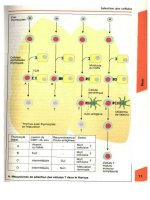
Figure 2 Diabetes leads to loss of the tight junctions while glucocorticoids induce assembly
of tight junctions. In diabetes, VEGF produced from the neural retina as well as inflammatory
cytokines cause phosphorylation of occludin, tight junction disassembly, and loss of tight
junction proteins. Steroids induce the synthesis of tight junction proteins, assembly of tight
junctions at the cell border, and dephosphorylation of occludin associated with increased
barrier properties in vitro and may induce barrier formation in vivo. Abbreviation: VEGF,
vascular endothelial growth factor.
34 Antonetti et al.
3. Cunha-Vaz JG, Shakib M, Ashton N. Studies on the permeability of the blood–retinal
barrier. I. On the existence, development, and site of a blood–retinal barrier. Br J
Ophthalmol 1966; 50:441–453.
4. Demeuse P, Kerkhofs A, Struys-Ponsar C, Knoops B, Remacle C, van den Bosch de
Aguilar P. Compartmentalized coculture of rat brain endothelial cells and astrocytes:
a syngenic model to study the blood–brain barrier. J Neurosci Methods 2002; 121:
21–31.
5. Duport S, Robert F, Muller D, Grau G, Parisi L, Stoppini L. An in vitro blood–brain
barrier model: cocultures between endothelial cells and organotypic brain slice cultures.
Proc Natl Acad Sci USA 1998; 95:1840–1845.
6. Stanness KA, Guatteo E, Janigro D. A dynamic model of the blood–brain barrier
‘‘in vitro.’’ Neurotoxicology 1996; 17:481–496.
7. Rubin LL, Hall DE, Porter S, et al. A cell culture model of the blood–brain barrier. J Cell
Biol 1991; 115:1725–1735.
8. Abbott NJ. Astrocyte–endothelial interactions and blood–brain barrier permeability.
J Anat 2002; 200:629–638.
9. Tout S, Chan-Ling T, Hollander H, Stone J. The role of Mu
¨
ller cells in the formation
of the blood–retinal barrier. Neuroscience 1993; 55:291–301.
10. Klein R, Klein BEK, Moss SE, Cruscishanks KJ. The wisconsin epidemiologic study
of diabetid retinopathy. XV. The long-term incidence of macular edema. Ophthalmology
1995; 102:7–16.
11. Vitale S, Maguire MG, Murphy RP, et al. Clinically significant macular edema in type I
diabetes. Incidence and risk factors. Ophthalmology 1995; 102:1170–1176.
12. Anonymous. Progression of retinopathy with intensive versus conventional treatment in
the diabetes control and complications trial. Diabetes control and complications trial
research group. Ophthalmology 1995; 102:647–661.
13. Moss SE, Klein R, Klein BE. The 14-year incidence of visual loss in a diabetic popula-
tion. Ophthalmology 1998; 105:998–1003.
14. Do Carmo A, Ramos P, Reis A, Proenca R, Cunha-vaz JG. Breakdown of the inner and
outer blood retinal barrier in streptozotocin-induced diabetes. Exp Eye Res 1998; 67:
569–575.
15. Aiello LP. Vascular endothelial growth factor and the eye: biochemical mechanisms of
action and implications for novel therapies. Ophthal Res 1997; 29:354–362.
16. Miller JW. Vascular endothelial growth factor and ocular neovascularization. Am J
Pathol 1997; 151:13–23.
17. Miller JW, Adamis AP, Aiello LP. Vascular endothelial growth factor in ocular
neovascularization and proliferative diabetic retinopathy. Diabetes Metab Rev 1997; 13:
37–50.
18. Paques M, Massin P, Gaudric A. Growth factors and diabetic retinopathy. Diabetes
Metab 1997; 23:125–130.
19. Gilbert RE, Vranes D, Berka JL, et al. Vascular endothelial growth factor and its recep-
tors in control and diabetic rat eyes. Lab Invest 1998; 78:1017–1027.
20. Hammes HP, Lin J, Bretzel RG, Brownlee M, Breier G. Upregulation of the vascular
endothelial growth factor/vascular endothelial growth factor receptor system in experi-
mental background diabetic retinopathy of the rat. Diabetes 1998; 47:401–406.
21. Murata T, Nakagawa K, Khalil A, Ishibashi T, Inomata H, Sueishi K. The relation
between expression of vascular endothelial growth factor and breakdown of the blood-
retinal barrier in diabetic rat retinas. Lab Invest 1996; 74:819–825.
22. Adamis AP, Miller JW, Bernal MT, et al. Increased vascular endothelial growth factor
levels in the vitreous of eyes with proliferative diabetic retinopathy. Am J Ophthalmol
1994; 118:445–450.
23. Aiello LP, Avery RL, Arrigg PG, et al. Vascular endothelial growth factor in ocular fluid
of patients with diabetic retinopathy and other retinal disorders. N Engl J Med 1994;
331:1480–1487.
Blood–Retinal Barrier 35
24. Chen YS, Hackett SF, Schoenfeld CL, Vinores MA, Vinores SA, Campochiaro PA.
Localization of vascular endothelial growth factor and its receptors to cells of vascular
and avascular epiretinal membranes. Br J Ophthalmol 1997; 81:919–926.
25. Amin RH, Frank RN, Kennedy A, Eliott D, Puklin JE, Abrams GW. Vascular endothe-
lial growth factor is present in glial cells of the retina and optic nerve of human subjects
with nonproliferative diabetic retinopathy. Invest Ophthalmal Vis Sci 1997; 38:36–47.
26. Lutty GA, McLeod DS, Merges C, Diggs A, Ploue
´
t J. Localization of vascular endo-
thelial growth factor in human retina and choroid. Arch Ophthalmol 1996; 114:
971–977.
27. Miyamoto K, Khosrof S, Bursell SE, et al. Prevention of leukostasis and vascular leakage
in streptozotocin-induced diabetic retinopathy via intercellular adhesion molecule-1 inhi-
bition. Proc Natl Acad Sci USA 1999; 96:10,836–10,841.
28. Luna JD, Chan CC, Derevjanik NL, et al. Blood-retinal barrier (BRB) breakdown in
experimental autoimmune uveoretinitis: comparison with VEGF, TNF, and IL1-B
mediated breakdown. J Neurosci Res 1997; 49:268–280.
29. Bailey TA, Kanuga N, Romero IA, Greenwood J, Luthert PJ, Cheetham ME. Oxidative
stress affects the junctional integrity of retinal pigment epithelial cells. Invest Ophthalmol
Vis Sci 2004; 45:675–684.
30. Kevil CG, Oshima T, Alexander JS. The role of p38 MAP kinase in hydrogen peroxide
mediated endothelial solute permeability. Endothelium 2001; 8:107–116.
31. Kevil CG, Oshima T, Alexander B, Coe LL, Alexander JS. H(2)O(2)-mediated perme-
ability: role of MAPK and occludin. Am J Physiol Cell Physiol 2000; 279:C21–C30.
32. D’Atri F, Nadalutti F, Citi S. Evidence for a functional interaction between cingulin and
ZO-1 in cultured cells. J Biol Chem 2002; 277:27,757–27,764.
33. Cordenonsi M, D’Atri F, Hammar E, et al. Cingulin contains globular and coiled-coil
domains and interacts with ZO-1, ZO-2, ZO-3, and myosin. J Cell Biol 1999; 147:
1569–1582.
34. Nishimura M, Kakizaki M, Ono Y, et al. JEAP, a novel component of tight junctions in
exocrine cells. J Biol Chem 2002; 277:5583–5587.
35. Mitic LL, van Itallie CM, Anderson JM. Molecular physiology and pathophysiology of
tight junctions—I. Tight junction structure and function: lessons from mutant animals
and proteins. Am J Physiol Gastrointest Liver Physiol 2000; 279:G250–G254.
36. Kniesel U, Wolburg H. Tight junctions of the blood–brain barrier. Cell Mol Neurobiol
2000; 20:57–76.
37. Fanning AS, Mitic LL, Anderson JM. Transmembrane proteins in the tight junction
barrier. J Am Soc Nephrol 1999; 10:1337–1345.
38. Tsukita S, Furuse M. Occludin and claudins in tight-junction strands: leading or support-
ing players? Trends Cell Biol 1999; 9:268–273.
39. Morita K, Furuse M, Fujimoto K, Tsukita S. Claudin multigene family encoding four-
transmembrane domain protein components of tight junction strands. Proc Natl Acad
Sci USA 1999; 96:511–516.
40. Li WY, Huey CL, Yu AS. Expression of claudin-7 and -8 along the mouse nephron.
Am J Physiol Renal Physiol 2004; 286:F1063–F1071.
41. Rahner C, Mitic LL, Anderson JM. Heterogeneity in expression and subcellular localiza-
tion of claudins 2, 3, 4, and 5 in the rat liver, pancreas, and gut. Gastroenterology 2001;
120:411–422.
42. Morita K, Sasaki H, Furuse M, Tsukita S. Endothelial claudin: claudin-5/TMVCF
constitutes tight junction strands in endothelial cells. J Cell Biol 1999; 147:185–194.
43. Barber AJ, Antonetti DA. Mapping the blood vessels with paracellular permeability in
the retinas of diabetic rats. Invest Ophthalmol Vis Sci 2003; 44:5410–5416.
44. Lippoldt A, Kniesel U, Liebner S, et al. Structural alterations of tight junctions are asso-
ciated with loss of polarity in stroke-prone spontaneously hypertensive rat blood–brain
barrier endothelial cells. Brain Res 2000; 885:251–261.
36 Antonetti et al.
45. Furuse M, Sasaki H, Fujimoto K, Tsukita S. A single gene product, claudin-1 or -2,
reconstitutes tight junction strands and recruits occludin in fibroblasts. J Cell Biol
1998; 143:391–401.
46. Furuse M, Sasaki H, Tsukita S. Manner of interaction of heterogeneous claudin species
within and between tight junction strands. J Cell Biol 1999; 147:891–903.
47. Furuse M, Hata M, Furuse K, et al. Claudin-based tight junctions are crucial for the
mammalian epidermal barrier: a lesson from claudin-1-deficient mice. J Cell Biol 2002;
156:1099–1111.
48. Nitta T, Hata M, Gotoh S, et al. Size-selective loosening of the blood–brain barrier in
claudin-5-deficient mice. J Cell Biol 2003; 161:653–660.
49. Van Itallie CM, Fanning AS, Anderson JM. Reversal of charge selectivity in cation or
anion-selective epithelial lines by expression of different claudins. Am J Physiol Renal
Physiol 2003; 285:F1078–F1084.
50. Muresan Z, Paul DL, Goodenough DA. Occludin 1B, a variant of the tight junction
protein occludin. Mol Biol Cell 2000; 11:627–634.
51. Mankertz J, Waller JS, Hillenbrand B, et al. Gene expression of the tight junction protein
occludin includes differential splicing and alternative promoter usage. Biochem Biophys
Res Commun 2002; 298:657–666.
52. Kevil CG, Okayama N, Trocha SD, et al. Expression of zonula occludens and adherens
junctional proteins in human venous and arterial endothelial cells: role of occludin in
endothelial solute barriers. Microcirculation 1998; 5:197–210.
53. Hirase T, Staddon JM, Saitou M, et al. Occludin as a possible determinant of tight junc-
tion permeability in endothelial cells. J Cell Sci 1997; 110:1603–1613.
54. Barber AJ, Antonetti DA, Gardner TW. Altered expression of retinal occludin and glial
fibrillary acidic protein in experimental diabetes. Invest Ophthalmol Vis Sci 2000;
41:3561–3568.
55. Balda MS, Whitney JA, Flores S, Gonzalez M, Cereijido M, Matter K. Functional dis-
sociation of paracellular permeability and transepithelial electrical resistance and disrup-
tion of the apical-basolateral intramembrane diffusion barrier by expression of a mutant
tight junction membrane protein. J Cell Biol 1996; 134:1031–1049.
56. Wong V, Gumbiner BM. A synthetic peptide corresponding to the extracellular domain of
occluding perturbs the tight junction permeability barrier. J Cell Biol 1997; 136:399–409.
57. Lacaz-Vieira F, Jaeger MM, Farshori P, Kachar B. Small synthetic peptides homologous
to segments of the first external loop of occludin impair tight junction resealing. J Membr
Biol 1999; 168:289–297.
58. Saitou M, Fujimoto K, Doi Y, et al. Occludin-deficient embryonic stem cells can differ-
entiate into polarized epithelial cells bearing tight junctions. J Cell Biol 1998; 141:
397–408.
59. Saitou M, Furuse M, Sasaki H, et al. Complex phenotype of mice lacking occludin, a
component of tight junction strands. Mol Biol Cell 2000; 11:4131–4142.
60. Yu AS, McCarthy KM, Francis SA, et al. Knock down of occludin expression leads to
diverse phenotypic alterations in epithelial cells. Am J Physiol Cell Physiol 2005;
288(6):C1231–C1241.
61. Furuse M, Itoh M, Hirase T, et al. Direct association of occludin with ZO-1 and its
possible involvement in the localization of occludin at tight junctions. J Cell Biol 1994;
127:1617–1626.
62. Fanning AS, Jameson BJ, Jesaitis LA, Anderson JM. The tight junction protein ZO-1
establishes a link between the transmembrane protein occludin and the actin cytoskele-
ton. J Biol Chem 1998; 273:29,745–29,753.
63. Haskins J, Gu L, Wittchen ES, Hibbard J, Stevenson BR. ZO-3, a novel member of
the MAGUK protein family found at the tight junction, interacts with ZO-1 and occlu-
din. J Cell Biol 1998; 141:199–208.
Blood–Retinal Barrier 37
64. Itoh M, Morita K, Tsukita S. Characterization of ZO-2 as a MAGUK family member
associated with tight as well as adherens junctions with a binding affinity to occludin
and alpha catenin. J Biol Chem 1999; 274:5981–5986.
65. Nusrat A, Chen JA, Foley CS, et al. The coiled-coil domain of occludin can act to orga-
nize structural and functional elements of the epithelial tight junction. J Biol Chem 2000;
275:29,816–29,822.
66. Traweger A, Fang D, Liu YC, et al. The tight junction-specific protein occludin is a func-
tional target of the E3 ubiquitin-protein ligase itch. J Biol Chem 2002; 277:10,201–10,208.
67. Antonetti DA, Barber AJ, Hollinger LA, Wolpert EB, Gardner TW. Vascular endothe-
lial growth factor induces rapid phosphorylation of tight junction proteins occludin and
zonula occluden 1. A potential mechanism for vascular permeability in diabetic retino-
pathy and tumors. J Biol Chem 1999; 274:23,463–23,467.
68. DeMaio L, Chang YS, Gardner TW, Tarbell JM, Antonetti DA. Shear stress regulates
occludin content and phosphorylation. Am J Physiol Heart Circ Physiol 2001; 281:
H105–H113.
69. Chen YH, Lu Q, Goodenough DA, Jeansonne B. Nonreceptor tyrosine kinase c-Yes
interacts with occludin during tight junction formation in canine kidney epithelial cells.
Mol Biol Cell 2002; 13:1227–1237.
70. Hirase T, Kawashima S, Wong EY, et al. Regulation of tight junction permeability and
occludin phosphorylation by Rhoa-p160ROCK-dependent and -independent mecha-
nisms. J Biol Chem 2001; 276:10,423–10,431.
71. Smales C, Ellis M, Baumber R, Hussain N, Desmond H, Staddon JM. Occludin phos-
phorylation: identification of an occludin kinase in brain and cell extracts as CK2. FEBS
Lett 2003; 545:161–166.
72. Jin M, Barron E, He S, Ryan SJ, Hinton DR. Regulation of RPE intercellular junction
integrity and function by hepatocyte growth factor. Invest Ophthalmol Vis Sci 2002;
43:2782–2790.
73. Harhaj NS, Barber AJ, Antonetti DA. Platelet-derived growth factor mediates tight
junction redistribution and increases permeability in MDCK cells. J Cell Physiol 2002;
193:349–364.
74. Morimoto S, Nishimura N, Terai T, et al. Rab13 mediates the continuous endocytic
recycling of occludin to the cell surface. J Biol Chem 2005; 280:2220–2228.
75. Ivanov AI, Nusrat A, Parkos CA. Endocytosis of epithelial apical junctional proteins by
a clathrin-mediated pathway into a unique storage compartment. Mol Biol Cell 2004; 15:
176–188.
76. Carrasquillo KG, Ricker JA, Rigas IK, Miller JW, Gragoudas ES, Adamis AP. Con-
trolled delivery of the anti-VEGF aptamer EYE001 with poly(lactic-co-glycolic)acid
microspheres. Invest Ophthalmol Vis Sci 2003; 44:290–299.
77. Qaum T, Xu Q, Joussen AM, et al. VEGF-initiated blood–retinal barrier breakdown in
early diabetes. Invest Ophthalmol Vis Sci 2001; 42:2408–2413.
78. Saishin Y, Saishin Y, Takahashi K, et al. VEGF-TRAP(R1R2) suppresses choroidal
neovascularization and VEGF-induced breakdown of the blood–retinal barrier. J Cell
Physiol 2003; 195:241–248.
79. Aiello LP, Bursell SE, Clermont A, et al. Vascular endothelial growth factor-induced
retinal permeability is mediated by protein kinase C in vivo and suppressed by an orally
effective ß-isoform-selective inhibitor. Diabetes 1997; 46:1473–1480.
80. Antonetti D, Barber A, Khin S, et al. Vascular permeability in experimental diabetes is
associated with reduced endothelial occludin content. Diabetes 1998; 47:1953–1959.
81. Wang W, Dentler WL, Borchardt RT. VEGF increases BMEC monolayer permeability
by affecting occludin expression and tight junction assembly. Am J Physiol Heart Circ
Physiol 2001; 280:H434–H440.
82. French L, Galicich J. The use of steriods for control of cerebral edema. Clin Neurosurg
1962; 10:212–223.
38 Antonetti et al.
83. Ruderman NB, Hall TC. Use of glucocorticoids in the palliative treatment of metastatic
brain tumors. Cancer 1965; 18:298–306.
84. Antonetti DA, Wolpert EB, DeMaio L, Harhaj NS, Scaduto RC. Hydrocortisone
decreases retinal endothelial cell water and solute flux coincident with increased content
and decreased phosphorylation of occludin. J Neurochem 2002; 80:667–677.
85. Hoheisel D, Nitz T, Franke H, et al. Hydrocortisone reinforces the blood–brain proper-
ties in a serum free cell culture system [corrected and republished article originally printed
in Biochem Biophys Res Commun 1998; 244(1):312–316]. Biochem Biophys Res
Commun 1998; 247:312–315.
86. Zettl KS, Sjaastad MD, Riskin PM, Parry G, Machen TE, Firestone GL. Glucocorti-
coid-induced formation of tight junctions in mouse mammary epithelial cells in vitro.
Proc Natl Acad Sci USA 1992; 89:9069–9073.
87. Buse P, Woo PL, Alexander DB, et al. Transforming growth factor-alpha abrogates
glucocorticoid-stimulated tight junction formation and growth suppression in rat mam-
mary epithelial tumor cells. J Biol Chem 1995; 270:6505–6514.
88. Singer KL, Stevenson BR, Woo PL, Firestone GL. Relationship of serine/threonine
phosphorylation/dephosphorylation signaling to glucocorticoid regulation of tight junc-
tion permeability and ZO-1 distribution in nontransformed mammary epithelial cells.
J Biol Chem 1994; 269:16,108–16,115.
89. Guan Y, Woo PL, Rubenstein NM, Firestone GL. Transforming growth factor-alpha
abrogates the glucocorticoid stimulation of tight junction formation and reverses the
steroid-induced down-regulation of fascin in rat mammary epithelial tumor cells by a
Ras-dependent pathway. Exp Cell Res 2002; 273:1–11.
90. Wong V, Ching D, McCrea PD, Firestone GL. Glucocorticoid down-regulation of fascin
protein expression is required for the steroid-induced formation of tight junctions and
cell–cell interactions in rat mammary epithelial tumor cells. J Biol Chem 1999; 274:
5443–553.
91. Stelwagen K, McFadden HA, Demmer J. Prolactin, alone or in combination with gluco-
corticoids, enhances tight junction formation and expression of the tight junction protein
occludin in mammary cells. Mol Cell Endocrinol 1999; 156:55–61.
Blood–Retinal Barrier 39
3
Neuroprotection
Dennis W. Rickman and Mel issa J. Mahoney
Departments of Ophthalmology and Neurobiology, Duke University Medical Center,
Durham, North Carolina, U.S.A.
INTRODUCTION
The mammalian retina comprises a rich, heterogeneous mosaic of neuronal morpho-
logical phenotypes intermeshed in an intricate pattern of synaptic connectivity. This
cellular diversity is further amplified by a wide variety of neurochemical phenotypes,
defined by specific expression patterns of numerous neurotransmitters, receptors, and
transporters, as well as intracellular regulators such as calcium binding proteins. This
complexity confers, in part, regional specializations in the retinal wiring and reflects
distinct regional metabolic requirements of retinal neurons. An implication of this
cellular diversity is that populations of retinal neurons exhibit differential vulnerabil-
ity to a variety of diseases or injuries, including genetic, environmental, and metabolic
insults. The endpoint for all of these insults is neuronal cell death (either necrotic or
apoptotic), and a major challenge in ophthalmology is to prevent or delay retinal
neuron loss, even in the face of a continued disease process—hence, neuroprotection.
Differential, or selective, vulnerability of retinal neurons also suggests multiple
potential targets for cell- or pharmacological-based neuroprotective interventions.
The goal of this chapter is to review the expression patterns of a number of cellular
and molecular targets (i.e., cell surface receptors, intracellular regulators of cell
death, and differential sensitivity to trophic factors) that may underlie a particular
nerve cell’s predilection for survival or death in the face of disease.
EXCITOTOXICITY AS A STIMULUS FOR NEURONAL CEL L DEATH
Excitatory neurotransmission in the central nervous system (CNS), including the
retina, is accomplished primarily by the amino acid glutamate. In the retina, gluta-
mate is released by photoreceptors, bipolar cells, and ganglion cells, presynaptically,
in normal neurotransmission (1–6). Normally, glutamate is rapidly removed from
the extracellular space following its release at the synapse. Glutamate removal is
accomplished both by binding to specific postsynaptic receptors that mediate activa-
tion of the postsynaptic cell and removal by glutamate transporters located in the
41
plasma membranes of both neurons and Mu
¨
ller glial cells. Thus, glutamate in the
synaptic cleft is typically maintained at a low level (7,8).
Multiple glutamate receptor subtypes have been identified, and these are charac-
terized based on their sensitivities to different glutamate receptor analogs (Table 1) (1).
Glutamate binds to both ionotropic receptors, which, in heteromeric association,
form ion channels in the neuronal plasma membrane, and metabotropic receptors,
which are coupled to G-protein-mediated pathways. Ionotropic glutamate receptors
(iGluR) can be further subdivided into those that bind glutamate and its analog
N-methyl-
D-aspartate (NMDA, i.e., NMDA receptors) and those that are sensitive
to kainate, alpha-amino-3-hydroxy-5-methyl-4-isoxazoleproprionic acid (AMPA),
and quisqualate (i.e., non-NMDA receptors). Binding to the NMDA receptor is
further characterized by a preferential increase in Ca
2þ
permeability. In the retina,
NMDA-type glutamate receptors are localized to ganglion cells and to some amacrine
cells (9–13), and their responses can be blocked by the selective antagonists MK-801,
AP-5 (2-amino-5-phosphonopentanoic acid), and AP-7 (2-amino-7-phosphoheptanoic
acid) (14,15).
There is considerable evidence that overstimulation of glutamate receptors
promotes cell death in a number of retinal disease processes. Glutamate overstimula-
tion may be particularly important in acute ischemic injuries, but it also may play a
role in diabetic retinopathy (16) and chronic neurodegenerative processes such as
glaucoma (17). Evidence for this includes the observation that glutamate levels are
elevated in the vitreous of patients with these conditions (16,17).
Retinal ischemic injury, for example, has been shown to result in the oversti-
mulation of ionotr opic glutamate receptors following the extracell ular accumulation
of glutamate (18,19). This effect appears to involve, in particular, the NMDA
class of glutamate receptors. The subsequent excessive influx of calcium results in
Table 1 Glutamate Receptor Functional Subtypes and Gene Subunits
Note: Glutamate receptors are subdivided into two main functional classes, those that regulate ion
channels (ionotropic receptors) and those that activate second messenger systems through activation of
G-coupled proteins (metabotropic receptors). The ionotropic receptors are further subdivided into sub-
types, based on the specificity of their activation by specific ligands (NMDA, AMPA, or kainate). The
molecular structure of these subtypes is conferred by the patterns of expression of specific genes. Similarly,
the metabotropic receptors either stimulate the generation of IP
3
(and diacylglycerol) resulting in an
increase in intracellular Ca
2þ
(Class I) or, alternatively, inhibit adenylate cyclase and stimulate the genera-
tion of cAMP (Classes II and III). Likewise, these classes are conferred by the specific gene expression.
Abbreviations: NMDA, N-methyl-
D-aspartate; AMPA, alpha-amino-3-hydroxy-5-methyl-4-isozazole
proprionic acid.
42 Rickman and Mahoney
the activation of intracellular pathways that trigger cell death, including the apopto-
tic cascade and generation of free radicals (described later).
Based on evidence that glutamate overstimulation is impor tant in ocular
neuronal cell death, an initial extracellular target for neurop rotective intervention
is the glutamate receptor. In the retina, a component of neurochemical diversity is
conferred by the differential distribution of glutamate receptor subunits to a variety
of retinal neurons. Thus, retinal neurons may have differential vulnerabilities to
injury. This suggests specific cellular targets for neuroprotection. Indeed, glutamate
receptor antagonists, such as memantine and MK-801, prolong survival of neurons
in glaucoma and ischemic injury, respectively (20–22). Memantine may have more
therapeutic benefit, in as much as it is a more specific uncompetitive antagonist and
has a voltage-dependent fast off rate, making it therapeutically safer.
Experimentally and therapeutically, a delicate balance must be reached
between blocking excitotoxicity while maintaining normal neurotransmitter receptor–
ligand interaction. Ultimately, this balance may be achieved by specifically targeting
glutamate receptor subunits at t he molecular level as op posed to a ‘‘sho tgun’’ app roach
with more gen eralized phar macological a ntagonists. Therefore, d elivery o f a gents that
target gene expression (e.g., viral constructs) or mRNA translation (e.g., antisense
oligonucleotides) may prove to offer more selective neuroprotection while preserving
overall neurotransmitter function. This approach assumes, however, a more complete
characterization of the populations of retinal neurons that display particular vulnerabil-
ities to excitotoxic damage.
INTRACELLULAR EFFECTORS OF CELL DEATH
Glutamate receptor overactivation that results from ischemic injury, and perhaps from
chronic neurodegenerative disease as well, is enhanced during reperfusion. Further-
more, there may be activation of effector pathways that result in the stimulation of
the apoptotic cascade (23,24,30). In particular, ionotrophic receptor overstimulation
appears to activate pathways that lead to cell death modulated at the level of the mito-
chondria. The process of programmed cell death, or apoptosis, is in part regulated by a
family of molecules related to the B cell leukemia-2 (bcl-2) gene product (Bcl-2) (21).
This proposed regulatory scheme is summarized in Figure 1. These molecules share lim-
ited sequences within three Bcl-2 homology domains (BH1, BH2, and BH3). Regulators
of cell death that enhance survival are Bcl-2, Bcl-X, and its splice variant Bcl-X
L
. Those
that induce apoptosis include Bax, Bak, and Bad (25–29). Molecules that interact at the
level of the inner mitochondrial membrane include the cell death promoter, Bax, and
the cell survival promoter, Bcl-2 (and closely related family members). In general,
homodimerization of Bax at the inner mitochondrial membrane creates ion channels
that allow the influx of ions into the mitochondrial matrix, swelling of the mitochon-
dria, and the subsequent release of cytochrome c into the cytoplasm. This further
activates cascades of cysteine proteases (caspases) that causes DNA fragmentation
and disrupts cytoskeletal integrity—hallmarks of apoptosis (23,24).
Additionally, Bax can form heterodimers with Bcl-2 or Bcl-X
L
. This leads to
the subsequent inability of Bcl-2 or Bcl-X
L
to homodimerize, resulting in a suppres-
sion of their protective effects against cell death. Recently, further characterization
of the Bcl-2 family has revealed that unbound (unphosphorylated) cytoplasmic
Bad also selectively heterodimerizes with Bcl-2 or Bcl-X
L
, displacing Bax and
promoting cell death by creating a cytoplasmic pool of free Bax (25–29).
Neuroprotection 43
On the other hand, Bcl-2 serves as a checkpoint for Bax activation by confer-
ring a protective effect through its heterodimerization with Bax, thus preventing
formation of ion channels in the mitochondrial membrane. Indeed, overexpression
of Bcl-2 results in abnormally high numbers of neurons surviving beyond the perina-
tal period, while Bcl-2-targeted deletion results in increased neuronal cell death
(described later).
Several members of the Bcl-2 family have been characterized in the mammalian
retina (32–42), and analyses of mice with targeted deletion or overexpression of
bcl-2-related genes are consistent with the model described previously. For instance,
bcl-2-deficient mice display a protracted loss of retinal ganglion cell axons well after
the period of developmental programmed cell death (35), whereas deletion of bax
results in a substantial increase in the number of ganglion cell axons in adult mice
(36,37). In contrast, bcl-2 overexpression increases the number of ganglion cells that
survive to adulthood and prevents ganglion cell death following optic nerve injury
(38–41). Furthermore, Isenmann et al. (33) reported that, following optic nerve
injury in the rat, there is an upregulation of Bax protein by retinal ganglion cells.
This increase preceded DNA fragmentation, supporting the notion that Bax is a reg-
ulator of retinal ganglion cell death. In this system, Bcl-2 expression by ganglion cells
appeared unchanged, further suggesting that the ratio of Bax to Bcl-2 favors cell
death in an optic nerve crush model.
Several lines of evidence suggest that an additional, finer level of control over
Bcl-2–Bax interaction may be achieved by the participation of related family mem-
bers such as Bad. In the retina, Bad is expressed predominantly by ganglion cells
(42). Normally, Bad is sequestered in the cytoplasm by the 14-3-3 class of proteins.
Extracellular growth or survival factors appear to affect survival, in part, through
Figure 1 Mechanisms of neuronal cell death. Homodimerization of a Bcl-2 family member,
Bax, in the mitochondrial membrane allows the influx of ions and the subsequent release
of cytochrome c from mitochondria. This activates cytoplasmic caspases, resulting in DNA
fragmentation and disruption of the cytoskeleton. The deleterious action of Bax can be inter-
rupted by the heterodimerization of Bax with Bcl-2 (or as shown here a related family member
Bcl-X
L
), resulting in cell survival. The availability of Bcl-2 is regulated, in part, by its hetero-
dimerization with Bad. This association promotes cell death by creating a free cytoplasmic
pool of Bax. See text for a complete description.
44 Rickman and Mahoney
activation of a pathway involving phosphoinositide 3 kinase (PI3-K) (43,44).
PI3-K, in turn, activates a serine–threonine protein kinase, Akt, that phosphorylates
Bad (45–47) and promotes its associati on with the 14-3-3 protein family (29). Thus
sequestered, Bad is unable to prevent the heterodimerization of Bcl-X
L
with Bax,
favoring cell survival.
In the adult rat brain, Bad is expressed exclusively by epithelial cells of the
choroid plexus (48), suggesting that Bad may play a critical role in regulating
the brain’s sensitivity to vascular-mediated environmental changes, including altera-
tions in the oxygen concentration of the blood. In the developing brain, howeve r,
Bad is expressed in neurons throughout the hippocampus and cerebral cortex,
neuronal populations that are particularly sensitive to ischemic insult, even in the
adult. Likewise, in the developing retina, Bad is highly expressed in ganglion cells
and numerous neurons in the inner nuclear layer—cells that may be particularly
vulnerable in ischemic retinopathies.
Interestingly, the pro-apoptotic effects of Bad are blocked by the immunosup-
pressants cyclosporin (CsA) and FK506. In a model of transient ischemia/reperfusion
following middle cerebral artery occlusion, both compounds reduced cerebral infarct
volume to 30% of control (49). Blockade of calcineurin-mediated dephosphorylation
of Bad is a potential mechanism for this effect. Thus phosphorylated, Bad remains
sequestered in the cytoplasm and is unable to bind to Bcl-2 (or Bcl-X
L
), thereby allow-
ing Bcl-2 to exert its protective effect. Regulating expression of specific members of
the Bcl-2 family by targeted gene expression is another potential therapeutic tool. This
approach is covered in Chapter 11.
OXIDATIVE STRESS AND THE GENERATION OF FREE RADICALS
One downstream consequence of neuronal injury, either from an acute ischemic event,
as the result of a chronic neurodegenerative process (such as a long-term elevation of
intraocular pressure) (50), or even from the normal aging process, is the generation
of reactive oxygen species (ROS) and free oxygen radicals (i.e., oxygen-containing
species with an unpaired electron, including
O
2
À
, OH,
NO, and ONOO
À
).
Normally, natural antioxidant mechanisms prevent interaction of free radicals with
cellular constituents, such as fatty acid side chains of membrane lipids (that could
be subjected to lipid peroxidation). They also protect the cell from nucleic acid break-
down and damage to cellular proteins (51). These na tural defenses include the anti-
oxidant enzyme superoxide dismutase (SOD), glutathione reductase and catalase,
and vitamins E and C. Indeed, treatments with various antioxidant compounds have
proved effective in maintaining retinal function following ischemia/reperfusion injury
(52–54). In fact, natural nutritional and antioxidant supplements have been suggested
to protect against photoreceptor loss in age-related macular degeneration and other
degenerative processes of aging (55–57).
Several murine models support a role for oxidative stress in neuronal degenera-
tion. For example, overexpression of SOD isoenzymes reduces both global and focal
ischemic injury in models of traumatic brain injury (58–60). Conversely, targeted
deletion of Cu, Zn-SOD and extracellular (EC)-SOD worsens the outcome of focal
ischemia (61,62). Recently, an especially intriguing protective effect has been
observed in a model of cerebral ischemia using middl e cerebral artery occlusion
(63–65). Application of the EC-SOD mimic, AEOL10113 (a metalloporphyrin cata-
lytic antioxidant) [manganese(III) meso-tetrakis (N-ethylpyridinium-2-yl) porphyrin],
Neuroprotection 45