Báo cáo y học: "Exome sequencing identifies a novel missense variant in RRM2B associated with autosomal recessive progressive external ophthalmoplegia" docx
Bạn đang xem bản rút gọn của tài liệu. Xem và tải ngay bản đầy đủ của tài liệu tại đây (613.98 KB, 7 trang )
RESEARCH Open Access
Exome sequencing identifies a novel missense
variant in RRM2B associated with autosomal
recessive progressive external ophthalmoplegia
Atsushi Takata
1,2,3
, Maiko Kato
4
, Masayuki Nakamura
4
, Takeo Yoshikawa
3
, Shigenobu Kanba
2
, Akira Sano
4
and
Tadafumi Kato
1*
Abstract
Background: Whole-exome sequencing using next-generation technologies has been previously demonstrated to
be able to detect rare dis ease-causing variants. Progressive external ophthalmoplegia (PEO) is an inherited
mitochondrial disease that follows either autosomal dominant or recessive forms of inheritan ce (adPEO or arPEO).
AdPEO is a genetically heterogeneous disease and several genes, including POLG1 and C10orf2/Twinkle, have been
identified as responsible genes. On the other hand, POLG1 was the only established gene causing arPEO with
mitochondrial DNA deletions. We previously reported a case of PEO with unidentified genetic etiology. The patient
was born of a first-cousin marriage. Therefore, the recessive form of inheritance was suspected.
Results: To identify the disease-causing variant in this patient, we subjected the patient’s DNA to whole-exome
sequencing and narrowed down the candidate variants using public data and runs of homozygosity analysis.
A total of 35 novel, putatively functional variants were detected in the homozygous segments. When we sorted
these variants by the conservation score, a novel missense variant in RRM2B, whose heterozygous rare variant had
been known to cause adPEO, was ranked at the top. The list of novel, putatively functional variants did not contain
any other variant in genes encoding mitochondrial proteins registered in MitoCarta.
Conclusions: Exome sequencing efficiently and effectively identified a novel, homozygous missense variant in
RRM2B, which was strongly suggested to be causative for arPEO. The findings in this study indicate arPEO to be a
genetically heterogeneous disorder, as is the case for adPEO.
Background
Massively parallel sequencing, also known as next gen-
eration-sequencing, is a revolutionary technology that
enables us to obtain large amounts of genomic sequence
information in an incomparably more rapid and less
expensive manner than before [ 1]. This te chnology is
applicable for various investi gations, includi ng resequen-
cing of full genomes or more targeted parts thereof for
discovery of genomic variations, genome-wide mapping
of structural rearrangements, transcriptome sequencing,
genome-wide epigenetic analysis, metagenomic sequen-
cing, and so on [2]. Whole-genome and whole-exome
(sequences of all protein-coding regions) resequencing
aiming at identification of causative variants for ra re,
inherited diseases is one of these applications, and have
demon strated t heir efficiency and effectiveness (reviewed
in [3]).
Previously, we reported a patient who had been born of
a first-c ousin marriage and was suspected to be affected
by inherited progre ssive external ophthalmoplegia (PEO)
[4]. Inherited PEO is a form of mitochondrial disease that
follows either autosomal dominant or recessive forms of
inheritance (adPEO (MIM 157640; 609283; 609286;
610131, 613077) or arPEO (MIM 258450)). The charac-
teristic findings of inherited PEOs are multiple mito-
chondri al DNA (mtDNA) deletions and ragged red fibers
in the muscle biopsy [5]. Typical clinical symptoms are
bilateral ptosis and paralysis of the extraocular muscle.
Other symptoms include exercise intolerance, cataracts,
hearing loss, sensory axonal neuropathy, optic atrophy,
* Correspondence:
1
Laboratory for Molecular Dynamics of Mental Disorders, RIKEN Brain Science
Institute, 2-1 Hirosawa, Wako-shi, Saitama 351-0198, Japan
Full list of author information is available at the end of the article
Takata et al. Genome Biology 2011, 12:R92
/>© 2011 Takata et al.; licensee BioMed Central Ltd. This is an open access article distributed under the terms of the Creative Com mons
Attribution License ( which perm its unrestricted use, distribution, and reproduction in
any medium, provided the original work is properly cited.
ataxia, depression, hypogonadism, and Parkinsonism
[6-10].
In the present case, the recessive form of inheritance
was suspected because of the patient’ s family history.
However, no pathogenic variant in POLG1 (MIM 174763),
which encodes a mitochondrial DNA polymerase and was
theonlyestablishedgenewhosevariantswereknownto
cause arPEO so far, was identified [4].
Theprobandinthisstudywastheonlychildandthe
available genetic information from family members was
limited. Therefore, it was almost impossible to identify the
causative variant using linkage analysis. On the other
hand, exome sequencing using a next-generation sequen-
cer has demonstrated its utility to detect causative variants
of rare disease using a small number of samples, especially
in the case of consanguineous family. Here, we performed
exome sequencing in combination with runs of homozyg-
osity (ROH) analysis in order to identify the causative
variant in this patient.
Results
Exome sequencing identifies a novel, homozygous
missense variant in RRM2B
A total of 3.2 Gb of sequence was generated from one lane
of sequencing using the Illumina Genome Analyzer II
(Illumina, San Diego, CA, USA). The proportion of the
targeted exome covered at 1×, 5× and 10× was 96.3%,
88.0% and 78 .3%, respectively. The mean coverage was
37.2×. A total of 19, 215 variants were detected in the cod-
ing regions defined by RefSeq Gene [11] and their flanking
splice sites. The number of detected coding variants does
not deviated greatly from that in previous repor ts [3,12].
After removing variants registered on the public database
of sequence variants (dbSNP, build 130) or found in eight
exomes of HapMap individuals [12] or the exome of a sin-
gle, healthy, unrelated Japanese individual, which was ana-
lyzed in the same run of Illumina Genome Analyzer II
sequencing, 1, 336 variant s remained. Among these, 592
variants, including 141 homozygous ones, were functional
(nonsense, missense, frameshift or splice site). Next, we
performed ROH analysis to narrow down the candidate
regions, using the base calling data on single nucleotide
variants in this patient. To enhance the accuracy of the
variant calling used for this analysis, 1) only the data of
single nucleotide variants were used and insertion/deletion
variants were excluded because of lower reliability of the
detection of insertion/deletion variants [13], 2) variants
called with coverage less than 8× w ere excluded, 3 ) var-
iants called with a coverage of more than 100× were
excluded because genomic regions that are known to be
duplicated or have similar sequences such as pseudogenes
tend to be read with high coverage. Because the primary
aim of this analysis was not to evaluate ROH segments
precisely, but to narrow down the list of candidate variants
without overlooking the causative variant, we used relaxed
criteria of RO H segments. The total size of RO H regions
was 992 Mb (about 32% of the genome), which was signifi-
cantly larger than the expected total size of ROH segments
in an offs pring born from a first cousin marriage (one-
eighth of the genome). A total of 35 novel and func tional
variants in 33 genes were identified in ROH segments. A
summary of the filtering strategy is given in Table 1.
When we sorted these listed variants by a conserva-
tion score (phyloP score) to identify those that were
most likely to be functional, a novel missense variant in
RRM2B (g.341G > A, p.P33S), whose rare, heterozygous
variant had been known to cause adPEO, was ranked at
the top (Table 2).
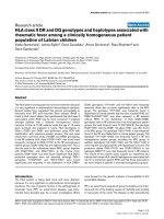
The existence of the RRM2B variant in the patient’ s
DNA was confirmed by Sanger sequencing (Figure 1a). As
expected, each of the parents had this variant in the het-
erozygous state. This varia nt changes an amino acid resi-
due that is highly conserved across 44 vertebrates (Figure
1b). Among 359 control subjects (718 chromosomes) of
Japanese origin, one subject carried this variant in the het-
erozygous state.
Exclusion of other variants that could cause PEO
In the list of 35 novel and functional variants in the ROH
segments, no other variants in genes encoding mitochon-
drial proteins were registered in Human MitoCarta [14].
We could not find any pathogenic mutations in other
genes known to cause mitochondrial diseases with multi-
ple mtDNA deletions (POLG1, POLG2 (MIM 604983),
C10orf2 (MIM 606075), SLC25A4 (MIM 103220), OPA1
(MIM 605290), TYMP (MIM 131222) and WFS1 (MIM
606201)) in exome analysis, as was observed in a previous
study using Sanger sequencing [4]. Although the mtDNA
sequence was not targete d by the SureSelect Human All
Exon Kit (Agilent, Santa Clara, CA, USA), 16, 558 of 16,
568 (99.9%) bases in mtDNA were read four or more
times due to its higher copy numbe r than nuclear DNA,
and no known pathogenic variant was found. Because of
the family history of the patient, we suspected that his
disease was caused by a recessive mutation. However,
there was another possibility that de novo variants affect
him in a dominant manner. To test this possibility, we
investigated whether he had de novo variants that could
explain his symptoms. In the list of 592 novel a nd puta-
tively functional variants, there were 26 heterozygous
variants in genes registered in MitoCarta. Among them,
five v ariants were not found in dbSNP132 or 1000 Gen-
ome Project data [15] (SNP calls released in June 2011),
and were located at conserved base positions (phyloP
score > 2 ). By performing Sanger sequencing, w e con-
firmed that all of these variants were not de novo,but
inherited from either of his healthy parents or found as a
false positive (Table 3).
Takata et al. Genome Biology 2011, 12:R92
/>Page 2 of 7
Evaluation of the amount of mtDNA
The mtDNA copy number relative to nuclear DNA in
the patient’ s skeletal muscle was not decreased, but
rather increased (Figure 2). As expected, the ND4/RNa-
seP ratio was lower than the ND1/RNaseP ratio in the
patient, which suggests increased levels of mtDNA dele-
tions that include the ND4 region, such as the 4, 977-bp
common mtDNA deletion [16]. This result indicated
that the clinical manifestation in the present patient was
not due to mtDNA depletion.
Discussion
In this study, we subjected DNA from a PEO patient with
unidentified genetic etiology to exome sequencing and
detected a novel, homozygous missense variant in
RRM2B. RRM2B encodes p53-inducible ribonucleotide
reductase small subunit 2-like protein (p53R2) and this
protein plays an essential role in the maintenance of
mtDNA by reducing ribonucleotides in the cytosol [17],
as is indicated by the fact that rare variants in this gene
cause various forms of mitochondrial diseases character-
ized by mtDNA depletion and deletions. To o ur knowl-
edge, 15 cases of mitochondrial depleti on syndrome
(MIM 612075) from 11 families [18-22] and one sporadic
case of mitochondrial neurogastrointestinal encephalopa-
thy [23] (MIM 603041) associated with homozygous or
compound heterozygous rare variants in RRM2B have
been reported. More recently, two families with adPEO
due to a heterozygous nonsense variant were described
[24]. In the screening of RRM2B variants in 50 mitochon-
drial disease patients without causative variants in
POLG1 and C10orf2, one Kearns-Sayre syndrome (MIM
530000) patient who carried two different novel missense
variants and one PEO patient who carried an in-frame
deletion were identified [25].
The clinical symptoms and findings in the muscle
biopsy of our case were typical for Mendelian-inherited
PEO. No members of his maternal family have shown
any neuromuscular symptoms, suggesting that the
mtDNA deletions of the patient were not maternally
inherited. Real-time quantitative PCR analysis revealed
that there was no mtDNA depletion. We did not observe
gastrointes tinal dysmotility, card iac conduction abnorm-
alities, pancreatic dysfunction and sensory ataxic neuro-
pathy, which are characteristic symptoms for other
mitochondrial diseases associated with mtDNA deletions,
namely mitochondrial neurogastrointestinal encephalo-
pathy, Kearns-Sayre syndrome, Pearson syndrome, and
Table 1 Summary of the filtering to narrow down the candidates for the causal variant
Criteria for the filtering Number of remaining variants
Coding variants 19, 215
Not in dbSNP130 2, 015
Not in eight HapMap exomes [12] 1, 833
Not in in-house data of a healthy Japanese individual 1, 336
Functional (missense, nonsense, frameshift and splice site) 592
In run-of-homozygosity regions 35 (in 33 genes)
The filtering was performed using the list ed criteria in descending order.
Table 2 List of novel and functional variants in run-of-homozygosity regions
Chromosome Position Reference allele Variant allele Variant calling/coverage Gene Amino acid change PhyloP score
8 103313660 G A 58/58 RRM2B Pro33Ser 6.741
1 39620317 G A 5/7* MACF1 Arg2523Gln; Arg3025Gln 5.329
4 107449465 A C 63/63 MGC16169 Asn34Lys 5.199
22 15980313 C T 5/5* LOC100287323 Val569Ile 4.997
11 64117795 G A 4/4* SLC22A12 Trp37Stp; Trp258Stp 4.945
10 29010439 G C 24/24 BAMBI Gly108Ala 4.878
20 49482400 G A 4/4* NFATC2 Ala778Val 4.437
1 238437608 C T 10/12 FMN2 Pro1101Leu 3.804
1 85362528 T - 65/69 WDR63 Splice site 3.503
3 99094433 A G 24/34 DKFZp667G2110 Lys546Glu 3.299
3 336547 T G 23/23 CHL1 Ser30Ala 3.014
3 46595758 C G 27/40* LRRC2 Arg41Gly 2.522
4 169335658 A C 9/13* ANXA10 Thr193Pro 2.257
5 140538797 C T 127/127 PCDHB8 Thr333Ile 2.011
Variants with PhyloP score > 2 are listed. Asterisks indicate variants with coverage < 8× or a variant calling/coverage ratio < 0.7; the reliability of these variant
calls is generally lower than that of the others.
Takata et al. Genome Biology 2011, 12:R92
/>Page 3 of 7
sensory ataxic neuropathy, dysarthria, and ophthalmopar-
esis (MIM 607459), respectively. Therefore, this patient
was diagnosed as having arPEO caused by a homozygous
missense variant of RRM2B.
Before this study, POLG1 had been the only estab-
lished gene responsible for arPEO, while adPEO is a
genetically heterogeneous disease, caused by rare var-
iants in POLG1, PO LG 2, C10orf2, S LC25A 4, OPA1 and
RRM2B. The resul ts of t his study identifying the second
responsible gene for arPEO indicate that arPEO is also a
genetically heterogeneous disease, as is the case for
adPEO.
The symptoms observed in this patient included major
depressive episodes. Frequent comorbidity of mood dis-
orders in patients of mito chondrial disease has been
generally re cognized [26] and several lines of evidences
have supported the possible involvement of mitochon-
drial dysfunctions in the pathophysiology of mood
g.341G>A, p.P33S
(a)
(b)
AND lortnoCAND tneitaP
L L S E E L L P E E
Figure 1 The identified disease-associated variant in RRM2B. (a) Partial seq uence of RRM2B in the p atient’ s DNA (left) and control DNA
(right). Red squares indicate the base position of the g.341G > A, p.P33S variant. (b) The substituted amino acid residue (red box) is highly
conserved across 44 vertebrate species (from the UCSC genome browser [31]).
Table 3 List of novel, putatively functional and heterozygous variants in mitochondrial genes
Chromosome Position Reference
allele
Variant
allele
Variant calling/
coverage
Gene Amino acid
change
PhyloP
score
Inheritance
7 30615756 G C 36/69 GARS Asp256His 6.494 Paternally inherited
10 104476790 T T 14/30 SFXN2 Leu73Pro 4.906 Maternally inherited
7 100670236 C C 20/51 FIS1 Ala90Pro 3.824 Maternally inherited
11 47620527 A A 3/8 MTCH2 Tyr23His 3.680 Not confirmed in Sanger
sequencing
1 10286026 C G 22/46 KIF1B Ile732Met 3.092 Maternally inherited
Variants with PhyloP score > 2 are listed.
Takata et al. Genome Biology 2011, 12:R92
/>Page 4 of 7
disorders [27]. So far, rare variants of POLG1, C10orf2
and SLC25A4 have been reported in inherited PEO ped-
igrees with frequent comorbidity of mood disorders
[28]. Given the typical symptoms of major depressive
disorder in the present case, RRM2B should be added to
the list of genes causa l for PEO associated with mood
disorders.
The identified P33S variant changes an amino acid resi-
due highly conserved among vertebrates. The amino-term-
inal region of p53R2, in which this altered amino acid is
located, is suggested to be crucial for interaction with p21
protein. p53R2 may contribute to DNA repair in coopera-
tion with p21 [29]. In its amino-terminal region, the
homozygous p.R41P variant was detected in a mitochon-
drial depletion syndrome case [21]. On the other hand,
other pathogenic missense variants have been located in
various sites of p53R2, including those involved in iron-
binding [18,20], those putatively crucial for homodimeriza-
tion of p53R2 [21,23] or heterotetramerization with the
RRM1 (ribonucleoside-diphosphate reductase large subu-
nit) homodimer [18,22], and so on. The relationships
between clinical phenotypes and the properties of variants,
as well as their underlying mechanisms, should be the sub-
ject of further investigations.
Conclusions
In this study, we describe a homozygous missense var-
iant in RRM2B that is strongly suggested to cause
arPEO. We were not only able to identify the disease-
associated variant, but could also exclude other candi-
dates (that is, variants in known PEO-related genes such
as POLG1, other mitochondrial genes in nucleic DNA
and mtDNA) using data from single exome sequencing.
This result further demonstrates the efficiency and effec-
tiveness of exome sequencing to detect causative var-
iants of rare , inherited, an d genetically heterogeneous
diseases.
Materials and methods
Clinical information of the patient
The detailed clinical history, family history and laboratory
data of t he studied subject are described elsewhere [4].
Briefly, a 43-year-old man presented with hearing loss,
bilateral ptosis, external ophthalmoplegia and muscle
weakness. Examinations revealed the existence of pigmen-
tary degeneration of the retina and gonadal atrophy. The
initial symptom of progressive h eari ng loss began at age
16 years. Depressive mood, anxiety and hypochondriacal
complaints were observed in his clinical course. His par-
ents were first cousins, he had no siblings, and no other
member of his family has a known history of neurological
illness. In the muscle biopsy, marked variation of muscle
fiber size, ragged red fibers, COX-negative fibers and mul-
tiple mtDNA deletions were detect ed. According to his
clinical history, family history and laboratory data, arPEO
was suspected.
The present study conformed to the Declaration of
Helsinki, and was approved by the RIKEN Wako Institute
Ethics Committee I, as well as the ethics committees of
Kagoshima U niversity Graduate School of Medical and
Dental Sciences and other pa rticipating institutes. Writ-
ten informed consent was obtained from every subject.
Exome sequencing and data analysis
TotalDNAwasobtainedfromperipheralbloodofthe
patient using st andard protocols. Total DNA (3 μg) was
sheared into approximately 300-bp fragments using a
Covaris sonicator (Covaris, Wobur n, MA, USA). A
paired-end exome library for Illumina sequencing was
prepared using the SureSelect Human All Exon Kit (Agi-
lent) following the manufacturer’s instructions. Massively
parallel sequencing was performed using one lane of the
Genome Analyzer II (Illumina) at RIKEN Omics Science
Center by the Life Science Acceler ator system. Base call-
ing was performed by the Illumina pipeline with default
parameters. Obtained reads were mapped against the
human reference genome (UCSC hg18/GRCh36) using
CLC Genomics Workbench v4.0.2 software (CLC Bio,
Aar hus, Denmark) with default parameters. Variant call -
ing was performed using the SNP and DIP detection
tools i n CLC Genomics Workbench v4.0.2 with default
parameters. Analysis o f ROH was performed using
PLINK software v1.0.7 [30]. The primary aim of this
0
0.5
1
1.5
2
2.5
ND1/RNaseP rati
o
ND4/RNaseP ratio
Patient
P
atient (rep)
Control-1
Control-2
Control-3
Control-4
Figure 2 Relative amounts of mtDNA in skeletal muscle tissues
from the patient and four control subjects. ND1/RNaseP and
ND4/RNaseP ratios calculated by real-time quantitative PCR were
used to evaluate mtDNA levels. The mtDNA level in the patient was
comparable to those of controls. Values are relative to the average
of all four controls.
Takata et al. Genome Biology 2011, 12:R92
/>Page 5 of 7
analysis was not to evaluate ROH segments precisely, but
to narrow down the list of candidate variants without
overlooking the causative variant. The refore, we used
relatively small (1, 000 kb) sliding windows for ROH seg-
ments, did not consid er local blocks of linkage disequili-
brium in the Japanese population, and did not exclude
thedataofvariantswhosefrequency was not registered
in dbSNP; those variants might not be polymorphic in
the Japanese population and possibly contributed to
extend the length of ROH. Conservation information for
the var iants among 44 vertebrate species (phyloP score)
was collected from the UCSC genome browser [31].
Sanger sequencing
Sanger sequencing of PCR amplicons was performed to
confirm the detected disease-associated variant using a
3730 × L DNA Analyser (Applied Biosystems, Foster City,
CA, USA). The primers used were: forward, 5’-AGGCA-
GACAGGCTCTCAAAC-3’; reverse, 5’-GGCAGAATTA-
GATGCCATTG-3’.
Real-time quantitative PCR
The amount of nuclear DNA and mtDNA in the skeletal
muscle of the patient and four age- and sex-matched con-
trols (all males aged 39 to 48 years) was evaluated by real-
time quantitative PCR analysis according to the previously
validated methods [32]. Briefly, copy numbers of RNaseP
(for nuclear DNA), ND1 and ND4 (for mtDNA) were eval-
uatedusingtheTaqManmethod(AppliedBiosystems).
Analysis of the patient’s tissue was performed in two inde-
pendent reactions, and each experiment was triplicated.
ND1/RNaseP and ND4/RNaseP ratios were calculated as 2
[Ct(RNaseP)-Ct(each gene)]
.
Data accessibility
The sequence data from this study have been submitted
to dbGaP [33] (study accession [phs000392.v1.p1]).
Abbreviations
adPEO: autosomal dominant progressive external ophthalmoplegia; arPEO:
autosomal recessive progressive external ophthalmoplegia; mtDNA:
mitochondrial DNA; PEO: progressive external ophthalmoplegia; ROH: runs of
homozygosity.
Acknowledgements
We would like to thank Dr Yu-ichi Goto (Department of Clinical Laboratory,
National Center Hospital for Mental, Nervous and Muscular Disorders,
National Center of Neurology and Psychiatry, Tokyo, Japan), who kindly
provided us with control muscle DNA samples from subjects without muscle
disease. We are grateful to the Support Unit for Bio-material Analysis
(Research Resources Center, RIKEN BSI) and LSA System Development Unit
(Omics Science Center, RIKEN Yokohama Institute) for performing exome
library construction and massively parallel sequencing.
Author details
1
Laboratory for Molecular Dynamics of Mental Disorders, RIKEN Brain Science
Institute, 2-1 Hirosawa, Wako-shi, Saitama 351-0198, Japan.
2
Department of
Neuropsychiatry, Graduate School of Medical Sciences, Kyushu University, 3-
1-1 Maidashi, Higashi-ku, Fukuoka 812-8582, Japan.
3
Laboratory for Molecular
Psychiatry, RIKEN Brain Science Institute, 2-1 Hirosawa, Wako-shi, Saitama
351-0198, Japan.
4
Department of Psychiatry, Kagoshima University Graduate
School of Medical and Dental Sciences, 8-35-1 Sakuragaoka, Kagoshima-shi,
Kagoshima 890-8520, Japan.
Authors’ contributions
AT and TK designed the study and drafted the manuscript. AT performed
data analysis and molecular experiments. MK, MN and AS performed clinical
assessment. MK, MN, TY and AS provided materials for experiments. TY, SK,
AS and TK coordinated the study and performed critical revision of the
manuscript. All authors read and approved the final manuscript.
Competing interests
The authors declare that they have no competing interests.
Received: 25 April 2011 Revised: 28 June 2011
Accepted: 28 September 2011 Published: 28 September 2011
References
1. Metzker ML: Sequencing technologies - the next generation. Nat Rev
2010, 11:31-46.
2. Shendure J, Ji H: Next-generation DNA sequencing. Nat Biotechnol 2008,
26:1135-1145.
3. Ng SB, Nickerson DA, Bamshad MJ, Shendure J: Massively parallel
sequencing and rare disease. Hum Mol Genet 2010, 19:R119-124.
4. Kato M, Nakamura M, Ichiba M, Tomiyasu A, Shimo H, Higuchi I, Ueno S,
Sano A: Mitochondrial DNA deletion mutations in patients with
neuropsychiatric symptoms. Neurosci Res 2010, 69:331-336.
5. Zeviani M, Servidei S, Gellera C, Bertini E, DiMauro S, DiDonato S: An
autosomal dominant disorder with multiple deletions of mitochondrial
DNA starting at the D-loop region. Nature 1989, 339:309-311.
6. Servidei S, Zeviani M, Manfredi G, Ricci E, Silvestri G, Bertini E, Gellera C, Di
Mauro S, Di Donato S, Tonali P: Dominantly inherited mitochondrial
myopathy with multiple deletions of mitochondrial DNA: clinical,
morphologic, and biochemical studies. Neurology 1991, 41:1053-1059.
7. Suomalainen A, Majander A, Haltia M, Somer H, Lonnqvist J, Savontaus ML,
Peltonen L: Multiple deletions of mitochondrial DNA in several tissues of
a patient with severe retarded depression and familial progressive
external ophthalmoplegia. J Clin Invest 1992, 90:61-66.
8. Melberg A, Lundberg PO, Henriksson KG, Olsson Y, Stalberg E: Muscle-
nerve involvement in autosomal dominant progressive external
ophthalmoplegia with hypogonadism. Muscle Nerve 1996, 19:751-757.
9. Suomalainen A, Majander A, Wallin M, Setala K, Kontula K, Leinonen H,
Salmi T, Paetau A, Haltia M, Valanne L, Lonnqvist J, Peltonen L, Somer H:
Autosomal dominant progressive external ophthalmoplegia with
multiple deletions of mtDNA: clinical, biochemical, and molecular
genetic features of the 10q-linked disease. Neurology 1997, 48:1244-1253.
10. Luoma P, Melberg A, Rinne JO, Kaukonen JA, Nupponen NN, Chalmers RM,
Oldfors A, Rautakorpi I, Peltonen L, Majamaa K, Somer H, Suomalainen A:
Parkinsonism, premature menopause, and mitochondrial DNA
polymerase gamma mutations: clinical and molecular genetic study.
Lancet 2004, 364:875-882.
11. The RefSeqGene Project [ />RSG/].
12. Ng SB, Turner EH, Robertson PD, Flygare SD, Bigham AW, Lee C, Shaffer T,
Wong M, Bhattacharjee A, Eichler EE, Bamshad M, Nickerson DA,
Shendure J: Targeted capture and massively parallel sequencing of 12
human exomes. Nature 2009, 461:272-276.
13. Ng SB, Bigham AW, Buckingham KJ, Hannibal MC, McMillin MJ,
Gildersleeve HI, Beck AE, Tabor HK, Cooper GM, Mefford HC, Lee C, Turner EH,
Smith JD, Rieder MJ, Yoshiura K, Matsumoto N, Ohta T, Niikawa N,
Nickerson DA, Bamshad MJ, Shendure J: Exome sequencing identifies MLL2
mutations as a cause of Kabuki syndrome. Nat Genet 2010, 42:790-793.
14. Pagliarini DJ, Calvo SE, Chang B, Sheth SA, Vafai SB, Ong SE, Walford GA,
Sugiana C, Boneh A, Chen WK, Hill DE, Vidal M, Evans JG, Thorburn DR,
Carr SA, Mootha VK: A mitochondrial protein compendium elucidates
complex I disease biology. Cell 2008, 134:112-123.
15. 1000
Genomes Project [ />16. He L, Chinnery PF, Durham SE, Blakely EL, Wardell TM, Borthwick GM,
Taylor RW, Turnbull DM: Detection and quantification of mitochondrial
Takata et al. Genome Biology 2011, 12:R92
/>Page 6 of 7
DNA deletions in individual cells by real-time PCR. Nucleic Acids Res 2002,
30:e68.
17. Pontarin G, Fijolek A, Pizzo P, Ferraro P, Rampazzo C, Pozzan T, Thelander L,
Reichard PA, Bianchi V: Ribonucleotide reduction is a cytosolic process in
mammalian cells independently of DNA damage. Proc Natl Acad Sci USA
2008, 105:17801-17806.
18. Bourdon A, Minai L, Serre V, Jais JP, Sarzi E, Aubert S, Chretien D, de
Lonlay P, Paquis-Flucklinger V, Arakawa H, Nakamura Y, Munnich A, Rotig A:
Mutation of RRM2B, encoding p53-controlled ribonucleotide reductase
(p53R2), causes severe mitochondrial DNA depletion. Nat Genet 2007,
39:776-780.
19. Acham-Roschitz B, Plecko B, Lindbichler F, Bittner R, Mache CJ, Sperl W,
Mayr JA: A novel mutation of the RRM2B gene in an infant with early
fatal encephalomyopathy, central hypomyelination, and tubulopathy.
Mol Genet Metab 2009, 98:300-304.
20. Kollberg G, Darin N, Benan K, Moslemi AR, Lindal S, Tulinius M, Oldfors A,
Holme E: A novel homozygous RRM2B missense mutation in association
with severe mtDNA depletion. Neuromuscul Disord 2009, 19:147-150.
21. Spinazzola A, Invernizzi F, Carrara F, Lamantea E, Donati A, Dirocco M,
Giordano I, Meznaric-Petrusa M, Baruffini E, Ferrero I, Zeviani M: Clinical and
molecular features of mitochondrial DNA depletion syndromes. J Inherit
Metab Dis 2009, 32:143-158.
22. Bornstein B, Area E, Flanigan KM, Ganesh J, Jayakar P, Swoboda KJ, Coku J,
Naini A, Shanske S, Tanji K, Hirano M, DiMauro S: Mitochondrial DNA
depletion syndrome due to mutations in the RRM2B gene. Neuromuscul
Disord 2008, 18:453-459.
23. Shaibani A, Shchelochkov OA, Zhang S, Katsonis P, Lichtarge O, Wong LJ,
Shinawi M: Mitochondrial neurogastrointestinal encephalopathy due to
mutations in RRM2B. Arch Neurol 2009, 66:1028-1032.
24. Tyynismaa H, Ylikallio E, Patel M, Molnar MJ, Haller RG, Suomalainen A: A
heterozygous truncating mutation in RRM2B causes autosomal-
dominant progressive external ophthalmoplegia with multiple mtDNA
deletions. Am J Hum Genet 2009, 85:290-295.
25. Pitceathly RD, Fassone E, Taanman JW, Sadowski M, Fratter C,
Mudanohwo EE, Woodward CE, Sweeney MG, Holton JL, Hanna MG,
Rahman S: Kearns-Sayre syndrome caused by defective R1/p53R2
assembly. J Med Genet 2011, 48:610-617.
26. Fattal O, Link J, Quinn K, Cohen BH, Franco K: Psychiatric comorbidity in
36 adults with mitochondrial cytopathies. CNS Spectrums 2007,
12:429-438.
27. Kato T: Molecular neurobiology of bipolar disorder: a disease of ‘mood-
stabilizing neurons’? Trends Neurosci 2008, 31:495-503.
28. Kasahara T, Kubota M, Miyauchi T, Noda Y, Mouri A, Nabeshima T, Kato T:
Mice with neuron-specific accumulation of mitochondrial DNA
mutations show mood disorder-like phenotypes. Mol Psychiatry 2006,
11:577-593, 523.
29. Xue L, Zhou B, Liu X, Heung Y, Chau J, Chu E, Li S, Jiang C, Un F, Yen Y:
Ribonucleotide reductase small subunit p53R2 facilitates p21 induction
of G1 arrest under UV irradiation. Cancer Res 2007, 67:16-21.
30. Purcell S, Neale B, Todd-Brown K, Thomas L, Ferreira MA, Bender D, Maller J,
Sklar P, de Bakker PI, Daly MJ, Sham PC: PLINK: a tool set for whole-
genome association and population-based linkage analyses. Am J Hum
Genet 2007, 81:559-575.
31. Genome browser at UCSC [].
32. Kakiuchi C, Ishiwata M, Kametani M, Nelson C, Iwamoto K, Kato T:
Quantitative analysis of mitochondrial DNA deletions in the brains of
patients with bipolar disorder and schizophrenia. Int J
Neuropsychopharmacol 2005, 8:515-522.
33. The Database of Genotypes and Phenotypes (dbGaP) [i.
nlm.nih.gov/gap].
doi:10.1186/gb-2011-12-9-r92
Cite this article as: Takata et al.: Exome sequencing identifies a novel
missense variant in RRM2B associated with autosomal recessive
progressive external ophthalmo plegia. Genome Biology 2011 12:R92.
Submit your next manuscript to BioMed Central
and take full advantage of:
• Convenient online submission
• Thorough peer review
• No space constraints or color figure charges
• Immediate publication on acceptance
• Inclusion in PubMed, CAS, Scopus and Google Scholar
• Research which is freely available for redistribution
Submit your manuscript at
www.biomedcentral.com/submit
Takata et al. Genome Biology 2011, 12:R92
/>Page 7 of 7