Fundamentals of Clinical Ophthalmology - part 6 pdf
Bạn đang xem bản rút gọn của tài liệu. Xem và tải ngay bản đầy đủ của tài liệu tại đây (433.28 KB, 23 trang )
40 Gayton JL, Sanders VN. Implanting two posterior
chamber intraocular lenses in a case of microphthalmos.
J Cataract Refract Surg 1993;19:776–7.
41 Hull CC, Liu CSC, Sciscio A. Image quality in
polypseudophakia for extremely short eyes. Br J
Ophthalmol 1999;83:656–63.
42 Shugar JK, Lewis C, Lee A. Implantation of multiple
foldable acrylic posterior chamber lenses in the capsular
bag for high hyperopia. J Cataract Refract Surg
1996;22(suppl 2):1368–72.
43 Findl O, Menapace R, Rainer G, Georgopoulos M.
Contact zone of piggyback acrylic intraocular lenses. J
Cataract Refract Surg 1999;25:860–2.
44 Gayton JL, Apple DJ, Peng Q, et al. Interlenticular
opacification: clinicopathological correlation of a
complication of posterior chamber piggyback intraocular
lenses. J Cataract Refract Surg 2000;26:330–6.
45 Shugar JK, Schwartz T. Interpseudophakos Elschnig
pearls associated with late hyperopic shift: a
complication of piggyback posterior chamber
intraocular lens implantation. J Cataract Refract Surg
1999;25:863–7.
46 Eleftheriadis H, Marcantonio J, Duncan G, Liu C.
Interlenticular opacification in piggyback AcrySof
intraocular lenses: explanation technique and laboratory
investigations. Br J Ophthalmol 2001;85:830–6.
47 Bradbury JA, Hillman JS, Cassells-Brown A. Optimal
postoperative refraction for good unaided near and
distance vision with monofocal intraocular lenses. Br J
Ophthalmol 1992;76:300–2.
48 Steinert RF, Aker BL, Trentacost DJ, Smith PJ,
Tarantino N. A prospective comparative study of the
AMO ARRAY zonal-progressive multifocal silicone
intraocular lens and a monofocal intraocular lens.
Ophthalmology 1999;106:1243–55.
49 Javitt JC, Wang F, Trentacost DJ, Rowe M, Tarantino
N. Outcomes of cataract extraction with multifocal
intraocular lens implantation: functional status and
quality of life. Ophthalmology 1997;104:589–99.
50 Cumming JS, Slade SG, Chayet A. Clinical evaluation
of the model AT-45 silicone accommodating intraocular
lens: results of feasibility and the initial phase of a Food
and Drug Administration clinical trial. Ophthalmology
2001;108:2005–9.
51 Arshinoff SA. Dispersive and cohesive viscoelastic
material in phacoemulsification. Ophthalmic Pract
1995;13:98–104.
52 Arshinoff SA. Dispersive-cohesive viscoelastic soft shell
technique. J Cataract Refract Surg 1999;25:167–173.
53 Corydon L, Thim K. Continuous circular capsulorhexis
and nucleus delivery in planned extracapsular cataract
extraction. J Cataract Refract Surg 1991;17:628–32.
54 Singh AD, Fang T, Rath R. Cartridge cracks during
foldable intraocular lens insertion. J Cataract Refract
Surg 1998;24:1220–1222.
55 Dick HB, Schwenn O, Fabian E, Neuhann T,
Eisenmann D. Cartridge cracks with different
viscoelastics. J Cataract Refract Surg 1998;25:463–465.
56 Miyake K, Ota I, Ichihashi S, Miyake S, Tanaka Y,
Terasaki H. New classification of capsular block
syndrome. J Cataract Refract Surg 1998;24:1230–4.
57 Masket S. Postoperative complications of capsulorhexis.
J Cataract Refract Surg 1993;19:721–4.
58 Oshika T, Nagata T, Ishii Y. Adhesion of lens capsule to
intraocular lenses of polymethyl methacrylate, silicone,
and acrylic foldable materials: an experimental study. Br
J Ophthalmol 1998;82:549–53.
59 Dua HS, Benedetto DA, Azuara-Blanco A. Protection
of corneal endothelium from irrigation damage: a
comparison of sodium hyaluronate and
hydroxypropylmethylcellulose. Eye 2000;14:88–92.
60 Henry JC, Olander K. Comparison of the effect of four
viscoelastic agents on early postoperative intraocular
pressure. J Cataract Refract Surg 1996;22:960–6.
61 Holzer MP, Tetz MR Auffarth GU, Welt R, Volcker
HE. Effect of Healon5 and 4 other viscoelastic
substances on intraocular pressure and endothelium
after cataract surgery. J Cataract Refract Surg 2001;27:
213–8.
62 Rainer G, Menapace R, Findl O, et al. Intraocular
pressure rise after small incision cataract surgery: a
randomised intraindividual comparison of two
dispersive viscoelastic agents. Br J Ophthalmol 2001;85:
139–42.
FOLDABLE INTRAOCULAR LENSES AND VISCOELASTICS
101
102
Before the widespread acceptance of
extracapsular techniques, the majority of
cataract surgery involved removal of the
cataractous lens, including its capsule, using the
intracapsular technique (Figure 8.1a).
Experience of extracapsular cataract extraction
(ECCE) had shown that if the posterior lens
capsule was preserved then it was likely to
become opaque, necessitating further surgery to
restore vision. However, correction of the high
degree of hypermetropia induced by
intracapsular cataract extraction (ICCE) was
not entirely satisfactory
1
because of the optical
properties of aphakic spectacles and difficulties
with contact lens usage in the age group prone to
cataract. The development of the intraocular
lens (IOL) made it possible to circumvent these
problems, but anterior chamber (Figure 8.2) or
iris fixated (Figures 8.2b and 8.3) lenses
implanted during intracapsular surgery were
associated with some ocular morbidity.
2
Attention then became focused on refining
the extracapsular technique to permit more
physiological lens implantation in the posterior
chamber. At about the same time, the
introduction of the neodymium : yttrium
aluminium garnet (Nd:YAG) laser permitted
outpatient management of posterior capsule
opacification. The improved extracapsular
technique (Figure 8.1b) permitted cataract
surgery to be timed according to patients’ visual
needs, unlike the intracapsular approach, in
which surgery was often deferred until visual loss
was marked and the physical properties of the
cataract were favourable to cryoextraction. As a
consequence, the extracapsular technique
became established as the principal means of
cataract extraction in the developed world.
However, advances in phacoemulsification
surgery and then foldable IOL technology have
provided more rapid visual rehabilitation and
fewer wound related complications. More
recently, this has resulted in a shift away from
use of the traditional extracapsular technique,
which has come to occupy a more circumscribed
role. Intracapsular extraction is now usually
reserved for unstable subluxed lenses in which
neither phacoemulsification or extracapsular
surgery is possible. An alternative to ICCE for
these cases is lensectomy (Figure 8.1c), in which
cataract surgery is combined with pars plana
vitrectomy.
Extracapsular cataract extraction
Indications for extracapsular technique
Although phacoemulsification is regarded as
the technique of choice for the bulk of cataract
surgical procedures, there are nonetheless
certain clinical contexts in which the
extracapsular approach may be preferred (Table
8.1). These include significant corneal opacity
that may preclude safe capsulorhexis or
phacoemulsification; marked endothelial cell loss,
in which postoperative corneal decompensation
8 Non-phacoemulsification
cataract surgery
may result; anterior capsular fibrosis preventing
capsulorhexis; and white or dark brown lenses,
which may be refractory to phacoemulsification.
In addition, if capsular complications or corneal
decompensation occur during phacoemulsification
surgery, then conversion to an extracapsular
approach may provide the best means of safely
completing the procedure. For these reasons, the
extracapsular technique represents an essential
skill for both trainee and trained surgeons.
In addition to these specific clinical
indications, there are circumstances in which the
extracapsular approach may be used for the
NON-PHACOEMULSIFICATION CATARACT SURGERY
103
a)
b)
c)
Figure 8.1 The various non-phacoemulsification
techniques for cataract extraction. (a) Intracapsular
cataract extraction: the entire lens, including the
capsule, is removed with a cryoprobe (arrow).
(b) Extracapsular cataract extraction: the anterior lens
capsule, lens nucleus and cortex are removed, and the
posterior lens capsule is left in situ. (c) Lensectomy:
during pars plana vitrectomy the lens is removed
using either ultrasound (fragmatome) or the vitrector
(seen here). Note that the anterior capsule may be
partly preserved.
a)
b)
c)
d)
Figure 8.2 Possible locations for an intraocular lens.
(a) Anterior chamber. (b) Iris fixated. (c) Sulcus
fixated. (d) Capsular bag.
majority of cataract surgery. Some surgeons
nearing retirement age who have a refined
extracapsular technique, may consider the
increased potential for surgical complications
associated with learning phacoemulsification
unjustified.
3
Alternatively, the capital outlay for
phacoemulsification equipment or the cost per
case of disposable surgical items may exceed
resources and prompt the adoption of an
extracapsular approach. Finally, in contrast to
phacoemulsification, extracapsular surgery can
be carried out with simple and portable
equipment, requiring little technical support;
this is an attractive attribute, especially in the
developing world.
4
Extracapsular technique
The widespread adoption of extracapsular
surgery throughout the world is reflected in the
diversity of its variations. The following account
of the technique therefore emphasises critical
phases in the procedure, outlines the approaches
that are commonly adopted in each phase, and
presents some of the factors that influence the
choice of approach, rather than specifying a
single technique.
Incision
The incision is made in the cornea, or at the
corneoscleral limbus, and is curved to maintain
a fixed point of entry into the anterior chamber
relative to the iris plane throughout its length.
Corneal incisions, being nearer the visual axis,
carry a greater risk of astigmatism than do limbal
incisions, but the potential for iris trauma may
be less. The length of the incision is based on the
size of the largest object to pass through it,
namely either the nucleus (if large and expressed
intact) or the IOL optic (if the nucleus is small
or techniques to reduce nuclear size are
adopted). Because the wound is curved, its
maximum dimension is not its circumferential
length but the straight line distance between its
ends (i.e. the chord length). The more the
wound is extended circumferentially, the less
the proportionate increase in chord length, but
the greater the potential for wound related
complications such as astigmatism (Figure 8.4).
It is thus desirable to keep the wound as short as
possible. Paradoxically, only a small increase in
wound length may permit expression of the
nucleus where previously it was not possible.
This is because the circumference of the wound
aperture increases by up to double the length
that the wound is enlarged (Figure 8.5).
The incision is commonly carried out as a two
stage procedure. The first is a partial thickness
cut in the limbus or cornea along the entire
length of the planned incision. At this stage, the
eye is firm and this assists accurate wound
construction. The small stab incision that
follows is sufficiently watertight to help preserve
anterior chamber depth during capsulotomy.
The second cut, converting the incision to a full
thickness wound, is made immediately before
CATARACT SURGERY
104
Figure 8.3 Iris clip lens in situ.
Table 8.1 Indications for extracapsular cataract
extraction
Eyes unfavourable to phacoemulsification:
Dense white/brown nucleus
Corneal opacity
Marked endothelial cell loss
Anterior capsular fibrosis
Intraoperative complications of phacoemulsification
Phacoemulsification learning curve complications
unacceptable
Phacoemulsification too expensive
Phacoemulsification logistically impractical
expression of the nucleus. The resulting incision
may be uniplanar, either perpendicular to
the cornea (Figure 8.6a) or backward (or
reverse) sloping to encourage a watertight seal
(Figure 8.6b). Alternatively, the incision may be
a biplanar construction, which also improves the
accuracy of wound apposition (Figure 8.6c).
Capsulotomy/capsulorhexis
An aperture can be made in the anterior lens
capsule by either capsulotomy or capsulorhexis,
commonly using a bent needle. Techniques for
capsulorhexis are discussed in Chapter 3. The
capsular edge of this type of opening is strong;
this property is useful in phacoemulsification
surgery, where integrity of the capsular bag is
essential for nuclear manipulation and in-the-
bag IOL insertion. In extracapsular surgery,
however, it may obstruct expression of the
nucleus, resulting in delivery of both nucleus
and capsule, so-called intracapsular delivery.
5
This creates difficulties in IOL placement and
risks vitreous complications. Radial relieving
incisions are therefore commonly made in the
capsulorhexis edge during extracapsular sugery
(Figure 8.7).
6
Capsulotomy may be performed either using
a “can opener” or endocapsular technique.
Can opener capsulotomy involves multiple
perforations made in a circular pattern in the
anterior lens capsule (Figure 8.8a), the centre of
which is then torn out (like a car tax disc; Figure
8.8b). This leaves a ragged capsular edge, which
presents little resistance to nucleus expression,
but may not be sufficient to secure placement of
the IOL inside the capsular bag. By contrast, the
technique used in endocapsular surgery employs
a linear capsulotomy (Figure 8.9), through
which the nucleus is expressed, cortex aspirated,
and the IOL inserted. Anterior capsulectomy is
then performed using either a can opener or
capsulorhexis-type approach. The endocapsular
NON-PHACOEMULSIFICATION CATARACT SURGERY
105
a) b) c)
Figure 8.4 The greater the circumferential extension (a–c) of the incision (solid line), the less the proportionate
increase in chord length (arrow) and maximum linear wound dimension.
a) b)
c) d)
Figure 8.5 Enlargement of the incision (solid line in
a) by a given amount (dotted line in c) produces double
the increase in wound circumference (b v d).
technique both protects the corneal endothelium
and facilitates placement of the IOL in the
capsular bag.
Nucleus manipulation
The separation of the nucleus from lens
cortex or capsule may assist its expression. In
part, this can be achieved by mechanically
dislocating the lens or injecting fluid between
capsule and lens (i.e. hydrodissection; see
Chapter 5). At this point the incision may
completed and the nucleus expressed. However,
given the desirability of minimising the length of
the incision, attempts may be made to reduce
the size of the lens before expression. The
nucleus may be separated from epinucleus
by injecting fluid between the two (i.e.
hydrodelamination; see Chapter 5) or by
mechanical fragmentation of the nucleus (in situ
nucleofractis),
7
for example with a wire snare
(Figure 8.10) or trisection. Such techniques may
permit expression of nuclear fragments through
a considerably smaller incision than would be
necessary to allow passage of the entire nucleus.
Expression is achieved either by application of
pressure to the eye, typically behind the
completed incision (Figure 8.11a), or by
injection of a viscous agent behind the nucleus
to expel it under positive pressure (i.e.
viscoexpression; Figure 8.11b).
Cortex aspiration
Following successful expression of the
nucleus, remnants of cortical lens matter
remain. These may be removed by manual
or automated systems, both of which
simultaneously maintain the anterior chamber
by fluid infusion and permit aspiration of soft
lens matter. By aspirating under the anterior lens
capsule, cortical lens matter is engaged, this is
then drawn centripetally and aspirated (see
Chapter 5). The process is repeated around the
CATARACT SURGERY
106
a) b) c)
Figure 8.6 Incision profiles. (a) Perpendicular to cornea. (b) Backward (reverse) sloping. (c) Stepped.
Figure 8.7 Continuous curvilinear capsulorhexis
with relieving incisions (arrows) to facilitate nucleus
expression and reduce the risk of intracapsular
delivery.
circumference of the capsular bag until no lens
matter remains. This requires a relatively
constant anterior chamber depth, and if this
cannot be achieved by appropriate construction
of the wound then it may be necessary to insert
temporary sutures to appose the wound.
Rigid intraocular lens insertion
To facilitate posterior chamber IOL insertion,
the capsular bag and anterior chamber should be
inflated with a viscoelastic agent. The incision
enables the implantation of a one-piece loop
haptic PMMA lens with a large optic diameter
(Figure 8.12). It is inserted along its long axis,
and once the leading haptic is in place, behind
the iris plane and within the capsular bag, the
trailing haptic may be rotated (or dialled) into
position (Figure 8.2d). Refilling the anterior
chamber with viscoelastic may facilitate this.
Some lens implants have dial holes drilled into
the optic to allow an instrument (for example, a
Sinskey hook) to obtain purchase on the IOL;
alternatively, the junction of the haptic and optic
is engaged. Dialling some polymethylmethacrylate
lenses into the capsular bag can be difficult,
particularly if there is an intact capsulorhexis. In
these cases the trailing haptic may be better
placed directly into the bag with forceps and a
bimanual technique employed, using a second
instrument to apply posterior pressure to the
lens optic. It is important to ensure that both
haptics are either in the bag or in the sulcus,
because if one haptic is in the bag and one in the
sulcus then the lens may become decentred.
Wound closure
The wound is closed, commonly with 10/0
monofilament nylon, using either multiple
interrupted sutures (Figure 8.13a) or as a single
continuous suture (Figure 8.13b). Continuous
sutures have the merit that suture tension is
distributed evenly along the wound, unlike
interrupted sutures, which may differ in tension.
There is, however some risk of translational
malposition of the wound with continuous
sutures, and selective suture removal to
counteract astigmatism is not possible (see
Chapter 12). Whichever technique is used,
knots and suture ends must lie beneath the
ocular surface to avoid irritation. This can be
achieved by rotating interrupted sutures after
they are tied, or by inserting a continuous
NON-PHACOEMULSIFICATION CATARACT SURGERY
107
a)
b)
Figure 8.8 “Can opener” capsulotomy. (a) Multiple
perforations (dotted circle) are made in the anterior
lens capsule with a bent needle. (b) The central
portion of the anterior lens capsule is avulsed with a
forceps, leaving a serrated edge to the anterior
capsular aperture.
CATARACT SURGERY
108
a) b) c)
d) e)
Figure 8.9 Endocapsular capsulotomy. A linear incision (a) is made in the anterior lens capsule with a needle.
Through this, nucleus expression, cortex aspiration, and intraocular lens implantation are carried out. The
residual anterior capsule may then be removed by can opener (b,c) or capsulorrhexis-type (d,e) technique.
b)
a)
c)
Figure 8.10 In situ mechanical nucleofractis with a
wire snare. (a) The snare is introduced into the
capsular bag. (b) It is looped around the nucleus. (c)
The snare is pulled tight, bisecting the nucleus.
b)
a)
Figure 8.11 Nucleus delivery. (a) Nucleus
expression. Delivery is achieved by pressure behind
the incision (arrow). (b) Viscoexpression. Delivery is
achieved by positive pressure from inferiorly injected
viscoelastic agent (dotted arrows).
NON-PHACOEMULSIFICATION CATARACT SURGERY
109
suture so that the knot is tied within the wound.
The viscoelastic agent should be aspirated,
because it may produce a postoperative rise in
intraocular pressure. The wound is then checked
to ensure that it is watertight and additional
sutures are inserted as necessary. Finally,
antibiotic and steroid may be injected
subconjunctivally for prophylaxis against
infection and inflammation.
Future developments in extracapsular
cataract surgery
Extracapsular surgery is unlikely to be
supplanted in the foreseeable future as a means
of cataract extraction in eyes in which
phacoemulsification would be difficult or has
been abandoned because of intraoperative
complications. The current shift away from
conventional extracapsular surgery toward
phacoemulsification is largely driven by the
reduced incidence of wound related complications
(Table 8.2) and accelerated visual rehabilitation
associated with smaller incision size. Techniques
such as mechanical nucleofractis, which permit
small incision cataract surgery without the
need for costly phacoemulsification equipment,
are attractive both in the developing world,
where financial constraints exist, and in the
Figure 8.12 Sinskey pattern polymethylmethacrylate
posterior chamber lens (Chiron Vision).
a)
b)
Figure 8.13 Comparison of suture patterns. (a)
Interrupted sutures. The sutures have been rotated
after tying so that the knots and loose ends lie under
the surface. (b) Continuous suture. Suturing starts
and ends in the incision so that the knot and suture
ends lie beneath the surface.
Table 8.2 Complications of cataract extraction
ECCE and ICCE Complications
Incision related Astigmatism
Loose suture
Suture track inflammation
Suture degradation and breakage
Wound dehiscence
Iris prolapse
Wound leakage
Suprachoroidal haemorrhage
Posterior capsule rupture
Postoperative uveitis
Endophthalmitis
Posterior capsule opacification
Macular oedema
Less incision related Retinal detachment
ECCE, extracapsular cataract extraction; ICCE,
intracapsular cataract extraction.
developed world, where an ageing population
places ever-increasing demands on funding for
health care.
Intracapsular cataract extraction
Indications for intracapsular technique
(Table 8.3)
ICCE, removal of the entire lens and capsule,
is commonly employed in the third world, but its
disadvantages mean that ECCE with posterior
chamber implantation is the preferred technique
where resources allow (see Chapter 13).
8
ICCE
has the advantage of no posterior capsule
opacification, but this precludes capsular or
unsutured sulcus IOL placement. Compared
with ECCE, there is also a higher risk of vitreous
loss and complications such as pupil block
glaucoma, cystoid macular oedema, and retinal
detachment.
9
ICCE also requires a larger
incision and has more potential for wound-
related complications (Table 8.2). There is the
additional risk of injury to structures such as the
cornea or the iris by the cryoprobe.
Where ICCE is not the standard method of
cataract extraction, it is usually reserved for
hard, subluxed, and unstable cataracts that
cannot be removed by either ECCE or
phacoemulsification.
10
Lensectomy is an
alternative treatment and is preferable in
patients with a high risk of retinal detachment,
for example those with Marfan’s syndrome and
high myopia. In children and young adults with
soft unstable lenses, lensectomy is also safer and
easier to perform. ICCE should be avoided if the
lens capsule has been ruptured or in cases where
vitreous is present in the anterior chamber and
vitreous traction may occur.
Intracapsular technique
The procedure may be performed under
general anaesthesia or local anaesthesia
(peribulbar, retrobulbar, or sub-Tenon’s). The
pupil is dilated preoperatively and a speculum
and superior rectus traction suture are inserted.
A scleral support ring may be sutured posterior
to the limbus in eyes with thin or weak sclera.
The principles and considerations of incision
construction described in the preceding section
on ECCE apply to ICCE (Figures 8.5–8.6),
except the wound is longer (12–14 mm or
160–180°). Preplaced 10/0 nylon sutures,
inserted before the incision is full thickness, may
reduce the risk of translational malposition
during wound closure. A 10/0 nylon traction
suture, at the mid-point of the anterior wound
edge, helps to retract the cornea during lens
delivery. A peripheral iridectomy is performed
after the incision to prevent pupil block
glaucoma and to allow injection of α-
chymotrypsin into the posterior chamber. This
dissolves the zonules, which should then be
irrigated to prevent blockage of the trabecular
meshwork. The iris is next dried gently, gently
retracted, and the wound opened to allow the
cryoprobe access to the anterior lens capsule.
CATARACT SURGERY
110
Table 8.3 Relative indications and contraindications for lensectomy and intracapsular cataract extraction
ICCE Lensectomy
Relative indications Subluxed unstable hard Subluxed unstable soft lens
lens (elderly) (children/young adults)
Inadequate resources for Cataract and need
ECCE or phacoemulsification for vitrectomy
Juvenile idiopathic arthritis
Proliferative vitreoretinopathy
Relative contraindications Trauma with capsule rupture Hard or mature cataract
Vitreous in anterior chamber
High risk of retinal detachment
ECCE, extracapsular cataract extraction; ICCE, intracapsular cataract extraction.
When firmly attached, rotary movement of the
probe breaks remaining zonule attachments and
the lens can gently be lifted out of the eye. In the
event of vitreous loss, an anterior vitrectomy is
performed. If an IOL is not to be inserted then
the wound is closed in the same manner as an
ECCE incision.
Anterior chamber lens insertion
Without capsular support either an anterior
chamber lens or posterior chamber sutured IOL
may be inserted (Table 8.4). Closed loop anterior
chamber IOLs developed a poor reputation,
particularly because of corneal endothelial
damage and decompensation.
1
Modern open
loop anterior chamber IOLs (Figure 8.14) appear
to have a lower risk of these complications, and
are less commonly explanted when compared
with closed loop lenses.
11
In the absence of
glaucoma and with adequate iris support, an open
loop anterior chamber IOL may be the lens of
choice in an older patient following ICCE.
12
This
avoids the risk of vitreous haemorrhage, retinal
trauma, and infection associated with sutured
lenses (see below).
An anterior chamber lens can usually be
successfully inserted without the need for a
second procedure.
13
The pupil should first be
constricted using acetylcholine (Miochol®;
Novartis) and the anterior chamber filled with
viscoelastic. A Sheet’s glide, placed anterior to
the iris, ensures that the IOL does not
accidentally enter the posterior chamber or snag
peripheral iris (Figure 8.15a). Once the leading
haptic is located in the angle, the glide is
removed (holding the lens in place). The trailing
haptic is then placed into the angle beneath the
incision, taking care not to catch the iris. A
bimanual technique, using forceps through the
main incision and a second instrument through
a paracentesis, can facilitate this manoeuvre
(Figure 8.18b).
NON-PHACOEMULSIFICATION CATARACT SURGERY
111
Figure 8.14 An open loop anterior chamber
intraocular lens after implantation.
Table 8.4 Choice of intraocular lens (IOL) in eyes
without capsular support
Anterior
chamber IOL Sutured IOL
Relative Old age Young age
indications
Patient intolerance Pre-existing
of prolonged glaucoma
surgery
Relative Risk of corneal Ciliary body
contraindications decompensation pathology
Abnormal angle
anatomy
a) b)
Figure 8.15 Anterior chamber intraocular lens
insertion technique. (Note peripheral iridectomy and
miosed pupil.) (a) Over a lens glide, the intraocular
lens is inserted so that the leading haptic is positioned
in the anterior chamber angle. The lens glide is then
removed. (b) The trailing haptic is carefully
positioned in the subincisional angle. This can be
facilitated by using a second instrument through a
paracentesis (as shown here).
Lensectomy
Indications for lensectomy (Table 8.3)
Lensectomy is most frequently performed
when cataract surgery is combined with pars
plana vitrectomy. It remains the method of
choice for removal of cataracts in juvenile
idiopathic arthritis related uveitis, in which an
anterior or complete vitrectomy is also performed
to prevent the development of a cyclitic
membrane and hypotony.
15,16
Lensectomy has
almost been superseded by phacoemulsification
combined with vitrectomy, particularly in other
patterns of uveitis. Unlike ECCE, the small
phacoemulsification wound is easily closed and
is unlikely to leak during vitrectomy, and
visualisation of the posterior segment is less
often compromised by corneal distortion or
pupil miosis. Phacoemulsification, combined
with pars plana vitrectomy, allows IOL insertion
into the capsular bag and retains the posterior
capsule.
16
However, lensectomy, may be indicated
where posterior segment disease exists and
placement of an IOL or retention of residual
capsule is not desired. This then necessitates
either contact lens use or secondary IOL
implantation (sutured posterior chamber IOL or
an anterior chamber IOL). More typically,
lensectomy preserves sufficient capsule to insert
a sulcus positioned IOL with lens implantation
through a corneal, corneoscleral, or scleral
incision at the end of the procedure. Subluxation
of the crystalline lens may prevent the
use of either an ECCE technique or
phacoemulsification. Because ICCE has a high
incidence of retinal detachment in patients who
are at risk of retinal complications with a
subluxed lens, for example those with Marfan’s
syndrome, lensectomy with vitrectomy is the
preferred procedure.
17
Indications for
lensectomy also include ocular trauma that
necessitates a vitrectomy, particularly in the
presence of capsule rupture or a posterior
segment intraocular foreign body. In these
circumstances, primary IOL implantation is not
usually anticipated, particularly because
biometry is not likely to be accurate. Lensectomy
is also the common form of lens surgery in
proliferative vitreoretinopathy, in which the
capsule is typically removed and primary IOL
implantation avoided.
Lensectomy technique
A standard three-port pars plana approach
with an infusion cannula is used. In soft lenses in
children and young adults, the vitrector can be
used directly to cut and aspirate the lens. In
harder lenses, ultrasonic fragmentation is
required to emulsify the lens. An MVR blade is
used to puncture the lens via the two superior
sclerostomies. The fragmatome, set on 10–15%
power with up to 300 mmHg suction, is passed
through the holes in the capsule and into
the lens. If small lens fragments fall posteriorly
then these can either be removed with
the fragmatome or aspirated using the cutter
and then crushed between it and the
endoilluminator. The posterior capsule can then
be removed using the cutter; although the
anterior capsule may be left intact, more usually
a central anterior capsulotomy is performed.
The choice and position of lens implant is in
part determined by the amount of residual lens
capsule available for support. If sufficient capsule
support exists, and providing the lens haptic
diameter is suitable (≥ 12·5 mm), it is possible to
insert a sulcus positioned foldable IOL. Although
silicone lenses should be avoided in the context
of vitrectomy, this allows the patient to benefit
from the advantages of small incision surgery. To
compensate for the relative anterior position of
the IOL when implanting a sulcus placed lens,
the optimal posterior chamber IOL power should
be reduced by 0·5 dioptres. In the absence of
sufficient capsule to support a sulcus placed IOL,
either an open loop anterior chamber IOL or a
sutured posterior chamber lens can be used.
Anterior chamber lenses have the advantages
over a sutured lens of simplicity and decreased
operating time.
18
However, in young patients or
CATARACT SURGERY
112
eyes with glaucoma, abnormal angle structures,
or insufficient iris support, a posterior chamber
lens is preferable.
Sutured intraocular lens
Since the late 1980s, a number of different
techniques have been described to fixate an IOL
with 10/0 prolene (polyester) sutures placed
through the ciliary sulcus.
19–21
Long and straight
or curved needles are available for this purpose,
and some have a loop of prolene attached, which
simplifies tying the suture to the lens. A
modified IOL with tying points (eyelets) ensures
that the sutures do not slip on the haptic
(Figure 8.16). Once tied, the knot may be buried
beneath the conjunctiva and Tenon’s fascia,
but this may erode and is associated with
endophthalmitis.
22
To minimise this risk the
suture should be buried either beneath a scleral
flap or within a groove cut in the sclera.
Alternatively, rotating the knot has been
described.
21
In most cases the lens is fixed at two
points using two triangular partial thickness
scleral flaps placed 180° apart, typically at 4
and 10 o’clock (Figure 8.17). It is easiest to
construct these flaps before opening the incision
for the IOL and reducing the intraocular
pressure.
The main variable in technique focuses on
whether the suture needle is passed from the
ciliary sulcus (ab interno) or from the sclera (ab
externo). The latter technique has been
advocated as more reproducible and reliable.
20
When the sutures are first tied to the lens an ab
interno technique is usually adopted, but an
insulin syringe can be placed through the sclera
into the sulcus (ab externo) to retrieve the
NON-PHACOEMULSIFICATION CATARACT SURGERY
113
Figure 8.16 Posterior chamber intraocular lens with
haptic eyelets to enable attachment of fixation sutures
(Alcon).
Figure 8.17 An example of a sutured intraocular
lens (IOL) technique: two partial thickness scleral
flaps are first constructed posterior to the limbus
directly opposite each other, followed by an incision
for the IOL. Prolene sutures tied to the IOL haptics
are then passed through the sclera via the flaps. The
IOL is positioned in the sulcus and the sutures tied.
Perpendicular
to sclera
Iris plane
Needle with
prolene suture
Insulin syringe
and needle
Figure 8.18 Ab externo technique using an insulin
syringe to retrieve the prolene suture: the syringe
needle is passed into the ciliary sulcus 1·5 mm behind
the limbus (at an oblique angle). The prolene suture
needle, which has been attached to the intraocular
lens, is inserted into the hollow needle. As the insulin
needle is withdrawn from the eye, the prolene suture
follows.
needle from the anterior chamber (Figure 8.18).
If a 50% scleral thickness flap is used the needle
should be placed through the sclera 1·2 mm
behind the surgical limbus, but without a scleral
flap the distance is increased to 1·5 mm.
The angle of the needle should be neither
perpendicular to the sclera nor parallel to the iris
plane, but in an oblique direction.
23
This
minimises the risk of damage to adjacent sutures
reducing, for example, the risk of vitreous
haemorrhage. Other complications not already
mentioned include iris dialysis, damage to the
angle structures with subsequent glaucoma, retinal
detachment, and IOL tilt or decentration.
24–26
Despite these risks, good visual results have been
reported with sutured lenses in appropriately
selected cases.
27
References
1 Kerr C. Clinical aspects of the correction of aphakia
with spectacles. Trans Ophthalmol Soc UK 1981;
101:440–5.
2 Apple DJ, Brems RN, Park RB, Norman DK, et al.
Anterior chamber lenses. Part I: complications and
pathology and a review of designs. J Cataract Refract
Surg 1987;13:157–74.
3 Ah-Fat FG, Sharma MK, Majid MA, Yang YC.
Vitreous loss during conversion from conventional
extracapsular cataract extraction to phacoemeulsification.
J Cataract Refract Surg 1998;24:801–5.
4 Gillies M, Brian G, La Nauze J, Le Mesurier R, et al.
Modern surgery for global cataract blindness. Arch
Ophthalmol 1998;116:90–2.
5 Harris DJ Jr, Specht CS. Intracapsular lens delivery
during attempted extracapsular cataract extraction.
Association with capsulorhexis. Ophthalmology 1991;
98:623–7.
6 Pande M. Continuous curvilinear (circular)
capsulorhexis and planned extracapsular cataract
extraction – are they compatible? Br J Ophthalmol 1993;
77:152–7.
7 Blumenthal M. Manual ECCE, the present state of the
art. Klin Monatsbl Augenheilkd 1994;205:266–70.
8 Ellwein LB. Kupfer C. Strategic issues in preventing
cataract blindness in developing countries. Bull World
Health Organ 1995;73:681–90.
9 Naeser K, Nielsen NE. Retinal detachment following
intracapsular and extracapsular cataract extraction.
J Cataract Refract Surgery 1995;21:127–31.
10 Lee SB, Au Eong KG, Yong VS. Management of
subluxated crystalline lenses with planned intracapsular
cataract extraction and anterior chamber intraocular
lens implantation. Singapore Med J 1999;40:352–5.
11 Lim ES, Apple DJ, Tsai JC, Morgan RC, Wasserman D,
Assia EI. An analysis of flexible anterior chamber lenses
with special reference to the normalised rate of lens
explanation. Ophthalmology 1991;98:243–6.
12 Hennig A, Johnson GJ, Evans JR, et al. Long term
clinical outcome of a randomised controlled trial of
anterior chamber lenses after high volume intracapsular
cataract surgery. Br J Ophthalmol 2001;85:11–7.
13 Bayramlar HS, Hepsen IF, Cekic O, Gunduz A.
Comparison of the results of primary and secondary
implantation of flexible open-loop anterior chamber
intraocular lens. Eye 1998;12:826–8.
14 Kanski JJ. Lensectomy for complicated cataract in
juvenile chronic iridocyclitis. Br J Ophthalmol 1992;76:
72–75.
15 Flynn HW Jr, Davis JL, Culbertson WW. Pars plana
lensectomy and vitrectomy for complicated cataracts in
juvenile rheumatoid arthritis. Ophthalmology 1988;95:
1114–9.
16 Koenig SB, Mieler WF, Han DP, Abrams GW.
Combined phacoemulsification, pars plana vitrectomy,
and posterior chamber intraocular lens insertion. Arch
Ophthalmol 1992;110:1101–4.
17 Hubbard AD, Charteris DG, Cooling RJ.
Vitreolensectomy in Marfan’s sydrome. Eye 1998;3A:
412–6.
18 Malinowski SM, Mieler WF, Koenig SB, Han DP,
Pulido JS. Combined pars plana vitrectomy-lensectomy
and open-loop anterior chamber lens implantations.
Ophthalmollogy 1995;102:211–5.
19 Stark WJ, Goodman G, Goodman D, Gottsch J.
Posterior chamber intraocular lens implantation in the
absence of posterior capsular support. Ophthalmic Surg
1988;19:240–3.
20 Lewis JS. Ab externo sulcus fixation. Ophthalmic Surg
1991;22:692–5.
21 Lewis JS. Sulcus fixation without flaps. Ophthalmology
1993;100:1346–50.
22 Heilskov T, Joondeph BC, Olsen KR, Blankenship GW.
Late endophthalmitis after transcleral fixation of a
posterior chamber intraocular lens. Arch Ophthalmol
1989;107:1427.
23 Yasukawa T, Suga K. Akita J, Okamoto N. Sulcus
fixation techniques. J Cataract Refract Surg 1998;24:
840–5.
24 McCluskey P, Harrisberg B. Long-term results using
scleral-fixated posterior chamber intraocular lenses.
J Cataract Refract Surg 1994;20:34–9.
25 Solomon K, Gussler JR, Gussler C, Van Meter WS.
Incidence and management of complications of
transsclerally sutured posterior chamber lenses.
J Cataract Refract Surg 1993;19:488–93.
26 Lee JG. Lee JH, Chung H. Factors contributing to
retinal detachment after transscleral fixation of posterior
chamber intraocular lenses. J Cataract Refract Surg
1998;24:697–702.
27 Uthoff D, Teichmann KD. Secondary implanation of
scleral-fixated intraocular lenses. J Cataract Refract Surg
1998;24:945–50.
CATARACT SURGERY
114
The aim of anaesthesia for cataract surgery should
be to make the procedure as safe and as pleasant as
possible for all concerned. Advances in
anaesthesia and surgery now permit cataract
extraction to be performed with minimal
physiological upset to the patient. In addition to
safety, analgesia, amnesia, anaesthesia, akinesia
and amaurosis are all factors to be considered. This
chapter outlines the options available (Box 9.1)
and the risks and benefits associated with each.
It emphasises the advantages of topical local
anaesthesia for small incision cataract day
surgery, which avoids the complications
described for injectional techniques. It also
recommends general anaesthesia, with carefully
titrated total intravenous anaesthesia with
propofol and a laryngeal mask airway, for those
cases in which local anaesthesia without sedation
is not possible.
Safety
The drive for maximum hospital efficiency has
led to over 70% of cataract surgical procedures in
the UK being performed under local anaesthesia
as day surgery.
1
The very small but real morbidity
and mortality associated with both local and
general anaesthesia still needs to be recognised.
An obese 66 year old man with ischaemic heart
disease died six days after cataract surgery in the
coronary care unit. A local anaesthetic had been
advised by the anaesthetist, but it appears that
the anaesthetist had been persuaded by the
surgeon to give a general anaesthetic.
A 68 year old man with known aortic stenosis had
a cataract extraction under local anaesthesia and
died thirty six hours later.
These two vignettes are the only deaths
directly related to cataract surgery out of 19 816
postoperative deaths from all causes studied by
the National Confidential Enquiry into
Perioperative Death in 1992 and 1993.
2
The
cases are mentioned to emphasise that,
irrespective of the choice of anaesthesia, care is
needed in making that decision. The mortality
rate following cataract extraction is unknown,
but the rarity of the event suggests that it is a
very safe surgical procedure.
Both general and local anaesthesia are not
without hazards (Tables 9.1 and 9.2).
3
Given that
the average patient undergoing cataract surgery
is elderly, it is not surprising that significant
115
9 Anaesthesia for cataract surgery
Box 9.1 Anaesthesia options for
cataract surgery
Local anaesthesia
Topical
Sub-Tenon’s
Peribulbar
Retrobulbar
Local anaesthesia with sedation
General anaesthesia
Spontaneous/assisted ventilation
Intubated/laryngeal mask airway
General anaesthesia with local anaesthesia
comorbidity may be present (Table 9.3).
4
This
includes, for example, occult hypertension,
diabetes mellitus, ischaemic heart disease, and
aortic stenosis. General anaesthesia itself poses
particular risks to the patient with morbid
obesity (body mass index in excess of 35 kg/m
2
)
or severe chronic respiratory disease.
Local anaesthesia causes less physiological
disturbance to the patient and allows more rapid
return to their daily routine. It also generally
allows more cases to be scheduled for a given list
because the turnaround time between cases is
shorter. The increasing move to day surgery also
favours local anaesthesia. Most physicians would
therefore try to encourage suitable patients to
have a local anaesthetic as a day case. Even so,
serious complications have been estimated to
occur once in 360 cases of local anaesthesia, and
life-threatening events once in 750 cases.
3
Thus,
the challenge must be to select the right
anaesthesia for the patient, the surgeon, and the
anaesthetist. This then allows adequate
preoperative preparation (Box 9.2).
5
Provided that patients living alone (even
without a telephone) or at a distance from the
site of surgery have adequate local support, they
need not be excluded from day case surgery
under local anaesthesia without sedation.
6
However, all patients should have a friend or
relative to accompany them to and from
surgery.
5
Preoperative investigations should only be
performed if they are likely to influence the
assessment of risks of anaesthesia or surgery;
they are not a substitute for an adequate history
and examination. Many patients are assessed
some time before their surgery, and it is
recommended that no more than three months
elapse between this assessment and surgery. On
CATARACT SURGERY
116
Table 9.1 Hazards of local anaesthesia
Type of hazard Examples
Drug related Overdose
Intravascular injection
Allergy
Vasovagal reaction
Central nervous system side
effects (brainstem
anaesthesia/fits)
Technique related Ecchymosis
Chemosis
Toxic keratopathy
Retrobulbar haemorrhage
Globe penetration/perforation
Amaurosis
Penetration of optic nerve sheath
Optic nerve damage/atrophy
Oculocardiac reflex
Myotoxicity/muscle palsy
Table 9.2 Hazards of general anaesthesia
Type of hazard Examples (where applicable)
Overdose
Allergy
Central nervous system Depression, amnesia,
cerebrovascular accident,
awareness, agitation,
confusion, disorientation
Cardiovascular Myocardial ischaemia,
myocardial infarction,
arrhythmia, oculocardiac and
vasovagal reflexes,
hypotension, hypertension
Respiratory Hypoxia, hypercapnia,
pulmonary aspiration,
barotrauma, oesophageal
intubation
Gastrointestinal Acid reflux, postoperative
nausea/vomiting
Urinary Postoperative urinary
retention
Skeletomuscular Malignant hyperpyrexia,
postoperative aches and
pains, fatigue
Skin Damage to lips, teeth, gums,
upper airway, extravascular
injection
Endocrine Greater physiological upset
Temperature Impaired control,
postoperative shivering,
hypothermia
Table 9.3 Comorbidity in patients undergoing
cataract surgery (mean age 75 years)
Percentage Comorbidity
84% One or more serious systemic disease
46% Hypertension
38% Ischaemic heart disease
18% Hypothyroidism history
16% Diabetes mellitus
3% New malignancy
the day of surgery any changes in medical
condition or medication must be identified.
Starvation is not usually necessary before local
anaesthesia for cataract surgery. Patients with
diabetes who are to undergo general anaesthesia
should generally be placed first on an operating
list so as to minimise the starvation period (six
hours for food, two hours for liquids). With
careful timing, little alteration to their usual
treatment is required. Patients on anticoagulants
should be reviewed with the aim of having an
INR (international normalised ratio for
prothrombin) within the therapeutic range on
the day of surgery.
5
Contraindications to local
anaesthesia?
It is not possible to undertake microsurgery
safely under local anaesthesia unless the patient
is adequately prepared and cooperative (Box 9.3).
Lack of cooperation may be predictable in
infants or be less obvious because of profound
deafness, lack of a common language,
intellectual impairment, or psychiatric disease.
Young adults, especially males, generally tolerate
local anaesthesia poorly, and so in the absence of
specific risks such patients are probably best
dealt with under general anaesthesia.
Cooperation may be unpredictable with
claustrophobia, orthopnoea, intractable coughing,
and musculoskeletal disorders (for example,
rheumatoid arthritis and kyphoscoliosis). None
of these may be evident until surgical drapes are
applied, the patient has been recumbent for
some time, and surgery has commenced.
Patients requiring sedation should also be
included in this category: these may “surface”
unexpectedly and in confusion, and may
therefore be less likely to follow instructions,
with potentially disastrous results.
7,8
ANAESTHESIA FOR CATARACT SURGERY
117
Box 9.2 Patient preparation for cataract surgery
• History and examination of cardiac and respiratory systems (particularly orthopnoea)
• Past medical history, in particular previous surgery
• Blood pressure, urinalysis, height / weight ratio
• Allergic history and current medications: continue on the day of surgery (oral anticoagulants or insulin;
see text)
• Investigations: none required unless specifically indicated
Electrocardiography: if significant cardiac history or examination
Chest radiography: if new symptoms or signs since last radiography
Haemoglobin: if anaemia suspected
Urea and electrolytes: if on diuretics, antihypertensive, or antiarrhythmic treatment, or if diabetic
Random blood sugar: if glycosuria present/diabetes suspected (known diabetes – check glycosylated
haemoglobin)
• Determine which anaesthetic technique is likely to provide optimal results, subject to confirmation by
anaesthetist (see Boxes 9.3 and 9.4)
• Preoperative information for patient and their carer on anaesthesia, surgery, and sequence of events on
day of surgery (transport, timing, clothing, etc.)
Box 9.3 Patients who are unsuitable
for local anaesthesia techniques
• Patient refusal despite adequate explanation
• Communication barrier or confusion*
• Infants and young adults
• Intractable coughing
• Tremor or abnormal body movements
• Orthopnoea or inability to lie flat
• Claustrophobia
• Uncontrolled anticoagulation
• Previous complication with local anaesthesia
• Previous reaction to local anaesthesia
* Local anaesthesia may still be the
procedure of choice.
Anticoagulation is not generally considered to be
a contraindication to local techniques, provided
that the INR is within the therapeutic range; the
risks associated with discontinuing therapy need
to be recognised.
9
Those with unstable angina
may find that mental stress may exacerbate their
symptoms; they may be better served by a
careful general anaesthetic combined with
topical anaesthesia.
If patients require cataract surgery and are
suitable for peribulbar anaesthesia, then they are
almost certainly also suitable for topical
anaesthesia, provided that appropriate surgical
skills are available. Although it makes sense to
select “best case” patients when establishing
experience with topical anaesthesia, continuing
success will rapidly allow the selection criteria to
be broadened. Indeed, if a patient is able to
tolerate Goldmann applanation tonometry
without the surgeon holding the eyelids open,
then they are very likely to tolerate small incision
cataract surgery under topical anaesthesia.
10
Contraindications to general
anaesthesia
Given the excellence of the local anaesthetic
techniques, which are now available to facilitate
cataract surgery (Table 9.1), general anaesthesia
has increasingly become an expensive second
choice, not only in terms of greater morbidity
but also in terms of reduced number of cases per
operating session and longer waiting lists.
General anaesthesia, despite its good safety
record, necessarily causes greater physiological
trespass. It is associated with more risk of minor
and major morbidity, especially if intubation
and assisted ventilation are required (Table 9.3).
Under these circumstances relative
contraindications to general anaesthesia are
extensive (Box 9.4). Unfortunately, general
anaesthesia is all too often chosen without a full
explanation of the risks and benefits of local
versus general anaesthesia. The informed
anaesthetist, being familiar with the advantages
and disadvantages of each technique, will be the
best guide as to which anaesthetic to use for the
individual patient. However, there may be
technical aspects of the surgery, such as the
anticipated difficulty of the operation, which the
ophthalmologist must consider.
Combined sedation with local
anaesthesia
Sedation is defined as the use of drug(s) that
produce a state of depression of the central
nervous system enabling treatment to be
performed, but throughout which constant
verbal contact with the patient is maintained. As
such, sedation necessarily depresses normal
physiological reflexes (including upper airway
protection) and differs fundamentally from
anxiolysis. It may also be linked with amnesia,
which may interfere with attempts to provide
postoperative instructions, especially following
day surgery.
Traditionally, the most popular sedatives have
been benzodiazepines, for example, midazolam.
These not only produce sedation but also dose
dependent respiratory depression, the duration
and extent of which is often unpredictable in
the elderly. Additionally, impaired hepatic
hydroxylation prolongs metabolism in some 5%
of the population. Flumazenil is a specific
benzodiazepine antagonist, but because its half-
life is shorter than most benzodiazepines, late
resedation is a real possibility and it cannot be
recommended for routine use.
Neurolept anaesthesia is a technique that
involves the administration of a sedative such as
CATARACT SURGERY
118
Box 9.4 Relative contraindications to
general anaesthesia
• Head injury, epilepsy
• Cardiac compromise
• Respiratory compromise
• Hiatus hernia, oesophageal reflux
• Severe musculoskeletal disorders
• Airway compromise
• Morbid obesity
• Diabetes mellitus
droperidol or haloperidol with an opioid, for
example phenoperidine or fentanyl. A
“neurovegetative block” ensues. The
unpredictable extent and duration of side effects,
as well as the arrival of newer improved
techniques, has meant that this method is now
seldom practised.
Propofol is a popular potent agent for total
intravenous anaesthesia because it produces
clear headed awakening following general
anaesthesia. It must be used with extra caution
in the elderly, in whom marked reductions in
blood pressure are common if the dose is not
given by infusion and carefully titrated to
response. At lower dosage it has become popular
among anaesthetists as a supplement to regional
or local anaesthetic techniques. Unfortunately,
biological variation means that a sedative dose of
propofol in one patient may cause loss of
consciousness in another.
In summary, the available techniques of
sedation lead to an unpredictable depression of
consciousness in the individual patient. This
may lead to either a sudden awakening of the
patient at a critical stage in the proceedings or
the inadvertent production of general
anaesthesia with an unprotected airway. In
either case, the consequences may be disastrous.
If a patient cannot tolerate ocular surgery
without sedation, then a general anaesthetic is
probably safer and more desirable.
General anaesthesia
Advantages of general anaesthesia for cataract
surgery include amnesia and analgesia for the
patient, while worries about the patient are
temporarily removed for the surgeon, with
benefits of an immobile eye, unhurried surgery,
and freedom of speech. Significant
improvements in both anaesthetic drugs and
equipment are mentioned below.
• Topical amethocaine or lignocaine/prilocaine
for painless intravenous cannulation
represents a significant contribution to patient
comfort, and is a boon for the 10% of patients
who are needle phobic.
• Sevoflurane, a vapour anaesthetic agent, has
become the agent of choice for gas induction
because of its very rapid onset and offset. It
has similar cardiovascular stability to
isoflurane, making it ideal for day surgery.
• Propofol anaesthesia helps to minimise
postoperative nausea and vomiting, and to
maximise early clear headed awakening as
compared with traditional thiopentone and
inhaled vapour anaesthetics. The addition of
small doses of a potent short acting opioid
(alfentanil) decreases further the dose of
propofol required to maintain general
anaesthesia. Target plasma levels of propofol
can be selected and achieved by computer
controlled infusion devices, which allows
rapid adjustments to the depth of anaesthesia.
• Short-acting opioid analgesics, including
alfentanil, help to minimise cough reflexes in
the spontaneously breathing patient or, in
higher dose, obtund the hypertensive
response to intubation.
• Short-acting muscle relaxants such as
atracurium, vecuronium, and mivacurium
have supplanted the older long-acting agents,
so minimising postoperative muscle weakness
due to residual partial muscle paralysis.
• Parenteral non-steroidal analgesics such as
ketorolac help to minimise postoperative
discomfort, without causing respiratory
depression. They should, however, be used
with caution in the elderly, who have reduced
renal reserve and may be more prone to their
gastrointestinal side effects.
• The reinforced laryngeal mask airway
(Figure 9.1) has avoided the need to intubate
most patients, so helping to reduce both major
and minor morbidity. Insertion and removal of
the airway cause less cardiovascular upset than
does intubation. Intermittent positive pressure
ventilation is possible if a tight seal is present
and high inflation pressures are avoided.
ANAESTHESIA FOR CATARACT SURGERY
119
• The universal use of standard monitoring, and
the more widespread use of end-tidal carbon
dioxide, anaesthetic agent, pressure–volume,
and muscle relaxation monitoring have all
contributed to the increased safety of general
anaesthesia.
When cataract surgery requires general
anaesthesia and repeated general anaesthesia
represents a risk to the patient, then a solution is
simultaneous bilateral cataract extraction.
11
This is a controversial issue,
12,13
principally
because of the potential risk of blinding bilateral
postoperative endophthalmitis. To minimise
risks, it is important that the operation on each
eye is completely separate and that the
intraocular lenses, intraocular fluids, and
viscoelastics are from different batches or
manufacturers. The non-disposable instruments
should also be from separately sterilised sets.
Which local anaesthetic technique
The debate has traditionally centred on
retrobulbar versus peribulbar anaesthesia with or
without adjuncts
14
such as hyaluronidase or
balloon compression devices. There is no
adequate prospective trial to justify the belief
that major complications, such as ocular
perforation, direct optic nerve damage, and
subarachnoid injection of anaesthetic (resulting
in respiratory arrest), or “minor” complications
such as bruising and retrobulbar haemorrhage
(which can be severe enough to result in optic
atrophy) are less common after peribulbar than
retrobulbar injections.
15.16
What is clear is that
such eye blocks should be performed with short
(25 mm) needles, or the needles should not be
inserted more than 25 mm. Both techniques run
the risk of direct myotoxicity to the extraocular
muscles with resultant complex oculomotor
disorders, which can be very difficult to manage.
The routine use of facial nerve blocks is
unnecessary and has largely been abandoned.
Ocular compression devices should only be used
for short periods at the lowest effective pressure
to minimise the risks of high pressures being
applied to the globe. Conventional local
anaesthetic techniques have the undoubted
advantages that they are widely familiar, their
complications and limitations are well known
(Table 9.2), and because they are often
performed by anaesthetists they may reduce the
time demands on the surgeon.
More recently, three innovative techniques
have been described: subconjunctival
anaesthesia,
17,18
sub-Tenon’s anaesthesia,
19
and
topical anaesthesia.
20–22
The first appears
attractive, but subconjunctival haemorrhage and
ocular perforation is possible. Although good
analgesia ensues,
23,24
the advantages of both the
sub-Tenon’s (akinesia and amaurosis) and
topical technique (ease and absence of needles)
are lost.
Sub-Tenon’s anaesthesia (Figure 9.2) starts
with topical drops of local anaesthesia and a
drop of epinephrine (adrenaline) to constrict
conjunctival vessels.
19
An incision is then made
in the inferonasal conjunctiva and a blunt needle
is passed into the posterior Tenon’s space, where
local anaesthetic agent is delivered intraconally.
This innovative technique produces deep
anaesthesia, good akinesia, and good amaurosis.
The technique is well tolerated as compared
with peribulbar injections (Figure 9.3), and
should reduce the risk of damage caused by
sharp needle techniques. The specially blunted
cannula may be difficult to pass behind the globe
CATARACT SURGERY
120
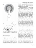
Figure 9.1 Reinforced laryngeal mask airway.
because of Tenon’s adhesions, whereas the
conjunctival incision may bleed or become
infected. Occasionally, the amaurosis may not
last as long as the akinesia and a period of
marked diplopia may occur as the agent wears
off. As yet, neither retrobulbar haemorrhage nor
rupture of staphylomatous sclera has been
reported. The technique does require somewhat
greater skill and patient cooperation than does
traditional local anaesthetic eye blocks, and the
true place of this technique in small incision
cataract surgery is not yet agreed.
ANAESTHESIA FOR CATARACT SURGERY
121
Topical anaesthesia for cataract extraction was
originally described using cocaine by Koller in
1884.
25
Preservative-free amethocaine or
prilocaine 1% topical anaesthesia avoids all of the
complications associated with injectional local
anaesthetics. Successful topical anaesthesia
nevertheless requires considerably more patient
education than most surgeons are used to
providing. As with any local anaesthetic technique
in which the patient is fully awake, it is also
sensible to avoid referring to cutting instruments
by names that the patient will understand;
Figure 9.2 Technique for sub-Tenon’s local anaesthetic. (a) Topical local anaesthetic drops (g.amethocaine)
are administered. (b) 5% povidine drops are administered. (c) The periocular skin and lids are cleaned with
povidine and a lid speculum inserted. (d) The conjunctiva and Tenon’s fascia is incised approximately 3 mm
posterior to the inferonasal limbus. (e) The sub-Tenon’s pocket is enlarged. (f) The blunt cannula is inserted
into the sub-Tenon’s space, advanced, and local anaesthetic injected.
a)
c)
d)
b)
e)
f)
CATARACT SURGERY
122
euphemisms or technical nomenclature easily
overcome this problem. Advantages and
limitations of topical anaesthesia are as follows:
• Need for rapport
• Analgesia without total anaesthesia
• No akinesia
• No amaurosis
• No orbicularis anaesthesia.
Pressure, touch and temperature, but not
pain, appear to be relatively preserved by surface
topical anaesthetics. The analogy with good
quality local anaesthesia for dental surgery is
useful (i.e. analgesia but not total anaesthesia).
Fortunately, it is possible to demonstrate the
analgesia to the patient, because the first drop to
the superior fornix usually stings and the second
drop two minutes later does not. The first drop
can be inserted in the anaesthetic room while an
intravenous cannula and monitoring are
established. Two drops are usually sufficient;
additional drops may reduce corneal clarity.
26
Most patients find it amazing that two drops
provide sufficient analgesia for surgery, as indeed
do their doctors.
There are three avoidable causes of potential
pain during topical anaesthesia:
• Pulling on the iris root
• Sudden increase in intraocular pressure
(avoidable by, for example, lowering the
bottle height when introducing the phaco tip
into the eye)
• Subconjunctival injections (avoided by the
use of prophylactic antibiotics in the irrigation
fluid or oral antibiotics).
Figure 9.3 Technique for single injection transconjunctival peribulbar local anaesthetic. (a) After local
anaesthetic and povidine iodine drops have been administered, the lower lid is retracted and needle (bevel toward
the globe) is inserted through the inferotemporal conjunctival below the globe. (b) The needle is inserted directly
posteriorly until the hub of the needle is parallel to the cornea (assuming a 25 mm needle is used and the eye has
an average axial length). (c) Once the needle tip is posterior to the globe equator, it is then directed posterior
to globe where the local anaesthetic injected. (d) A compression device is used to apply pressure to the eye for
5–10 minutes.
a)
b)
c)
d)
ANAESTHESIA FOR CATARACT SURGERY
123
Studies have confirmed that the level of
analgesia produced by topical anaesthesia is
comparable with that by injectional
techniques.
20,27,28
The addition of 0·5 ml
intracameral unpreserved lignocaine 1% injected
into the anterior chamber at the start of surgery
can further minimise intraoperative discomfort.
29
The lack of akinesia makes the technique
unsuitable for conventional large incision cataract
surgery, but any technique with a 5 mm or
smaller incision is suitable. The patient must not
forcibly close the contralateral eye, because any
Bell’s phenomenon will disadvantage the
surgeon. However, the ability to ask a patient to
deviate the globe in any desired direction is an
advantage,
28
removing the need for a superior
rectus bridle suture, and may reduce the
incidence of postoperative ptosis.
The lack of amaurosis is also both an
advantage and disadvantage. Patients need to be
reassured that they will not see details of their
surgery. The level of microscope illumination
can be increased, as the patient becomes
accustomed to the bright light. This produces a
significant after-image, lasting for up to two
hours after surgery, and emphasises the potential
danger of excessively bright coaxial illumination.
In contrast, the lack of amaurosis assists early
visual rehabilitation.
30
Perioperative monitoring
Routine monitoring for all patients
undergoing surgery has led to the recognition
that critical incidents (events that did or, if left
untreated, could have lead to harm to the
patient) are common during both local and
general anaesthesia (Table 9.4). In addition to a
contemporaneous record of events, all patients
undergoing cataract surgery with general
anaesthesia should, as a minimum, have
continuous electrocardiography, pulse oximetry,
regular non-invasive blood pressure
measurements, capnography, and vapour
analysis.
31
If a sharp needle local anaesthetic
technique or sedation in conjunction with local
anaesthesia is used, then similar monitoring is
required and intravenous access is essential.
5
Monitoring should always commence before
induction of anaesthesia (unless it is not possible
to attach a device, for example in an
uncooperative child). There is an emerging
consensus that where topical anaesthesia alone is
applied, pulse oximetry is sufficient, provided
that there is trained assistance immediately
available. Indeed, monitoring is of little benefit
unless those monitoring the patient have the
skill and expertise to recognise and treat
abnormalities before they become disasters. This
person should at least be trained in basic life
support.
If the “only” anaesthetic required for
phacoemulsification is two drops, then is the
presence of an anaesthetist essential? The role of
the anaesthetist is to monitor and attend to the
wellbeing of the patient; the surgeon’s is to
concentrate on the surgery. As the patient’s
“friend in court”, the anaesthetist can do much
Table 9.4 Critical incidents (n = 831) reported by
the National Confidential Enquiry into Perioperative
Deaths 1992–3 (of which 365 were multiple
occurrences)
2
Number Incident
493 Hypotension (>50% fall from baseline
values)
218 Cardiac arrest
150 Arrhythmia
137 Bradycardia (>50% of baseline value)
106 Hypoxia
55 Cyanosis
36 Pulmonary oedema
25 Hypertension (>150% of baseline
resting systolic values)
24 Bronchospasm
18 Respiratory arrest
14 Pulmonary aspiration
14 Airway obstruction
12 Pneumothorax
10 Misplaced tube
5 Convulsions
4 Anaphylaxis
3 Wrong drug
2 Air embolism
1 Disconnected
breathing tube
1 Hyperpyrexia (due to sepsis)