Hyperopia and Presbyopia - part 6 potx
Bạn đang xem bản rút gọn của tài liệu. Xem và tải ngay bản đầy đủ của tài liệu tại đây (1.72 MB, 34 trang )
160 Chalita and Krueger
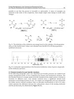
Figure 11 Spherical aberration after myopic treatment showing increased positive asphericity,
as represented by a sombrero hat.
REFERENCES
1. Applegate RA, Thibos LN, Hilmantel G. Optics of aberroscopy and super vision. J Cataract
Refract Surg 2001; 27:1093–1107.
2. Maeda N. Wavefront technology in ophthalmology. Curr Opin Ophthalmol 2001; 12:294–299.
3. Huang D. Physics of customized corneal ablation. In: MacRae SM, Krueger RR, Applegate
RA, eds. Customized Corneal Ablation: The Quest for Supervision. Thorofare NJ: Slack, 2001:
51–62.
4. Mrochen M, Kaemmerer M, Seiler T. Wavefront-guided laser in situ keratomileusis: early
results in three eyes. J Refract Surg 2000; 16:116–121.
5. Kaemmerer M, Mrochen M, Mierdel P, Krinke HE, Seiler T. Clinical experience with the
Tscherning aberrometer. J Refract Surg 2000; 16:S584–S587.
6. Krueger RR, Mrochen M, Kaemmerer M, Seiler T. Understanding refraction and accommoda-
tion through “retinal imaging” aberrometry. Ophthalmology 2001; 108:674–678.
7. Artal P. Understanding aberrations by using double-pass techniques. J Refract Surg 2000; 16:
S560–S562.
8. Schwiegerling J. Theoretical limits to visual performance. Surv Ophthalmol 2000; 45(2):
139–146.
9. Applegate RA. Limits to vision: Can we do better than nature? J Refract Surg 2000; 16:
S547–S551.
10. Williams D, Yoon GY, Porter J, Guirao A, Hofer H, Cox I. Visual benefit of correcting higher
order aberrations of the eye. J Refract Surg 2000; 16:S554–S559.
11. Thibos LN. The prospects for perfect vision. J Refract Surg 2000; 16:S540–S546.
12. Thibos L. Wavefront data reporting and terminology. J Refract Surg 2001; 17:S578–S583.
161Wavefront Changes After Hyperopia Surgery
13. Oshika T, Klyce SD, Applegate RA, Howland HC, Danasoury MAE. Comparison of corneal
wavefront aberrations after photorefractive keratectomy and laser in situ keratomileusis. Am
J Ophthalmol 1999; 127:1–7.
14. Mrochen M, Kaemmerer M, Mierdel P, Seiler T. Increased higher-order optical aberrations
after laser refractive surgery. A problem of subclinical decentration. J Cataract Refract Surg
2001; 27:362–369.
15. Mrochen M, Kaemmerer M, Seiler T. Clinical results of wavefront-guided laser in situ keratom-
ileusis 3 months after surgery. J Cataract Refract Surg 2001; 27:201–207.
16. Howland HC. The history and methods of ophthalmic wavefront sensing. J Refract Surg 2000;
16:S552–S553.
17. Mrochen M, Kaemmerer M, Mierdel P, Krinke HE, Seiler T. Principles of Tscherning aber-
rometry. J Refract Surg 2000; 16:S570–S571.
18. Platt BC, Shack R. History and principles of Shack-Hartmann wavefront sensing. J Refract
Surg 2001; 17:S573–S577.
19. Krueger RR. Technology requirements for Summit-Autonomus CustomCornea. J Refract Surg
2000; 16:S592–S601.
20. Thibos L. Principles of Shack-Hartmann aberrometry. J Refract Surg 2000; 16:S563–S565.
21. Roberts C, Dupps Jr WJ. Corneal biomechanics and their role in corneal ablative procedures.
In: MacRae SM, Krueger RR, Applegate RA eds. Customized Corneal Ablation: The Quest
for Supervision. Thorofare, NJ: Slack, 2001:109–131.
22. Argento CJ, Consentino MJ. Laser in situ keratomileusis for hyperopia. J Cataract Refract
Surg 1998; 24:1050–1058.
23. McDonald M. Summit—Autonomus CustomCornea Laser in situ keratomileusis outcomes. J
J Refract Surg 2000; 16:S617–S618.
24. Pettit GH, Campin J, Liedel K, Housand B. Clinical experience with the CustomCornea mea-
surement device. J Refract Surg 2000; 16:S581–S583.
16
Contrast Sensitivity Changes After
Hyperopia Surgery
LAVINIA C. COBAN-STEFLEA
Bucharest University Hospital and Carol Davila University of Medicine and
Pharmacy, Bucharest, Romania
TOMMY S. KORN
University of California–San Diego and Rees-Stealy Medical Group, San Diego,
California, U.S.A.
BRIAN S. BOXER WACHLER
Boxer Wachler Vision Institute, Beverly Hills, California, U.S.A.
A. INTRODUCTION
Understanding the importance of contrast sensitivity can be easier if we emphasize its
relationship to spatial vision, which is the core of the visual perception (1). Spatial fre-
quency theory of image processing is based on spatially extended patterns called sinusoidal
gratings, which are characterized by four parameters: spatial frequency, orientation, ampli-
tude, and phase. The contrast sensitivity function is a measure of the observer’s sensitivity
to gratings at different frequencies and is determined by the lowest contrast at which the
sinusoidal gratings can still be detected (2). Over 200 years ago, contrast sensitivity began
to be acknowledged as a clinical tool for doctors in studying visual disorders (3). In
1760 Bouguer defined and gave a value to the term light-difference threshold, the first
denomination of contrast threshold. Since then other researchers have made a great number
of contributions to this field: Bjerrum (1884) with letter charts, the first low-contrast letter
acuity tests, and Young (1918) with the ink spot test, an easy method to measure the light-
difference threshold. More recently Schade (1956) applied his knowledge of television
technology to contrast sensitivity testing. The work of Campbell and Green contributed
to a better understanding of the optical and neural mechanism of contrast sensitivity testing
and inspired further studies regarding alterations of contrast sensitivity in ocular diseases.
163
164 Coban-Steflea et al.
Correction of hyperopia has been a constant concern of ophthalmologists over the
past decades. Some of the surgical procedures that have been developed—hexagonal
keratotomy (4,5), keratophakia, keratomileusis, and epikeratophakia (6–9)—have been
abandoned because of limited applicability or side effects. Among current corrective proce-
dures undoubtedly laser-assisted in situ Keratomileusis (LASIK) and Ho:YAG laser ther-
mal keratoplasty (LTK) are the most widespread. Recently published clinical results em-
phasize the fact that LASIK is a procedure with good predictability, stability, efficacy,
and safety for the correction of low to moderate spherical hyperopia (10). Long-term
predictability with occurrence of undercorrection is influenced by the preoperative kerato-
metric values and ablation zone diameter (11). Other studies point out the importance of
corneal thickness and width of the flap for LASIK feasibility (12). The effectiveness of
LASIK for severe hyperopia and hyperopic astigmatism is reduced (13,14). For treatments
over ם5.00 D, the incidence of loss of best-corrected visual acuity was increased. Current
nomograms require the cut of a larger flap in order to enlarge the ablation zone and to
decrease the risk of halos, glare, and night vision difficulties for patients with high hyper-
opia and astigmatism (15). A lower predictability for astigmatic corrections was also
reported after LASIK for myopia (16) in spite of in situ axis alignment (17,18). Encourag-
ing results have been reported with respect to the safety, predictability, and stability of
LASIK correction, for small degrees of hyperopia that were secondary to previous radial
keratotomy (RK), and for automated lamellar keratoplasty (ALK) (19). The degree of
regression after H-LASIK was reported to be higher relative to myopic corrections but
lower, even in high hyperopia, than with the PRK procedure (20). Flap irregularities,
epithelium, infection, or nonspecific inflammation at the flap interface have been reported
complications of the LASIK procedure (21). Loss of vision can occur in cases of button-
holes, free cap, or amputation of the flap (22).
Correction of hyperopia and astigmatism by thermal keratoplasty was reported more
than 100 years ago (23–25). The actual mechanism by which this procedure alters the
anterior corneal curvature has been clarified with the discovery of shrinkage temperature
of corneal collagen by Stringer and Parr (26). In 1970s and 1980s, keratoconus was the
focus of theromokeratoplasty technology. A number of clinical studies done have evaluated
thermal keratoplasty potential to replace penetrating keratoplasty in keratoconus treatment
(27–30). In spite of the fact that initial flattening of the cone followed the procedure,
regression occurred within a few weeks postoperatively. It was not uncommon for these
keratoconus treatments to be accompanied by complications such as corneal scarring,
vascularization, and bullous keratopathy. Additionally, poor predictability and stability
contributed to the withdrawing of the procedure from clinical use for keratoconus.
A more recent approach to thermal keratoplasty is credited to Fyodorov, who devel-
oped a technique, using controlled thermal burns of corneal stroma with a retractable probe
tip heated to 600ЊC and applied in a radial pattern. The procedure was eventually abandoned
because of the high incidence of postoperative regression (31). In spite of repeated chal-
lenges to achieve predictable and stable refractive outcomes, researchers did not give up
on probe technology but took another avenue, which was the use of lasers to deliver
controllable amounts of energy to the stroma.
Lasers such as continuous CO
2
and cobalt magnesium fluoride have been used in
experimental studies on rabbit corneas, with transient results (32,33). Reports of clinical
studies that used the erbium:glass laser (34) have shown good results for hyperopia higher
than ם3.00 D. Over the past decade, ophthalmologists in the United States have directed
their work at evaluating two Ho:YAG laser systems: the noncontact system (Sunrise Tech-
165Changes After Hyperopia Surgery
nologies, Fremont, CA) and the contact system (Summit Technologies, Waltham, MA).
The Sunrise Ho:YAG is a pulsed laser that emits laser light at a wavelength of 2.13 m.
Other technical characteristics include pulse repetition frequency of 5 Hz and pulse energy
in the range of 226 to 258 in correlation with the amount of refractive correction required.
The energy is applied to the cornea in a noncontact mode through a fiberoptic slit-lamp
system; the treatment pattern is represented by rings of spots concentric to the pupil (35).
Sand, who was granted a patent for performing infrared LTK, was an important contributor
to the development of this technology. Initial in vitro investigations have been made on
swine and human cadaver eyes (36,37) in an attempt to establish a treatment protocol.
Further studies done on human poorly sighted eyes showed a mean change in corneal
curvature of 1.10 D followed by some amounts of regression (38). Results of clinical trials
done outside the United States, which used the eight-spot treatment pattern applied at
different diameters (6, 7, or 8 mm), had shown that the procedure works best up to ם3.00
D. They also proposed a treatment algorithm adjusted to variables such as age and central
corneal thickness (39). Other studies have demonstrated that the amount of refractive
change is increased when a two-ring treatment is applied at the 6- and 7-mm center line
in a radial instead of a staggered pattern (40,41). The U.S. phase III study protocol has
defined the efficacy criteria for the LTK procedure as improvement in distance UCVA
and reduction in hyperopia manifest refraction spherical equvalent (MRSE) Ͼ 0.5 D.
Evaluation at 2 years showed that 69.4% of patients had more than two lines of improve-
ment in distance uncontrolled visual acuity (UCVA) and no eyes had lost more than two
lines of best spectacle corrected visual acuity (BSCDVA) (35).
B. CONTRAST SENSITIVITY IN LASIK AND LTK
In understanding the outcomes of contrast sensitivity, we conducted a study to evaluate
the quality of vision through its changes in LASIK and noncontact Ho:YAG LTK for the
correction of low to moderate spherical hyperopia. We analyzed the results of two groups
of patients who had LASIK and LTK, respectively, as primary procedures. There was no
history of ocular diseases or surgery. We compared best-corrected contrast sensitivity
values preoperatively and at 3 months postoperatively. Contrast sensitivity was measured
with the self-calibrated, internally luminated CSV-1000E Vector Vision (Dayton, OH) at
12 cycles per degree (cpd) spatial frequency. The patient was instructed to identify whether
the bars were in the top circle, bottom circle, or neither. The last correct identification
has been taken as the contrast sensitivity. On the contrast sensitivity chart the numbers
represent normalized ratios where values greater than 1.0 correspond to percent contrasts
sensitivity above the population average and values below 1.0 represent percent of the
average contrast sensitivity below the population average (42). Visual acuity was measured
with the Vector Vision acuity chart using a scoring method of the U.S. Food and Drug
Administration for refractive surgery clinical trials (43). All visual function tests were
done with best spectacle-corrected visual acuity.
Data were analyzed with the StatView (SAS Institute Inc., Cary, NC) statistical
package. Visual acuity data were analyzed in logMAR values. Normalized contrast sensi-
tivity values were converted to log values and used for statistical analysis.
The LASIK study group comprised 94 eyes of 49 patients, 21 men and 28 women.
Mean patient age was 59.67 years ע7.95 SD, range 44 to 78 years. Preoperatively, mean
deviation from target manifest refraction was ם2.4 D ע1.2 D, SD, (range ם0.37 to
ם5.60 D). LASIK procedures were performed by the same surgeon (B.B.W.) using the
166 Coban-Steflea et al.
Table 1
H-LASIK Group—Preoperative and Postoperative Log Contrast Sensitivity Values and
Best Spectacle-Corrected LogMAR Visual Acuity Values
Mean Standard deviation Minimum Maximum
Preop log CS 1.30 0.22 0.61 1.69
Postop log CS 1.23 0.27 0.61 1.54
Preop logMAR VA Ϫ0.01 0.08 Ϫ0.20 0.20
Postop logMAR VA 0.02 0.10 Ϫ0.20 0.50
Moria LSK (Doylestown, PA) microkeratome and the Summit Apex Plus Laser (Summit
Technology Inc., Waltham, MA); the treatment zone was centered on the pupil. Results
have shown a mean postoperative deviation from target manifest refraction of מ0.09 D
ע0.88 D, SD, (range מ2.25 to ם2.00 D) at 3 months. Table 1 shows the mean preopera-
tive and postoperative log contrast sensitivity values, standard deviations, and maximum
and minimum values. At 3 months postoperatively the mean log contrast sensitivity value
was not statistically significantly different compared to preoperative levels (p ס 0.18). The
mean best spectacle-corrected logMAR visual acuity value at 3 months was statistically
significantly worse relative to preoperative value (p ס 0.008). However, the change was
not clinically significant, as the logMAR conversion was a loss of 1.5 letters on the acuity
chart. There was a statistically significant correlation between achieved refraction and
changes in log contrast sensitivity values (p ס 0.006) (Fig. 1) (r ס 0.29, p ס 0.006).
This indicated that higher amounts of hyperopic correction were associated with greater
loss of best-corrected contrast sensitivity. No statistically significant correlation was ob-
Figure 1 Correlation between changes in log contrast sensitivity values and achieved refraction
in the H-LASIK group.
167Changes After Hyperopia Surgery
Figure 2 Correlation between changes in best spectacle-corrected logMAR visual acuity values
and achieved refraction in the H-LASIK group.
served between achieved refraction and changes in best spectacle-corrected logMAR visual
acuity (r ס 0.05, p ס 0.58)(Fig. 2).
The LTK study group comprised 55 eyes of 35 patients, 16 males and 19 females.
Mean patient age was 57.61 years ע7.35, SD, with a range of 39 to 71 years; mean
deviation from target manifest refraction of treated eyes was ם1.5 D ע0.59 D, SD, range
0toם3.00 D. Noncontact Ho:YAG LTK treatments were performed by the same surgeon
(B.B.W.) using the Sunrise Hyperion Holmium Laser Corneal Shaping System (Sunrise
Technologies Inc., Fremont, CA). The treatment was centered on the corneal purkinje
image of the patient fixation light. The light reflex closely approximates the visual axis.
Therefore, in cases of positive angle kappa, the treatment was not centered on the pupil.
Laser parameters included wavelength, 2.13 m; pulse duration, 250 s; pulse repetition
frequency, 5 Hz; pulse energy, adjustable from 226 to 258 mJ/pulse. In the current study
we used a two concentric radial 8-spot ring treatment pattern centered around the fixation
light reflex on the cornea. Postoperatively, results showed a mean deviation from target
manifest refraction of מ0.36 D ע0.84 D, SD, range מ3.50 to ם1.25 D. Mean log
contrast sensitivity value was not statistically significantly decreased (p ס 0.07) (Table
2) and mean best spectacle-corrected logMAR visual acuity value was statistically signifi-
Table 2
LTK Group—Preoperative and Postoperative Log Contrast Sensitivity Values and Best
Spectacle-Corrected LogMAR Visual Acuity Values
Mean Standard deviation Minimum Maximum
Preop log CS 1.28 0.24 0.61 1.69
Postop log CS 1.19 0.29 0 1.84
Preop logMAR VA Ϫ0.01 0.08 Ϫ0.2 0.2
Postop logMAR VA 0.04 0.11 Ϫ0.1 0.6
168 Coban-Steflea et al.
Figure 3 Correlation between changes in log contrast sensitivity values and achieved refraction
in the LTK group.
cantly worse (p ס 0.0067) relative to preoperative values. The change in acuity was not
clinically significant as the change represented approximately four letters on the acuity
chart. No statistically significant correlation (R ס 0.16, p ס 0.25) was found between
achieved refraction and changes in log contrast sensitivity values (Fig. 3). Figure 4 shows
the lack of correlation between achieved refraction and best-spectacle corrected logMAR
visual acuity values (r ס 0.15, p ס 0.26).
Figure 4 Correlation between changes in best spectacle-corrected logMAR visual acuity values
and achieved refraction in the LTK group.
169Changes After Hyperopia Surgery
C. DISCUSSION
As new surgical procedures are added to the refractive surgery armamentarium, assessing
visual outcome becomes more difficult. Information regarding postoperative visual acuity
and refractive changes is no longer satisfactory to evaluate the quality of the image
projected on the retina (44). Contrast sensitivity, as a functional method, has been shown
to be directly affected by the distorted image following excimer laser surgery (45). Using
digitized retroillumination, Vinciguerra has shown that corneal distortion arising from
prominent flap striae may be overlooked by the customary slit-lamp examination (46).
Our results have shown a slight decrease in contrast sensitivity at 12 cpd spatial frequency
postoperatively after LASIK procedure. However the difference was not statistically signif-
icantly different (p ס 0.18). Previous literature data that have demonstrated that spatial
frequency of 12 cpd is mostly affected by degradation in optics, such as aberration or blur
(47). Other studies reported a loss of contrast sensitivity at 12 months after LASIK of up
to one line for low hyperopia and of more than two lines for high hyperopia with no
statistical significance (13). An interesting finding in the LASIK group was the significant
correlation between achieved refraction and change in contrast sensitivity, demonstrating
that larger amounts of correction are accompanied by larger loss of contrast sensitivity.
This indicates that with the Summit Apex Plus laser used for LASIK and centered on the
pupil, higher degrees of hyperopic treatment as associated with a higher risk of loss of
best-corrected contrast sensitivity.
Contrast sensitivity showed little change after the LTK procedure. The minimal
decrease observed was not statistically significant (p ס 0.07). Furthermore, contrast sensi-
tivity changes showed no correlation with the amount of spherical correction attempted.
Clinical trials at 1 and 2 years after LTK reported that mean contrast sensitivity increased
at all follow-up visits for the two-ring treatment group at Regan charts (40,48). Postopera-
tively visual acuity did not vary significantly (p ס 0.0067) and was not influenced by
the amount of correction, although the amount of hyperopia corrected in the LTK group
was less than that corrected in the LASIK group.
We conclude that measuring contrast sensitivity after refractive surgical procedures
should be encouraged and further developed in order to assess the limits of safety for
given procedures and devices used for such procedures. Studies should be directed at
identifying laser characteristics and treatment patterns that are able to optimize the optical
system of the eye, thus increasing safety.
REFERENCES
1. Palmer SE. Vision Science—Photons to Phenomenology. Cambridge, MA: MIT Press, 1999:
146–198.
2. Blakemore C, Campbell FW. On the existence of neurons in the human visual system selectively
responsive to the orientation and size of retinal images. J Physiol 1969; 203:237–260.
3. Shapley R, Man-Kit Lam D. Contrast Sensitivity: Proceedings of the Retina Research Founda-
tion Symposia. Vol. 5. Cambridge, MA: MIT Press, 1993: 253–266.
4. Werblin TP. Hexagonal keratotomy. Should we still be trying? J Refract Surg 1996; 12:
613–620.
5. Grandon SC, Sanders DR, Anello RD, Jacobs D, Biscaro M. Clinical evaluation of hexagonal
keratotomy for the treatment of primary hyperopia. J Cataract Refract Surg 1995; 21:140–149.
6. Swinger CA, Barraquer JI. Keratophakia and keratomileusis—clinical results. Ophthalmology
1981; 88:709–715.
170 Coban-Steflea et al.
7. Barraquer JI. Keratomileusis. Int Surg 1967; 48:103–117.
8. Morgan KS, Stephenson GS, McDonald MB, Kaufman HE. Epikeratophakia in children. Oph-
thalmology 1984; 91:780–784.
9. Anshutz T. Laser correction of hyperopia and presbyopia. In: ed. International Ophthalmology
Clinics. Refractive Surgery. Boston: Little, Brown, 1994:107–137.
10. Boxer Wachler BS, O’Brien TP, Tauber S. Vision Correction—Seeing the Future. Current
Laser Refractive and Surgical Alternatives for the Correction of Hyperopia. Oxford Institute
for Continuing Education, 2000:3–5.
11. Esquenazi S, Mendoza A. Two-year follow-up of laser in situ keratomileusis. J Refract Surg
1999; 15:648–652.
12. Rosa JL, Febbraro DS. Laser in situ keratomileusis for hyperopia. J Refract Surg 1999; 5(2
suppl):S212–S215.
13. Arbelaez MC, Knorz MC. Laser in situ keratomileusis for hyperopia and hyperopic astigma-
tism. J Refract Surg 1999; 15:406–414.
14. Barraquer C, Gutierrez AM. Results of laser in situ keratomileusis in hyperopic compound
astigmatism. J Cataract Refract Surg 1999; 25:1198–1204.
15. Lipner M. Distant visions: how practitioners outside the United States are treating hyperopia.
Eye World 1999; 4:17–18.
16. Knorz MC, Wiesinger B, Liebermann A, Seiberth V, Liesenhoff H. Laser in situ keratomileusis
for moderate and high myopia and myopic astigmatism. Ophthalmology 1998; 105:932–940.
17. Stevens JD. Astigmatic excimer laser treatment: theoretical effects of axis misalignment. Eur
J Implant Ref Surg 1994; 6:310–318.
18. Vajpayee RB, McCarthy CA, Taylor HR. Evaluation of axis alignment system for correction
of myopic astigmatism with the excimer laser. J Cataract Refract Surg 1998; 24:911–916.
19. Buzard KA, Fundingsland BR. Excimer laser assisted in situ keratomileusis for hyperopia. J
Cataract Refract Surg 1999; 25:197–204.
20. Reviglio VE, Luna JD, Rodriguez ML, Garcia FE, Juarez CP. Laser in situ keratomileusis
using the LaserSight 200 laser: results of 950 consecutive cases. J Cataract Refract Surg 1999;
25:1062–1068.
21. Wilson SE. LASIK: management of common complications (review). Cornea 1998; 17:
459–467.
22. Farah SG, Azar DT, Gurdal C, Wong J. Laser in situ keratomileusis: literature review of a
developing technique (review). J Cataract Refract Surg 1998; 24:989–1006.
23. Lans LJ. Experimentelle Untersuchungen uber die Entstehung von Astigmatismus durch nicht-
perforierende Corneawunden. Graefes Arch Clin Exp Ophthalmol 1898; 45:117–152.
24. Wray C. Case of 6 D of hypermetropic astigmatism cured by the cautery. Trans Ophthalmol
Soc UK 1914; 34:109–110.
25. O’Connor R. Corneal cautery for high myopic astigmatism. Am J Ophthalmol 1933; 16:337.
26. Striger H, Parr J. Shrinkage temperature of eye collagen. Nature 1964; 204:1307.
27. Gasset AR. Thermokeratoplasty in the treatment of keratoconus. Am J Ophthalmol 1975; 79:
226–232.
28. Aquavella JV, Buxton JN, Shaw EL. Thermokeratoplasty in the treatment of persistent corneal
hydrops. Arch Ophthalmol 1977; 95:81–84.
29. Itoi M. Computer photokeratometry changes following thermokeratoplasty. In: Schachar RA,
Levy NS, Schachar L, eds. Refractive Modulation of the Cornea. Denison, TX LAL Publishers,
1981.
30. Rowsey JJ, Doss JD. Preliminary report of Los Alamos keratoplasty techniques. Ophthalmol-
ogy 1981; 88:755–760.
31. Caster AI. The Fyodorov technique of hyperopia correction by thermal coagulation: a prelimi-
nary report. J Refract Surg 1988; 4:105–108.
32. Peyman GA, Larson B, Raichand M, Andrews AH. Modification of rabbit corneal curvature
with use of carbon dioxide laser burns. Ophthalmic Surg 1980; 11:325–329.
171Changes After Hyperopia Surgery
33. Horn G, Spears KG, Lopez O, Lewicky A, Yang X, Riaz M, Wang R, Silva D, Serafin J.
New refractive method for laser thermal keratoplasty with the Co: MgF2 laser. J Cataract
Refract Surg 1990; 16:611–616.
34. Kanoda AN, Sorokin AS. Laser correction of hypermetropic refraction. In: Fyodorov SN, ed.
Microsurgery of the Eye: Main Aspects. Moscow: MIR Publishers, 1987:147–154.
35. Aker AB, Boxer Wachler BS, Brown DC. Vision Correction—Seeing the Future. Noncontact
Holmium:YAG Laser Thermal Keratoplasty for the Treatment of Hyperopia. Oxford Institute
for Continuing Education, 2000:3–5.
36. Koch DD, Berry MJ, Vassiliadis AJ, Abarca AA, Villarreal R, Haft EA. Noncontact holmium:
YAG laser thermal keratoplasy. In: Salz JJ, ed. Corneal Laser Surgery. Philadelphia: Mosby,
1995:247–254.
37. Moreira H, Campos M, Sawusch MR, McDonnell JM, Sand B, McDonnell PJ. Holmium laser
thermokeratoplasty. Ophthalmology 1993; 100:752–761.
38. Ariayasu RG, Sand B, Menefee R, Hennings D, Rose C, Berry M, Garbus JJ, McDonnell PJ.
Holmium laser thermal keratoplasty of 10 poorly sighted eyes. Refract Surg 1995; 11:358–365.
39. Alio JL, Ismail MM, Sanchez Pego JL. Correction of hyperopia with noncontact Ho:YAG
laser thermal keratoplasty. J Refract Surg 1997; 13:17–22.
40. Koch DD, Kohnen T, McDonnell PJ, Menefee RF, Berry MJ. Hyperopia correction by noncon-
tact holmium:YAG laser thermal keratoplasty. US phase IIA clinical study with a 1-year
follow-up. Ophthalmology 1996; 103:1525–1536.
41. Vinciguerra P, Kohnen T, Azzolini M, Radice P, Epstein D, Koch DD. Radial and staggered
patterns to correct hyperopia using noncontact holmium:YAG laser thermal keratoplasty. J
Cataract Refract Surg 1998; 24:21–30.
42. Boxer Wachler BS, Kruger RR. Normalized contrast sensitivity: a new notation for mainstream
contrast sensitivity testing in refractive surgery. Invest Ophthalmol Vis Sci 1997; 38:530.
43. Boxer Wachler BS, Durrie DS, Assil KK, Kruger RR. Role of clearance and treatment zones
in contrast sensitivity: significance in refractive surgery. J Cataract Refract Surg 1999; 25:
16–23.
44. Pallikaris IG. Quality of vision in refractive surgery. J Refract Surg 1998; 14:551–557.
45. Boxer Wachler BS, Frankel RA, Kruger RR, Durrie DS, Assil KK. Contrast sensitivity and
patient satisfaction following photorefractive keratectomy and radial keratotomy. Invest Oph-
thalmol Vis Sci 1996; 37(suppl):S19.
46. Vinciguerra P, Azzollini M, Radice P. A new corneal analysis after excimer laser ablation:
digitized retroillumination. In: Pallikaris IG, Siganos DS, eds. LASIK. Thorofare, NJ: Slack,
1997:331–337.
47. Campbell FW, Green DS. Optical and retinal factors affecting visual resolution. J Physiol
1965; 181:576–593.
48. Koch D, Abarca A, Villarreal R, Menefee R, Kohnen T, Vassiliadis A, Berry M. Hyperopia
correction by noncontact holmium:YAG laser thermal keratoplasty. Clinical study with two-
year follow-up. Ophthalmology 1996; 103:731–740.
17
Wound Healing After Hyperopic
Corneal Surgery
Why There Is Greater Regression in the
Treatment of Hyperopia
RENATO AMBRO
´
SIO, JR.
University of Washington, Seattle, Washington, U.S.A., University of Sa
˜
o Paolo,
Sa
˜
o Paolo, and Clı
´
nica e Microcirurgia Oftalmolo
´
gica Renato Ambro
´
sio, Rio de
Janeiro, Brazil
STEVEN E. WILSON
University of Washington, Seattle, Washington, U.S.A.
A. INTRODUCTION
Biological diversity in the corneal wound-healing response is a major factor in the out-
comes of all keratorefractive surgical procedures (1,2). It is one of the most important
determinants for overcorrection, undercorrection, and other complications, such as haze
(3) and irregular astigmatism, which occur with laser-assisted in situ keratotomileusis
(LASIK) and photorefractive keratectomy (PRK) in the treatment of myopia (4,5), hyper-
opia (6,7), or astigmatism (8,9).
This response is very similar in different species, facilitating the creation of animal
models for better characterization of the wound-healing response. There are quantitative
and qualitative variations in specific processes that comprise the cascade. There is also
variability depending on the inciting injury within a species. For example, thermal, inci-
sional, lamellar, and surface scrape injuries are followed by wound-healing responses that
are similar in some respects but different in others.
Corneal wound healing following correction of hyperopia may be more complex
than that associated with corrections of myopia (10). Steepening of the central cornea is
required for hyperopic treatments. This leads to the creation of a corneal contour with a
steeper central area and a flatter paracentral area.
173
174 Ambro
´
sio and Wilson
Refractive regression is defined as a gradual, partial, or total loss of the initial
correction. It limits the predictability of all refractive surgery procedures performed on
the cornea. It has been hypothesized that changes occurring as a result of corneal wound
healing lead to addition of new tissue. Epithelial hyperplasia and stromal remodeling are
the two mechanisms that are thought to underlie this phenomenon (3,11,12).
1. Keratocytes Disappear in Response to Epithelial
Injury—Keratocyte Apoptosis
One of the earliest observations that debunked the prior dogma regarding the quiescence
of keratocytes was detection of disappearance of superficial keratocytes following corneal
epithelial scrape injury. This observation was made first by Dohlman and coworkers in
1968 (16). Studies by later investigators confirmed that keratocytes in the anterior stroma
disappear following corneal epithelial scrape injury (17–20) as well as thermokeratoplasty
(21). The mechanism of disappearance of the keratocytes was not elucidated in these
studies. The authors of these studies suggested that the disappearance of the keratocytes
was attributable to several factors, such as osmotic changes from the loss of epithelium,
exposure to the atmosphere, or even artifact.
In 1996, Wilson and coworkers (20) first demonstrated that the early disappearance
of keratocytes that follows epithelial injury is mediated by apoptosis (13–15,22–29). Cell
shrinkage, blebbing with formation of membrane bound bodies, condensation, fragmenta-
tion of the chromatin, and DNA fragmentation consistent with apoptosis were detected
in anterior stromal keratocytes after epithelial scrape wounds by transmission electron
microscopy. Nuclear DNA fragmentation was confirmed by the TUNEL assay for 3′-
hydroxyl DNA ends.
Apoptosis is a programmed form of cell death that occurs without the release of
lysosomal enzymes or other intracellular components that could damage the surrounding
tissue or cells. Uncontrolled release of cellular contents is characteristic of necrotic cell
death (26). Studies have suggested that apoptosis is mediated by cytokines released from
the injured epithelium, such as interleukin 1 (IL-1) (22), the Fas/Fas ligand system (27),
bone morphogenic proteins (BMP) 2 and 4 (28), or tumor necrosis factor (TNF) alpha
(29).
Virtually any type of epithelial injury induces keratocyte apoptosis. These include
mechanical scrapes (22–25), corneal surgical procedures like PRK and LASIK (24), herpes
simplex keratitis (14), incisions (25), and even a plastic ring pressed firmly against the
epithelial surface (24).
Keratocytes undergo apoptosis after epithelial injury to a depth of one-third to one-
half the stromal thickness, depending on the species and the type of injury. Cellular pro-
cesses, known as gap junctions, connect keratocytes in the unwounded cornea to form a
syncytium (31,32). It is possible that signals transmitted by cytokines to the most superficial
keratocytes are relayed to deeper keratocytes via these intercellular communication chan-
nels. Alternatively, the proapoptotic cytokines may penetrate into the stroma after injury.
The keratocyte apoptosis response in the stroma varies with the type of corneal
epithelial injury (25). Thus, injuries such as scraping of the epithelium (25) or viral infec-
tion of the epithelium (14) triggers keratocyte apoptosis in the superficial stroma. A lamel-
lar cut across the cornea produced by a microkeratome also induces keratocyte apoptosis.
This can be detected at the site of epithelial injury and along the lamellar interface (Figure
175Wound Healing After Corneal Surgery
Figure 1 (A) Apoptosis detected along the lamellar interface by TUNEL assay in rabbit eye that
had LASIK and (B) on the surface in rabbit eye that had PRK.
1). Localization of keratocyte apoptosis in LASIK is thought to be attributable to tracking
of epithelial material, including proapoptotic cytokines, into the interface by the microkera-
tome blade (22–25). Alternatively, cytokines from the injured peripheral epithelium could
diffuse along the lamellar interface and into the central stroma (22–25).
Apoptosis has also been correlated with severe complications. Meitz et al. (33)
reported a severe case of acute corneal necrosis following PRK for hyperopia that required
penetrating keratoplasty. Histopathological studies of the excised tissue were negative for
micro-organisms. Utilizing light microscopy, an anterior zone of corneal necrosis was
found to be present, with a moderate amount of acute inflammation at the interface between
necrotic and viable corneal stroma; in addition, keratocytes with typical features of
apoptosis were detected by TUNEL assay and electron microscopy (Figure 2).
2. Keratocyte Proliferation and Migration: Myofibroblasts
After the loss of keratocytes caused by apoptosis within the first few hours of corneal
epithelial injury, there will be an area of stroma devoid of keratocytes. Zieske and cowork-
ers (34) demonstrated that remaining keratocytes in the posterior and peripheral cornea
begin to undergo mitosis about 12 to 24 hours after the injury (34). Keratocyte mitosis
can be detected using bromodeoxyuridine incorporation or immunocytochemical staining
for a mitosis-specific antigen called Ki-67 (34).
176 Ambro
´
sio and Wilson
Figure 2 Transmission electron microscopy (TEM) of rabbit cornea, 24 hours after Hi PRK
(מ9.0D): Keratocyte apoptosis and a PMN.
The cell types derived from the keratocytes that undergo mitosis following corneal
epithelial injury remain to be completely characterized. Studies have suggested that myofi-
broblasts are an important cell type generated following injury (38–41). These studies,
however, are primarily in vitro tissue culture-based investigations. Little information is
available regarding the fate of the cells that undergo mitosis following PRK (41). Nothing
has been reported about the status of these cells following LASIK.
3. Resolution of The Wound-Healing Response—Return to
“Normalcy”
In the months following injury to the cornea, the wound-healing response is completed
and there is a return to normal morphology and function. This process is associated with
elimination of some of the cells associated with wound healing and remodeling of disor-
dered collagen that was produced by myofibroblasts or keratocytes during the wound-
healing process (54–55). This process begins within a few weeks after injury and can
continue for years following severe injury.
The corneal epithelium may undergo hyperplasia following corneal injury (1,56)
as well as refractive surgery (11,12,21,57–59) as a part of the wound-healing response.
Hyperplasia may vary between individuals, the eyes of a single individual, and with differ-
ent types and levels of refractive correction. This is thought to be an important mechanism
for regression of many keratorefractive procedures (1,12,56–59). There may be a return
to a normal epithelial thickness over a period of months to years, and this may result in
instability of the refractive effect of PRK or LASIK. The regulatory mechanisms that
modulate this return to normal corneal epithelial morphology have not been characterized.
B. CONSIDERATIONS ON HYPEROPIC CORRECTIONS: WHY ARE
THEY DIFFERENT FROM MYOPIC CORRECTIONS?
The surgical correction of hyperopia remains challenging, especially for corrections greater
than 4 to 5 D. While corneal surgery for myopia requires flattening the cornea with an
177Wound Healing After Corneal Surgery
Table 1 Classification of Hyperopic Refractive Surgery
1. Excimer laser procedures
2. Collagen shrinkage procedures
3. Corneal implants and inlays
4. Phakic intraocular lens (IOL)
5. Clear lens extraction with IOL (also piggyback; multifocal IOL)
appropriate effective optical zone, hyperopic treatments require steepening of the central
cornea. This leads to the creation of more complex compound curves, which are steeper
in the center and flatter in the paracentral area.
Currently, options for refractive surgery to treat hyperopic patients can be separated
into five categories (Table 1). The present chapter discusses only the first two options.
The excimer laser allows reshaping of the corneal surface to a desired contour
with submicron precision and reproducibility (65). Several issues must be considered
in differentiating hyperopic and myopic corrections using the excimer laser. In myopic
corrections, the laser is applied in the center of the cornea. Hyperopic treatment with the
excimer laser consists of an annular zone of ablation to cause a relative flattening of the
corneal periphery and a concomitant relative steepening of the center (optical zone) to
achieve the desired refractive effect. Hyperopic corrections require more complex laser
delivery systems (66). Since the treatment is typically longer and performed in the periph-
ery, careful alignment of the laser beam is critical in order to prevent decentration. Thus,
a greater chance of decentration may be noted. Optical zone and ablation zone sizes are
fundamental to the efficacy of these procedures. A blend transitional zone must be created
to avoid abrupt steps on the corneal surface, which would be likely to lead to regression
via epithelial hyperplasia (67). The maximum ablation depth will be in an annulus between
the optical zone and the outer diameter of the ablation zone. Larger outer zones may
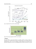
provide for less regression of the refractive effect (68,69) (Figure 3). However, Aron-
Rosa and Febbraro noted that when using an ablation zone of 5.5 ן 8.25 mm with LASIK,
Figure 3 Diagram showing epithelial hyperplasia after hyperopic cornea surgery.
178 Ambro
´
sio and Wilson
there was better predictability and stability than with an ablation zone of 5.5 ן 9.0 mm
(70). One possible explanation for this observation is that the corneal flap size may have
been smaller than the periphery of the hyperopic treatment. In such settings, a smaller
ablation zone may be preferable.
Excimer laser surgery for hyperopia may induce more astigmatism than for myopia.
Significant change in the astigmatism power and axis was noted 3 months following
hyperopic spherical LASIK in a two-step approach for treating hyperopic or mixed astig-
matism (71). This could be related to centration issues in the treatment of hyperopia relative
to myopia.
Attempts to shrink the peripheral corneal collagen with thermal energy (thermokera-
toplasty) were first reported by Lans over a century ago (72). Central steepening of the
cornea is achieved by thermal shrinkage of the midperipheral corneal tissue. The use of
different types of lasers and radiofrequency energy in the corneal stroma to shrink the
collagen lamellae is an active topic of study and is discussed elsewhere in this book.
Recent reports have shown that these procedures may be effective in correcting low hyper-
opia, although corrections were subject to regression (73). Age-dependent corneal factors
were shown to influence the effectiveness of thermal energy on stromal collagen and
regression (74). Stability following thermokeratoplasty may be related to the type of lesion
produced. A perfect thermal lesion, delivered at the perfect depth, with a perfect geometry,
and for the perfect length of time would cause a permanent change in the collagen fibers
in the cornea, so that regression would be less likely to occur. It remains to be seen whether
such a “perfect thermal lesion” that is permanent can be created or whether ever-vigilant
keratocytes will eventually detect these anomalies in the collagen fibers and repair them.
Corneal iron pigmentation lines or rings can be observed after hyperopic corneal
surgery (75–78). Corneal iron deposition has been seen in the normal cornea with aging
(Hudson-Stahli line) and in pathological corneal conditions such as keratoconus (Fleischer
ring), pterygia (Stocker-Busacca line), and filtering blebs (Ferry’s line). Stellate iron lines
were also described after radial keratotomy (79) and in cases of central island (80). The
most likely explanation for the formation of such lines is that the iron is derived from the
tear film and deposited in the corneal epithelium in those areas where there is tear pooling.
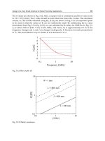
Since keratorefractive procedure for hyperopia sculpts the cornea to resemble a convex
lens, a furrow-like ring zone in the corneal periphery is produced. This can be observed
when looking at the corneal elevation map after H-LASIK. (Figure 4). Tear pooling occurs
and subsequently triggers iron deposition. It may also prolong the exposure time to tear
film cytokines (81,82) causing epithelial hyperplasia in this midtransition zone (junction
of the optical and ablated zones) (11).
C. MECHANISMS OS REGRESSION
A complete understanding of the mechanisms underlying regression after keratorefractive
surgery in vivo require the study of the wound-healing response and factors related to
biomechanics. A thorough understanding of corneal microstructure can now be obtained
using new methods. High-frequency (50-MHz) ultrasound biomicroscopy (UBM and
VHF) (83–86) (Figure 5) and optical coherence tomography (OCT) (87–89) are two
promising technologies that have the capacity to measure the thickness of each layer within
the cornea. These measurements could help us to distinguish between epithelial hyperplasia
and stromal remodeling as the cause of the refractive regression in individual eyes. Confo-
cal microscopy allows for optical sectioning through intact living cornea, obtaining images
179Wound Healing After Corneal Surgery
Figure 4 Elevation map before and after hyperopic LASIK.
of the cornea at its cellular level in four dimensions (x, y, z, and t-time) (3,10,90,91). It
has been difficult, however, to obtain reliable measurements of epithelial thickness using
this technology. Slit-based videokeratography instruments like the Orbscan (Bausch &
Lomb, Orbtek, Inc., Salt Lake City, UT) may be useful for assessing pachymetric values
through the entire cornea as well as for measuring posterior curvature (92,93). However,
uncertainty regarding the meaning of values derived from the posterior surface of the
cornea is a limiting factor. Studies have shown that corneal thickness measurements are
inaccurate with this instrument (94,95). At the present time, therefore, it appears that
high-frequency ultrasound or OCT provides the best opportunity for monitoring epithelial
thickness following refractive surgery procedures. Studies are in progress using these
methods.
Animal model studies have been performed to characterize corneal wound healing
following surgery for hyperopia (11,21,96–99). It is important to recognize the possible
limitations of the rabbit model in assessing the nature of the wound-healing response in
humans. Wound healing is thought to be more vigorous in rabbits, and qualitative as well
as quantitative differences may exist. It is feasible to perform studies in patients who
180 Ambro
´
sio and Wilson
Figure 5 Corneal image using ultrasound biomicroscopy.
undergo surgery for enucleation or exenteration, as well as before penetrating keratoplasty,
to clarify these potential differences between humans and animal models (21).
Our working hypothesis at the present time is that regression after LASIK or PRK
surgery for hyperopia is due to a combination of epithelial hyperplasia and stromal re-
growth in the ablation zone. Using confocal microscopy and histological examination in
a rabbit model, Hosoda at al. detected subepithelial proliferative changes in the ablated
zone that progressed for 1 month after surgery, then decreased by the third month (96).
In a similar study by Dierick et al. (11), mean stromal regrowth after 10-D hyperopic
PRK was 50% of ablated tissue. Deposition showed a lenticular pattern and could account
for up to 5.00 D of regression (11). In addition, the epithelium thickened 20% at the
midtransition zone (junction of the optical and ablated zones), contributing to more refrac-
tive regression (11).
A key question is whether the epithelial hyperplasia is attributable to an increased
wound healing response due to the size of the ablation zone, the altered surface topography
associated with steepening the central contour, or a combination of both these factors.
With smaller ablation zone diameters that have been tested in the past, rapid regression
may have been largely due to abrupt changes in corneal curvature in the midperiphery of
the ablation. With wider ablations that allow a more gradual transition than with smaller
ablation zones, there is less tendency for regression, suggesting that the influence of this
factor has been reduced. Differences in tear pooling and distribution on the corneal surface
between smaller and larger ablation zone diameters could play a role. Well-controlled
studies of varying ablations with careful measurements of epithelial hyperplasia and stro-
181Wound Healing After Corneal Surgery
mal regrowth should help to increase our understanding of regression associated with the
laser correction of hyperopia.
Other sources of regression may be a greater than average wound-healing response
in individual patients or variations in surgery that promote increased healing. For example,
a thin flap may be associated with regression, since the stromal wound-healing response
and epithelium-modulating modulating growth factor production are more likely to be in
proximity to the epithelium (13). This is probably a major factor promoting epithelial
hyperplasia. Other factors such as epithelial defects produced by the microkeratome and
diffuse interface keratitis may also be associated with a stronger wound-healing response
and therefore regression. The rate of enhancement in a recent series was significantly
higher (53 versus 16%; pס0.02) following DLK than for eyes that did not have DLK
(Wilson and Ambro
´
sio, unpublished data, 2001). Since the treatment for hyperopia is
typically performed in the periphery of the cornea, closer to the limbus, it is likely that a
stronger inflammatory reaction will follow those surgeries. A study involving an animal
model comparing hyperopic and myopic PRK, using specific antibodies for inflammatory
cells as well as cytokines, might be helpful for elucidating this hypothesis.
The higher the level of correction attempted for hyperopia, the more likely regression
due to wound healing will occur. In our experience with hyperopic LASIK and PRK,
regression is most common in eyes where the attempted correction is over 4 to 5 D.
Intraocular pressure could be a factor in the regression of hyperopic LASIK in some
cases with high-pressure increases. A case of acute angle-closure glaucoma was reported
by Paciuc et al. 1 year after hyperopic LASIK (100). The glaucoma attack was treated
with laser peripheral iridotomy and a prophylactic iridotomy was performed in the fellow
eye. Corneal topography was performed 2, 5, and 18 weeks after the acute episode and
a myopic shift occurred in the eye that had angle closure. This resolved over 3 months.
It is important to consider that the eye blinks over 10,000 times per day (101) at lid
velocities up to 30 cm/s (102). Each blink has enough force to raise intraocular pressure
10 to 70 mmHg (103).
Koch and coworkers (21) studied Ho:YAG LTK on three human corneas 1 day
before their removal at penetrating keratoplasty in patients with corneal edema secondary
to Fuchs endothelial dystrophy (without bullous epithelial changes) and on six New
Zealand white rabbit corneas followed for up to 3 months. The pulse radiant energy level
was noted to be proportional to the acute tissue injury. In human corneas, changes in the
irradiated zones included epithelial cell injury and death, loss of fine filamentous structure
in Bowman’s layer, disruption of stromal lamellae, and keratocyte injury and death. A
cone-shaped zone of increased stromal hematoxylin uptake extending posteriorly for 90%
of stromal thickness was noted in the treatment areas. Special immunohistochemical stains
to detect apoptosis were not used, although transmission electron microscopy findings
suggested that they might play a role. In the rabbit corneas, similar acute changes were
noted. By 3 weeks, epithelial hyperplasia and stromal contraction were present. Wound
healing in the rabbits included repair of the epithelial attachment complex, keratocyte
activation, synthesis of type I collagen, and partial restoration of stromal keratin sulfate
and type VI collagen. There was also a marked endothelial proliferative response in the
rabbit corneas. Attempted corrections with LTK of greater than 2 D are associated with
significant regression. This is likely related to stromal remodeling, with the keratocytes
functioning to repair the altered collagen over time.
182 Ambro
´
sio and Wilson
D. FUTURE DIRECTIONS AND CONCLUSIONS
The ability to modulate corneal wound healing to achieve better clinical outcomes would
be beneficial to extend the efficacy and safety of keratorefractive corrections of hyperopia.
Apoptosis is the first detected event in the complex cascade of the corneal wound healing.
Differences in this initiator and subsequent events in healing between eyes likely is a
major determinant of variation between eyes following laser correction for hyperopia.
Development of methods to control this first event may be useful for normalizing the
response between patients.
A better understanding of the mechanisms associated with regression, especially
differentiating between the key determinants epithelial hyperplasia and stromal remodel-
ing, would provide specific strategies to improve stability.
Corneal implants and inlays may become an option for hyperopic treatment in the
future. New alloplastic materials with acceptable permeability for corneal tissue, with
refractive indices and clarity equal to those of the cornea, may provide a reversible refrac-
tive procedure for hyperopia. Intracorneal lenses with higher refractive indexes than the
cornea and therefore intrinsic refractive power would not rely on changing the cornea’s
shape. They could attenuate epithelial hyperplasia as a factor in regression.
Corneal surgery for hyperopia has lagged behind that of myopia primarily due to
issues related to efficacy, stability, and safety. Several procedures were abandoned during
the past decade. Understanding and respecting the limits of the available procedures is
key for achieving success with hyperopic patients. Intraocular procedures for hyperopia,
such as phakic intraocular lenses and clear lens extraction, may have an important role
in treating this group of patients if safety can be improved.
ACKNOWLEDGMENTS
Supported in part by an unrestricted grant from Research to Prevent Blindness, New York,
N.Y., and U.S. Public Health Service grant EY 10056 and EYO1730 from the National
Eye Institute, National Institutes of Health, Bethesda, Maryland.
PROPRIETARY INTEREST STATEMENT
The authors have no proprietary or financial interest in relation to this manuscript.
REFERENCES
1. Wilson SE, Mohan RR, Hong JW, Lee JS, Choi R, Mohan RR. The wound healing response
after laser in situ keratomileusis and photorefractive keratectomy: elusive control of biological
variability and effect on custom laser vision correction. Arch Ophthalmol 2001; 119:889–896.
2. Wilson SE, Mohan RR, Mohan RR, Ambrosio R Jr, Hong J, Lee J. The corneal wound
healing response: cytokine-mediated interaction of the epithelium, stroma, and inflammatory
cells. Prog Ret Eye Res. 2001 9; 20(5):625–637.
3. Moller-Pedersen T, Cavanagh HD, Petroll WM, Jester JV. Stromal wound healing explains
refractive instability and haze development after photorefractive keratectomy: a 1-year confo-
cal microscopic study. Ophthalmology 2000; 107:1235–1245.
4. Kim JH, Kim MS, Hahn TW, Lee YC, Sah WJ, Park CK. Five year results of photorefractive
keratectomy for myopia. J Cataract Refract Surg 1997; 23:731–735.
183Wound Healing After Corneal Surgery
5. Shah SS, Kapadia MS, Meisler DM, Wilson SE. Photorefractive keratectomy using the summit
SVS Apex laser with or without astigmatic keratotomy. Cornea 1998; 17:508–516.
6. Esquenazi S, Mendoza A. Two-year follow-up of laser in situ keratomileusis for hyperopia.
J Refract Surg 1999; 15:648–652.
7. Zadok D, Maskaleris G, Montes M, Shah S, Garcia V, Chayet A. Hyperopic laser in situ
keratomileusis with the Nidek EC–5000 excimer laser. Ophthalmology 2000; 107:
1132–1137.
8. Hersh PS, Abbassi R. Surgically induced astigmatism after photorefractive keratectomy and
laser in situ keratomileusis. Summit PRK-LASIK Study Group. J Cataract Refract Surg 1999;
25:389–398.
9. Kapadia MS, Krishna R, Shah S, Wilson SE. Surgically induced astigmatism after photorefrac-
tive keratectomy with the excimer laser. Cornea 2000; 19:174–179.
10. Vesaluoma MH, Petroll WM, Perez-Santonja JJ, Valle TU, Alio JL, Tervo TM. Laser in situ
keratomileusis flap margin: wound healing and complications imaged by in vivo confocal
microscopy. Am J Ophthalmol 2000; 130:564–573.
11. Dierick HG, Van Mellaert CE, Missotten L. Histology of rabbit corneas after 10-diopter
photorefractive keratectomy for hyperopia. J Refract Surg 1999; 15:459–468.
12. Lohmann CP, Guell JL. Regression after LASIK for the treatment of myopia: the role of the
corneal epithelium. Semin Ophthalmol 1998; 13:79–82.
13. Wilson SE, Mohan RR, Mohan RR, Ambrosio Jr R, Hong J-W, Lee J-S. The corneal wound
healing response: Cytokine-mediated interaction of the epithelium, stroma, and inflammatory
cells. Prog Ret Eye Res 2001; 20:625–637.
14. Wilson SE, Pedroza L, Beuerman R, Hill JM. Herpes simplex virus type–1 infection of
corneal epithelial cells induces apoptosis of the underlying keratocytes. Exp Eye Res 1997;
64:775–779.
15. Wilson SE. Role of apoptosis in wound healing in the cornea. Cornea 2000; 19(suppl):
S7–S12.
16. Dohlman CH, Gasset AR, Rose J. The effect of the absence of corneal epithelium or endothe-
lium on stromal keratocytes. Invest Ophthalmol Vis Sci 1968; 7:520–534.
17. Nakayasu K. Stromal changes following removal of epithelium in rat cornea. Jpn J Ophthalmol
1988; 32:113–125.
18. Campos M, Szerenyi K, Lee M, McDonnell JM, McDonnell PJ. Keratocyte loss after corneal
deepithelialization in primates and rabbits. Arch Ophthalmol. 1994; 112:254–260.
19. Szerenyi KD, Wang X, Gabrielian K, McDonnell PJ. Keratocyte loss and repopulation of
anterior corneal stroma after de-epithelialization. Arch Ophthalmol 1994; 112:973–976.
20. Polack FM. Keratocyte loss after corneal deepithelialization in primates and rabbits. Arch
Ophthalmol 1994; 112:1509.
21. Koch DD, Kohnen T, Anderson JA, Binder PS, Moore MN, Menefee RF, Valderamma GL,
Berry MJ. Histologic changes and wound healing response following 10-pulse noncontact
holmium: YAG laser thermal keratoplasty. J Refract Surg 1996; 12:623–634.
22. Wilson SE, He Y-G, Weng J, Li Q, Vital M, Chwang EL. Epithelial injury induces keratocyte
apoptosis: hypothesized role for the interleukin–1 system in the modulation of corneal tissue
organization. Exp Eye Res 1996; 62:325–338.
23. Wilson SE. Molecular cell biology for the refractive corneal surgeon: programmed cell death
and wound healing. J Refract Surg 1997; 13:171–175.
24. Wilson SE. Keratocyte apoptosis in refractive surgery: Everett Kinsey Lecture. CLAO J 1998;
24:181–185.
25. Helena MC, Baerveldt F, Kim W-J, Wilson SE. Keratocyte apoptosis after corneal surgery.
Invest Ophthalmol Vis Sci 1998; 39:276–283.
26. Kim WJ, Mohan RR, Mohan RR, Wilson SE. Caspase inhibitor z-VAD-FMK inhibits kerato-
cyte apoptosis, but promotes keratocyte necrosis, after corneal epithelial scrape. Exp Eye Res
2000; 71:225–232.
184 Ambro
´
sio and Wilson
27. Mohan RR, Liang Q, Kim W-J, Helena MC, Baerveldt F, Wilson SE. Apoptosis in the cornea:
Further characterization of Fas/Fas ligand system. Exp Eye Res 1997; 65:575–589.
28. Mohan RR, Kim W-J, Mohan RR, Chen L, Wilson SE. Bone morphogenic proteins 2 and 4
and their receptors in the adult human cornea. Invest Ophthalmol Vis Sci 1998; 3926–2636.
29. Mohan RR, Mohan RR, Kim WJ, Wilson SE. Modulation of TNF-alpha–induced apoptosis
in corneal fibroblasts by transcription factor NF-kb. Invest Ophthalmol Vis Sci 2000; 41:
1327–1336.
30. Gao J, Gelber-Schwalb TA, Addeo JV, Stern ME. Apoptosis in the rabbit cornea after photore-
fractive keratectomy. Cornea 1997; 16:200–208.
31. Watsky MA. Keratocyte gap junctional communication in normal and wounded rabbit corneas
and human corneas. Invest Ophthalmol Vis Sci 1995; 36:2568–2576.
32. Spanakis SG, Petridou S, Masur SK. Functional gap junctions in corneal fibroblasts and
myofibroblasts. Invest Ophthalmol Vis Sci 1998; 39:1320–1328.
33. Mietz H, Severin M, Seifert P, Esser P, Krieglstein GK. Acute corneal necrosis after excimer
laser keratectomy for hyperopia. Ophthalmology 1999; 106:490–496.
34. Zieske JD, Guimaraes SR, Hutcheon AEK. Kinetics of keratocyte proliferation in response
to epithelial debridement. Exp. Eye Res 2001; 72:33–39.
35. Kamiyama K, Iguchi I, Wang X, Imanishi J. Effects of PDGF on the migration of rabbit
corneal fibroblasts and epithelial cells. Cornea 1998; 17:315–325.
36. Andresen JL, Ehlers N. Chemotaxis of human keratocytes is increased by platelet-derived
growth factor-BB, epidermal growth factor, transforming growth factor-alpha, acidic fibro-
blast growth factor, insulin-like growth factor-I, and transforming growth factor-beta. Curr
Eye Res 1009; 17:79–87.
37. Kim W-J, Mohan RR, Mohan RR, Wilson SE. Effect of PDGF, IL–1 alpha, and BMP2/4
on corneal fibroblast chemotaxis: expression of the platelet-derived growth factor system in
the cornea. Invest Ophthalmol Vis Sci 1999; 40:1364–1372.
38. Masur S, Dewal HS, Dinh TT, Erenburg I, Petridou S. Myofibroblasts differentiate from
fibroblasts when plated at low density. Proc Natl Acad Sci USA 1996; 93:4219–4223.
39. Jester JV, Huang J, Barry-Lane PA, Kao WW, Petroll WM, Cavanagh HD. Transforming
growth factor (beta)-mediated corneal myofibroblast differentiation requires actin and fibro-
nectin assembly. Invest Ophthalmol Vis Sci 1990; 40:1959–1967.
40. Jester JV, Petroll WM, Cavanagh HD. Corneal stromal wound healing in refractive surgery:
the role of myofibroblasts. Prog Retin Eye Res 1999; 18:311–356.
41. Moller-Pedersen T, Cavanagh HD, Petroll WM, Jester JV. Neutralizing antibody to TGF beta
modulates stromal fibrosis but not regression of photoablative effect following PRK. Curr
Eye Res 1998; 17:736–747.
42. Jester JV, Moller-Pedersen T, Huang J, Sax CM, Kays WT, Cavanagh HD, Petroll WM,
Piatigorsky J. The cellular basis of corneal transparency: evidence for “corneal crystallins.”
J Cell Sci 1999; 112:613–622.
43. Weng J, Mohan RR, Li Q, Wilson SE. IL-1 upregulates keratinocyte growth factor and
hepatocyte growth factor mRNA and protein production by cultured stromal fibroblast cells:
interleukin-1 beta expression in the cornea. Cornea 1996; 16:465–471.
44. Kaji Y, Obata H, Usui T, Soya K, Machinami R, Tsuru T, Yamashita H. Three-dimensional
organization of collagen fibrils during corneal stromal wound healing after excimer laser
keratectomy. J Cataract Refract Surg 1998; 24:1441–1446.
45. El-Shabrawi Y, Kublin CL, Cintron C. mRNA levels of alpha1(VI) collagen, alpha1(XII)
collagen, and beta ig in rabbit cornea during normal development and healing. Invest Ophthal-
mol Vis Sci 1998; 39:36–44.
46. Girard MT, Matsubara M, Fini ME. Transforming growth factor-beta and interleukin-1 modu-
late metalloproteinase expression by corneal stromal cells. Invest Ophthalmol Vis Sci 1991;
32:2441–2454.