NEUROENGINEERING - PART 3 pot
Bạn đang xem bản rút gọn của tài liệu. Xem và tải ngay bản đầy đủ của tài liệu tại đây (1.89 MB, 40 trang )
4
-6
Neuroengineering
to the patient’s skull. Carbon fiber posts and MRI/CT-compatible pins are used. The MRI scan consists
of a contrast-enhanced T1-weighted volume acquisition using axial 1.3-mm slices with zero slice-gap.
This is followed by a whole-head CT scan using 3-mm slices with zero slice-gap. The two data sets are
imported over the local network to a computer workstation (BrainLab, Heimstetten, Germany). After
fusing MR and CT data, both target and trajectory are defined. A probe view algorithm is used to
maximize the distance between any surface veins and the depth lead at the cortical entry point.
At this point, the patient is appropriately positioned on the operating table by attaching the base-
ring to the Mayfield holder. The craniotomy incision is marked with gentian violet, and the surgical
area is prepped and draped in a standard sterile fashion. The previously marked incision is now
infiltrated with a local anesthetic and a horseshoe-shaped craniotomy flap is turned. The Leksell
stereotactic arc system is attached to the base-ring, and a drill guide tube is then advanced through
the incision down to the skull and antibiotic irrigation is flushed through the tube. The appropriate
burr hole is outlined on the skull and a high-speed, air-driven drill (Midas Rex, Fort Worth, TX) is
used. Attention must be given to the diameter of the burr hole to exactly match the diameter of the
Navigus (Image-guided Neurologics, Melbourne, FL) securing device, which will be used for securing
the implanted depth lead (Figure 4.3). The underlying dura is opened in a linear fashion. At this point,
a 2.1 mm inner-diameter guide block is introduced and the dura and pia are cauterized with a mono-
polar electrode (AdTech, Racine, WI). Next, a 14-gauge depth electrode cannula (AdTech, Racine, WI)
is passed through the same guide block to the target point. Intraoperative fluoroscopy is used to verify
proper placement. The cannula and guide tube are then withdrawn. A Navigus cranial base and cap
device is then implanted over the burr hole and secured by using the two self-tapping screws provided.
Attention must be paid to align the exit groove on the base in the postero-lateral direction, in the
same direction that the subcutaneous portion of the lead will later be directed. An insertion tool
(AdTech, Racine, WI) is then passed through a large diameter guide block and inserted into the slot
and the Navigus device. The insertion tool and guide block are then removed. The depth lead is then
carefully inserted to the target point. Fluoroscopy is used again to verify proper position of the
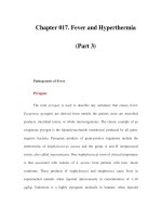
FIGURE 4.3
Intraoperative picture demonstrating the implanted Navigus electrode lead securing device
(Image-guided Neurologics, Melbourne, FL) in its final position.
8174_C004.fm Page 6 Saturday, November 3, 2007 7:40 AM
Responsive Neurostimulation for Epilepsy — Neurosurgical Experience
4
-7
implanted lead. The stylet of the lead is removed and the distal shaft of the implanted lead is secured
into the Navigus device.
4.3.2.3 Subdural Lead Placement
If a subdural strip lead is to be placed, the dura is opened linearly and the 1
×
4 cortical lead is inserted
through the dural opening. Fluoroscopy is used to verify proper placement. The distal shaft of the
implanted lead is safely secured to the Navigus device.
4.3.2.4 Pulse Generator Implantation
At this point, the provided ferrule is placed on the exposed bone and the desired bony defect is outlined
(Figure 4.4). The outlined bone is drilled out by a high-speed, air-driven drill with attention not to
traumatize the underlying dura (Figure 4.5). The bony edges are smoothed and meticulous hemostasis
is performed if necessary by applying bone wax. Thorough irrigation of the wound is of great importance
for removing any residual bone dust. The provided ferrule is then implanted and secured to the adjacent
bone at four points with the provided self-tapping mini-screws (Figure 4.2). The pulse generator is
connected to the distal end of the already implanted lead or leads, and then is secured in the implanted
ferrule (Figure 4.6). At this point, the programmer is used with the sterile covered telemetry wand to
interrogate the implanted pulse generator, measure the impedances of all lead contacts, and perform
electrocorticography to verify proper function of the implanted RNS
™
neurostimulator system. The
surgical wound is thoroughly irrigated with bacitracin solution and then is closed in anatomical layers.
The wound is covered with a sterile dressing.
The patient is transported to the neurosurgical ward for observation and discharged within two to
three days. Before discharge, a postoperative head CT scan and skull plain x-rays (antero-posterior and
lateral views) are obtained to provide baseline imaging studies for future reference (Figures 4.7A
and 4.7B). The first interrogation and seizure detection programming session is usually performed on
the third postoperative day. Further interrogations and programming sessions are usually required for
fine adjustment of the implanted RNS
™
neurostimulator detection and stimulation parameters.
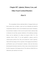
FIGURE 4.4
Intraoperative picture demonstrating the outlining of the pulse generator on the bone.
8174_C004.fm Page 7 Saturday, November 3, 2007 7:40 AM
4
-8
Neuroengineering
FIGURE 4.5
Intraoperative picture demonstrating the bony defect created for implanting the ferrule and the
pulse generator.
FIGURE 4.6 (See color insert following page 15-4).
Intraoperative picture demonstrating the pulse generator
connected to an implanted depth lead and secured to the underlying ferrule.
8174_C004.fm Page 8 Saturday, November 3, 2007 7:40 AM
Responsive Neurostimulation for Epilepsy — Neurosurgical Experience
4
-9
A
B
FIGURE 4.7
(A) Postoperative x-rays (lateral view) of one of our patients demonstrating the implanted RNS system
with two subdural leads. (B) Postoperative x-rays (lateral view) of one of our patients demonstrating the implanted
RNS system with two depth leads.
8174_C004.fm Page 9 Saturday, November 3, 2007 7:40 AM
4
-10
Neuroengineering
The operative blood loss is usually minimal (in all of our cases has been maintained at less than 100 cc)
while the mean duration of our surgical procedure of implantation is 3.2 hours (range 2 to 4.5 hours).
4.4 Conclusions
The implantable, local, closed-loop RNS
™
neurostimulator system is an investigational option in patients
with well-localized, focal, medically refractory epilepsy, who are not candidates for surgical resection.
A multiinstitutional, prospective, clinical study is underway to evaluate the clinical safety and efficacy of
this novel treatment modality. Technical improvement of this system along with accumulation of expe-
rience from its clinical use could lead to the development of a system that would accurately detect and
efficiently abort any detected epileptiform activity.
Acknowledgments
The authors wish to thank David Greene from NeuroPace, Inc. (Mountain View, CA) for his valuable
assistance. The authors also wish to acknowledge their appreciation and thanks to Aaron Barth and Stacy
Perry for assistance in the preparation of this chapter.
References
1. Aarabi, A., Wallois, F., and Grebe, R. (2006). Automated neonatal seizure detection: a multistage
classification system through feature selection based on relevance and redundancy analysis.
Clin.
Neurophysiol.
, 117(2):328–340.
2. Arai, A. and Lynch, G. (1998). AMPA receptor desensitization modulates synaptic responses
induced by repetitive afferent stimulation in hippocampal slices.
Brain Res.
, 799(2):235–242.
3. Benabid, A L., Koudsie, A., Chabardes, S., et al. (2004). Subthalamic nucleus and substantia nigra
pars reticulata stimulation: the Grenoble experience. In Luders, H.O., Ed.,
Deep Brain Stimulation
and Epilepsy
. Marting Dunitz, London, p. 335–348.
4. Bikson, M., Lian, J., Hahn, P.J., et al. (2001). Suppression of epileptiform activity by high frequency
sinusoidal fields in rat hippocampal slices.
J. Physiol.
, 531(Pt 1):181–191.
5. Brown-Sequard, C.E. (1856–1857). Researches on epilepsy: its artificial production in animals,
and its etiology, nature and treatment in man.
Boston Med. Surg. J.
: p. 55–57.
6. Chernihovskyi, A., Mormann, F., Muller, M., et al. (2005). EEG analysis with nonlinear excitable
media.
J. Clin. Neurophysiol.
, 22(5):314–329.
7. Chkhenkeli, S.A., Sramka, M., Lortkipandze, G.S., et al. (2004). Electrophysiological effects and
clinical results of direct brain stimulation for intractable epilepsy.
Clin. Neurol. Neurosurg.
,
106(4):318–329.
8. Contzen, R. and Witte, O.W. (1994). Epileptic activity can induce both long-lasting potentiation
and long-lasting depression.
Brain Res.
, 653(1–2):340–344.
9. Cooper, I.S., Amin, I., Riklan, M., et al. (1976). Chronic cerebellar stimulation in epilepsy. Clinical
and anatomical studies.
Arch. Neurol.,
33(8):559–570.
10. Cooper, I.S. and Upton, A.R. (1978). Effects of cerebellar stimulation on epilepsy, the EEG and
cerebral palsy in man.
Electroencephalogr. Clin. Neurophysiol. Suppl.,
34:349–354.
11. Cooper, I.S., Upton, A.R., and Amin, I. (1980). Reversibility of chronic neurologic deficits. Some
effects of electrical stimulation of the thalamus and internal capsule in man.
Appl. Neurophysiol.
,
43(3–5):244–258.
12. Dudek, S.M. and Bear, M.F. (1993). Bidirectional long-term modification of synaptic effectiveness
in the adult and immature hippocampus.
J. Neurosci.,
13(7):2910–2918.
13. Dudek, F.E., Yasumura, T., and Rash, J.E. (1998). “Non-synaptic” mechanisms in seizure and
epileptogenesis.
Cell. Biol. Int.
22:793–805.
14. Durand, D.M. and Warman, E.N. (1994). Desynchronization of epileptiform activity by extracel-
lular current pulses in rat hippocampal slices.
J. Physiol.
, 480(Pt. 3):527–537.
8174_C004.fm Page 10 Saturday, November 3, 2007 7:40 AM
Responsive Neurostimulation for Epilepsy — Neurosurgical Experience
4
-11
15. Estellar, R., Echauz, J., Tcheng, T., et al. (2001). Line length: an efficient feature of seizure onset
detection. Proc. 23rd Annu. Conf. IEEE Eng. Med. Bio. Soc., pp. 1707–1709.
16. Fisher, R.S., Uematsu, S., Krauss, G.L., et al. (1992). Placebo-controlled pilot study of centromedian
thalamic stimulation in treatment of intractable seizures.
Epilepsia,
33(5):841–851.
17. Fountas, K.N., Smith, J.R., Murro, A.M., et al. (2005). Implantation of a closed-loop stimulation
in the management of medically refractory focal epilepsy. A Technical Note.
Stereotact. Funct.
Neurosurg.
, 83:153–158.
18. Fountas, K.N., Smith, J.R., Murro, A.M., et al. (2005). Closed-loop stimulation implantable system
for the management of focal, medically refractory epilepsy: implantation technique and prelimi-
nary results.
Epilepsia
, 46(S8):240–241.
19. Franaszczuk, P.J., Kudela, P., and Bergey, G.K. (2003). External excitatory stimuli can terminate
bursting in neural network models.
Epilepsy Res.
, 53(1–2):65–80.
20. Siegel, R.E. (1976). Galen on the affected parts (de locis affectis). Book III. S. Karger, Basel,
Switzerland, pp. 15–90.
21. Gowers, W.R. (1885). Epilepsy and Other Chronic Convulsive Diseases: Their Causes, Symptoms
and Treatment. William Wood, New York, p. 235–236.
22. Hauser, W.A. (1995). Epidemiology of Epilepsy in Children. In Adelson, P.D. and Black, P.M., Eds.,
Neurosurgery Clinics of North America
. W.B. Saunders Co., Philadelphia, 6(3):419–429.
23. Hauser, W.A. and Hesdorffer, D.C. (2001). Epidemiology of intractable epilepsy. In Luders, H.O. and
Comair, Y.G., Eds.,
Epilepsy Surgery, 2nd edition
. Lippincott Williams & Wilkins, Philadelphia, p. 55–61.
24. Hodaie, M., Wennberg, R.A., Dostrovsky, J.O., and Lozano, A.M. (2002). Chronic anterior thalamus
stimulation for intractable epilepsy.
Epilepsia
, 43(6):603–608.
25. Jackson, J.H. (1868). Case of convulsive attacks arrested by stopping the aura.
Lancet
, 1:618–619.
26. Kang-Park, M.H., Sarda, M.A., Jones, K.H., et al. (2001). Protein phosphatases mediate depoten-
tiation induced by high-intensity theraburst stimulation.
J. Neurophysiol.
, 89:684–690.
27. Kerrigan, J.F., Litt, B., Fisher, R.S., Craunston, S., et al. (2004). Electrical stimulation of the anterior
nucleus of the thalamus for the treatment of intractable epilepsy.
Epilepsia
, 45(4):346–354.
28. Kinoshita, M., Ikeda, A., Matsumoto, R., et al. (2004). Electrical stimulation on human cortex
suppresses fast cortical activity and epileptic spikes.
Epilepsia
, 45(7):787–791.
29. Kiymik, M.K., Subasi, A., and Ozcalik, H.R. (2004). Neural networks with periodogram and
autoregressive spectral analysis methods in detection of epileptic seizure.
J. Med. Syst.
,
28(6):511–522.
30. Labar, D. (2004). Vagal nerve stimulation: effects on seizures. In Luders, H.O., Ed.,
Deep Brain
Stimulation and Epilepsy
. Martin Dunitz, London.
31. Lesser, R.P., Kim, S.H., Beyderman, L., et al. (1999). Brief bursts of pulse stimulation terminate
after-discharges caused by cortical stimulation.
Neurology
, 53(9):2073–2081.
32. Liang, F., Isackson, P.J., and Jones, E.G. (1996). Stimulus-dependent, reciprocal up- and down-
regulation of glutamic acid decarboxylase and Ca
2+
/calmodulin-dependent protein kinase II gene
expression in rat cerebral cortex.
Exp. Brain Res.
, 110(2):163–174.
33. Meng, L., Frei, M.G., Osorio, I., Strang, G., and Nguyen, T.Q. (2004). Gaussian mixture models
of ECoG signal features for improved detection of epileptic seizures.
Med. Eng. Phys.
,
26(5):379–393.
34. Morrell, M. (2006). Brain stimulation for epilepsy: can scheduled or responsive neurostimulation
stop seizures?
Curr. Opin. Neurol.
, 19(2):164–168.
35. Motamedi, G.K., Lesser, R.P., Miglioretti, D.L., et al. (2002). Optimizing parameters for terminating
cortical after-discharges with pulse stimulation.
Epilepsia
, 43(8):836–846.
36. Munz, M., Sweasey, R., Barrett, C., et al. (2003). Preclinical testing of an implantable responsive
neurostimulator system in a sheep model. In
Society for Neuroscience
. New Orleans, 2003.
37. Munz, M., Sweasey, R., Barret, C., et al. (2003). Implantation and testing of responsive neuro-
stimulator (RNS) system for epilepsy. In
American Society for Stereotactic and Functional Neuro-
surgery
, New York.
8174_C004.fm Page 11 Saturday, November 3, 2007 7:40 AM
4
-12
Neuroengineering
38. Murro, A.M., Park, Y.D., Bergey, G.K., et al. (2003). Multicenter study of acute responsive stimu-
lation in patients with intractable epilepsy.
Epilepsia,
44(Suppl. 9):326.
39. Murro, A., Park, Y., Greene, D., et al. (2002). Closed-loop neuro-stimulation in patient with
intractable epilepsy. In
American Clinical Neurophysiology Society,
New Orleans, LA.
40. Nair, D.R., Matsumoto, R., Luders, H.O., et al. (2004). Direct cortical electrical stimulation in the
treatment of epilepsy. In Luders, H.O., Ed.,
Deep Brain Stimulation and Epilepsy
. Martin Dunitz,
London.
41. Neme, S., Montgomery, E.B., Rezai, A., et al. (2004). Subthalamic nucleus stimulation in patients
with intractable epilepsy: the Cleveland experience. In Luders, H.O., Ed.,
Deep Brain Stimulation
and Epilepsy
. Martin Dunitz, London, p. 349–358.
42. Netoff, T.I. and Schiff, S.J. (2002). Decreased neuronal synchronization during experimental
seizures.
J. Neurosci.
, 22(16):7297–7307.
43. Niederhauser, J.J., Esteller, R., Echauz, J., et al. (2003). Detection of seizure precursors from
depth-EEG using a sign periodogram transform.
IEEE Trans. Biomed. Eng.
, 50(4):449–458.
44. Oommen, J., Morrell, M., and Fisher, R.S. (2005). Experimental electrical stimulation therapy for
epilepsy.
Curr. Opin. Neurol.
, 7(4):261–271.
45. Osorio, I., Frei, M.G., Manly, B.F., et al. (2001). An introduction to contingent (closed-loop) brain
electrical stimulation for seizure blockage, to ultra-short term clinical trials, and to multidimen-
sional statistical analysis of therapeutic efficacy.
J. Clin. Neurophysiol.
, 18(6):533–544.
46. Osorio, I., Frei, M.G., Sunderam, S., et al. (2005). Automated seizure abatement in humans using
electrical stimulation.
Ann. Neurol.
, 57(2):258–268.
47. Peters, T.E., Bhavaraju, N.C., Frei, M.G., and Osorio, I. (2001). Network system for automated
seizure detection and contingent delivery of therapy.
J. Clin. Neurophysiol.
, 18(6):545–549.
48. Poliakov, A.V., Powers, R.K., Sawczuk, A., and Binder, M.D. (1996). Effects of background noise
on the response of rat and cat motoneurones to excitatory current transients.
J. Physiol.
,
495(Pt.1):147–157.
49. Saab, M.E. and Gotman, J. (2005). A system to detect the onset of epileptic seizures in scalp EEG.
Clin. Neurophysiol.,
116(2):427–442.
50. Sheng, M. and Kim, M.J. (2002). Postsynaptic signaling and plasticity mechanisms.
Science,
298(5594):776–780.
51. Slutzky, M.W., Cvitanovic, P., and Mogul, D.J. (2003). Manipulating epileptiform bursting in the
rat hippocampus using chaos control and adaptive techniques.
IEEE Trans. Biomed. Eng.
,
50(5):559–570.
52. Sramka, M., Fritz, G., Gajadosova, D., and Nadvornik, P. (1980). Central stimulation treatment
of epilepsy.
Acta Neurochir. Suppl. (Wien.),
30:183–187.
53. Sramka, M., Fritz, G., Galanda, M., and Nadvornik, P. (1976). Some observations in treatment
stimulation of epilepsy.
Acta Neurochir. (Wien.), (23 Suppl.):257–262.
54. Stacey, W. and Durand, D.M. (2001). Synaptic noise improves detection of subthreshold signals
in hippocampal CA1 neurons. J. Neurophysiol., 86(3):1104–1112.
55. Stevens, C.F. and Tsujimoto, T. (1995). Estimates for the pool size of releasable quanta at a single
central synapse and for the time required to refill the pool. Proc. Natl. Acad. Sci., U.S.A.,
92(3):846–849.
56. Subasi, A. (2006). Application of adaptive neuro-fuzzy inference system for epileptic seizure
detection using wavelet feature extraction. Comput. Biol. Med., February 8; [Epub ahead of print].
57. Subasi, A. and Ercelebi, E. (2005). Classification of EEG signals using neural network and logistic
regression. Comput. Meth. Prog. Biomed., 78(2):87–99.
58. Velasco, F., Carrillo-Ruiz, J.D., Brito, F., et al. (2005). Double-blind, randomized controlled pilot
study of bi-lateral cerebellar stimulation for treatment of intractable motor seizures. Epilepsia,
46:1071–1081.
59. Velasco, F., Velasco, M., Jimenez, F., et al.(2001). Stimulation of the central median thalamic nucleus
for epilepsy. Stereotact. Funct. Neurosurg., 77(1–4):228–232.
8174_C004.fm Page 12 Saturday, November 3, 2007 7:40 AM
Responsive Neurostimulation for Epilepsy — Neurosurgical Experience 4-13
60. Velasco, F., Velasco, M., Ogarrio, C., and Fanghanel, G. (1987). Electrical stimulation of the
centromedian thalamic nucleus in the treatment of convulsive seizures: a preliminary report.
Epilepsia, 28(4):421–430.
61. Velasco, M., Velasco, F., and Velasco, A.L. (2001). Centromedian-thalamic and hippocampal
electrical stimulation for the control of intractable epileptic seizures. J. Clin. Neurophysiol.,
18(6):495–513.
62. Velasco, F., Velasco, M., Velasco, A.L., et al. (2001). Electrical stimulation for epilepsy: stimulation
of hippocampal foci. Stereotact. Funct. Neurosurg., 77(1–4):223–227.
63. Wagenaar, D.A., Madhavan, R., Pine, J., and Potter, S.M. (2005). Controlling bursting in cortical
cultures with closed-loop multi-electrode stimulation. J. Neurosci., 25(3):680–688.
64. Zucker, R.S. and Regehr, W.G. (2002). Short-term synaptic plasticity. Annu. Rev. Physiol.,
64:355–405.
8174_C004.fm Page 13 Saturday, November 3, 2007 7:40 AM
8174_C004.fm Page 14 Saturday, November 3, 2007 7:40 AM
5
-1
5
Responsive
Neurostimulation for
Epilepsy: RNS
™
Technology and
Clinical Studies
5.1 Introduction and Background
5
-1
Clinical Market and Relevance of the Therapy ·
Review of Other Technologies
5.2 Fundamental Neuroscience: Mechanism of Action
5
-2
Mechanisms and Physiology of Epilepsy
5.3 Technological Innovation
5
-3
Origin of Responsive Neurostimulation Technology ·
Description and Implementation of Technology
5.4 Clinical Studies
5
-8
eRNS Study · Implantable RNS™ System Feasibility Study ·
Implantable RNS™ System Pivotal Study
5.5 Conclusions, Discussion, and Future Directions
5
-9
References
5
-10
5.1 Introduction and Background
5.1.1 Clinical Market and Relevance of the Therapy
Epilepsy is a neurological disorder that affects 2.3 million people in the United States and as many as
fifty million people worldwide [Begley et al., 2000]. Perhaps half have intractable epilepsy; that is, seizures
cannot be controlled by antiepileptic drug (AED) therapy, and/or there are side effects from AEDs that
adversely impact quality of life. The ketogenic diet, the vagus nerve stimulator, and epilepsy surgery are
other treatment options. However, many persons with epilepsy are left without treatment that is effica-
cious, tolerable, and acceptable. Device-based therapies may provide additional therapeutic options. One
approach to treating medically intractable localization-related epilepsy with partial onset seizures is to
provide focal stimulation in response to electrographic epileptiform activity. The NeuroPace
®
RNS
™
system includes a cranially implanted responsive neurostimulator that continuously monitors electro-
corticographic (ECoG) activity from intracranial electrodes, detects electrographic events of significance
according to programmable detection algorithms, and provides responsive stimulation. The intent is to
modify abnormal electrographic activity in an effort to prevent or terminate clinically evident seizures.
Thomas K. Tcheng and
Martha Morrell
8174_C005.fm Page 1 Monday, October 29, 2007 2:27 PM
5
-2
Neuroengineering
The RNS
™
system includes a fully implantable, patient-specific, field-programmable responsive neuro-
stimulator with multiple electrodes surgically placed into selected brain target areas.
Responsive neurostimulation technology is designed to benefit epilepsy patients and their families by
potentially improving long-term health, decreasing disability, and reducing healthcare costs. Improved
long-term health could result from a reduction in the damaging effects of epileptic seizures, psychiatric
disorders, and reproductive dysfunction associated with epilepsy. In addition, patients may also benefit
from a reduction or elimination of side effects from concurrent therapies by potentially enabling a
reduction or elimination of the dosage of these therapies. Patients and their employers may benefit from
increased productivity as they return to the workplace. Epilepsy patients and third-party healthcare payers
may benefit from a reduced need for medical care associated with seizures and health complications
associated with epilepsy.
NeuroPace’s development of responsive neurostimulation technology could also benefit the medical
device industry by demonstrating the feasibility and utility of responsive neurostimulation.
5.1.2 Review of Other Technologies
The current practice for treating epilepsy includes AEDs, vagus nerve stimulation, the ketogenic diet,
and resective surgery. There are currently no FDA-approved implantable direct brain stimulation devices
available to treat epilepsy. In the research setting, direct brain stimulation for epilepsy has had mixed
results. The vast majority of this work used noncontingent continuous or on–off cycling stimulation,
also referred to as open-loop stimulation.
5.1.2.1 Vagus Nerve Stimulation
Cyberonics currently offers vagus nerve stimulation therapy. The vagus nerve stimulator provides inter-
mittent, regularly scheduled electrical stimulation to the left vagus nerve at the level of the external
carotid artery. The FDA approved the vagus nerve stimulator therapy system indicated for use as an
adjunctive therapy in reducing the frequency of seizures in adults and adolescents over 12 years of age
with partial onset seizures, which are refractory to antiepileptic medications.
In two randomized clinical trials in people with intractable partial seizures, therapy with the vagus
nerve stimulator was associated with a median percentage reduction in seizures of 23%. In addition,
23 to 30% of patients achieved a 50% or greater reduction in seizures leading to marketing approval,
therapy with the vagus nerve stimulator reduced partial seizures [Cyberonics, 2002]. Long-term uncon-
trolled follow-up of patients participating in the investigational trials suggests that persons continuing
vagus nerve stimulation over one to two years continue to benefit. However, fewer than 5% achieve
freedom from seizures. Adverse effects arise in approximately 30% of patients, including hoarseness,
headache, muscle pain, throat pain, coughing, and nausea [Schachter and Schmidt, 2001].
5.1.2.2 Thalamic Stimulation
Stimulation of the anterior nucleus of the thalamus for epilepsy is currently being evaluated by an FDA-
approved randomized, controlled trial (Medtronic). In this multicenter study, patients with at least six
partial seizures per month, with or without secondary generalization, are treated with regular intermittent
bilateral high-frequency stimulation of the anterior thalamic nucleus [Oommen et al., 2005]. The results
of this investigation were not available at the time this manuscript was prepared.
5.2 Fundamental Neuroscience: Mechanism of Action
5.2.1 Mechanisms and Physiology of Epilepsy
Epilepsy is a complex neurological disease characterized by seizures. Epileptic seizures are sudden,
excessive, and temporary neurological events. “Sudden” means a transition from background to abnormal
activity. “Excessive” means increased excitation due to a shift in the balance between inhibition and
excitation. “Temporary” indicates that the typical seizure is less than 60 seconds. Seizures are usually
8174_C005.fm Page 2 Monday, October 29, 2007 2:27 PM
Responsive Neurostimulation for Epilepsy: RNS™ Technology and Clinical Studies
5
-3
accompanied by electrographic features that are apparent in electroencephalographic (EEG) recordings.
Additionally, abnormal electrographic discharges are often observed interictally (between seizures).
In partial onset epilepsy, a seizure onset zone is defined as the brain area where the seizure begins. Seizures
can arise in any region of cortex. There are often structural and functional brain abnormalities that can
be detected using diagnostic imaging methods. Another common observation is a neurochemical
imbalance. These structural, functional, and neurochemical abnormalities are believed to contribute to
abnormal electrographic activity that is characteristic of seizures, such as hypersynchronous oscillations.
Direct electrical stimulation can affect neurons and neural systems in different ways, depending on
the stimulation parameters and the brain area being stimulated. Continuous high-frequency stimulation
is theorized to inhibit neural activity, either by causing preferential release of inhibitory neurotrans-
mitters, by depleting neurotransmitter in the presynaptic terminal, or by causing a depolarization block
[Buerrier et al., 2001; Lee et al., 2004; Benabid et al., 2005; Mantovani et al., 2006]. In contrast, short
bursts of stimulation delivered in response to detection of a seizure onset are thought to disrupt the
oscillatory neural dynamics underlying the seizure process.
5.3 Technological Innovation
5.3.1 Origin of Responsive Neurostimulation Technology
Responsive neurostimulation technology was invented by Robert and David Fischell and neurologist
Adrian Upton. In 1997, they founded NeuroPace to design, develop, manufacture, and market the
responsive neurostimulator and other implantable devices for the treatment of neurological disorders.
Many of the developers and engineers involved in the development of the RNS
™
system came from the
cardiac defibrillator industry, where pattern detection and responsive stimulation technologies were
developed to treat cardiac arrhythmias. These technologies were further developed by NeuroPace to
detect and treat the abnormal electrographic brain activity associated with epilepsy. The Half Wave, Line
Length, and Area detection tools were based on successful seizure and spike detection tools documented
in the literature. These detection tools were simplified and adapted to fit the low power and low
computational complexity constraints of an implantable medical device. Stimulation features were devel-
oped to be appropriate for direct brain stimulation. These included a current-controlled, charge-
balanced, biphasic rectangular waveform, as well as a wide range of stimulation amplitudes, pulse widths,
frequencies, and burst durations. Additionally, the developers included the ability to stimulate different
brain areas using different stimulation parameters, depending on the pattern or location of the abnormal
activity detected. Because responsive stimulation is new technology, a number of safety features were
also incorporated into the RNS
™
system.
5.3.1.1 Preliminary Stimulation Studies
Responsive stimulation has been effective in terminating seizure activity in a wide range of studies from
rat hippocampal preparations to limited human clinical studies. A recurring finding throughout many
of these studies is that the earlier the stimulation is applied, relative to the seizure, the greater the
likelihood of success [Lesser et al., 1999]. This observation lent support to the hypothesis that responsive
stimulation may be an effective therapy for epilepsy.
5.3.1.1.1 Animal Studies of Closed-Loop Stimulation
In animal studies, Psatta [1983] studied interictal spiking activity in epileptic foci in cats. Responsive
stimulation was automatically delivered to the caudate nucleus when a 1-sec spike burst was detected,
resulting in spike depression. No comparable effects were seen with random stimulation. In a rat
hippocampal slice preparation, Nakagawa and Durand [1991] applied subthreshold electrical currents
to the stratum pyramidale upon detection of abnormal electrical activity. This resulted in complete
suppression of interictal bursts in 90% of the slices. Further confirmation of the potential efficacy of
responsive stimulation was demonstrated by Vercueil et al. [1998], who demonstrated the ability of
short bursts (5 sec) of high-frequency (130 Hz) bilateral stimulation of the subthalamic nuclei to
8174_C005.fm Page 3 Monday, October 29, 2007 2:27 PM
5
-4
Neuroengineering
decrease the duration of absence seizures in rats. In their study, no decrease in epileptiform activity
was observed when continuous bilateral stimulation was applied. To the extent that animal studies are
valid models for human epilepsy, these studies lend support for the use of responsive electrical stimu-
lation to treat epilepsy.
•
Initial closed-loop stimulation studies.
The groundbreaking work of Penfield and Jasper [1954]
showed that spontaneous epileptiform activity can be interrupted using electrical stimulation.
Since then, progress has been made in the application of responsive stimulation to control seizures
in epilepsy patients. Rajna and Lona [1989] observed that epileptic seizures were promptly inhib-
ited in 79 of 139 observations in nineteen patients when acoustical stimuli were delivered at the
onset of absence seizures.
•
Human Trials of Closed-loop Stimulation.
Chkhenkeli [1997] reported that manually triggered
stimulation of the caudate terminated electrically evoked seizures in humans. Lesser et al. [1999]
found that electrical stimulation was effective in terminating epileptiform activity (afterdischarges)
caused by brain-mapping stimulation. Osorio et al. [2001] has provided one of the most direct
demonstrations of the effectiveness of responsive stimulation in focal or remote locations of the
brain using stimulation frequencies higher than 100 Hz in eight patients. As Litt and Baltuch
[2001] concluded in their review, past efforts point to intelligent closed-loop brain stimulators as
the next logical step in the treatment of epilepsy.
5.3.2 Description and Implementation of Technology
The RNS
™
system consists of an implantable responsive neurostimulator (Figure 5.1), electrode leads, a
wireless telemetry wand, a patient data transmitter, and a physician programmer. The neurostimulator
is designed with a thickness and curvature matching that of the skull so that it replaces an identically
shaped section of bone that is surgically removed. During implant, a craniectomy the same size and
shape of the neurostimulator is made, and the neurostimulator is placed into a ferrule that is inserted
into the craniectomy and anchored to the skull with titanium screws.
Two lead designs can be used with the RNS
™
system; either cortical strip leads or depth leads. Each
lead contains four electrodes. The leads are surgically positioned so that the electrodes are as close to
the seizure focus as possible. Depth leads are usually implanted stereotactically through a burr hole, and
cortical strip leads are usually implanted through a craniectomy. The implanted parts of the RNS
™
system
are shown in Figure 5.2.
The neurostimulator is capable of communicating with an external wand via wireless telemetry. The
telemetry wand is connected to either a physician programmer or a patient data transmitter. The
programmer is used by clinicians to upload device data (interrogation) and adjust neurostimulator
settings (programming), while the patient data transmitter is used by patients for interrogation only.
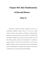
FIGURE 5.1
The NeuroPace
®
RNS
™
neurostimulator.
8174_C005.fm Page 4 Monday, October 29, 2007 2:27 PM
Responsive Neurostimulation for Epilepsy: RNS™ Technology and Clinical Studies
5
-5
Both the programmer and patient data transmitter communicate via the Internet with a central patient
data management system where device data are stored and can be reviewed by physicians. Patients can
interrogate their neurostimulator at home on a daily basis using the wand and patient data transmitter,
then upload device data to the patient data management system. This allows physicians to view the latest
patient data without an office visit. The components of the RNS
™
system are summarized in Table 5.1.
5.3.2.1 RNS
™
Neurostimulator Capabilities
The RNS
™
neurostimulator is a multifunctional neurostimulation device that includes the following
capabilities:
• Electrographic sensing
• Electrographic event detection
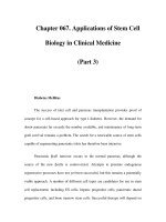
FIGURE 5.2
Schematic illustration of the implanted RNS
™
neurostimulator, depth lead, and cortical strip lead.
TABLE 5.1
Summary of RNS
™
System Components
RNS
™
System Component Description
Neurostimulator Fully implantable responsive neurostimulator (42 mm wide, 60 mm long, 8.4 mm thick)
Cortical strip lead Cortical strip lead with four disk electrodes spaced 10 mm apart
Depth lead Depth lead with four cylindrical electrodes available with electrode spacings in the
range of 3.5 mm to 10 mm apart
Telemetry wand Hand-held external wireless telemetry transceiver
Physician programmer Laptop computer with physician programmer software used for interrogating and
programming the neurostimulator
Patient data transmitter Laptop computer with patient data transmitter software used for interrogating the
neurostimulator and uploading device data to patient data management system
Patient data management system Database and clinician website
8174_C005.fm Page 5 Monday, October 29, 2007 2:27 PM
5
-6
Neuroengineering
• Responsive electrical stimulation
• ECoG storage
• Detection and stimulation event storage
• Electrode impedance measurement
• Battery voltage measurement
• Wireless telemetry
5.3.2.1.1 Clinical Use Example
An example of how the RNS
™
system’s intended clinical use will demonstrate how the different capabilities
work together to treat epilepsy. Prior to implant, the patient’s seizure focus is localized. This can be done
using a range of diagnostic methods, including scalp EEG and intracranial EEG; imaging methods such
as magnetic resonance imaging (MRI), positron emission spectroscopy (PET), and single photon emis-
sion computed tomography (SPECT); observation of seizure semiology; and neuropsychological testing.
If the seizure focus is adequately localized, leads are implanted such that the electrodes are as close as
possible to the seizure focus. The proximal ends of the leads are connected to the neurostimulator, which
is implanted in the skull. During the implant procedure, the programmer and wand are used to test lead
impedance, view real-time ECoG signals, and test neurostimulator functionality. After implant, the
programmer and wand are used to program detection settings into the neurostimulator. The neurostim-
ulator then detects electrographic events that meet the programmed detection criteria. When an event
is detected, information describing the event is stored, and an ECoG record containing the event may
be captured. In between follow-up visits, the patient data transmitter and wand can be used by the patient
to upload event information and ECoGs from the neurostimulator to the data transmitter. These device
data are subsequently uploaded from the patient data transmitter via the Internet to the patient data
management system. Patient device data can then be reviewed by the physician using a Web browser.
During follow-up visits, the programmer is used to interrogate and program the neurostimulator.
Detection settings can be reviewed and refined based on prior ECoG data uploaded from the neurostim-
ulator using a detection simulator built into the programmer. Once satisfactory detection specificity and
sensitivity have been achieved, responsive therapy can be enabled. Prior to enabling responsive therapy,
test stimulation can be provided interactively using the programmer to evaluate patient tolerability.
Responsive stimulation settings are then configured by the physician and programmed into the neuro-
stimulator to be delivered in response to electrographic event detection. Information describing therapy
delivery is also stored in the device and uploaded to the patient data management system along with
detection data and ECoGs. During subsequent follow-up visits, the physician can further refine and
update detection and therapy settings and optimize therapy to reduce seizure frequency and severity.
5.3.2.2 Electrographic Sensing and ECoG Storage
Electrographic activity is sensed through the cortical strip and depth electrodes using four differential
amplifiers. Because two leads can be connected to the neurostimulator, there are eight electrodes that
can be used for sensing. Electrodes are commonly assigned to amplifiers in adjacent pairs as shown in
Table 5.2. This provides an easily interpretable representation of electrographic neural activity at each
of four different locations and informs the selection of stimulation electrodes.
Electrographic data can be continuously collected on all four channels and stored within the neuro-
stimulator as ECoG records. The electrographic data are also used as input to the event detection
algorithm. Amplifier channels that are assigned to electrographic event detection also have noise and
saturation detectors enabled. An example of one channel from a stored ECoG, along with its spectrogram,
is shown in Figure 5.3. ECoGs can be stored in response to a variety of triggers, including detection,
therapy, noise, saturation, and time of day.
5.3.2.3 Electrographic Event Detection
Electrographic events are detected using a detection algorithm that continuously monitors electrographic
brain activity. The event detection system is capable of specific and sensitive detection. Three detection
tools — Half Wave, Line Length, and Area — are used by the detection algorithm. These detection tools
8174_C005.fm Page 6 Monday, October 29, 2007 2:27 PM
Responsive Neurostimulation for Epilepsy: RNS™ Technology and Clinical Studies
5
-7
can be applied independently or in combination with a single ECoG channel, and up to two ECoG
channels can be configured for detection.
•
Half Wave Tool.
The Half Wave Tool identifies local minima and maxima within the ECoG
waveform and uses them to measure half waves. The half wave amplitude is defined as the voltage
difference between adjacent minima and maxima, and the half wave duration is defined as the
length of time between adjacent minima and maxima. The Half Wave Tool is used to detect
electrographic activity with specific frequency and amplitude characteristics.
•
Line Length Tool.
The Line Length Tool estimates a measure of complexity or fractal dimension
within the ECoG signal. The line length is calculated as the sum of the unsigned amplitude changes
within a time window. Either the absolute difference or the ratio of the average line length between
a short window and a long window is used to determine whether signal complexity is increasing
or decreasing. The Line Length Tool is sensitive to changes in amplitude or frequency.
•
Area Tool.
The Area Tool estimates a measure of energy within the ECoG signal. The area is
calculated as the sum of the unsigned area under the curve within a time window. Either the
absolute difference or the ratio of the average area between a short window and a long window
is used to determine whether energy is increasing or decreasing. The Area Tool is sensitive to
changes in amplitude but minimally sensitive to changes in frequency.
5.3.2.4 Responsive Stimulation
Electrical stimulation is delivered in response to electrographic event detection. A number of stimulation
parameters can be adjusted to control stimulation, including current amplitude, pulse width, pulse
frequency, and burst duration. Stimulation can be delivered in either a monopolar or bipolar montage.
Monopolar stimulation is delivered between any combination of electrodes and the neurostimulator case.
TABLE 5.2
Typical Amplifier Configuration
Electrode
Amp. 1 Amp. 2 Amp. 3 Amp. 4
Lead 1, electrode 1 +
Lead 1, electrode 2 -
Lead 1, electrode 3 +
Lead 1, electrode 4 -
Lead 2, electrode 1 +
Lead 2, electrode 2 -
Lead 2, electrode 3 +
Lead 2, electrode 4 -
FIGURE 5.3 (See color insert following page 15-4).
Example of one channel from a stored ECoG, along with its
spectrogram. The x-axis is in seconds. The upper panel displays the FFT spectrogram to represent normalized
spectral power (red = high, blue = low). The y-axis on the upper panel is frequency (in Hertz). The lower panel
displays the time-series trace of the ECoG. The y-axis on the lower panel indicates the relative amplitude of the signal
in percent full scale. In the time series trace, the “B” label and the vertical blue line indicate when a detection by a
Line Length Tool occurred, the light blue background indicates a detection episode, and the yellow background,
barely visible in the lower trace starting at 78 seconds, indicates saturation.
100
75
50
25
50
0
–50
Percent
Hz
0102030405060708090
B
8174_C005.fm Page 7 Monday, October 29, 2007 2:27 PM
5
-8
Neuroengineering
For monopolar stimulation, all the electrodes must be assigned the same polarity, either negative
(cathodic) or positive (anodic), while the case is assigned the opposite polarity. For bipolar stimulation,
one set of electrodes is defined as cathodic and another set of electrodes is defined as anodic. Stimulation
is configured in a patient-specific manner to reduce seizure frequency and severity. In general, stimulation
is delivered to electrodes where abnormal electrographic activity is observed. In responsive stimulation,
it is important to note that event detection is the primary means by which therapy is allocated over time.
One strategy for allocating therapy is to configure detection so that therapy is delivered as early as possible
during an electrographic seizure. With redetection during an event, therapy can be delivered repeatedly.
An example of where responsive stimulation appears to terminate a seizure is shown in Figure 5.4.
5.4 Clinical Studies
NeuroPace
®
RNS
™
neurostimulation technology has been used in several human clinical trials. An
investigation of manually triggered stimulation to terminate afterdischarges produced during brain
mapping with intracranial electrodes was completed in 2003. A safety investigation using an external
responsive neurostimulator (eRNS) in an epilepsy monitoring unit setting was completed in 2005.
A feasibility study using an implanted neurostimulator (model RNS-300) was initiated in 2004. The data
from this study provide sufficient evidence of safety and preliminary evidence of efficacy supporting the
December 2005 commencement of a pivotal multicenter, double-blind trial of responsive stimulation
safety and efficacy using the implanted neurostimulator.
5.4.1 eRNS Study
In the eRNS study, an external responsive neurostimulator provided automatic seizure detection and
stimulation. The study subjects were patients being evaluated for epilepsy surgery with intracranial
electrodes. Seizure detection algorithms were optimized over several days while electrographic data
were acquired for seizure localization. Automatic responsive stimulation was applied to the seizure
onset zone at the end of the intracranial study prior to removal of the mapping electrodes. No
unanticipated serious device-related adverse events were reported. Four subjects had a perception of
stimulation, which was eliminated by adjustment of the stimulation settings. NeuroPace’s experience
with the eRNS study suggested that it is important for stimulation to occur early in the electrographic
FIGURE 5.4 (See color insert following page 15-4).
Example of a possible seizure termination by responsive
stimulation. The upper panel shows the FFT spectrogram and time series of the entire ECoG channel. The lower
panel shows a close-up of the time series around the time of termination. In the time series plots, detection is indicated
by the vertical blue line and the “B” label, a detection episode is indicated by the blue background, and responsive
stimulation is indicated by the vertical red lines and the “Tr” labels (the first is partly obscured by the “B” label).
These data are from the same subject as in Figure 5.3.
100
75
50
25
50
–50
01020304050607080
0
Percent Hz
TrTr
Tr Tr
64 65 66 67 68 69 70 71 72
Br
8174_C005.fm Page 8 Monday, October 29, 2007 2:27 PM
Responsive Neurostimulation for Epilepsy: RNS™ Technology and Clinical Studies
5
-9
event, and that to accomplish this, electrodes should be positioned to capture the earliest electrographic
onset. Additionally, multiple electrodes in the vicinity of the seizure onset should be used for delivery
of responsive stimulation.
5.4.2 Implantable RNS
™
System Feasibility Study
The implantable RNS
™
system feasibility study was a multicenter feasibility investigation to assess the
safety and preliminary efficacy of a cranially implanted, programmable responsive neurostimulator as
adjunctive therapy for medically refractory epilepsy in adults. Automatic detection settings were initially
optimized without stimulation, and then responsive stimulation therapy was enabled or subjects were
randomized into the stimulation OFF group. In a subsequent open extension period, stimulation was
enabled for all subjects. There have been no unanticipated device-related serious adverse events, and
no serious surgical complications in this study. NeuroPace has accumulated more than 140 patient years
of experience with the implanted responsive neurostimulator system.
5.4.3 Implantable RNS
™
System Pivotal Study
The implantable RNS
™
system pivotal investigation is currently underway. This study is a randomized,
double-blind, multicenter, controlled clinical investigation. The overall objective of this study is to
demonstrate that the RNS
™
system is safe and effective in reducing the frequency of disabling seizures
in adults with medically intractable partial onset seizures who have not responded to two or more
antiepileptic drugs. In this study, following completion of the baseline, implant, and subsequent stabi-
lization periods, the patients are randomized to one of two treatment conditions: therapy ON or therapy
OFF. The patients are blind to their treatment condition. At the end of the blind evaluation period, the
patients enter the Open Label extension period and stimulation can be enabled for all patients in the
trial. The intent of this study is to demonstrate safety and effectiveness, and to gather additional infor-
mation on long-term safety.
5.5 Conclusions, Discussion, and Future Directions
The NeuroPace
®
RNS
™
system is the first fully implantable, closed-loop responsive neurostimulator to
be evaluated in clinical trials. Responsive neurostimulation may become an option for adults with
medically intractable partial seizures. Because responsive stimulation can be targeted to specific brain
regions and therapy is delivered only in response to specific brain activity, responsive stimulation can
avoid the systemic side effects of AEDs and can be specifically applied to brain areas where the risks of
resective surgery are unacceptably high. Responsive neurostimulation is designed to deliver targeted
stimulation less often than continuous or on–off cycling neurostimulation methods. Potential advantages
of responsive neurostimulation include:
1. Specifically treating the pathological events
2. Avoiding habituation and desensitization, which may occur with continuous stimulation
3. Reducing side effects of stimulation
4. Preserving battery life reducing follow-up surgical procedures
The technology incorporated into the RNS
™
system allows physicians to review and evaluate electro-
corticographic activity in their patients, and adjust event detection and responsive stimulation settings
in a patient-specific manner. This flexibility allows for evaluation of a range of treatment strategies in
the same patient without additional surgery or neurostimulator replacement. The feasibility clinical
experience with the RNS
™
system provides preliminary data indicating that the neurostimulator and
leads can be safely implanted and that responsive stimulation can be safely applied to many regions of
the cortex. Epilepsy is the first clinical indication being treated by responsive stimulation technology.
The ability to monitor brain activity, detect events of interest, and respond to those events with custom-
izable therapy may be a general strategy that can be applied to a variety of neurological indications.
8174_C005.fm Page 9 Monday, October 29, 2007 2:27 PM
5
-10
Neuroengineering
References
Begley, C.E., Famulari, M., Annegers, J.F., Lairson, D.R., Reynolds, T.F., Coan, S., Dubinsky, S., Newmark,
M.E., Leibson, C., So, E.L., and Rocca, W.A. The cost of epilepsy in the United States: an estimate
from population-based clinical and survey data.
Epilepsia
, 41:342–351 (2000).
Chkhenkeli, S.A. and Chkhenkeli, I.S. Effects of therapeutic stimulation of nucleus caudatus on
epileptic electrical activity of brain in patients with intractable epilepsy.
Stereotact. Funct. Neurosurg
.,
69(1–4 Pt. 2):221–224 (1997).
Cyberonics.
Physician’s Manual: NeuroCybernetic Prosthesis SystemNCP
®
Pulse Generator Models 100 and
101
. Cyberonics (2002).
Lesser, R.P., Kim, S.H., Beyderman, L., Miglioretti, D.L., Webber, W.R., Bare, M., Cysyk, B., Krauss, G.,
and Gordon, B. Brief bursts of pulse stimulation terminate afterdischarges caused by cortical
stimulation.
Neurology
, 53(9):2073–2081 (1999).
Litt, B. and Baltuch, G. Brain stimulation for epilepsy.
Epilepsy and Behavior
,
2(3):S61–S67 (2001).
Nakagawa, M. and Durand, D. Suppression of spontaneous epileptiform activity with applied currents.
Brain Res
., 567(2):241–247 (1991).
Oommen, J., Morrell, M., and Fischer, R.S. Experimental electrical stimulation therapy for epilepsy.
Curr. Treat. Options Neurol
., 7(4):261–271 (2005).
Osorio, I., Frei, M.G., Manly, B.F., Sunderam, S., Bhavaraju, N.C., and Wilkinson, S.B. An introduction
to contingent (closed-loop) brain electrical stimulation for seizure blockage, to ultra-short-term
clinical trials, and to multidimensional statistical analysis of therapeutic efficacy.
J. Clin. Neuro-
physiol
., 18(6):533–544 (2001).
Penfield, W. and Jasper, H.
Epilepsy and the Functional Anatomy of the Human Brain
. Boston: Little,
Brown (1954).
Psatta, D.M. Control of chronic experimental focal epilepsy by feedback caudatum stimulations.
Epilepsia
.
24(4):444–454 (1983).
Rajna, P. and Lona, C. Sensory stimulation for inhibition of epileptic seizures.
Epilepsia
. 30(2):168–174
(1989).
Schachter, C.S. and Schmidt, D.
Vagus Nerve Stimulation
. London: Taylor & Francis (2001).
Vercueil, L., Benazzouz, A., Deransart, C., Bressand, K., Marescaux, C., Depaulis, A., and Benabid, A.L.
High-frequency stimulation of the subthalamic nucleus suppresses absence seizures in the rat:
comparison with neurotoxic lesions.
Epilepsy Res
., 31:39–46 (1998).
8174_C005.fm Page 10 Monday, October 29, 2007 2:27 PM
6
-1
6
Deep Brain Stimulation
for Pain Management
6.1 Introduction
6
-1
6.2 Neurophysiology of Periventricular and
Periaqueductal Pain Modulation
6
-3
6.3 Neurophysiology of Ventral Posterior Lateralis,
Ventral Posterior Medialis, and Internal
Capsule Stimulation
6
-4
6.4 Electrode and Pulse Generator Considerations
6
-6
6.5 Indications and Patient Selection
6
-7
6.6 Technique
6
-9
Pariventricular Gray Electrode Placement · Thalamic and
Internal Capsule Placement · Chronic Stimulation
6.7 Troubleshooting
6
-13
6.8 Complications and Side Effects
6
-14
6.9 Results
6
-14
6.10 Discussion
6
-15
References
6
-16
6.1 Introduction
Electrical stimulation of the brain has been used as an investigational technique since the days of Horsley
and Clarke at the turn of the past century, to activate excitable tissue and determine its function from
the resulting physiologic changes (18). The therapeutic use of electrical stimulation in the central nervous
system is a relatively recent addition to the armamentarium for the treatment of otherwise intractable
pain and suffering, and has been pursued vigorously because of the low potential for side effects and the
potential to affect previously untreatable central and deafferentation pain.
There have been reports of sporadic cases of pain relief related to electrical stimulation primarily used
for other reasons. Heath, in the early 1950s, found that stimulation of the septal area in a patient with
terminal cancer produced pain relief, and also produced dysphoria and euphoria which helped reduce
the stress and depression of her terminal illness (17). In his extensive recording of the electrical activity
and the effect of electrical activation of multiple sites in the brains of chronic schizophrenics, this was
the only patient who had a significant pain problem, but it did demonstrate that pain could be relieved
by stimulation. In 1956, Pool et al. also reported that activation of the septal area in the region of the
anterior third ventricle produced pain relief (36). Ervin et al. in 1966, reported the relief of pain in a
patient by stimulation of the thalamus (11). In 1967, Gol reported a small series of patients treated for
pain with electrical stimulation in the septal area, which was only moderately successful (13). In 1969,
Reynolds reported that stimulation of the periaqueductal gray produced deep analgesia in the rat suffi-
cient, in fact, to allow surgical procedures to be performed without any evidence of pain or discomfort
Donald E. Richardson
8174_C006.fm Page 1 Tuesday, November 6, 2007 8:51 AM
6
-2
Neuroengineering
(38). In 1971, Mayer et al. in John Liebskind’s laboratory at the University of California in Los Angeles
reported periaqueductal gray (PAG) stimulation produced analgesia (SPA) in the rat, which spurred a
large number of animal studies to investigate the mechanism of stimulation-induced analgesia in animals
(27). I had observed that stimulation in one patient during a steriotaxic leisoning procedure for the relief
of cancer pain that prelesion stimulation produced pleasant olfactory hallucinations along with pain
relief. Since the electrode was misdirected, the exact site of stimulation was never determined however;
it did prove to us that SPA was possible in the human (Richardson, unpublished data).
In 1970, based on these studies, Akil and I began a systematic acute stimulation study of sites in the
periventricular (PVG) and periaqueductal (PAG) areas in humans to determine sites to be used for pain
relief. These acute stimulation studies were carried out in five patients as a prelude to placing stereotaxic
lesions in the centrum median for chronic pain. The results led us to believe that the entire area along
the wall of the third ventricle, extending from a centimeter above the intercommissural line caudally
into the area of the raphae nuclei, could be used for electrical stimulation analgesia in the human (41)
(Figure 6.1). We chose a site in the periventricular area for chronic studies because of the minimal side-
effects in this area, in contrast to stimulation at sites farther caudal in the brainstem (41, 42). Our chronic
studies began in 1971 with the cooperation of Medtronic, Inc., Minneapolis, MN, which provided
modified their induction spinal cord stimulation system for our use and later made electrodes for long-
term stimulation in human patients (42). Since the early 1970s, many investigators have shown that
stimulation in what is now thought to be the endogenous opiate system in the PVG and PAG is effective
for relieving certain types of chronic pain.
FIGURE 6.1
Stimulation paths through the pariventricular and pariaquiductal gray (PVG, PAG) in five patients; all
patients received pain relief in the PVG. AQ = aqueduct of Sylvius, TH = thalamus, COS = superior colliculus,
COI = inferior colliculus, RU = nucleus rubber. (
J. Neurosurg
. 47:178–183, 1977.)
HPTH
Patient 1
2
3
4
5
TH
RU
COS
COI
CBL
AQ
8174_C006.fm Page 2 Tuesday, November 6, 2007 8:51 AM
Deep Brain Stimulation for Pain Management
6
-3
The second site used for relief of chronic pain has been the primary sensory relay nuclei of the thalamus.
Dorsal column stimulation and peripheral nerve stimulation have proven to be effective for relief of pain,
and the ventral posterior lateralis (VPL) and ventral posterior medialis (VPM) nuclei of the thalamus are
extensions of the main touch and proprioception sensory tracts (lemniscal system) from the dorsal
columns into the central nervous system. In 1960, Mazars et al. reported that stimulation of the soma-
tosensory pathways in VPL gave pain relief (28), and our studies in the cat showed that painful stimulation
could be reduced by VPL/VPM electrical stimulation (39). Subsequently, Hosobuchi et al. in the early
1970s, began to use chronic VPM and internal capsule (IC) stimulation, initially, for the relief of anesthesia
dolorosa following fifth nerve section for trigeminal neuralgia (19). The stimulation of these nuclei
produces paresthesia in the area of pain with subsequent loss of the chronic burning paresthetic and aching
quality of the pain, characteristic of deafferentation pain.
Young et al. in 1992 reported chronic stimulation of the lateral upper brainstem, more lateral than the
periaqueductal gray, in the nuclear mass of Kolliker–Fuse, produced analgesia in three of his patients (56).
6.2 Neurophysiology of Periventricular and
Periaqueductal Pain Modulation
Physiologically, stimulation-produced analgesia (SPA) is different for each of the sites selected. The best-
understood, but perhaps most complex, is the mechanism for SPA produced by stimulation of the PVG
and PAG. Initially, it was thought that stimulation of these sites produced direct effects on pain trans-
mission, but when SPA produced by PVG stimulation was found by Akil and Liebeskind to be inhibited
by naloxone, a specific opiate antagonist, understanding the physiology of pain modulation became more
complex (4). Watson et al. have shown in the rodent that the cell bodies for the central opiate mechanism
(mu system) are in the arcuate, infundibular, and periventricular nucleus of the hypothalamus, with
axons extending from these nuclei anteriorly through the septal area and then superiorly and posteriorly
through the septal area and medial to the thalamus in the PVG to the raphe nuclei in the ventral
periaqueductal gray and inferior to the locus ceruleus, where this tract terminates (52). This promelano-
opio-cortin nucleus and tract (also called the beta-endorphin system) has also been identified in the
human brain by Pilcher et al (34). We have demonstrated, in patients, that stimulation anywhere along
this system produces analgesia but with markedly different side effects. In the periaquiductal gray
osilopsia and symptoms identical to the epigastric rising syndrome are so noxious that patients will not
allow stimulation longer than a few seconds. Further in the anterior PVG and septal area, mild euphoric
is produced, and more inferiorly in the area of the basal forebrain and hypothalamus, elevation of blood
pressure is prompt and often dramatic (44) (Figure 6.2). This has given us several potential alternate
targets for use in patients who need a different site for electrode placement because of technical reasons,
with some obvious restrictions.
The secondary serotonergic fibers from the raphae nuclei and the norepinergic fibers from the locus
ceruleus then descend through the dorsolateral funiculus of the spinal cord to impinge on the dorsal
horn of the spinal cord (8, 9). Evidence suggests that there is an intermediate opioid interneuron that is
activated by these descending tracts that then inhibits pain at the first synapse in the dorsal horn (9).
Thus, the primary effect of stimulation of the PVG/PAG area is to activate the same system that is
activated by systemic opioids of the mu type (5, 47), which secondarily activates the descending inhib-
itory tracts emanating from the locus ceruleus and raphe nuclei and probably the Kolliker–Fuse and
parabrachiai nucleus, to inhibit input from somatic origin at the first synapse for pain impulses in the
dorsal horn (56).
There is also evidence that this inhibition occurs at the thalamic level and perhaps at the brainstem
level in the reticular formation, inhibiting propagation of impulses reaching awareness for pain through
both the spinoreticular tracts and the spinothalamic tracts. The evidence for the central mechanism,
however, is much less compelling than the evidence for inhibition of input to the spinal cord. Some
controversies have been raised about whether activation of these tracts produces analgesia by an opiate
mechanism. Our studies with Akil et al. (6, 7) have shown that activation of this system releases endogenous
8174_C006.fm Page 3 Tuesday, November 6, 2007 8:51 AM
6
-4
Neuroengineering
opiates into the ventricular fluid and analgesia is blocked by naloxone in animals (4), and is reduced in
humans (21, 42). Questions have been raised by Fessler et al. (12) about the use of contrast material for
visualization of the ventricular system during surgery to allow electrode placement, but in our studies
the baseline ventricular opiate levels and the post-stimulation opiate levels were both taken long after
the administration of contrast material (6, 7). In addition, studies by Yaksh et al. using direct opiate
activation of the cells in the periaqueducatal and periventricular gray have shown activation of seroton-
ergic and norepinergic mechanisms in the cord, and the blocking of these monoamine mechanisms
chemically reduces the effect of SPA (53, 54, 55) and later with Young et al. in the human patient measured
release of beta-endorphin and methionin enkephlelin during PVG stimulation (57, 58). Thus, at the
present time, it is thought that activation of target sites in the PVG and PAG activates opiate fibers
extending from the periventricular nuclei in the hypothalamus to the raphe nucleus and locus ceruleus,
where they terminate and activate serotonergic and norepinergic fibers descending through the dorso-
lateral funiculus, which terminate in the dorsal horn to inhibit pain at the first synapse, perhaps via an
opiate interneuron (Figure 6.3).
The Kolliker–Fuse nucleus, which is in the parabrachial nuclei area, has been described as having
norepinephrinergic cell bodies which project inferiorly to the spinal cord. Thus, stimulation of this area
may produce direct activation of the descending norepinephrine inhibitor fibers to the dorsal horn with
resulting pain relief. Stimulation of areas such as this would, therefore, reduce the chain of events by one
synapse, because it directly stimulates the descending norepinephrine fibers and thus is more effective
in patients who have a defective opiate mechanism, habituation, or high tolerance to opiates. Young has
reported that stimulation produces minimal adverse side effects and was efficacious for pain relief in
three of six patients (56).
6.3 Neurophysiology of Ventral Posterior Lateralis,
Ventral Posterior Medialis, and Internal Capsule Stimulation
The inhibitory mechanism of stimulation of the main sensory relay nuclei of the thalamus and internal
capsule remains obscure, at least in detail. These structures are cephalad extensions of the dorsal columns
extending upward through the nucleus Cuniatus and Gracilis from the spinal cord, stimulation of which
is known to relieve pain. Our studies in the cat reveal that sectioning of the dorsal columns below the
level of activation does not completely obliterate the pain-reducing effects of dorsal column stimula-
tion, indicating an additional more central inhibitory mechanism (39). Complete deactivation of pain
FIGURE 6.2
Stimulation sites tested in the human melano-opio-cortin (beta-endorphin) pathway. (
Appl. Neuro-
physiol
. 45:116–122, 1982.)
Stimulation Sites
β-Endorphine
Tract
β-Endorphine
Cell Group
Raphe Nuclei
Descending
Serotonin
Pathway
8174_C006.fm Page 4 Tuesday, November 6, 2007 8:51 AM
Deep Brain Stimulation for Pain Management
6
-5
inhibition by dorsal column stimulation can only be obtained by sectioning the dorsal column above
and below the level of activation (Richardson, unpublished data). In addition, single-cell recordings in
the sensory thalamus reveal an on/off mechanism activated by dorsal column stimulation, which would
indicate a complex inhibitory mechanism of interaction between the proprioception, touch, and posi-
tion sense fibers in the dorsal columns and the pain transmission system in the dorsal horn at the origin
of the ventrolateral spino-reticular and spino-thalamic tracts spinal cord, as well as in the synapses in
the brainstem, thalamus, and cortex of the brain (40). Mazars et al. (28) in 1960 reported that stimu-
lation of the somatosensory pathways in the cord gave pain relief and Ervin et al. (11) reported pain
relief in one patient in 1966 when stimulating the sensory thalamus.
Use of chronic thalamic stimulation was reported by Hosobuchi et al. for anesthesia dolorosa following
trigeminal rhizotomy (19). They reported, however, that this stimulation tended to lose its effectiveness
over time, and it is necessary to use a programmed type of stimulation with ramping or intermittent
stimulation to obtain good pain relief continuously over a long period of time. Placement of electrodes
for pain relief in this area is also facilitated by the fact that activation of these centers produces paresthesia
in the area of the patient’s pain, thus requiring accurate placement of the electrodes.
Hosobuchi et al. (20) suggested using the internal capsule (IC) for neospinothalamic stimulation.
I agree that this is more effective and less likely to produce sensory loss following electrode insertion
than placement in the main sensory nucleus, the penetration of which may produce small lesions resulting
in areas of focal sensory loss, which can be troubling if they involve the face or hand. Insertion of
electrodes into the internal capsule does not seem to produce this, although the reason is not clear at
this time, but probably indicates that fibers are less vulnerable than cell bodies to the trauma of electrode
insertion. This will be addressed in more detail in the discussion of technical development.
FIGURE 6.3 (See color insert following page 15-4).
The beta-endorphin serotonin norepinephrin pain modulation
system. (
Neurosurg. Clin. N. Am
. 6(1):135–144, 1995.)
alamus
Paraventricular
Opiate Bundle
Opiate
Dorsal Raphe Nucleus
Serotonin (5-HT)
Locus Ceruleus
Norepinephrine (NE)
Parabrachial Nucleus
Norepinephrine (NE)
Descending Inhibitory Tract
Serotonin (5-HT) and Norepinephrine (NE)
Raphe Magnus Nucleus
Serotonin (5-HT)
Hypothalamic Nucleus
Proopiomelanocortin (POMC)
8174_C006.fm Page 5 Tuesday, November 6, 2007 8:51 AM
6
-6
Neuroengineering
The inhibitory mechanism of stimulation of the dorsal columns, main sensory relay nuclei of the
thalamus and internal capsule which produces paresthesia in the area of the patient’s pain is based on
the original studies of Melzack and Wall (29) showing that inhibition of pain could be produced by
activation of touch and proprioception fibers, originally called the “gate theory of pain transmission.”
The effect of VPL/VPM/IC as well as dorsal column stimulation are multilevel. The inhibitory mechanism
appears to be locally driven at each synapse of the pain pathway through the dorsal horn, brainstem,
and thalamus and do not depend on inhibitory mechanisms involving the entry mechanism in the spinal
cord alone as in the opiate system (39). Thus, activation of the VPL, VPM, and IC is more effective for
relieving deafferentation pain, as well as pain generated in the nervous system due to its ability to inhibit
pain neurons at multiple levels, if these structures are intact (Figure 6.4).
6.4 Electrode and Pulse Generator Considerations
There have been several interesting observations over the years during development of the equipment
and electrical systems used for stimulation. The original electrode we used on our first patient was solid
silver with silver balls (1 mm in diameter) at the tip. We were warned that this electrode would last only
a few weeks due to chloridization of the silver ball contacts, which makes them great recording electrodes,
but produces degradation of contact between the electrode balls and the brain tissue, as well as the wiring
and the silver/silver chloride ball contacts. This makeshift electrode system was connected to a Medtronic
external radio-frequency coupled spinal cord stimulation system. Much to our surprise this system lasted
between 3 and 4 years before requiring replacement (silver chloride is an excellent electrical conductor)
(Figure 6.5).
The first-generation chronic electrode-produced by Medtronic were Schriver-type four-contact pull-
down electrodes made from platinum/iridium wire. Soon after their release, we began to notice electrode
migration and loss of optimum stimulation; the migration was almost always 5 mm deep to the original
placement. We then realized that when the dura is opened, the CSF loss allows the brain to sag; and
when CSF volume is restored, the brain floated forward and upward, allowing the semirigid electrode
FIGURE 6.4 (See color insert following page 15-4).
Theoretical interaction of touch/proprioception and pain fibers
at each pain synapse. Based on animal studies. (
Surg. Forum
21:447–449, 1970.)
8174_C006.fm Page 6 Tuesday, November 6, 2007 8:51 AM