NEUROENGINEERING - PART 9 pot
Bạn đang xem bản rút gọn của tài liệu. Xem và tải ngay bản đầy đủ của tài liệu tại đây (1.81 MB, 40 trang )
Transient Optical Nerve Stimulation
21
-3
This technique uses radiant exposures at wavelengths that are more strongly absorbed than in LLLT to
directly stimulate neural tissue. As discussed later in this chapter, we have preliminary evidence that the
induction of a temperature gradient (
dT
/
dz
or
dT
/
dt
) is, in fact, required to induce an action potential.
Similarly, the term “optical stimulation” in neural tissue can be used to describe the use of light to activate
caged compounds or phototransduction in visual cortex mapping; using the above definition, we do not
consider these applications a form of optical stimulation. Nevertheless, we have demonstrated that the
radiant exposure needed to induce neural stimulation is well below the threshold for inducing permanent
damage to the tissue. We refer to the radiant exposure needed for optical stimulation of neural tissue as
“low level” relative to the conventional therapeutic laser applications that lead to tissue coagulation and
ablation. A final distinction arises in the literature for modulation of the excitability of nerves using light
(Wu et al., 1987; Balaban et al., 1992; Bragard et al., 1996). Here, laser stimulation means applied
light
acting to modulate that signal
or potential, which is
produced spontaneously or by some other means
(electrical stimulation), rather than light stimulation being the primary source of that signal. In contrast,
our definition of laser “stimulation” involves the direct incidence of light on the neural tissue
resulting
in an evoked potential
from the neural tissue. In this case, the laser light is not modulating an existing
potential; rather, it is the
means by which a signal is produced
. This distinction clearly separates these two
uses for a laser incident on neural tissue.
21.1.3 Previous Work in Optical Stimulation
Although no reports of low-level, direct laser stimulation of neural tissue exist, it is instructive to review
literature pertaining to high-energy, transient laser irradiation of the nervous system. Optical stimulation
was first reported (Fork, 1971) as action potentials generated in
Aplysia
neurons (pigmented) through
a reversible mechanism. This was the first indication that optical irradiance of nerve cells could perhaps
induce neural stimulation in the form of an elicited action potential. In a different study, a bundle of
rat CNS fibers in the medial lemniscus and cuneate bundle in the spinal cord (recording from the thalamic
VPN) was reported as a side effect to ablation using a short pulse, ultraviolet excimer laser (Allegre et
al., 1994). The stimulation radiant exposures (1.0 J/cm
2
) were greater than the tissue damage threshold
(0.9 J/cm
2
); nonetheless, animal movements were observed in response to pulsed laser energy. Hirase et
al. (2002) reported that a high-intensity, mode-locked infrared femtosecond laser induced depolarization
and subsequent action potential firing in transiently irradiated pyramidal neurons. However, prior to
our work described in the subsequent sections of this chapter, there had been no systematic studies
published on the application of optical energy for neural activation. In particular, there is no evidence
in the literature on the concept of using low levels of pulsed infrared light to chronically stimulate neural
potentials
in vivo
for future clinical as well as research applications.
21.2 Optical Stimulation
The basis of this work is that delivery of pulsed laser light can be used for contact-free, damage-free,
artifact-free stimulation of discrete populations of neural fibers. We have previously shown that a pulsed,
low-energy laser beam elicits compound nerve and muscle action potentials, with resultant muscle
contraction, which is indistinguishable from responses obtained with conventional bipolar, electrical
stimulation of the rat sciatic nerve
in vivo
(Wells et al., 2005a). The stimulation threshold (0.3 to 0.4 J/
cm
2
) at optimal wavelengths in the infrared (1.87, 2.1, 4.0
μ
m) is at least two times less than the threshold
at which any histological tissue damage occurs (0.8 to 1.0 J/cm
2
). Optical nerve stimulation has three
fundamental advantages over electrical stimulation (Wells et al., 2005b) that make it ideal for a number
of procedures that currently employ electrical means as the standard of care:
1. The precision of optically delivered energy is far superior to electrical stimulation techniques and
can easily be confined to individual nerve fascicles without requiring separation between the area
of stimulation and other areas.
8174_C021.fm Page 3 Saturday, November 3, 2007 8:17 AM
21
-4
Neuroengineering
2. Optical stimulation does not produce a stimulation artifact, whereas electrical stimulation inher-
ently results in noise in the recorded signal.
3. Optical stimulation is achieved in a noncontact fashion, a technical advantage that can minimize
the risk of nerve trauma or metal–tissue interface concerns.
The following section describes the methodology and fundamental considerations that one must under-
stand to benefit from these advantages without causing tissue damage. It should be noted that the work
described here primarily deals with the peripheral nervous system. To date we have focused on inducing
motor responses. In other studies in collaboration with Richter and Walsh at Northwestern University,
this has been extended to the sensory nervous system (spiral ganglion cells in the cochlea) (Izzo et al.,
2005; Richter, 2005a,b; Izzo, 2006a,b).
21.2.1 Introduction to the Feasibility, Methodology, and
Physiological Validity
Initially, to demonstrate the ability to stimulate peripheral nerves with a pulsed laser, a proof of concept
study was performed
in vivo
on the sciatic nerve of a frog. Shortly thereafter, we demonstrated feasibility
within our current mammalian peripheral nerve model, the rat sciatic nerve. The typical experimental
setup to perform optical stimulation with electrical recording of the nerve and muscle potentials is
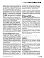
depicted in Figure 21.1.
In general, an infrared pulsed laser source is optically manipulated to a small focal spot utilized for
optical stimulation of the peripheral nerve. For these experiments, the holmium:YAG laser operating at
a wavelength of 2.12
μ
m and pulse duration of 350
μ
s was used. This wavelength has been shown to be
optimal for peripheral nerve stimulation. The importance of this parameter is discussed in detail in
Section 21.2.4. Delivery to the tissue is accomplished with an optical fiber, waveguide, or simply a free-
beam incident on the nerve surface. Wavelengths that transmit through optical fibers (<2.5
μ
m) are
considered ideal because the tip of the fiber can be easily manipulated in three dimensions for precise
delivery to the nerve. Stimulation experiments in the rat sciatic nerve reveal that a 400- to 600-
μ
m fiber
diameter can most efficiently result in excitation while maintaining precision in stimulation, although
FIGURE 21.1
Typical experimental setup for optical stimulation and recording in the rat sciatic nerve.
Laser
Pulse Energy
Detector
MM2000
Energy Meter
Trigger
(2 msec)
EMG
ENG
Recording Software
and Display
Muscle
Recording
System
Nerve
Recording
System
Electrical
Stimulator
Pulsed IR Light
90%
Focusing Lens
3-D Micro-Manipulator
Optical Fiber
(600 μm Diameter)
Sciatic Nerve
(Dorsocaudal Region)
Innervated
Muscles
y
x
z
10%
8174_C021.fm Page 4 Saturday, November 3, 2007 8:17 AM
Transient Optical Nerve Stimulation
21
-5
the optimal fiber diameter will vary according to the thickness of the given peripheral nerve bundle.
While not discussed here, the theoretical limits for both delivery methods are on the order of a few
micrometers. Radiant exposures required to stimulate vary, depending on the wavelength of the laser
source used (see wavelength dependence section). Electrical stimulation and recording of the compound
nerve and muscle potentials can be employed to verify the validity of the evoked response from laser
stimulation and compare this to the standard electrical stimulation methods.
Several experiments were performed
in vivo
, initially on the frog sciatic nerve, and subsequently in
mammals using a rat model, to verify the physiologic validity of optical stimulation. To confirm the
direct stimulatory effect of low-level optical energy, the nerve was optically isolated from its surrounding
tissues using an opaque material and stimulated. A consistent evoked response was recorded, indicating
that the incident light is directly responsible for the compound nerve (CNAP) and muscle action
potentials (CMAP) observed. Both signals were lost when the delivery of optical energy was blocked
with a shutter, indicating that stimulation was not due to artifacts associated with the trigger pulse or
other electrical interference synchronous with acquisition. Application of a depolarizing neuromuscular
blocker (succinylcholine) resulted in a measurable CNAP and loss of CMAP, confirming the involvement
of normal propagation of impulses from nerve to muscle upon optical stimulation.
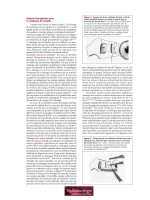
In a proof of principle study, CNAPs and CMAPs were consistently observed and recorded using
conventional electrical recordings (Figure 21.2) from both electrical and optical peripheral nerve exci-
tation methods. CNAP responses were amplified 5000X and filtered using a high-pass filter (>20 Hz)
and a low-pass filter (<3 kHz). CMAP responses were amplified 1000X and filtered using a high-pass
filter (>0.05 Hz) and low-pass filter (<5 kHz). The similarity in the shape and timing of the signals from
optical and electrical stimulus in Figure 21.2 show that conduction velocities, represented by the time
between the CNAP and CMAP, are equal. These traces imply that the motor fiber types recruited and
seen in the recorded compound action potentials are identical, regardless of excitation mechanism. That
is, based solely on observation of the physiologic portions of recorded signals (nerve and muscle), one
cannot discern between the two stimulation techniques. However, two important signal characteristics
manifest in Figure 21.2 that allow one to differentiate between optically and electrically evoked potentials.
One is the inherent electrical stimulation artifact that is only seen in the electrically stimulated peripheral
nerve recordings. The other is the superior spatial selectivity, or precise and localized number of axons
recruited with optical stimulation when compared to electrical stimulation. This phenomenon is realized
by the order of magnitude difference in amplitude (proportional to the number of axons recruited)
between electrical and optical recordings. In the following sections, each of these unique advantages
associated with optical stimulation is explored in more detail.
21.2.2 Generation of an Artifact-Free Nerve Potential Recording
The standard method for peripheral nerve stimulation requires that the stimulation technique occurs in
the same domain as the recording technique, through electrical means. Therefore, an inescapable artifact,
the amplitude of which is much greater than the physiological signal, is inherent to any electrically
stimulated nerve recording for the first 1 to 2 ms. Considering the speed at which action potentials are
propagated, it is clear that this artifact may obscure measurement of this signal. The lack of stimulation
artifact intrinsic to traditional electrical methodology for nerve stimulation is a unique advantage with
the optical stimulation methods. The artifact associated with electrical stimulation prevents scientists
from recording neural potentials near the site of stimulation. The electrical noise magnitude increases
proportionally to the stimulus intensity. Consequently, it is not possible to make interpretations or
observations on excitability characteristics of tissue with recording electrodes near the stimuli. This
fundamental limitation of adjacent electrical stimulation and recording processes is demonstrated in
Figure 21.2b. This plot contains the CNAP response recorded from the rat sciatic nerve following electrical
stimulus. Recording occurs 22 mm away from the site of stimulation. A large electrical artifact completely
conceals the nerve response for over 1 ms following stimulation. Thus, the onset time — and in some
8174_C021.fm Page 5 Saturday, November 3, 2007 8:17 AM
21
-6
Neuroengineering
cases peak amplitude of the response — is very difficult to distinguish from background, and therefore
no relevant response characteristics or signal processing can be inferred.
In contrast, Figure 21.2a depicts the nerve response to optical stimulation (same stimulation and
recording site as electrical) using laser radiant exposures above stimulation threshold intensities, which
do not contain a noise artifact. Now the nerve conduction velocities from the fast and slower conducting
motor fibers within the sciatic nerve can be quantified in terms of timing and amplitude. The distance
from stimulation to recording in the nerve was 22 mm, and two peaks are seen at 0.6 and 2.5 ms following
the laser stimulus (
t
= 1.8 ms) yielding conduction velocities measured to be 36.7 m/s with fast conducting
axons and slower conduction fiber velocity of 8.8 m/s. Peak amplitudes of the CNAP response from all
three fiber types are manifest. It is worth noting that the velocity of conduction within the nerve
subsequent to laser stimulation falls within the normal range for the rat sciatic nerve fast-conducting
A
α
motor neurons and slower-conducting A
γ
motor neurons. Thus, this new modality for nerve exci-
tation enables simultaneous stimulation and recording from adjacent portions of a nerve, a phenomenon
that is infeasible using electrical means for activation. These results also imply that all motor fiber types
are excitable with pulsed laser irradiation using optimal laser parameters.
FIGURE 21.2
Compound nerve and muscle action potentials recorded from sciatic nerve in rat. (a) CNAP recorded
using optical stimulation at 2.12
μ
m; (b) CNAP from electrical stimulation; (c) biceps femoris CMAP recorded using
optical stimulation at 2.12
μ
m; and (d) biceps femoris CMAP using electrical stimulation. The stimulation time for
all recordings occurred at
t
= 1.8 ms.
Time (msec)
Nerve-Optical
Nerve-Electrical
Muscle-Optical
Muscle-Electrical
0.1
5
0.1
0.05
Volts
Volts
Volts
Volts
0
0 2 4 6 8 10 12 14 16
0 2 4 6 8 10 12 14 16
0 2 4 6 8 10 12 14 16
0 2 4 6 8 10 12 14 16
–0.05
–0.1
–0.15
3.0
2.0
1.0
0.0
–1.0
–2.0
–3.0
1.2
0.8
0.4
0
–0.4
–0.8
–1.2
10.0
5.0
0.0
–5.0
–10.0
(a)
(b)
(c)
(d)
8174_C021.fm Page 6 Saturday, November 3, 2007 8:17 AM
Transient Optical Nerve Stimulation
21
-7
21.2.3 Spatial Selectivity in Optical Stimulation
It is well known in electrophysiology that electrical stimulation has an unconfined spread of charge
radiating far from the electrode. In the case of peripheral nerve stimulation, as the injected current
required for stimulation increases, the volume of tissue affected by the electric field increases propor-
tionally. Therefore, modulating the electrical stimulation intensity will lead to a graded response when
stimulating excitable tissue (for a review, see Palanker et al., 2005). From data obtained with electrical
stimulation, the greater the energy applied, the more fibers recruited, resulting in larger amplitude
compound potentials. Thus, the CNAP and CMAP represent a population response to stimulation, made
up of individual all-or-none responses from constituent axons, where a linear relationship exists between
stimulation intensity and strength of the CNAP response (Geddes and Bourland, 1985a). The electrical
current density necessary to evoke potentials in this tissue is significant, and the associated extent of the
electric field affects tissue a considerable distance from the electrode. Thus, a minimum value for spatial
selectivity in activation exists, and appreciably limits the precision of electrical stimulation (Geddes and
Bourland, 1985b). In contrast, lasers excel in applications necessitating a precisely controlled and quan-
tifiable volume of action in biological tissue (van Hillegersberg, 1997; Vogel and Venugopalan, 2003).
Laser distribution in tissue (i.e., volume of excited axons) depends on penetration depth, spot size, and
laser radiant exposure. Each of these constitutes a variable parameter. The wavelength of light determines
the penetration depth of photons from a laser; thus, the depth of axons recruited in optical stimulation
can be controlled very precisely over a large range of depth by modifying the laser wavelength. The next
section discusses this phenomenon in detail. The laser spot size incident on the nerve can be decreased
to an extremely small area (several micrometers). As a result of a small spot and lack of radial diffusion
in tissue, optical stimulation allows for more selective excitation of fascicles, resulting in isolated, specific
muscle contraction. Thus, theoretically, these parameters (wavelength, spot size, and radiant exposure)
can be optimized for efficient stimulation of any tissue geometry by changing the wavelength, optical
fiber diameter, or laser intensity used.
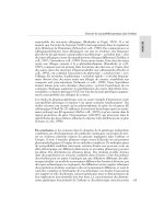
As a demonstration of the spatial discrimination innate to optical stimulation, CMAP recordings from
electrical and optical stimulation were compared within the rat sciatic nerve using threshold energies
for each modality. Figure 21.3 depicts the difference in selective activation for electrical vs. optical
stimulation. CMAP recording electrodes were placed within the gastrocenemius and biceps femoris
approximately 40 and 55 mm from the site of stimulation, respectively. Electrical stimulation with
threshold energy (1.02 A/cm
2
) was delivered proximal to the first nerve branch point on the fascicle
leading to the gastrocenemius and the muscular responses within gastrocenemius and biceps femoris
were simultaneously recorded. Note that using the minimum energy required to stimulate contraction
of the gastrocenemius still results in stimulation of the neighboring biceps femoris fascicle (causing biceps
femoris contraction). The change in voltage for these CMAPs was 1.495 and 0.492 V, respectively, seen
in Figure 21.3a. Laser stimulation at threshold (0.4 J/cm
2
) is shown for comparison with a voltage change
0.102 V recorded in the gastrocenemius and no response observed in the biceps femoris (Figure 21.3b).
Grossly, the electrical stimulation results in excitation of the entire nerve and a subsequent twitch response
from all innervated muscles. In contrast, the optical stimulation results in a muscle twitch of the muscle
innervated by the targeted nerve fascicle. By moving the laser spot across the nerve, different individual
muscle groups can indeed be stimulated. The precision and spatial specificity with optical activation
demonstrates selective recruitment of nerve fibers, as indicated by comparing the relative magnitudes of
nerve and muscle potentials (Figure 21.3) elicited from optical and electrical stimulation. These results
collected with optical nerve stimulation in mammals unequivocally confirm that optical nerve activation
exhibits significant spatial specificity, or lack of spread of stimulus to axons not directly irradiated by
the optical source.
Another noteworthy observation from our studies is that optical stimulation with less than 1 J/cm
2
can produce extremely precise stimulation of individual fascicles in a volume of axons considerably
smaller than that attainable with threshold electrical stimulation. As in electrical stimulation, increasing
optical energy results in a linear increase in recruitment of axons. The linear relationship suggests that
8174_C021.fm Page 7 Saturday, November 3, 2007 8:17 AM
21
-8
Neuroengineering
the energy is confined to a tissue volume immediately beneath the laser spot and has limited diffusion
to surrounding tissue, unlike electrical stimulation. A limit to laser excitation does exist at about 2 J/cm
2
stimulation radiant exposures, where a decrease in the physiologic response occurs. This is attributed to
axon damage within the nerve as stimulation energies approach the laser thermal damage and ablation
threshold, affecting the tissue’s ability to generate and propagate action potentials.
21.2.4 Threshold for Stimulation Dependence on Wavelength
When applying laser light to biological tissue, a variety of complex interactions can occur. Although a
comprehensive review of all aspects of laser–tissue interaction is clearly beyond the scope this chapter,
some important concepts must be discussed to understand the light distribution in neural tissue. Both
tissue characteristics and laser parameters contribute to this diversity. Tissue optical properties, refractive
index and the wavelength-dependent coefficients of absorption and scattering, govern how light will
interact with and propagate within the irradiated tissue. Alternatively, the following parameters are given
by the laser radiation itself: wavelength, exposure time, laser power, applied energy, spot size, radiant
exposure (energy/unit area), and irradiance (power/unit area).
In describing the optical properties and light propagation in tissues, light is treated as photons. The
primary reason for this approach is that biological tissue is an inhomogeneous mix of compounds, many
FIGURE 21.3
Selective recruitment of isolated nerve fascicles within a large peripheral nerve using electrical vs.
optical stimulation techniques. (a) Electrical stimulation with threshold energy (1.02 A/cm
2
) delivered to the fascicle
leading to the gastrocenemius. Muscular responses within gastrocnemius and biceps femoris were simultaneously
recorded. (b) Laser stimulation at threshold (0.4 J/cm
2
) recorded in the gastrocnemius and no response observed in
the biceps femoris.
Electrical
Stimulator
a.
b.
Gastrocnemius
Fascicle
1
0.5
CMAP(V)
CMAP(V)
CMAP(V)
CMAP(V)
0
–0.5
–1
Rat Sciatic Nerve
Foot Fascicle
Foot Fascicle
Biceps Femoris
Fascicle
Gastrocnemius
Fascicle
Fiber Coupled
Laser
Optical Fiber
Rat Sciatic Nerve
Biceps Femoris
Fascicle
1
0.5
0
–0.5
0.12
0.08
0.04
0
0.12
0.08
0.04
0
–1
0
24
6
8
10
12
14 16
0
24
6
8
10
12
14 16
0 2 4 6 8 10121416
02468
Time (msec)
Time (msec)
Time (msec)
Time (msec)
10 12 14 16
8174_C021.fm Page 8 Saturday, November 3, 2007 8:17 AM
Transient Optical Nerve Stimulation
21
-9
with unknown properties. Hence, analytical solutions to Maxwell’s equations (basic electromagnetic
[EM] theory that treats light as an EM wave induced by an oscillating dipole moment) in this medium
poses an intractable mathematical problem. The representation of light as photons presents the oppor-
tunity to apply probabilistic approaches that lend themselves particularly well to numerical solutions
that are manageable in computer simulations. Photons in a turbid medium such as tissue can move
randomly in all directions and may be scattered (described by its scattering coefficient
μ
s
[m
–1
]) or
absorbed (described by its absorption coefficient
μ
a
[
m
–1
]). These coefficients, along with anisotropy
(i.e., the direction in which a photon is scattered) and index of refraction, are referred to as the optical
properties of a material. If photons impinge on tissue, several things can happen; some photons will
reflect off the surface of the material (Fresnel reflection) and the majority of the photons will enter the
tissue. In the latter case, the photon is absorbed (and can be converted to heat, trigger a chemical reaction,
or cause fluorescence emission), or the photon is scattered (bumps into a particle and changes direction
but continues to exist and has the same energy). Although light scattering does occur in soft biological
tissues, such as the peripheral nerve, in the infrared (IR). For the purposes of this discussion we assume
that scattering is negligible relative to absorption. Thus, as a first-order approximation, light penetration
in peripheral nerve tissue can be described by the wavelength-dependent property of tissue absorption.
Because of this, we can also assume that the light propagation into the tissue will be confined to regions
directly under the irradiated spot on the nerve surface.
In tissue optics, absorption of photons is a crucial event because it allows a laser to cause a potentially
therapeutic (or damaging) effect on a tissue. Without absorption, there is no energy transfer to the tissue
and the tissue is left unaffected by the light. Molecules that absorb light are called
chromophores
. In the
IR tissue absorption is dominated by water absorption, so the major chromophore in the peripheral
nerve is water. The absorption of light can be characterized using Beer’s law, which predicts that the light
intensity in a material decays exponentially with depth (
z
):
where
E
0
is the incident irradiance [W/m
2
],
E
(
z
) is the irradiance through some distance
z
of the medium,
and
μ
a
(
λ
) is the wavelength-dependent absorption coefficient.
For a photon traveling over an infinitesimal distance
Δ
z
, the probability of absorption is given by
μ
a
∗Δ
z
, where
μ
a
is defined as the absorption coefficient (
m
–1
) (i.e., 1/
μ
a
is the mean free path a photon
travels before an absorption event takes place) (Welch and Gemert, 1995). A related and useful parameter
is the penetration depth, defined as the depth in the medium at which the energy or irradiance is reduced
to 1/
e
times (~37%) the incident irradiance at the surface. By definition, the penetration depth equals
1/
μ
a
in cases where there is no scattering.
The irradiance (power per unit area [W/m
2
]) gives us information about how much light made it to
a certain point in the tissue, but it does not tell us how much of that light is absorbed at that point. We
define a new term called the heat source term or “rate of heat generation” (
S
) as the number of photons
absorbed per unit volume [W/m
3
]. Note that number of photons absorbed can be related to amount of
heat generated, that is, heat source. Mathematically, heat source can be written as the product of the
irradiance at some point in the tissue,
E
(
z
), and the probability of absorption of that light at that point,
μ
a
:
Once the power density
S
(
z
) [W/m
3
] is known, the energy density
Q
(
z
) [J/m
3
] is easily calculated by
multiplying the power density by the exposure duration,
Δ
t
:
Ez Ee
a
z
()
()
=
−
0
μλ
Sz Ee Ez
a
z
a
a
() ()==
−
μμ
μ
0
Qz Sz t() ()=Δ
8174_C021.fm Page 9 Saturday, November 3, 2007 8:17 AM
21
-10
Neuroengineering
Then the laser induced temperature rise is given by:
where
ρ
is the density [
kg
/
m
3
] and
c
is the specific heat [
J/kg•K] of the irradiated material.
With this as background, theoretically the most appropriate wavelengths for stimulation will depend
on the tissue geometry of the target tissue (i.e., here the peripheral nerve). A typical rat sciatic nerve
section stimulated in this study was approximately 1.5 mm in diameter, with a 100- to– 200-μm epineural
and perineural sheath between the actual axons and the nerve surface. Despite the fact that the number
of fascicles per nerve varies greatly across all mammalian species, the typical fascicle thickness is constant
and tends to be between 200 and 400 μm (Paxinos, 2004). Thus, to theoretically achieve selective
stimulation of individual fascicles within the main nerve the penetration depth of the laser must be
greater than the thickness of the outer protective tissue (200 μm) and in between the thickness of the
underlying fascicle (penetration depth of 300 to 500 μm). In general, ultraviolet wavelengths (λ = 100
to 400 nm) are strongly absorbed by tissue constituents such as amino acids, fats, proteins, and nucleic
acids, while in the visible part of the spectrum (λ = 400 to 700 nm), absorption is dominated by
(oxy)hemoglobin and melanin. The near-infrared part of the spectrum (700 to 1300 nm) represents an
area where light is relatively poorly absorbed (this is referred to as the tissue absorption window, allowing
deep penetration) while in the mid- to far-infrared (> 1400 nm), absorption by tissue water dominates
and results in shallow penetration (Vogel and Venugopalan, 2003). By irradiating the nerve surface
overlying the target fascicle for stimulation within the main branch, infrared laser light may provide
profound selectivity (in terms of spot size and optical penetration depth) in excitation of individual
fascicles, resulting in isolated muscle contraction without thermal damage to tissue if the appropriate
wavelength and spot size are utilized.
To test this hypothesis, a continuously tunable, pulsed infrared laser source in the form of a free
electron laser (FEL) was employed (Edwards and Hutson, 2003). The FEL is a tunable laser that operates
in the 2- to 10-μm IR region, and emits a pulse with a duration of 5 μs. Wavelengths at or near relative
peaks and valleys of the IR tissue absorption spectrum (λ = 2.1, 3.0, 4.0, 4.5, 5.0, and 6.1 μm) (Hale
and Querry, 1973) were chosen for this study to facilitate recognition of general trends in stimulation
thresholds compared to tissue absorption. While the FEL is an excellent source for gathering experi-
mental data and exploring the wavelength dependence of the interaction owing to its tunability, it is
neither easy to use nor clinically viable. Nevertheless, experimental data gathered with this tunable light
source can provide guidance for the design of an appropriate and optimized turnkey benchtop laser
system for optical nerve stimulation.
The stimulation threshold is defined as the minimum radiant exposure required for a visible muscle
contraction occurring with each laser pulse. The ablation threshold is defined as the minimum radiant
exposure required for visible cavitation or ejection of material from the nerve, observed using an
operating microscope, with ten laser pulses delivered at 2 Hz. The stimulation threshold exhibits a
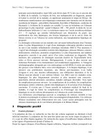
wavelength dependence that mirrors the inverse of the soft tissue absorption curve. This trend is clearly
illustrated in Figure 21.4a, which shows the stimulation and ablation threshold radiant exposures for
five trials with each of the six wavelengths used in this study. The water absorption spectrum is included
to discern general trends. Suitable wavelengths for optimal stimulation, those with maximum efficacy
and minimum damage, can be inferred. The wavelength dependence of the optical stimulation thresholds
yields pertinent wavelengths for the most favorable stimulation values based on the optical properties
of the target neural tissue. Because absorption dominates scattering in the IR, the hypothesis was that
at wavelengths where absorption is least, light penetration depth (i.e., 1/absorption) is maximized; thus,
the nerve is more efficiently stimulated with less damage because photons are distributed over a greater
tissue volume to minimize thermal injury. As one would expect based on a photothermal mechanism,
the ablation threshold for neural tissue is inversely proportional to the water absorption curve, or directly
proportional to the depth of laser penetration in the tissue. We see that the stimulation threshold is lower
ΔTz
Qz
c
()
()
=
ρ
8174_C021.fm Page 10 Saturday, November 3, 2007 8:17 AM
Transient Optical Nerve Stimulation 21-11
at wavelengths with high absorption, but it is also easier to ablate tissue (less radiant exposure required)
at these wavelengths. Thus, a more useful indicator of optimal wavelengths is the safety ratio, defined as
the ratio of threshold radiant exposure for ablation to that for stimulation.
This ratio (Figure 21.4b) identifies spectral regions with a large margin between radiant exposures
required for excitation and damage, and thus of safety. Results indicate that the highest safety ratios (>6)
are obtained at 2.1 and 4.0 μm, which correspond to valleys in tissue absorption and have nearly
equivalent absorption coefficients. We can conclude that clinically relevant wavelengths for optimal
stimulation, at least in the peripheral nerves and their anatomy/geometry, will not occur at peaks in
tissue absorption because the energy required to produce action potentials within the nerve is roughly
equal to the energy at which tissue damage occurs. For example, the penetration depth at λ = 3 μm is
roughly 1 μm in soft tissue. In this case, the axons can only be stimulated by heat that has diffused from
the point of absorption in the outer layers of connective tissue surrounding the nerve or from the
propagation of a laser-induced pressure wave. We can also predict that absolute valleys in the absorption
curve (i.e., visible and NIR region, 400 to 1400 nm) will not yield optimal wavelengths because the low
absorption, owing to lack of endogenous chromophores for these wavelengths in neural tissue, will
distribute the light over a large volume, leading to insufficient energy being delivered to the nerve fibers
for an elicited response. Results show that the most appropriate wavelengths for stimulation of the sciatic
nerve occur at relative valleys in IR soft tissue absorption, which produce an optical penetration depth
of 300 to 500 μm (corresponding to the optical penetration depth at λ = 2.12 μm). In this scenario, the
a
b
FIGURE 21.4 Wavelength dependence of the (a) stimulation vs. the ablation thresholds (b) the safety ratio = ablation
threshold/stimulation threshold. The solid line in both figures indicates the optical penetration depth (left y-axis).
In figure (b), the safety ratio obtained for the Ho:YAG laser is shown in stripes.
1
0.00001
2.1 2.5 3 3.5 4
Wavelength (Microns)
4.5 5 5.5 6.1
0
1
2
3
4
5
6
7
8
0.0001
1/Absorption (cm)
reshold (J/cm
2
)
0.001
0.01
Stim Avg
Abl Avg
0.1
2.12
0.00001
1
0.1
0.01
0.001
0.0001
Wavelength (μm)
2.1 2.5 3 3.5 4 4.5 5 5.5 6.1
0
1
Safety Ratio
1/Absorption (cm)
2
3
4
5
6
7
8174_C021.fm Page 11 Saturday, November 3, 2007 8:17 AM
21-12 Neuroengineering
optical penetration depth matches up with the target geometry to stimulate one fascicle within the nerve.
Note that the laser spot size can be adjusted to give precision of stimulation in all three dimensions of
tissue volume.
By matching the absorption values of the wavelengths yielding the highest safety ratio with commercially
available pulsed lasers, a clinically useful benchtop laser becomes a possibility. There are few lasers that
emit light at 4.0 μm in wavelength, and fiber-optic delivery at this wavelength is problematic as regular
glass fibers do not transmit beyond 2.5 μm. However, the holmium:YAG (Ho:YAG) laser at 2.12 μm is
commercially available and is currently used for a variety of clinical applications (Razvi et al., 1995; Topaz
et al., 1995; Kabalin et al., 1998; Fong et al., 1999; Jones et al., 1999). Although the inherent pulse duration
and pulse structure of this laser differs from the FEL, light at this wavelength can be delivered via optical
fibers, thus facilitating the clinical utility of this laser. The Ho:YAG laser was successfully used for neural
stimulation, with an average stimulation threshold radiant exposure of 0.32 J/cm
2
and an associated
ablation threshold of 2.0 J/cm
2
(n = 10), yielding a safety ratio of greater than 6.
21.2.5 Nerve Histological Analysis
Information obtained from the wavelength dependence study clearly suggests that the penetration depth
in nerve tissue using the Ho:YAG can provide the desired stimulatory effect with the lowest radiant
exposure compared to that required for tissue ablation. While tissue ablation served as a good indicator
for safe wavelengths by allowing calculation of a safety ratio for stimulation, this phenomenon is not a
synonym for thermal damage resulting in altered tissue morphology and function. It is essential to define
an exact range of “safe” laser radiant exposures, or the values between threshold and the upper end of
radiant exposures, which do not result in permanent tissue damage to strictly define what is appropriate
for clinical use. To this end, nerves were prepared for histological analysis by a neuropathologist special-
izing in assessment of thermal changes in tissue resulting from laser irradiation.
To quantify (thermal) damage induced by optical stimulation in peripheral nerve tissue, histological
analysis was performed on excised rat sciatic nerves, extracted acutely (less than one hour after stimu-
lation) or three to five days following stimulation. In acute studies, the radiant exposure was varied but
always larger than the stimulation threshold, and ten laser pulses at this radiant exposure were delivered
to each site. For a positive control, a damaging lesion was induced using radiant exposures well over the
ablation threshold in a location adjacent to the stimulation site. In survival studies, muscle and skin were
sutured following stimulation and the animal was allowed to survive for a period of three to five days
before nerves were harvested to assess any delayed neuronal damage and Wallerian degeneration. A sham
procedure with no stimulation was performed in the contra-lateral leg as negative control. None of the
shams showed any signs of damage, verifying a sound surgical technique and minimal tissue dehydration
due to the surgical procedure alone. Indications of damage include, but are not limited to, collagen
hyalinization, collagen swelling, coagulated collagen, decrease or loss of birefringence image intensity,
spindling of cells in perineurium and in nerves (thermal coagulation of cytoskeleton), disruption and
vacuolization of myelin sheaths of nerves, disruption of axons, and ablation crater formation. These
criteria help define a four-point grading scheme assigned by a pathologist blinded to the treatment of a
given sample to each acute specimen indicating extent of damage at the site of optical stimulation: 0 – no
visible thermal changes, 1 – thermal changes in perineurium, no nerve damage, 2 – thermal damage in
perineurium extending to the interface of the perineurium and the nerve, 3 – thermal damage in
perineurium and in nerve. Survival scoring was reported as damage or no damage to the nerve.
Figure 21.5 shows sample histological images (H&E stain) of the rat sciatic nerve from the acute
experiments following Ho:YAG laser stimulation. Results indicate that none of the ten nerves studied
showed any signs of acute thermal tissue damage at the site of stimulation with radiant exposures up to
two times stimulation threshold (Wells et al., 2005a,b). Histological examination of nerves from the
survival study do not reveal damage to the nerve or surrounding perineurium in eight of the ten
specimens, with damage occurring at radiant exposures above two times threshold. These histological
findings suggest that nerves can be consistently stimulated using optical means at or near threshold
8174_C021.fm Page 12 Saturday, November 3, 2007 8:17 AM
Transient Optical Nerve Stimulation 21-13
without causing any neural tissue damage. These findings are further corroborated by a functional
analysis of toe spreading in the survival animals. No functional neurological deficits were seen in any of
the animals stimulated at less than two times the stimulation threshold.
21.3 Mechanism
While our studies have shown that optical stimulation is an effective and advantageous method for
stimulation of neural tissue, the obvious and intriguing question of the underlying mechanism is largely
unanswered. Exactly what biophysical stimulus is induced in the tissue by the absorbed laser light that
ultimately results in an action potential and given this biophysical stimulus, what is the biological
mechanism responsible for the transduction into action potentials? To a large extent, unraveling these
mechanisms is still in its infancy. To get a grasp on this question, it is important to build a conceptual
understanding of the laser tissue interactions that occur during optical stimulation to refine the optimal
parameter set for this technique, as well as identify both the possible clinical applications and limitations
for this nerve stimulation modality. The best strategy for determining the biophysical mechanism respon-
sible for optical stimulation is to take a process of elimination approach to prove or disprove the possibility
of the various types of photobiological interactions that may occur. Before we discuss our hypothesis
for the underlying photobiological effect resulting in laser excitation of the peripheral nerve, it is appro-
priate to review some basic concepts regarding light–tissue interactions.
In general, many studies have been conducted investigating potential interaction effects using all types
of laser systems and tissue targets. Although the number of possible combinations for the experimental
parameters is unlimited, three main interaction mechanisms are classified today: (1) photochemical,
(2) photothermal, and (3) photomechanical. It is worth noting here that chemical, thermal, and mechan-
ical means have all been previously shown to produce action potentials in neurons. Before going into
detail, an interesting observation deserves to be stated. All these seemingly different interaction types
share a common property: the characteristic radiant exposure [J/cm
2
] ranges from approximately 1 to
1000 J/cm
2
. This is surprising because the irradiance itself [W/cm
2
] varies over more than fifteen orders
of magnitude. Thus, a single parameter distinguishes and primarily controls these processes: the duration
of the laser exposure, which is largely similar to the interaction time itself (Niemz, 2004). According to
a graph of the laser radiant exposure vs. the duration of pulse width the time scale can roughly be divided
in three major sections; (1) continuous wave or exposure times greater than 1 s for photochemical
interactions, (2) 100 s down to 1 μs for photothermal interactions, and (3) 1 μs and shorter for
photomechanical interactions. It should be clear, however, that these boundaries are not strict, and
adjacent interaction types cannot always be separated. Thus, overlap in these main regions does exist.
For example, in the range of 1 to several hundreds of microseconds, the interaction mechanisms typically
have photothermal as well as photomechanical components to them, while many photochemical inter-
actions also exhibit photothermal components.
FIGURE 21.5 (See color insert following page 15-4). Histological images (H&E stain or 5 μm tissue section) of the
rat sciatic nerve from the acute experiments following Ho:YAG laser stimulation. (a) Normal nerve tissue sample with
no laser irradiation. (b) Laser irradiation with ten pulses at 2 Hz using radiant exposures slightly above stimulation
threshold (0.5 J/cm
2
). (c) Laser irradiation with ten pulses at 2 Hz using radiant exposures above damage threshold
producing a lesion in neural tissue (2.5 J/cm). Shaded boxes represent relative size of laser spot (<1 mm diameter).
a. b. c.
8174_C021.fm Page 13 Saturday, November 3, 2007 8:17 AM
21-14 Neuroengineering
In brief, the group of photochemical interactions is based on the fact that light can induce chemical
effects and reactions within macromolecules or tissues. The most obvious example of this is created by
nature itself: photosynthesis. In the field of medical laser applications, photochemical interaction mech-
anisms play a role during photodynamic therapy (PDT) (Takahashi et al., 2002; Ionita et al., 2003;
Yamamoto et al., 2003). Frequently, biostimulation is also attributed to photochemical interactions,
although this is not scientifically ascertained. Photochemical interactions take place at very low irradi-
ances (typically 1 W/cm
2
) and long exposure times ranging from seconds to tens of minutes. Recent and
exciting developments that rely on photochemical interactions include experimental applications in the
field of photostimulation of neurons where light may be used to activate “caged compounds (McCray
and Trentham, 1989; Eder et al., 2002, 2004). In this scenario, stimulatory neurotransmitters are linked
to an inactivating group (a “caged” compound). Upon UV light exposure, cleavage of the neurotrans-
mitter from its “cage” is achieved, rendering the active form of the stimulatory neurotransmitter only
there where light exposure is activated. This technique takes advantage of the high spatial resolution of
uncaging molecules with light.
We examined whether the mechanism for optical nerve stimulation is a result of photochemical effects
from laser–tissue interaction. In essence, the stimulation thresholds in the infrared part of the spectrum
follow the water absorption curve (Wells, 2005a,b), suggesting that no “magical wavelength” has been
identified, effectively excluding a single tissue chromophore responsible for any direct photochemical
effects. This also provides some evidence that the effect is directly thermally mediated or a secondary
effect to photothermal interactions (i.e., photomechanical effects) as tissue absorption from laser
irradiation can be directly related to the heat load experienced by the tissue. Theoretically, one can predict
that a photochemical phenomenon is not responsible because infrared photon energy (<0.1 eV) is too
low for a direct photochemical effect of laser–tissue interactions and the laser radiant exposures used
are insufficient for any multiphoton effects (Thomsen, 1991).
Maxwell’s EM theory suggests an inherent electric field exists within laser light, which is associated
with the propagation of light itself and driven by a time and space varying electric and magnetic field
(Waldman, 1983). We questioned whether the electric field within the light beam used to irradiate and
stimulate the peripheral nerve is large enough to directly initiate action potentials, considering the
standard method of stimulation is through electrical means. To test this proposition, we used an alex-
andrite laser operating at 750 nm (near-infrared light) to attempt stimulation of the peripheral nerve.
This wavelength, unlike the Ho:YAG wavelength, has minimal absorption in soft tissue; however, the
electric field of intensity is similar regardless of wavelength. Thus, any stimulation reported with a low
absorption wavelength would indicate that the electric field of the laser light mediates stimulation. Results
explicitly prove a direct electrical field effect due to laser radiation traversing the tissue is highly unlikely
as a means for optical stimulation because light from the alexandrite laser did not stimulate even at
radiant exposures fifty times higher than those used for the Ho:YAG laser. Experimental calculations
further illustrate this point. Consider the equation: S
threshold
=
1
/2 c ε
o
E
max
2
, where the threshold laser
radiant exposure (S
threshold
) = 0.32 J/cm
2
, the speed of light (c) = 3.10
8
m/s, and the permittivity of neural
tissue (ε
o
) in units of A-s/V-m (c ε
o
= 0.002634). The calculated value for the maximum instantaneous
intensity of the electric field (E
max
) at the tissue surface is 0.155 V/mm
2
, or 0.05 mA/mm
2
. This theoretical
prediction is well below the electrical stimulation threshold of the peripheral nerve found in our previous
studies, where 0.95 ± 0.58 A/cm
2
was required for surface stimulation. Moreover, it is important to realize
that the electric field owing to light oscillates at 10
14
to 10
15
Hz, which is an order of magnitude higher
than the typical electrical stimulation field oscillator frequency.
Photomechanical effects are secondary to rapid heating with short laser pulses (<1 µs) that produce
forces, such as explosive events and laser-induced pressure waves, able to disrupt cells and tissue. Because
we are operating well below the ablation threshold, ablative recoil can be excluded as a source of
mechanical effects. In contrast, tissue heating will always result in thermoelastic expansion. Nerve stim-
ulation using pressure waves (rapid mechanical displacement, ultrasound) is well documented in the
literature (Shusterman et al., 2002; Norton, 2003). We sought to prove or disprove photomechanical
effects (thermoelastic expansion or pressure wave generation) leading to optical stimulation.
8174_C021.fm Page 14 Saturday, November 3, 2007 8:17 AM
Transient Optical Nerve Stimulation 21-15
Contributions from pressure waves to optically stimulate the peripheral nerve were studied by exam-
ining the effect of pulse duration on stimulation threshold. It is clear from our results that the stimulation
threshold radiant exposure required for stimulation at this wavelength does not change with pulse width
through almost three orders of magnitude (5 μs to 5 ms). Moreover, all pulse durations lie well outside
the stress confinement zone. Given that, there is strong evidence that laser-induced pressure waves are
not implicated in the optical stimulation mechanism. Because pressure wave generation has been dis-
carded as a plausible means, tissue displacement during the laser pulse was measured next using a phase-
sensitive OCT setup (Rylander et al., 2004) to test the actual magnitude of thermoelastic expansion of
the tissue resulting from optical stimulation. The change in surface displacement of the rat sciatic nerve
(ex vivo) upon irradiation with Ho:YAG radiant exposures slightly above threshold (0.4 J/cm
2
) were
measured to be 300 nm. Displacement of 300 nm in a 350-μsec pulse width is small, but not negligible.
Nevertheless, while at this point we cannot exclude contributions of the thermoelastic expansion, this
effect is thermal in origin.
Through this process of elimination we have systematically shown that the electric field, and photo-
chemical and photomechanical effects from laser tissue interactions do not result in excitation of neural
tissue. Thus, we have arrived at the hypothesis that laser stimulation of neural tissue is mediated by some
photothermal process resulting from transient irradiation of peripheral nerves using infrared light.
Photothermal interactions include a large group of interaction types resulting from the transformation
of absorbed light energy to heat, leading to a local temperature increase and thus a temperature gradient
both in time and space. While photochemical processes are often governed by a specific reaction pathway,
photothermal effects generally tend to be nonspecific and are mediated primarily by absorption of optical
energy and secondly governed by fundamental principles of heat transport. Depending on the duration
and peak value of the temperature achieved, different effects such as coagulation, vaporization, melting,
or carbonization may be distinguished. An excellent overview of these interaction regimes can be found
in Jacques (1992). It is essential to emphasize that thermal interactions in tissue are typically governed
by rate processes; that is, it is not just the temperature that plays a role, but also the duration for which
the tissue is exposed to a particular temperature is a parameter of major importance. Once deposited in
tissue and given sufficient time, the traditional mechanism of heat transfer applies to laser-irradiated
biological tissues. Heat flows in biological tissue whenever a temperature difference exists. The transfer
of thermal energy is governed by the laws of thermodynamics: (1) energy is conserved, and (2) heat
flows from areas of high temperature to areas of low temperature. The primary mechanisms of heat
transfer to consider include conduction, convection, and radiation (Incropera, 2002).
Two-dimensional radiometry of the irradiated tissue surface was performed to gain a better under-
standing of the thermal processes and actual tissue temperature values required for optical nerve stim-
ulation. Using this technique, the temperature profile in space and time was observed. We measured a
peak temperature rise at the center of the spot of 8.95°C, yielding an average temperature rise of 3.66°C
across the Gaussian laser spot. The peripheral nerve temperature profile in time was also observed using
the infrared camera from laser stimulation. The thermal relaxation time is defined as the time required
for the temperature of the tissue to return to 1/e (37%) of the maximum tissue temperature change. In
the case of the rat peripheral nerve, we measured the thermal relaxation time to be about 90 ms, which
corresponds well with the theoretical value of about 100 ms. We can infer that the pulse width of light
delivered to the tissue must be less than 90 ms in duration to result in the desired stimulation effect. We
can also infer that temperature superposition will begin to occur at higher repetition rates (>5 Hz) as
the tissue requires slightly greater than 200 ms to return to baseline temperature. At repetition rates
greater than 5 Hz, tissue temperatures will become additive with each ensuing laser pulse and resulting
tissue damage may begin to occur with long-term stimulation.
Based on results from measurements of tissue temperature as a function of radiant exposure, nerve
temperature clearly increases linearly with laser radiant exposure. Recent literature suggests that slight
thermal changes to mitochondria begin to occur as low as 43°C (protein denaturation begins at tissue
temperature close to 57°C). This temperature corresponds to the onset of thermal damage radiant
exposure found from histological analysis of short-term laser nerve stimulation (0.8 to 1.0 J/cm
2
). These
8174_C021.fm Page 15 Saturday, November 3, 2007 8:17 AM
21-16 Neuroengineering
results imply that optical stimulation of peripheral nerves is mediated through a thermal gradient as a
result of laser tissue interaction and that this phenomenon is safe at radiant exposures of at least two
times the threshold required for action potential generation. In the case of nonhydrated tissue, the
temperature as a function of radiant exposure shifts upward 6°C. Here, the mitochondrial damage will
theoretically begin to occur between 0.5 and 0.6 J/cm
2
, thus illustrating the importance of tissue hydration
for safe and efficient nerve excitation.
21.4 Impact
21.4.1 Applications
Optical neural activation has three fundamental advantages over electrical stimulation that make it ideal
for a number of procedures that currently employ electrical stimulation as the standard of care. First,
the precision of optically delivered energy is far superior to electrical stimulation techniques. Examples
of limitations in electrical stimulation techniques arise when precision stimulation of neural structures
are required for peripheral nerve surgery, during which small clusters of nerve fibers are stimulated to
determine their viability in peripheral nerve repair (Weiner, 2003). In peripheral nerve surgery, electrical
stimulation is utilized to identify the connectivity and functionality of specific nerve roots to selectively
avoid or resect. This usually requires dissecting apart the nerve bundles to determine which ones conduct
through a damaged area and which bundles do not. The optical method could confine the stimulation
easily to segments of a nerve without requiring separation between the intended area to be stimulated
and other areas. Similarly, surgeries involving cranial nerves would benefit from precise functional testing,
such as differentiating nerve tissue from tumor in small areas such as the central pontine angle through
which the vestibular and facial nerve traverse. Auditory nerve stimulation could be significantly enhanced
with a larger number of distinct stimulation sites along the cochlea than is currently possible using
electrical means. This suggests that it may be possible to develop a better cochlear implant.
Second, optical stimulation does not produce an electrical artifact during stimulation, whereas elec-
trical stimulation inherently results in an artifact in the recorded signal. To record small nerve potentials
in response to the electrical stimulation, usually the recording electrodes are located a sufficient distance
away from the stimulation source. Furthermore, signal averaging techniques are frequently used for
discerning electrical responses contained within large electric field artifacts (Fiore et al., 1996; Wagenaar
and Potter, 2002; Andreasen and Struijk, 2003). Optical stimulation, on the other hand, produces no
stimulation artifact in the recorded response, and therefore the recorded response can be very close to
the stimulation source. Clinically, this results in neural potentials that can be more easily recorded near
the source of stimulation. Also, fewer stimuli need to be applied due a decreased need for signal averaging
(which requires usually hundreds of stimuli), which in turn facilitates higher throughput of mapping.
Third, electrical stimulation requires contact between the electrode and the tissue being stimulated. It is
susceptible to all the properties of impedance, current shunting, and field distortion around the area of
contact between the electrode and the tissue in the acute setting (e.g., in current cochlear implants,
a maximum of six to nine channels/electrodes are used, owing to the fact that along the basilar membrane
each electrode affects an area of approximately 4 to 8 mm) (Palanker et al., 2005). In the chronic setting,
issues of half-cell potential differences, metal toxicity, and tissue reaction to various implanted electrodes
significantly limit the materials and sizes of materials used for chronic electrode implants. Optical stimu-
lation, on the other hand, does not require direct contact with the tissue being stimulated, thereby mini-
mizing tissue disturbance. Furthermore, plating and deplating of metal in an ionic medium (interstitial
fluid) is not an issue with optical stimulation. It would seem more likely that chronically implanted optical
stimulating probes would be more precise due to the lack of any stimulating current spread, and also more
tolerable as a chronic implant due to longer tissue stability (no unstable impedance characteristics) and
safer (i.e., inert) interface materials (glass/fiber-optic cable vs. metal) (Agnew et al., 1989).
In summary, the capability of optical energy to yield a contact-free, spatially selective, artifact-free
method of stimulation has significant advantages over electrical methods for a variety of diagnostic and
therapeutic clinical applications.
8174_C021.fm Page 16 Saturday, November 3, 2007 8:17 AM
Transient Optical Nerve Stimulation 21-17
21.4.2 Future Directions
Optical stimulation presents a paradigm shift in neural activation that has major implications for clinical
neural stimulation as well as fundamental neurophysiology and neuroscience. To date, this concept has
been demonstrated using large, cumbersome, and expensive laboratory laser sources (FEL, Ho:YAG).
For optical stimulation to find its way to practical utility and clinical use, a simple, user-friendly, portable,
reliable, and low-cost device must be developed. Aculight, a company that specializes in the design and
manufacturing of innovative solid-state lasers for the defense and medical markets, in collaboration with
Vanderbilt University, has demonstrated the utility and unique capability of this concept by developing
a portable optical stimulator for routine use in laboratory settings, with the ultimate goal of delivering
a clinically usable product. The prototype of the optical stimulator is a pulsed 1.85 to 1.87 μm (resulting
in similar absorption as Ho:YAG laser in soft tissue) diode laser with a fiber-coupled output, representing
a >95% reduction in size compared to the device used for initial testing at Vanderbilt University. The
laser-based stimulator, which appears similar to telecom products offered today, has the advantages of
compactness and portability, high reliability, and low life-cycle cost. Furthermore, the unit plugs into a
regular power outlet (110 V) and has no special power or cooling requirements Experiments described
above have been repeated with this laser system and, as expected, show very similar results.
In summary, we have shown a novel alternative to electrical stimulation to interface with the neural
system using light. This method provides several unique advantages over traditional methods. However,
transient optical stimulation of neural tissue is in its infancy and many questions remain open with
regard to the underlying mechanism, the limitations of its utility, and applications that have not even
been thought of at this time. Moving this field forward will require multidisciplinary approaches and
intense research efforts on all fronts.
References
Agnew, W. F., McCreery, D.B., et al. (1989). Histologic and physiologic evaluation of electrically stimu-
lated peripheral nerve: considerations for the selection of parameters. Ann. Biomed. Eng.,
17(1):39–60.
Allegre, G., Avrillier, S., et al. (1994). Stimulation in the rat of a nerve fiber bundle by a short UV pulse
from an excimer laser. Neurosci. Lett., 180(2):261–264.
Andreasen, L.N. and Struijk, J.J. (2003). Artefact reduction with alternative cuff configurations. IEEE
Trans. Biomed. Eng., 50(10):1160–1166.
Balaban, P., Esenaliev, R., et al. (1992). He-Ne laser irradiation of single identified neurons. Lasers Surg.
Med., 12(3):329–337.
Bragard, D., Chen, A.C., et al. (1996). Direct isolation of ultra-late (C-fibre) evoked brain potentials by
CO
2
laser stimulation of tiny cutaneous surface areas in man. Neurosci. Lett., 209(2):81–84.
Chen, Y.S., Hsu, S.F., et al. (2005). Effect of low-power pulsed laser on peripheral nerve regeneration in
rats. Microsurgery, 25(1):83–89.
Eder, M., Zieglgansberger, W., et al. (2002). Neocortical long-term potentiation and long-term depression:
site of expression investigated by infrared-guided laser stimulation. J. Neurosci., 22(17):7558–7568.
Eder, M., Zieglgansberger, W., et al. (2004). Shining light on neurons-elucidation of neuronal functions
by photostimulation. Rev. Neurosci., 15(3):167–183.
Edwards, G. S. and Hutson, M.S. (2003). Advantage of the Mark-III FEL for biophysical research and
biomedical applications. J. Synchrotron. Radiat., 10(Pt. 5):354–357.
Fiore, L., Corsini, G., et al. (1996). Application of non-linear filters based on the median filter to
experimental and simulated multiunit neural recordings. J. Neurosci. Methods, 70(2):177–184.
Fong, M., Clarke, K., et al. (1999). Clinical applications of the holmium:YAG laser in disorders of the
paediatric airway. J. Otolaryngol., 28(6):337–343.
Fork, R.L. (1971). Laser stimulation of nerve cells in Aplysia. Science, 171(974):907–908.
Geddes, L.A. and Bourland, J.D. (1985a). The strength-duration curve. IEEE Trans. Biomed. Eng.,
32(6):458–459.
8174_C021.fm Page 17 Saturday, November 3, 2007 8:17 AM
21-18 Neuroengineering
Geddes, L.A. and Bourland, J.D. (1985b). Tissue stimulation: theoretical considerations and practical
applications. Med. Biol. Eng. Comput., 23(2):131–137.
Hale, G.M. and Querry, M.R. (1973). Optical-constants of water in 200-nm to 0.2-mm wavelength region.
Appl. Optics, 12(3):555–563.
Hirase, H., Nikolenko, V., et al. (2002). Multiphoton stimulation of neurons. J. Neurobiol., 51(3):237–247.
Incropera, F.P. (2002). Fundamentals of Heat and Mass Transfer. New York: John Wiley & Sons.
Ionita, M.A., Ion, R.M., et al. (2003). Photochemical and photodynamic properties of vitamin B2 —
riboflavin and liposomes. Oftalmologia, 58(3):29–34.
Izzo, A.D., Jansen, E.D., and Walsh, J.T. (2006b). Laser stimulation of the auditory nerve. Lasers Surg.
Med., (submitted).
Izzo, A.D., Suh, E., Walsh, J.T., et al. (2006a). Selectivity of optical stimulation in the auditory system.
SPIE, San Jose, CA.
Izzo, A.D., Walsh, J.T., and Jansen, E.D. (2005). Safe ranges for optical cochlear neuron stimulation. Mid-
Winter Meeting for the Association for Research in Otolaryngology (ARO), New Orleans, LA.
Jacques, S.L. (1992). Laser-tissue interactions. Photochemical, photothermal, and photomechanical. Surg.
Clin. N. Am., 72(3):531–558.
Jansen, E.D., Asshauer, T., et al. (1996). Effect of pulse duration on bubble formation and laser-induced
pressure waves during holmium laser ablation. Lasers Surg. Med., 18(3):278–293.
Jones, J.W., Schmidt, S.E., et al. (1999). Holmium:YAG laser transmyocardial revascularization relieves
angina and improves functional status. Ann. Thorac. Surg., 67(6):1596–1601; discussion 1601-2.
Kabalin, J.N., Gilling, P.J., et al. (1998). Application of the holmium:YAG laser for prostatectomy. J. Clin.
Laser Med. Surg., 16(1):21–22.
Kanjani, N., Jacob, S., et al. (2004). Wavefront- and topography-guided ablation in myopic eyes using
Zyoptix. J. Cataract Refract. Surg., 30(2):398–402.
Lu, J. and Waite, P. (1999). Advances in spinal cord regeneration. Spine, 24(9):926–930.
McCray, J.A. and Trentham, D.R. (1989). Properties and uses of photoreactive caged compounds. Annu.
Rev. Biophys. Biophys. Chem., 18:239–270.
McGill, K., Cummins, K.L., et al. (1982). On the nature and elimination of stimulus artifact in nerve
signals evoked and recorded using surface electrodes. IEEE Trans. Biomed. Eng., 29(2):129–137.
Miller, C.A., Abbas, P.J., et al. (2000). An improved method of reducing stimulus artifact in the electrically
evoked whole-nerve potential. Ear Hear., 21(4):280–290.
Niemz, M.H. (2004). Laser-Tissue Interactions, Berlin: Springer.
Norton, S.J. (2003). Can ultrasound be used to stimulate nerve tissue? Biomed. Eng. Online, 2: 6.
Palanker, D., Vankov, A., et al. (2005). Design of a high-resolution optoelectronic retinal prosthesis.
J. Neural Eng., 2(1):S105–S120.
Paxinos, G. (2004). The Rat Nervous System. Sydney, Australia: Elsevier.
Razvi, H.A., Chun, S.S., et al. (1995). Soft-tissue applications of the holmium:YAG laser in urology.
J. Endourol., 9(5):387–390.
Richter C.P., Walsh, J.T., and Jansen, E.D. (2005a). Optical Stimulation of the Auditory System. NIH
Symposium on Neural Interfaces, Bethesda, MD.
Richter, C.P., Walsh, J.T., and Jansen, E.D. (2005b). Optically-evoked acoustic nerve activity. Mid-Winter
Meeting for the Association for Research in Otolaryngology (ARO), New Orleans, LA.
Roehm, P.C. and Hansen, M.R. (2005). Strategies to preserve or regenerate spiral ganglion neurons. Curr.
Opin. Otolaryngol. Head Neck Surg., 13(5):294–300.
Rylander, C.G., Dave, D.P., et al. (2004). Quantitative phase-contrast imaging of cells with phase-sensitive
optical coherence microscopy. Opt. Lett., 29(13):1509–1511.
Shusterman, V., Jannetta, P.J., et al. (2002). Direct mechanical stimulation of brainstem modulates cardiac
rhythm and repolarization in humans. J. Electrocardiol., 35 Suppl:247–256.
Sisken, B.F., Walker, J., et al. (1993). Prospects on clinical applications of electrical stimulation for nerve
regeneration.
J. Cell. Biochem., 51(4):404–409.
8174_C021.fm Page 18 Saturday, November 3, 2007 8:17 AM
Transient Optical Nerve Stimulation 21-19
Takahashi, M., Nagao, T., et al. (2002). Roles of reactive oxygen species in monocyte activation induced
by photochemical reactions during photodynamic therapy. Front Med. Biol. Eng., 11(4):279–294.
Thomsen, S. (1991). Pathologic analysis of photothermal and photomechanical effects of laser-tissue
interactions. Photochem. Photobiol., 53(6):825–835.
Topaz, O., Rozenbaum, E.A., et al. (1995). Laser-assisted coronary angioplasty in patients with severely
depressed left ventricular function: quantitative coronary angiography and clinical results. J. Interv.
Cardiol., 8(6):661–669.
van Hillegersberg, R. (1997). Fundamentals of laser surgery. Eur. J. Surg., 163(1):3–12.
Vogel, A. and Venugopalan, V. (2003). Mechanisms of pulsed laser ablation of biological tissues. Chem.
Rev., 103(2):577–644.
Wagenaar, D.A. and Potter, S.M. (2002). Real-time multi-channel stimulus artifact suppression by local
curve fitting. J. Neurosci. Methods, 120(2):113–120.
Waldman, G. (1983). Introduction to Light: The Physics of Light, Vision, and Color. Englewood Cliffs, NJ:
Prentice Hall.
Walsh, L.J. (1997). The current status of low level laser therapy in dentistry. 1. Soft tissue applications.
Aust. Dent. J., 42(4):247–254.
Weiner, R.L. (2003). Peripheral nerve neurostimulation. Neurosurg. Clin. N. Am., 14(3):401–408.
Welch, A.J. and v. Gemert, M.J.C. (1995). Optical-Thermal Response of Laser-Irradiated Tissue. New York:
Plenum Press.
Welch, A.J., Motamedi, M., et al. (1991). Laser thermal ablation. Photochem. Photobiol., 53(6):815–823.
Wells, J.D., Kao, C., et al. (2005). Application of Infrared Light for in vivo Neural Stimulation. J. Biomed.
Optics, 10: 064003.
Wells, J.D., Kao, C., et al. (2005). Optical Stimulation of Neural Tissue in vivo. Optics Letters, 30(5):
504–507.
Wietholt, D., Alberty, J., et al. (1992). Nd-YAG laser-photocoagulation — acute electrophysiological,
hemodynamic, and morphological effects in large irradiated areas. Pace-Pacing and Clin. Electro-
physiol., 15(1):52–59.
Wu, W.H., Ponnudurai, R., et al. (1987). Failure to confirm report of light-evoked response of peripheral
nerve to low power helium-neon laser light stimulus. Brain Res., 401(2):407–408.
Yamamoto, M., Nagano, T., et al. (2003). Production of singlet oxygen on irradiation of a photodynamic
therapy agent, zinc-coproporphyrin III, with low host toxicity. Biometals, 16(4):591–597.
8174_C021.fm Page 19 Saturday, November 3, 2007 8:17 AM
8174_C021.fm Page 20 Saturday, November 3, 2007 8:17 AM
22
-1
22
Transcranial Magnetic
Stimulation of
Deep Brain Regions
22.1 Introduction
22
-1
22.2 Basic Principles of TMS
22
-2
22.3 Deep TMS Coils: Design Principles
22
-4
22.4 A Coil for Stimulation of Deep Brain Regions
Related to Mood Disorders: Simulations and
Phantom Measurements
22
-6
Methods · Measurements of the Electrical Field Induced
in a Phantom Brain · Results
22.5 Transcranial Magnetic Stimulation of Deep Brain
Regions: Evidence for Efficacy of the H-Coil
22
-14
Methods · Results · Discussion
22.6 Transcranial Magnetic Stimulation of
Deep Prefrontal Regions
22
-19
Comparison of Electric Field Distributions
References
22
-23
22.1 Introduction
Transcranial magnetic stimulation (TMS) is a noninvasive technique used to apply brief magnetic pulses
to the brain. The pulses are administered by passing high currents through an electromagnetic coil
placed upon the scalp that can induce electrical currents in the underlying cortical tissue, thereby
producing a localized axonal depolarization. Neuronal stimulation by TMS was first demonstrated in
1985 (Barker et al., 1985), when a circular coil was placed over a normal subject vertex and evoked
action potentials from the abductor digiti minimi. Since then this technique has been applied to studying
nerve conduction, excitability, and conductivity in the brain and peripheral nerves, and to studying
and treating various neurobehavioral disorders, primarily mood disorders (Kircaldie et al., 1997;
Wassermann and Lisanby, 2001).
The ability of the TMS technique to elicit neuronal response has until recently been limited to brain
cortex. The coils used for TMS (such as round or a figure-of-eight coil) induce stimulation in cortical
regions mainly just superficially under the windings of the coil. The intensity of the electric field drops
dramatically deeper in the brain as a function of the distance from the coil (Maccabee et al., 1990; Tofts,
1990; Tofts and Branston, 1991; Eaton, 1992). Therefore, to stimulate deep brain regions, a very high
intensity would be necessary. Such intensity cannot be reached by standard magnetic stimulators, using
the regular figure-of-eight or circular coils. Stimulation of regions at depths of 3 to 4 cm, such as the
Yiftach Roth and
Abraham Zangen
8174_C022.fm Page 1 Saturday, November 3, 2007 8:20 AM
22
-2
Neuroengineering
leg motor area, can be achieved using coils such as the double-cone coil (Stokic et al., 1997; Terao et al.,
1994, 2000), which is a larger figure of eight with an angle of about 95° between the two wings. However,
the intensity needed to stimulate deeper brain regions effectively would stimulate cortical regions and
facial nerves over the level that might lead to facial pain, facial and cervical muscle contractions, and
may cause epileptic seizures and other undesirable side effects.
This chapter describes the principles and design of TMS coils for deep brain stimulation. The con-
struction of such coils should meet several goals simultaneously:
1. High enough electric field intensity in the desired deep brain region that will surpass the threshold
for neuronal activation
2. High percentage of electric field in the desired deep brain region relative to the maximal intensity
in the cortex
3. Minimal aversive side effects during stimulation, such as pain and activation of facial muscles
22.2 Basic Principles of TMS
The TMS stimulation circuit consists of a high-voltage power supply that charges a bank of capacitors,
which are then rapidly discharged via an electronic switch into the TMS coil to create the briefly changing
magnetic field pulse. A typical circuit is shown in Figure 22.1, where low-voltage AC is transformed into
high-voltage DC, which charges the capacitors. A crucial component is the thyristor switch, which must
traverse very high current at very short times of 50 to 250
μ
s. The cycle time depends on the capacitance
(typically 10 to 250
μ
F) and on the coil inductance (typically 10 to 30
μ
H). Typical peak currents and
voltages are 5000 A and 1500 V, respectively.
Most TMS stimulators produce a biphasic pulse of electric current. During the discharge cycle, the
TMS circuit behaves like an RCL circuit, and the current
I
is given by:
(22.1)
where
α
=
R
/2
L
, , and
R
,
C
, and
L
are the total values of the resistance, capacitance,
and inductance, respectively, in the circuit. The inductance is mainly the coil inductance but there is an
additional contribution from the cables, and the resistance includes contributions from the thyristor and
the coil.
Biologically, the most relevant parameter for neuronal activation is the induced electric field, which
is proportional to the rate of change of the current (
dI
/
dt
). The brief strong current generates a time-
varying magnetic field B. An electric field E is generated in every point in space with direction perpen-
dicular to the magnetic field, with amplitude proportional to the time-rate of change of the vector
potential
A
(
r
).
FIGURE 22.1
Typical magnetic stimulation circuit, including high-voltage transformer, capacitor, resistor, thyristor
trigger, and stimulating coil.
It
V
wL
twt
()
=−
()()
exp sinα
wLC=−
−
()
12
α
8174_C022.fm Page 2 Saturday, November 3, 2007 8:20 AM
Transcranial Magnetic Stimulation of Deep Brain Regions
22
-3
The vector potential
A
(
r
) in position
r
is related to the current in the coil
I
by the expression:
(22.2)
where
μ
0
= 4
π
∗
10
–7
Tm
/
A
is the permeability of free space, the integral of
dl
′
is over the wire path, and
r
′
is a vector indicating the position of the wire element. The magnetic and electric fields are related to
the vector potential through the expressions:
(22.3)
(22.4)
The only quantity that is changing with time is the current
I
. Hence, the electric field
E
A
can be written as:
(22.5)
Because brain tissue has conducting properties, while the air and skull are almost complete insulators,
the vector potential will induce accumulation of electric charge at the brain surface. This charge is another
source for electric field, which can be expressed as:
(22.6)
where
Φ
is the scalar potential produced by the surface electrostatic charge.
The total field in the brain tissue
E
is the vectorial sum of these two fields:
(22.7)
The influence of the electrostatic field
E
Φ
is, in general, to oppose the induced field
E
A
and
consequently to reduce the total field
E
. The amount of surface charge produced and hence the
magnitude of
E
Φ
depend strongly on coil configuration and orientation. This issue will be elaborated
in the following sections.
Figure 22.2 demonstrates the electric field pulse produced by a figure-of-eight coil, as measured by a
two-wire probe in a brain phantom filled with saline solution at physiologic concentration. In repetitive
TMS (rTMS), several such pulses are administered in a train of between 1 and 20 Hz.
This electric field produces action potential in excitable neuronal cells, which might result in acti-
vation of neuronal circuits when applied above a certain threshold. The neuronal response depends
not only on the electric field strength, but also on the pulse duration, through a strength-duration
curve of the form:
(22.8)
where
E
th
is the threshold electric field required to induce neuronal response, and
τ
is the duration the
field was above this threshold. The biological parameters determining neural response are the threshold
at infinite duration, called the rheobase (
b
, measured in V/m), and the duration at which the threshold
is twice the rheobase, called the chronaxie (
c
, in
μ
s
). Motor and sensory curves as reported by Bourland
Ar
uI
dl
rr
()
=
′
−
′
∫
0
4π
BA
A
=∇×
E
A
t
A
=
−∂
∂
E
I
t
dl
rr
A
=
−∂
∂
′
−
′
∫
μ
π
0
4
E
Φ
Φ=−∇
EE E
A
=+
Φ
Ebc
th
=+
()
1 τ
8174_C022.fm Page 3 Saturday, November 3, 2007 8:20 AM
22
-4
Neuroengineering
et al. (1996) are shown in Figure 22.3. These curves should be treated as illustrative only, because the
chronaxie and rheobase depend on many biological and experimental factors, such as whether or not
the nerves are myelinated (hence peripheral and cortical parameters should be different), or train
frequency in rTMS, which in general reduces the threshold for stimulation.
As shown by Heller and Van Hulstein (1992), the three-dimensional maximum of the electric field
intensity will always be located at the brain surface, for any configuration or superposition of TMS coils.
It is possible, however, to increase considerably the depth penetration and the percentage of electric field
intensity in deep brain regions, relative to the maximal field at the cortex. The next section outlines the
construction principles for efficient deep brain stimulation, and subsequent sections demonstrate several
examples of TMS coils designed to accomplish this goal.
22.3 Deep TMS Coils: Design Principles
While the activation of peripheral nerves depends mainly on the derivative of the electric field along the
nerve fiber (Maccabee et al., 1993), the most relevant parameter for activation of brain structures seems
to be the electric field intensity (Thielscher and Kammer, 2002; Amassian et al., 1992). In both cases,
FIGURE 22.2
The induced electric field of a figure-of-eight (figure-8) coil vs. time over a TMS pulse cycle. The time
scale is 100
μ
s.
FIGURE 22.3
Neural strength-duration curve depicting stimulation threshold vs. duration.
Strength-Duration Curve
0
50
100
150
200
250
Duration (Microsec)
Electric Field (V/m)
Sensory
Motor
10 60 110 160 210 260 310 360
8174_C022.fm Page 4 Saturday, November 3, 2007 8:20 AM
Transcranial Magnetic Stimulation of Deep Brain Regions
22
-5
however, physiological studies indicate that optimal activation occurs when the field is oriented in the
same direction as the nerve fiber (Durand et al., 1989; Roth and Basser, 1990; Basser and Roth, 1991;
Brasil-Neto et al., 1992; Mills et al., 1992; Pascual-Leone et al., 1994; Niehaus et al., 2000; Kammer et al.,
2001). Hence, to stimulate deep brain regions, it is necessary to use coils in such an orientation that they
will produce a significant field in the preferable direction to activate the neuronal structures or axons
under consideration.
In light of these findings, the geometrical features of each specific design depend primarily on:
• The location and size of the deep brain region or regions intended for activation
• The preferred direction or directions one wants to stimulate
The design of a specific coil is dictated by these goals. Nevertheless, all deep TMS coils have to share the
following important features:
1.
Base complementary to the human head.
The part of the coil close to the head (the base) must be
optimally complementary to the human skull at the desired region. In some coils, the base may
be flexible and able to receive the shape of an individual patient, and in other coils it may be more
robust, namely arcuated in a shape that fits the average human skull at the desired region. In the
latter case, there may be a few similar models designed to fit smaller and larger heads.
2.
Proper orientation of stimulating coil elements.
Coils must be oriented such that they will produce a
considerable field in a direction tangential to the surface, which should also be the preferable direction
to activate the neurons under consideration. That is, the wires of the coils are directed in one or
more directions, which results in a preferred activation of neuronal structures orientated in these
particular directions. In some cases, there is one preferred direction along the length or width axis;
and in other cases, there are two preferred directions along both the length and width axes.
3.
Summation of electric impulses.
The induced electric field in the desired deep brain regions is
obtained by optimal summation of electric fields, induced by several coil elements with common
direction, located in different locations around the skull. The principle of summation can be applied
either in time or in space, or in a combination of both. The main kinds of summation include:
a.
One-point spatial summation.
In this kind of summation, coil elements carrying current in the
desired direction are placed in various locations around the head, in such a configuration to
create a high electric field intensity in a specific deep brain region, which is simultaneously a
high percent of the maximal electric field at the brain cortex.
b.
Morphological line spatial summation.
The goal of this summation is to induce an electric field
at several points along a certain neuronal structure. This line should not be straight and may
have a complex bent path. The application of diffusion tensor imaging (DTI) in MRI for fiber
tracking is an evolving field that may improve significantly the efficacy of TMS treatment.
If, for example, we know the path of a certain axonal bundle, a coil can be designed in a
configuration that will produce a significant electric field at several points along the bundle.
This configuration can enable induction of an action potential in this bundle, while minimizing
the activation of other brain regions. For example, the TMS coil can be activated in an intensity
that will induce a subthreshold electric field at most brain regions, which will not induce an
action potential; while the induction of a subthreshold field along the desired path can induce
an action potential in this bundle, thus increasing the specificity of the TMS treatment.
c.
Temporal summation
.
The various coil elements can be stimulated consecutively and not
simultaneously. As shown in Figure 22.3, the neuronal activation threshold depends on both
electric field intensity and stimulation duration. The TMS coil can be designed in such a
configuration that the various elements are scattered around the desired region or path, so
that passing a current in each element will produce a significant field at the desired deep brain
region. In such a case, the coil can be stimulated consecutively so that at each time period only
a certain element or a group of elements is activated. In this way, a significant electric field
will be induced in the desired deep brain region at all time periods; while in more cortical
8174_C022.fm Page 5 Saturday, November 3, 2007 8:20 AM
22
-6
Neuroengineering
regions, a significant field will be induced mainly at certain periods, when proximate coil
elements are activated. This will enable stimulation of the deep brain structure while mini-
mizing stimulation of other brain regions, and specifically of cortical regions.
A detailed study
of the neural response to trains with interpulse intervals of milliseconds
(instead of hundreds
of milliseconds as in rTMS) will aid in refining this technique.
4.
Minimization of radial components.
Coil construction is meant to minimize wire elements carrying
current components that are nontangential to the skull. The electric field intensity in the tissue
to be stimulated and the rate of decrease of the electrical field as a function of distance from the
coil depend on the orientation of the coil elements relative to the tissue surface. It has been shown
that coil elements that are nontangential to the surface induce accumulation of surface charge,
which leads to the cancellation of the perpendicular component of the induced field at all points
within the tissue, and reduction of the electrical field in all other directions. At each specific point,
the produced electric field is affected by the lengths of the nontangential components, and their
distances from this point. Thus, the length of coil elements that are not tangential to the brain
tissue surface should be minimized. Furthermore, the nontangential coil elements should be as
small as possible and placed as far as possible from the deep region to be activated.
5.
Remote location of return paths.
The wires leading the current in a direction opposite to the
preferred direction (the return paths) should be located far from the base and the desired brain
region. This enables higher absolute electric field in the desired brain region. In some cases, the
return paths may be in the air, namely far from the head. In other cases, part of the return paths
may be adjacent to a different region in the head that is distant from the desired brain region.
6.
Shielding
. Feature 5 enables the possibility of screening. Because the return paths are far from the
main base, it is possible to screen all or part of their field by inserting a shield around them or
between them and the base. The shield consists of a material with high magnetic permeability,
capable of inhibiting or diverting a magnetic field, such as mu metal, iron, or steel core. Alterna-
tively, the shield is comprised of a metal with high conductivity, which can cause electric currents
or charge accumulation that can oppose the effect produced by the return portions.
Specific deep TMS coils for stimulating different deep brain regions are described in the next sections.
22.4 A Coil for Stimulation of Deep Brain Regions Related to
Mood Disorders: Simulations and Phantom Measurements
Accumulating evidence suggests that the nucleus accumbens plays a major role in mediating reward and
motivation (Self and Nestler, 1995; Schultz et al., 1997; Breiter and Rosen, 1999; Ikemoto and Panksepp,
1999; Kalivas and Nakamura, 1999). Functional MRI (fMRI) and positron emission tomographic (PET)
studies showed that the nucleus accumbens is activated in cocaine addicts in response to cocaine admin-
istration (Lyons et al., 1996; Breiter et al., 1997). Other brain regions are also associated with reward
circuits, such as the ventral tegmental area, amygdala, and medial prefrontal, cingulate, and orbitofrontal
cortices (Breiter and Rosen, 1999; Kalivas and Nakamura, 1999). Moreover, neuronal fibers connecting
the medial prefrontal, cingulate, or orbitofrontal cortex with the nucleus accumbens may have an
important role in reward and motivation (Jentsch and Taylor, 1999; Volkow and Fowler, 2000). The
nucleus accumbens is also connected to the amygdala and the ventral tegmental area. Therefore, activation
of these brain regions may affect neuronal circuits, mediating reward and motivation. In rats and monkeys
and even in humans, electrical stimulation of the median forebrain bundle is rewarding; and when a
stimulating electrode is inserted into various parts of that bundle (including the ventral tegmental area,
the median prefrontal cortex, and the nucleus accumbens septi), compulsive self-stimulation can be
obtained (Milner, 1991; Jacques, 1999). The new coil (called the
Hesed coil, H-coil) is designed to
stimulate effectively deeper brain regions without increasing the electrical field intensity in the superficial
cortical regions. Numeric simulations and phantom measurements of the total electrical field produced
by the Hesed coil inside a homogeneous spherical volume conductor are presented and compared with
8174_C022.fm Page 6 Saturday, November 3, 2007 8:20 AM
Transcranial Magnetic Stimulation of Deep Brain Regions
22
-7
results from a circular coil in different orientations and from the double-cone coil. The decrease in the
electrical field in the brain as a function of the distance from the new coil is much slower compared with
previous coils. It is hoped that such a coil can stimulate deeper regions, such as the nucleus accumbens
and the fibers connecting the medial prefrontal or cingulate cortex with the nucleus accumbens. Acti-
vation of these fibers may induce reward, and chronic treatment may have antidepressant properties or
serve as a new strategy against drug addiction.
22.4.1 Methods
22.4.1.1 Numerical Simulations
The simulations were conducted using a Mathematica program (Wolfram, 1999). The head was modeled
as a spherical homogeneous volume conductor with a radius of 7 cm. The induced and electrostatic field
at a specific point inside the spherical volume were computed for several coil configurations, using the
method presented by Eaton (1992), and the total electric fields in the
x
,
y
, and
z
directions were calculated.
The vector potential
A
and scalar potential
Φ
can be expanded in terms of spherical harmonic functions
up to
N
order. After enforcing the boundary conditions at the sphere boundary, the final expressions for
the total electric field in the three Cartesian directions are:
(22.9)
where the induced field in each direction is given by:
(22.10)
where
Y
lm
(
θ,ϕ)
are spherical harmonic functions;
r
,
θ
, and
ϕ
are spherical coordinates of the point inside
the conductive sphere where the electric field is calculated (see Figure 22.4); and are
j
-components
of the integration over the coil path:
(22.11)
where * means complex conjugate;
r
′
,
θ′
, and
ϕ′
are spherical coordinates of the coil element (Figure 22.4);
and
dlj
is the
j
-component of the differential element of the coil.
FIGURE 22.4 The relation between the spherical coordinate system and the Cartesian coordinate system in which
the field components in every point were calculated. R is the radius vector to the point inside the sphere where the
field is computed, and r
′
is the vector to the differential coil element on which the integration is performed.
Coil Element
r´
r
Y
X
Z
θ
´
´
´
EE E jxyz
jAj j
=+ =
Φ
,,
E
I
t
YCjxyz
Aj lm lm
j
m
l
l
N
=
−∂
∂
()
=
=−=
∑∑
μ
θϕ
0
10
,,,
C
lm
j
C
Ydlj
lr
jxyz
lm
j
lm
l
coil
=
′′
()
+
()
′
=
+
∫
*
,
,,
θϕ
21
1
8174_C022.fm Page 7 Saturday, November 3, 2007 8:20 AM